Encyclopedia of geology, five volume set, volume 1 5 (encyclopedia of geology series) ( PDFDrive ) 96
Bạn đang xem bản rút gọn của tài liệu. Xem và tải ngay bản đầy đủ của tài liệu tại đây (61.69 KB, 1 trang )
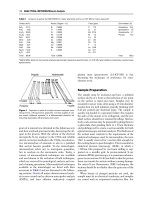
ANALYTICAL METHODS/Geochemical Analysis (Including X-ray) 57
Other techniques use chromatography, the time taken
for one substance to move through another or through
a capillary under a given gradient. A wide family of
geochemical analytical techniques use mass spectrometry to split propelled material, converted into
charged particles, using electromagnets. Other techniques involve examining the products of heating
Earth materials under controlled conditions and
studying either the evolved fluids or the changes in
the properties of the residual solids.
X-ray Techniques
Origin of X-rays
X-ray technologies have proved to be useful in geochemical analysis (Table 2). X-rays are part of the
electromagnetic spectrum (Figure 3) and have wavelengths ranging between 0.01 nm and 10 nm (0.1–
˚ ). They are waveforms that are part of a family
100 A
that includes light, infrared, and radio waves. Since
X-rays have no mass and no electrical charge, they are
not influenced by electrical or magnetic fields and
travel in straight lines. X-rays, like all parts of the
electromagnetic spectrum, possess a dual character,
being both particles and waves. The name that has
been given to the small packets of energy with these
characteristics is photon.
The simple model of the atom, proposed by Niels
Bohr in 1915, is not completely correct, but it has
many features that are approximately correct. The
modern theory of the atom is called quantum mechanics; the Bohr model is an approximation to quantum mechanics that has the virtue of being much
simpler than the full theory. In the Bohr model neutrons and protons occupy a dense central region (the
nucleus), and electrons orbit the nucleus. The basic
feature of quantum mechanics that is incorporated in
the Bohr model is that the energies of the electrons
in the Bohr atom are restricted to certain discrete
values (the energy is quantized) – only certain electron
orbits with certain radii are allowed.
X-rays are generated when free electrons from
an electron gun give up some of their energy when
they interact with the orbital electrons or the nucleus
of an atom (Figure 4). The energy given up by the
electron during this interaction reappears as emitted
electromagnetic energy, known as X-radiation. Two
different atomic processes can produce X-ray
photons. One is called bremsstrahlung and the other
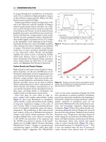
is called electron-shell emission (Figure 5). Bremsstrahlung means ‘braking rays’. When an electron
approaches an atom, it is affected by the negative
force from the electrons of the atom, and it may be
slowed or completely stopped. The energy absorbed
by the atom during the slowing of the electrons
is excessive to the atom and will be radiated as
X-radiation of equal energy to that absorbed. Bremsstrahlung X-rays tend to have a broad range of energies since the degree of slowing can be variable and
materials composed of mixtures have atoms with
different properties (Figure 6). Bremsstrahlung tend
not to be used for geochemical analysis; that is the
preserve of electron-shell emission.
Analysis of X-rays: Electron-Shell Emission
A common geochemical application of X-ray analysis
is to direct a focussed electron beam at a polished rock
or mineral surface and then collect and quantify the
resulting secondary characteristic X-rays (Figure 7).
The secondary X-rays help to reveal the elements
present in that part of the sample that is directly
under the electron beam. This technique is known as
electron-beam microanalysis, or microprobe analysis,
and gives spatially resolved major- and trace-element
geochemical data from solid samples, including rocks,
minerals, sediments, soils, and glass. Many ordinary
electron microscopes are fitted with a secondary
X-ray detector, making them suitable for geochemical
analysis. All of these devices rely on electron optics,
using electromagnetic lenses to focus and direct a
stream of electrons, generated by an electron gun,
onto a polished mineral or rock surface (Figure 7).
The focused electron beam has a variable radius, but
can typically be maintained at slightly greater than
about 1 mm. The spatial resolution of a microprobe
is actually somewhat greater than 1 mm. The impinging electron stream interacts with the polished surface and produces a wide range of signals, including
secondary and backscattered electron and cathodoluminescence (light) as well as the secondary X-rays of
concern here. There is an activation volume from
which X-rays are generated, below the polished surface, which is several times larger than the primary
beam. Samples must be highly polished (flat) to avoid
scattering.
When a sample is bombarded by an electron beam,
some electrons are knocked out of their quantum shells
in a process called inner-shell ionization (Figure 5).
Outer-shell electrons fall in to fill a vacancy in a process
of self-neutralization. The shells are termed K, L, M,
and N starting from the innermost most strongly bound
shell.
Electrons moving from one shell to another produce characteristic X-rays. K-shell ionizations are
commonly filled by electrons from the L shell (Ka
radiation) or M shell (Kb radiation). There are two
Ka peaks (Ka1 and Kb2) corresponding to two discrete states of the in-falling electron. When outer-shell
electrons drop into inner shells, they emit a quantized