- Trang chủ >>
- Khoa Học Tự Nhiên >>
- Vật lý
Sulfide assisted growth of silicon nano wires by thermal evaporation of sulfur powders
Bạn đang xem bản rút gọn của tài liệu. Xem và tải ngay bản đầy đủ của tài liệu tại đây (292.4 KB, 4 trang )
www.elsevier.com/locate/ph
y
se
Physica E 24 (2004) 278–281
Sulfide-assisted growth of silicon nano-wires by
thermal evaporation of sulfur powders
Junjie Niu
a
, Jian Sha
a,b
, Deren Yang
a,
*
a
State Key Lab of Silicon Materials, Zhejiang University, Hangzhou 310027, People’s Republic of China
b
Department of Physics, Zhejiang University, Hangzhou 310027, People’s Republic of China
Received 23 March 2004; accepted 11 May 2004
Available online 10 July 2004
Abstract
Silicon nanowires (SiNWs) with a diameter of B20 nm were synthesized by the thermal evaporation of
sulfur powders on silicon wafers. The source of the SiNWs came from the silicon substrates. It is considered
that the generated SiS compound assisted the formation of SiNWs. Finally, the Raman shift of SiNWs was
discussed.
r 2004 Elsevier B.V. All rights reserved.
PACS: 71.55.Cn; 81.05.Ys
Keywords: Silicon; Nanowires; Sulfide assisted
1. Introduction
Recently the one-dimensional nano-materials,
especially silicon nanowires (SiNWs), have stimu-
lated much interest because of their different
properties in comparison with the corresponding
bulk materials [1–6]. Several synthesized methods
for SiNWs have been reported, including laser
ablation [7], chemical-vapor-deposition (CVD) via
vapor–liquid–solid (VLS) mechanism [8–13], ther-
mal evaporation via oxygen-assisted [14–17] and
solid–liquid–solid (SLS) mechanisms [18–20], and
electronic-chemical method [21].
In this paper, the thermal evaporation of
sulfur powders on silicon wafers used to grow
SiNWs is reported. Compared with the other
approach, this process was simple and the
source of SiNWs was from the silicon wafer
substrates but not from the silane gas [11]
and silicon oxide [14,17]. It is also found that
sulfide played an important role in the formation
of SiNWs, therefore, a sulfide-assisted growth
mechanism was suggested. In the experiments,
the samples were checked by filed emission
scanning electron microscopy (FESEM), transmis-
sion electron microscopy (TEM), and X-ray
diffraction (XRD), respectively. Finally, the Ra-
man spectroscopy was also used to investigate the
SiNWs.
ARTICLE IN PRESS
*Corresponding author. Tel./fax: +86-571-879-523-22.
E-mail address: (D. Yang).
1386-9477/$ - see front matter r 2004 Elsevier B.V. All rights reserved.
doi:10.1016/j.physe.2004.05.002
2. Experimental
SiNWs were produced on p-type (1 1 1) silicon
wafers with a resistivity of about 0.001 O cm by
means of a low-vacuum CVD system. First,
several pieces of silicon wafers and plenty of sulfur
powders were placed in a semi-sealed alumina boat
which was put at the center of a horizontal quartz
tube furnace. Then the furnace was evacuated to
reach 30 Pa by a mechanical pump. The tempera-
ture of the system was then raised to 900
Cata
heating rate of 25
C min
À1
and continually up to
1250
C at a heating rate of 10
C min
À1
, and held
at 1250
C for 30 min at a constant pressure of
30 Pa. After reaction, the weak black and yellow
substrates with the as-grown materials were
removed from the furnace and characterized by
FESEM (FEI, Sirion), TEM (JEOL, JEM200CX),
XRD (Rigaku, D/MAX-rA), and Raman scatter-
ing spectroscopy (Nicolet Almega), respectively.
The possible chemical composition of the as-
grown materials on the wafers was investigated
by using energy-dispersive X-ray spectroscopy
(EDX) attached to the FESEM.
3. Results and discussion
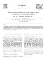
The FESEM morphology of the SiNWs with a
diameter of B20 nm (that of a very few SiNWs is
over 50 nm) is shown in Fig. 1. The EDX (the inset
of Fig. 1) taken from the corresponding nanowires
indicates that the nanowires were silicon. How-
ever, oxygen could also be detected. The oxygen is
considered to have come mainly from the surface
oxidation of nanowires. This suggests that the
nanowires were composed of silicon and silicon
oxide as sheath. Actually, some SiNWs could be
oxidized to be silicon oxide nanowires because of
the low-vacuum system and high temperature, as
found in our previous work [6]. The TEM image of
a number of curved SiNWs is shown in Fig. 2.It
can be seen that the average diameter of the
SiNWs was B20 nm, while the minimum one was
less than 10 nm. The structure of the SiNWs is
indicated by the selected area electric diffraction
(SAED) image in the top right of Fig. 2. The weak
electric diffraction spots proved that the SiNWs
did not crystallize well. This might be due to the
fast growth rate of the SiNWs. The thorough
analysis of crystal nature is indicated by the XRD
data as shown in Fig. 3. The sharp peaks of Si
(1 1 1), Si (3 1 1), Si (4 0 0), and Si (3 3 1) indicates
that the SiNWs were crystalline. Some crystal Al
peaks and low-intensity peaks of SiS
2
(3 0 1) and
SiS
2
(2 1 3) are simultaneously observed. Al peaks
ARTICLE IN PRESS
Fig. 1. FESEM image of the SiNWs on a silicon wafer. The
lower left inset is the EDX taken from the corresponding
sample.
Fig. 2. TEM image of the SiNWs. The top right inset is the
SAED taken from one of the SiNWs. On top left is the
magnified TEM image of a SiNW with tip.
J. Niu et al. / Physica E 24 (2004) 278–281 279
come from the sample preparation process for
the XRD analysis, while SiS
2
comes mainly from
the decomposition of SiS. The discussion of the
growth mechanism is displayed below.
As the function of silicon oxide in the oxygen-
assisted mechanism [15,16], in our experiments,
the silicon sulfide (SiS) also played a key role
in assisting the growth of SiNWs. Therefore, a
sulfide-assisted model for SiNW growth is sug-
gested here.
The reaction procedure mainly took two steps.
One was that the sulfur reacted with the silicon
substrate and generated SiS compound at the
lower temperature (B900
C). The next step was
that the SiS decomposed to be Si and SiS
2
at
higher temperature (B1000
C). During the de-
composition, SiNWs grew up from the generated
Si as source. The reaction equations are shown
below.
S þ O
2
¼ SO
2
m ð1Þ
S þ Si ¼ SiS ðB900
CÞð2Þ
2SiSm ¼ Si þ SiS
2
ðB1000
CÞð3Þ
and
SiS
2
þ 2H
2
O ¼ SiO
2
þ2H
2
Sm: ð4Þ
When the temperature reached B900
C, Eq. (2)
happened and the SiS film was produced. Con-
tinually, the SiS is decomposed into Si and SiS
2
at
higher temperature (B1000
C), which could be
confirmed by the XRD spectrum in Fig. 3.
Therefore, it is believed that the SiNWs generated
from the SiS acted as nucleation centers which
were located at the tip of the SiNWs, as shown in
the top left of Fig. 2. Thus, the tip should contain
SiS
2
. But there was no S signal in the EDX
spectrum (the inset of Fig. 1) and only weak SiS
2
peaks are displayed in XRD data (Fig. 3). This is
because the SiS
2
could sublimate and disappear
when the temperature was higher than 1090
C.
Furthermore, SiS
2
was also easy to react with
H
2
O, which exists in air, as illustrated in Eq. (4).
Surely, not only SiS but also other sulfides such
as zinc sulfide, ferric sulfide, etc, which form
silicon sulfides by the reaction with silicon, can
also be used to assist the growth of SiNWs. In
principle, all of silicon compounds, such as silicon
sulfide shown in this paper and silicon oxide [17],
can assist the formation of SiNWs. It is called the
silicon compound-assisted mechanism for SiNWs
growth.
The SiNWs and bulk silicon in comparison were
also checked using Raman spectroscopy (Fig. 4).
It is clear that the 510.5 cm
À1
peak of the
SiNWs (Fig. 4(a)) shows a B 10 cm
À1
downshift
compared with the 520.3 cm
À1
peak of bulk silicon
(Fig. 4(b)). Usually, the peak of 510.5 cm
À1
was
regarded to be the first-order transverse optical
phonon mode (TO). The downshift might be
ARTICLE IN PRESS
30 40 50 60 70 80
0
500
1000
1500
2000
Al(111)
Al(200)
SiS
2
(213)
SiS
2
(301)
Al(220)
Al(311)
Si(331)
Si(400)
Si(311)
Si(111)
Intensity (CPS)
2θ (degrees)
Fig. 3. XRD spectrum of the as-grown SiNWs.
400 450 500 550 600 650 700 750
0
1000
2000
3000
4000
b
a
510.5cm
-1
520.3cm
-1
Intensity
Raman Shift (cm
-1
)
Fig. 4. Raman spectra of SiNWs (a) and bulk silicon (b).
J. Niu et al. / Physica E 24 (2004) 278–281280
associated with the quantum confinement effect
and laser heating effect [22,23].
4. Conclusion
In summary, silicon nanowires (SiNWs) with a
diameter of B20 nm were successfully synthesized
on silicon wafers by thermal evaporation of sulfur
powders. It is considered that the decomposition
of SiS resulted in the formation of SiNWs.
Furthermore, a sulfide-assisted growth model of
SiNWs was suggested. At last, the Raman shift of
SiNWs was also discussed.
Acknowledgements
This work was supported by the National
Natural Science Foundation of China (Nos.
50272057 and 60225010) and the Key Project
of Chinese Ministry of Education. The authors
would like to thank Prof. Youwen Wang and
Mr. Z.C. Chen for their great help in the
measurements of TEM and Raman spectroscopy.
References
[1] Xiangfeng Duan, Yu Huang, Yi Cui, Jiangfang Wang,
Charles M. Lieber, Nature 409 (2001) 66.
[2] Z.Q. Liu, W.Y. Zhou, L.F. Sun, D.S. Tang, X.P. Zou,
Y.B. Li, C.Y. Wang, G. Wang, S.S. Xie, Chem. Phys. Lett.
341 (2001) 523.
[3] S. Nihonyanagi, Y. Kanemitsa, Physica E 17 (2003) 183.
[4] N. Clement, D. Tonneau, H. Dallaporta, V. Bouchiat,
D. Fraboulet, D. Mariole, J. Gautier, V. Safaror, Physica
E 13 (2002) 999.
[5] J. Sha, J.J. Niu, X.Y. Ma, J. Xu, X.B. Zhang, Q. Yang,
D.R. Yang, Adv. Mater. 14 (2002) 1219.
[6] J.J. Niu, J. Sha, N.S. Zhang, Y.J. Ji, X.Y. Ma, D.R. Yang,
Physica E 23 (2004) 1.
[7] W.S. Shi, H.Y. Peng, Y.F. Zheng, N. Wang, N.G. Shang,
Z.W. Pan, C.S. Lee, S.T. Lee, Adv. Mater. 12 (2000) 1343.
[8] K.K. Lew, J.M. Redwing, J. Cryst. Growth 254 (2003) 14.
[9] M. Lu, M.K. Li, L.B. Kong, X.Y. Guo, H.L. Li, Chem.
Phys. Lett. 374 (2003) 542.
[10] X.Y. Zhang, L.D. Zhang, G.W. Meng, G.H. Li,
N.Y.J. Phillipp, F. Phillipp, Adv. Mater. 13 (2001) 1238.
[11] J.J. Niu, J. Sha, X.Y. Ma, J. Xu, D.R. Yang, Chem. Phys.
Lett. 367 (2003) 528.
[12] J.J. Niu, J. Sha, Q. Yang, D.R. Yang, Jpn. J. Appl. Phys.
43 (2004).
[13] J.J. Niu, J. Sha, Y.W. Wang, X.Y. Ma, D.R. Yang,
Microelectron. Eng. 66 (2003) 65.
[14] R.Q. Zhang, T.S. Chu, H.F. Cheung, N. Wang, S.T. Lee,
Mater. Sci. Eng. C 16 (2001) 31.
[15] Z. Zhang, X.H. Fan, L. Xu, C.S. Lee, S.T. Lee, Chem.
Phys. Lett. 337 (2001) 18.
[16] Q.L. Hu, G.Q. Li, H.S. Suzuki, H.S. Araki, N. Ishikawa,
W. Yang, T. Noda, J. Cryst. Growth 246 (2002) 64.
[17] J.J. Niu, J. Sha, D.R. Yang, Physica E 23 (2004) 131.
[18] T.I. Kamins, R. Stanley Williams, T. Hesjedal, J.S. Harris,
Physica E 13 (2002) 995.
[19] D.P. Yu, Z.G. Bai, Y. Ding, Q.L. Hang, H.Z. Zhang,
J.J. Wang, Y.H. Zou, W. Qian, G.C. Xiong, H.T. Zhou,
S.Q. Feng, Appl. Phys. Lett. 72 (1998) 3458.
[20] D.P. Yu, Y.J. Xing, Q.L. Hang, H.F. Yan, J. Xu, Z.H. Xi,
S.Q. Feng, Physica E 9 (2001) 305.
[21] Y.J. Zhang, Q. Zhang, N.L. Wang, Y.J. Yan, H.H. Zhou,
J. Zhu, J. Cryst. Growth 226 (2001) 185.
[22] M.J. Konstantinovic, S. Bersier, X. Wang, M. Hayne,
P. Lievens, R.E. Silverans, V.V. Moshchalkov, Phys. Rev.
B 66 (2002) 161311 (R).
[23] R. Gupta, Q. Xiong, C.K. Adu, U.J. Kim, P.C. Eklund,
Nano Lett. 3 (2003) 627.
ARTICLE IN PRESS
J. Niu et al. / Physica E 24 (2004) 278–281 281