Báo cáo khóa học: Functional properties of the protein disulfide oxidoreductase from the archaeon Pyrococcus furiosus A member of a novel protein family related to protein disulfide-isomerase doc
Bạn đang xem bản rút gọn của tài liệu. Xem và tải ngay bản đầy đủ của tài liệu tại đây (466.7 KB, 12 trang )
Functional properties of the protein disulfide oxidoreductase
from the archaeon
Pyrococcus furiosus
A member of a novel protein family related to protein disulfide-isomerase
Emilia Pedone
1
, Bin Ren
2
, Rudolf Ladenstein
2
, Mose
`
Rossi
3,4
and Simonetta Bartolucci
3
1
Istituto di Biostrutture e Bioimmagini, C.N.R., Napoli, Italy;
2
Center for Structural Biochemistry, Karolinska Institutet, Huddinge,
Sweden;
3
Dipartimento di Chimica Biologica, Universita
`
degli Studi di Napoli Federico II, Napoli, Italy;
4
Istituto di Biochimica
delle Proteine, C.N.R., Napoli, Italy
Protein disulfide oxidoreductases are ubiquitous redox
enzymes that catalyse dithiol–disulfide exchange reactions
with a CXXC sequence motif at their active site. A disulfide
oxidoreductase, a highly thermostable protein, was isolated
from Pyrococcus furiosus (PfPDO), which is characterized
by two redox sites (CXXC) and an unusual molecular mass.
Its 3D structure at high resolution suggests th at i t may be
related to the multidomain protein disulfide-isomerase
(PDI), which is currently known only in eukaryotes. This
work focuses o n the functional characterization of PfPDO
as well as its r elation to the eukaryotic PDIs. Assays of
oxidative, reductive, and isomerase activities of PfPDO were
performed, which revealed that the archaeal protein not only
has o xidative and reductive activity, but also isomerase
activity. On the basis of structural data, two single mutants
(C35S and C146S) and a double mutant (C35S/C146S) of
PfPDO were constructed and analyzed to elucidate the
specific roles of the two redox sites. The results indicate that
the CPYC site in the C-terminal half of the protein is
fundamental to reductive/oxidative activity, whereas iso-
merase activity requires both a ctive sites. I n comparison w ith
PDI,theATPaseactivitywastestedforPfPDO, which was
found to be cation-dependent with a basic pH optimum and
an optimum temperature of 9 0 °C. These results and an
investigation on genomic sequence d atabases indicate that
PfPDO may be an ancestor of the eukaryotic PDI and
belongs to a novel protein disulfide oxidoreductase family.
Keywords: A rchaea; p rotein disulfide-isomerase; protein
disulfide oxidoreductase; Pyrococcus furiosus; redox sites.
Protein disulfide oxidoreductases are ubiquitous redox
enzymes that catalyse d ithiol–disulfide exchange reactions.
These enzymes share a CXXC sequence motif at their active
sites. The two cysteines can undergo reversible oxidation–
reduction by shuttling between a dithiol and a disulfide form
in the catalytic process. Protein disulfide oxidoreductases
comprise the families o f t hioredoxin, g lutaredoxin, protein
disulfide-isomerase (PDI), and DsbA (disulfide-bond form-
ing) and their homologs. Whereas thioredoxin and
glutaredoxin mainly catalyse the reduction of disulfides,
PDI and DsbA catalyse the formation or rearrangement of
disulfide bridges in the protein-folding process.
Protein d isulfide oxidoreductases have been well studied
in bacteria and eukarya, although to date only a few
archaeal members of this protein f amily have been isolated,
and therefore very little is known about protein disulfide
oxidoreductases in archaea.
A small redox protein w ith a molecular m ass of 12 kDa
was purified from the archaeon Methanobacterium
thermoautotrophicum by McFarlan et al.[1].Thisprotein
can catalyse the reduction of insulin disulfides and function
as a hydrogen donor for Escherichia coli ribonucleotide
reductase. The presence of the active-site motif CPYC,
which is conserved in all glutaredoxins, suggested that it acts
as a g lutaredoxin-like p rotein. Surprisingly, however, the
reduced enzyme does not react with either thioredoxin
reductase or glutathione differently from other thioredoxins
and glutaredoxins [2]. I n the hyperth ermophilic archaeon
Methanococcus jannaschii [3], a thioredoxin homologue was
identified (Mj0307) [4] that has the sequence CPHC, which
had never before been observed in e ither thioredoxins o r
glutaredoxins. It exhibits biochemical activities similar to
thioredoxin, although its structure is more similar to
glutaredoxin. The observation that a single thioredoxin
system is present in M. jannaschii and Mb. thermoauto-
trophicum suggested that a single t hioredoxin-like protein
with a glutaredoxin-like structure is enough to maintain
redox homeostasis in the a rchaeal methanogen [5].
Guagliardi et al . [6] p urified a protein d isulfide oxido-
reductase from the hyperthermophilic archaeon Sulfolobus
solfataricus. Given its ability to catalyse the reduction of
insulin disulfides in the presence of dithiothreitol, t he
protein was named t hioredoxin. The monomeric form of
the enzyme h as an unusual molecular m ass of a bout
26 kDa, compared with that observed in thioredoxin and
glutaredoxin (12 kDa).
A homologous protein disulfide oxidoreductase was
purified from the hyperthermophilic archaeon Pyrococcus
Correspondence to S. Bartolucci, Dipartimento di Chimica Biologica,
Universita
`
degli Studi di Napoli Federico II, via Mezzocannone 16,
80134 Napoli, Italy. Fax: +39 81 2534614, Tel.: +39 81 2534732,
E-mail:
Abbreviations: PfPDO, protein disulfide oxidoreductase from the
archeon Pyrococcus furiosus; DMS, dimethyl suberimidate; MgATP,
5m
M
MgCl
2
,2m
M
ATP; PDI, protein disulfide-isomerase.
(Received 19 May 2004, revised 29 June 2004, accepted 8 July 2004)
Eur. J. Biochem. 271, 3437–3448 (2004) Ó FEBS 2004 doi:10.1111/j.1432-1033.2004.04282.x
furiosus (PfPDO) [7]. PfPDO showed close similarity to the
S. solfataricus p rotein in molecular mass (25 648 Da ) and
dithiothreitol-dependent insulin reduction activity. In addi-
tion, both proteins displayed thiol transferase activity by
catalysing the reduction of disulfide bonds in
L
-cysteine
[7,8]. The PfPDO primary structure does not show any
overall sequence similarity to known protein disulfide
oxidoreductases. Interestingly, it has two potential active
sites with the conserved CXXC sequence motif. A CPYC
sequence is located at the C-terminal half of PfPDO, which
is the conserved active sequence of the glutaredoxin family,
usually l ocated at the N-terminus. In a ddition, a CQYC
sequence, which has never been observed in any other
protein disulfide oxidoreductase, is present at the
N-terminal half of the protein. The PfPDO crystal structure
provides some intriguing challenges to the understan ding of
the enzyme’s function [9–11]. The protein consists of two
homologous units with low sequence identity ( 18%). Each
unit contains a thioredoxin fold, and the accessibilities of the
two CXXC active sites are rather different. The presence of
two homologous units in the same protein resembles the
structure of PDI; in fact, the PDI molecule possesses two
thioredoxin-like domains with two active sites. Interestingly,
whereas thioredoxins and glutaredoxins were identified in
both prokaryotes and eukaryotes, DsbA was only found in
prokaryotes. PDIs, with multiple thioredoxin/glutaredoxin
domains within a single polypeptide are known in e ukary-
otes, a nd it is likely that t he first step in their molecular
evolution was the duplication of an ancestral thioredoxin/
glutaredoxin domain [12]. The unusual structural features of
PfPDO suggest that this enzyme probably represents a new
member of the protein disulfide oxidoreductase superfamily
and a new form of isomerase compared with PDI and
DsbA. Functional studies of PfPDO are essential to support
this finding, but have not yet been conducted. Therefore,
this work focuses on the functional characterization o f the
PfPDO p rotein in an attempt t o elucidate its relation with
the eukaryotic multidomain PDI. Functional data revealed
that the archaeal protein not only has oxidative and
reductive activity, but also isomerase activity. T his is the
first example of an archaeal protein characterized with
disulfide isomerase activity.
To investigate the specific roles of each PfPDO redox site,
two single mutants (C35S and C146S) were constructed, in
which the N-terminal active-site cysteine residue (Cys3 5 or
Cys146) was replaced by serine, and a double mutant
(C35S/C146S). All mutants w ere e xpressed, purified, and
their activities compared with that of the wild-type protein.
To compare t he PfPDO w ith PDI for ATP bindin g and
hydrolysis, the archaeal protein was also tested for its
ATPase activity.
Experimental Procedures
Materials
Bovine insulin, glutathione disulfide (GSSG), glutathione
(GSH), bovine liver PDI, horse liver alcohol dehydro-
genase, bovine pancreas scrambled RNase and all the other
reagents used were from Sigma. Molecular-mass s tandards
for SDS/PAGE were obtained from Pharmacia or Bio-
Rad. E. coli strain JM101 was purchased from Boehringer.
Expression vector pET22(b+), E. coli strain BL21(DE3),
and CJ236 E. coli strain were fro m AMS Biotechnology
(Abingdon, UK). R adioactive materials were obtained
from New England Nuclear/Life Science (Boston, MA,
USA). 8-Azido-[
32
P]ATP[aP] was obtained from ICN.
Deoxynucleotides and restriction and modification
enzymes were from Boehringer. All materials used for
gene amplification were supplied by Stratagene Cloning
Systems. All synthetic oligonucleotides and the peptide
designed by Ruddock et al. [13] were from PRIMM
(Milan, Italy). Bacterial cultures, plasmid purifications,
and transformations were performed as described by
Sambrook et al . [14].
Construction of
E. coli Pf
PDO and mutants
Pf
PDO(C35S),
Pf
PDO(C146S) and the double mutant
(C35S/C146S)
Isolation of chr omosomal DNA from P. furiosus was
performed as described by Barker [15]. From the PfPDO
amino-acid sequence from r esidues 1–7, t he following
oligonucleotides were designed and u sed as primers in the
PCR gene amplification procedure, using the chromosomal
DNA (200 ng) a s template: forward p rimer, 5¢-GGAATT
catatgGGATTGATTAGTGACGCTG-3¢, contained a
5¢-NdeI site (indicated in lowercase); reverse primer housed
the PfPDO stop c odon 3¢ of a unique BamHI (indicated
in lowercase) 5¢-GGAATTcatatgGGATTAGTGACGC
TG-3¢. The amplification was performed as described by
Saiki [16] for 35 cycles at 45 °C annealing temperature, on a
Perkin–Elmer Cetus Cycler Temp using Pfx polymerase
(Stratagene). The amplified DNA fragment (PfPDO),
opportunely digested, was inserted into the pET22(b+)
plasmid. The r ecombinant clone, designated pET-PfPDO
wild-type, represented the expression vector.
The mutations Cys35Ser (C35S) and Cys146Ser (C146S)
were introduced into the PfPDO DNA by the method of
Kunkel [17]. The amplified genes, opportunely digested,
were ligated to the cloning pET22(b+) plasmid. Insertion of
the correct mutations was confirmed b y DNA sequencing
using S anger’s dideoxy method, with a Sequenase Sequen-
cing Kit from Amersham [18].
Expression and purification of recombinant
Pf
PDO
mutants
Competent E. coli BL21(DE3) cells were transformed with
pET-PfPDO wild-type, C35S, C146S, and C35S/C146S,
and grown at 37 °C to different densities in 500 mL terrific-
broth medium; isopropyl thio-b-
D
-galactoside was added to
1m
M
final concentration, varying the induction time from
2to24h.E. coli BL21DE3 cells transformed with
pET22(b+) represented a negative control. Optimized
overexpression of all the proteins was obtained by exposing
the c ells to 1 m
M
isopropyl t hio-b-
D
-galactoside at a cell
density o f A
600
¼ 2.5 for 18 h. Cell pellets from 500 mL
cultures were resuspended in 5 m L 10 m
M
Tris/HCl,
pH 8.4, and crude extracts were prepared by disrupting
the cells with 20 min pulses at 20 H z (Sonicator Ultrasonic
liquid processor; Heat System Ultrasonics Inc., F arming-
dale, NY, USA ) and ultracentrifugation a t 160 000 g for
30 min. Recombinant wild-type protein and its mutants
3438 E. Pedone et al.(Eur. J. Biochem. 271) Ó FEBS 2004
were purified in a similar way. The crude extracts were
subjected to heat treatment at 80 °C and then centrifuged at
5000 g at 4 °C for 15 min, removing almost 70% of the
mesophilic host proteins. The crude extracts were applied to
a2.6cm· 60 cm column (HiLoad Superdex 75; Pharma-
cia) connected to an FPLC system (Pharmacia) and eluted
with 10 m
M
Tris/HCl (pH 8.4)/0.2
M
NaCl at a flow rate of
2mLÆmin
)1
. The active fractions were pooled, concentra-
ted, and extensively dialysed against 10 m
M
Tris/HCl,
pH 8.4. They were then loaded on an anion-exchange
Mono Q column in 10 m
M
Tris/HCl, pH 8.4, connected to
an FPLC system (Pharmac ia), and eluted with a linear
gradient (0/0.3
M
NaCl) in 30 min at a flow r ate of
0.5 m LÆmin
)1
. A single peak was observed on RP-HPLC
and a single protein b and on SDS/PAGE.
Analytical methods for protein characterization
Protein concentration was determined using BSA as the
standard [19]. T he molar a bsorption coefficient, obtained
by the method used by the Schepertz laboratory (http://
paris.chem.yale.edu), was 1 9 724
M
)1
Æcm
)1
.
Protein homogeneity was a ssessed by SDS/PAGE
[12.5% (w/v) gels] using the silver staining procedure of
Rabilloud et al. [20]. In addition, proteins were analysed by
nondenaturing electrophoresis [12.5% (w/v) polyacrylamide
slab gel].
The molecular mass of the proteins was estimated usin g
electrospray mass spectra recorded on a B io-Q triple
quadrupole instrument (Micromass). Samples were dis-
solved in 1 % (v/v) acetic acid/50% (v/v) acetonitrile a nd
injected into the i on source at a flow rate of 10 mLÆmin
)1
using a Phoenix syringe pump. Spectra were collected and
elaborated using
MASSLYNX
software provided by the
manufacturer. Calibration of the mass spectrometer was
performed with horse heart myoglobin ( 16 951.5 Da).
UV-CD spectra in 10 m
M
sodium phosphate, pH 7 .0,
using a 1-mm path-length cell at 185–260 n m at 25 °C, were
recorded on a Jasco J-710 spectropolarimeter equipped with
a Peltier thermostatic cell holder (Jasco, model PTC-343)
for all the proteins.
Counting integral numbers of residues by chemical
modification
The procedure of H ollecker & C reighton [21] was used to
detect the different exposure of the cysteine residues. All the
proteins (PfPDO and mutants at a final concentration of
200 m
M
) were incubated in a final volume of 1 mL for
30 min a t 37 °Cin10 m
M
Tris/HCl (pH 8.0)/10 m
M
EDTA
(pH 7 .0) in native, reduced (10 m
M
dithiothreitol), and
reduced and denatured (10 m
M
dithiothreitol and 8
M
urea)
conditions. Successively in a final volume of 10 lL, five
different solutions co ntaining 0.25
M
iodoacetate (in 0.25
M
Tris/HCl, pH 8.0, and 0.25
M
KOH) and 0.25
M
iodoacet-
amide (in 0.25
M
Tris/HCl, pH 8 .0) were prepared in the
following ratios: 0 : 1 (250 m
M
); (250 m
M
)1:0;1:1(each
125 m
M
); (187.5 m
M
)3:1(62.5m
M
); (225 m
M
)9:1
(25 m
M
). At the end of the incubation, 40 lL of the mixture
was added to each of the five solutions; these were then left
to react on ice for 5 min. The reaction mixtures w ere
analysed by nondenaturing electrophoresis [12.5% (w/v)
polyacrylamide slab g el]. The ÔladderÕ or control i s repre-
sented by a mixture of 10 mL taken from each of the five
reaction mixtures. The method consists of adding various
iodoacetamide and iodoacetate ratios to portions of the
protein to generate a complete spectrum of protein mole-
cules with 0, 1, or 4 acidic carboxymethyl groups, where 4 is
the integral number of cysteine residues. Protein in which
all thiol groups were blocked with iodoacetate, if well
exposed, migrated more slowly than that blocked with
iodoacetamide, because of the acidic carboxymethyl groups.
Cross-linking with dimethyl suberimidate (DMS)
Following the procedure of Davies & Stark [22], 10 lg
PfPDO was incubated f or 2 h at room temperature with
differentquantitiesofDMS(1:1,1:2.5,1:5,1:10)to
determine the best protein to DMS ratio. Molecular mass
and yield were checked by SDS/PAGE [ 12.5% (w/v)
polyacrylamide gel].
Assay of enzyme activities
Insulin reductase activity. Reductase activity was a ssayed
by Holmgren’s turbidimetric method [23] with a few
modifications. T he catalytic reduction of insulin disulfide
bonds was m easured at 30 °C.Proteinwasaddedin1mL
100 m
M
sodium phosphate buffer, pH 7.0, containing
2m
M
EDTA and 1 mg bovine insulin. A control cuvette
contained only buffer and insulin. The reaction was started
by the addition of 2 m
M
dithiothreitol to both cuvettes.
Increasing turbidity from precipitation of the insulin B
chain was recorded at 650 nm. The stock solution of insulin
(10 m gÆmL
)1
) was prepared according to the Holmgren
protocol.
Oxidation activity. The disulfide bond-forming activity of
the proteins was monitor ed using the s ynthetic decapep-
tide NRCSQGSCWN containing two cysteine residues at
position 3 and 8 design ed by Ruddock et al.[13].The
peptide contains a fluorescent group (tryptophan) on one
side of one cysteine residue and a protonated g roup
(arginine) on the other side of the s econd cysteine residue,
and the two cysteine residues are separated by a flexible
linker region. The linker is long enough to permit the
formation of an unstrained disulfide bond, and the
peptide i s s mall and water soluble. Oxidation o f t his
dithiol peptide to the disulfide st ate is accompanied by a
change in tryptophan fluorescence emission intensity. In
fact, on oxidation, the fluorescent group and the
protonated group are b rought close t ogether, and
quenching on the fluorophore occurs where arginine is
the charged quencher. Fluorescence quen ching was used
as the basis for monitoring the disulfide bond-forming
activity of PfPDO.
Spectrofluorimetric analysis. The assay was performed in
McIlvaine buffer (0.2
M
disodium hydrogen phosphate/
0.1
M
citric acid, pH 7.0) with 2 m
M
GSH, 0.5 m
M
GSSG and 5 l
M
PfPDO. The reaction mixture was
placed in a fluorescence cuvette with a final assay
volume of 1 mL. After mixing, the cuvette was placed
in a thermostatically controlled Perkin–Elmer LS50B
Ó FEBS 2004 Archeal protein disulfide oxidoreductase/isomerase (Eur. J. Biochem. 271) 3439
spectrofluorimeter for 1 min to allow thermal equilibra-
tion of the solution to 50 °C. Next, 5 l
M
substrate
peptide was added, mixed, and the change in fluorescence
intensity ( excitation 295 nm, e mission 350 nm, slits
10/10 nm) was monitored over an a ppropriate time
(15 m in). As a c ontrol, the same experiment was carried
out in the absence of any protein; no decrease in
fluorescence intensity was observed [ 13].
HPLC analysis. Alternatively the oxidation activity was
measured by HPLC analysis (Varian). The reduced and
oxidized fo rms of the peptide have different retention times
and are eluted separately on reverse-phase chromatography
[L. Birolo and A. Tosco (1999) personal communication].
Thepeptidewaselutedinasinglepeakandstoredat)20 °C
in the elution buffer (30% acetonitrile in 0.1% trifluoro-
acetic acid; v/v/v) a t a concentration o f 1.05 m
M
.The
peptide concentration was determined spectrophotometi-
cally using an absorption c oefficient o f 5 600
M
)1
Æcm
)1
at
278 n m. The oxidized state of the peptide w as generated b y
incubating the peptide at a concentration of 50 l
M
in 0.2
M
Tris/HCl, pH 8.4, at 20 °C for 15 h. The reduced state was
generated by incubating the peptide in McIlvaine buffer
(0.2
M
disodium hydrogen phosphate/0.1
M
citric acid ,
pH 7.0) at a final c oncentration o f 50 l
M
and 1 m
M
dithiothreitol in a final v olume of 50 lL.
The a ssay mixture con tained 5 l
M
reduced peptide,
100 m
M
GSH (stock solution 60.1 mgÆmL
)1
), 25 m
M
GSSG
(stock solution 30.7 mgÆmL
)1
) and the protein PfPDO (final
concentration 10, 50, 100, 150 or 200 l
M
). The mixture was
incubated at different temperatures (50 °C, 60 °Cor70°C)
for different times in the presence of different concentrations
of the protein. After incubation, the mixture was loaded on
the HPLC reversed-phase Vydac C18 column equilibrated
in buffer A [0.1% (v/v) trifluoroacetic acid in water].
Chromatography was carried out with a linear gradient
0–100% buffer B (95% acetonitrile, 0.07% trifluoroacetic
acid; v/v/v) in buffer A at a flow rate of 1 mLÆmin
)1
for
35 min.
Re-activation of scrambled RNase. The isomerase activity
was assayed by Lambert’s method. Re-activation of
scrambled RNase was monitored after incubation of
PfPDO in 50 m
M
sodium phosphate, pH 7.5, in a total
volume of 0.9 mL, with 10 lL dithiothreitol (1 m
M
stock
solution, final concentration 10 l
M
) for 2 min at 30 °C[24].
A 0.1 mL portion of ÔscrambledÕ RNase (Sigma;
0.5 mgÆmL
)1
in 10 m
M
acetic acid, final concentration
4 l
M
) was added, and a t different times after this addition
10 lL samples were withdrawn and assayed for RNase
activity. Each sample was added to an assay mixture of
1mL 0.5mgÆmL
)1
RNA in 5 0 m
M
Tris/HCl, pH 7.5.
RNase a ctivity o n yeast R NA w as assayed by the method
outlined by Kunitz [25] with some modifications, and under
conditions in which the decrease in A
300
was linear for at
least 3–4 min. Yeast RNA was d issolved in water, and the
pH was kept neutral by performing the assay in 50 m
M
Tris/
HCl, pH 7.5. The positive control was re-activation of
scrambled RNase catalysed by PDI (bovine liver; Sigma).
Nonenzymatic reactivation of scrambled R Nase was cor-
rected for by using the same mixture without the addition of
any of the proteins.
Detection of ATP binding by CD
CD measurements were performed in a Jasco J-720
spectropolarimeter in 20 m
M
Tris/HCl (pH 7.5)/5 m
M
MgCl
2
at 25 °C. Each sample was scanned five times, noise
reduction was applied, and baseline buffer spectra were
subtracted from sample spectra before molar ellipticities
were calculated. To obtain spectra in the near-UV region
(250–320 nm), the cell path length was 1 cm and the protein
concentration 1 mgÆmL
)1
. T he CD spectra were evaluated
at 260 nm.
Cross-linking of
Pf
PDO with 8-Azido-[
32
P]ATP[aP]
To analyze the ability of PfPDOtocross-linkto8-azido-
ATP, 3 mg protein was incubated in the presence of 2 mCi
8-azido[
32
P]ATP[aP] for 30 min in 50 m
M
Tris/HCl, pH 8.0
or 10 m
M
Gly/NaOH, pH 10.0, containing 2 m
M
EDTA,
1m
M
dithiothreitol and 5 m
M
MgCl
2
at 6 0° or 70 °C. To
induce cross-linking, samples were exposed for 1 0 m in to
UV irradiation and then resolved by SDS/PAGE in 12%
polyacrilamide gel and visualized by radioauto graphy on a
Fuji medical X -ray film. The s ame procedure w as used for
incubation of PfPDO at pH 10.0 at 70 °C in the presence of
an increasing concentration o f unlabeled ATP (50 l
M
and
1m
M
)[26].
Fluorescence measurements
Samples of PfPDO (100 lg) were incubated for 10 min at
70 °C, 80 °C, or 90 °C in the presence of MgATP, and then
loaded on a Superdex 75 HiLoad column (Amersham
Pharmacia Biotech; 1 · 30 cm; eluent 10 m
M
Tris/HCl,
pH 7.5, 0.2
M
NaCl; flow rate 0.3 mLÆmin
)1
) to remove the
nucleotide excess. The protein samples recovered from t he
columns and a sample of native PfPDO were a nalyzed for
fluorescence at 3 l
M
final protein concentration (excitation
wavelength 280 nm; emission recorded between 310 and
410 n m) using a Perkin–Elmer LS50B spectrofluorimeter at
25 °C[27].
Assay of ATPase activity
A colorimetric assay was routinely used to measure ATPase
activity following the method of Lanzetta et al.[28].To
100 lL of s ample (water a nd 10 mg protein) was a dded
800 lL o f g reen malachite/ammonium molybdate in 1
M
HCl, followed by mixing. After 1 min, 100 lL34%citrate
was added a nd mixed. This solution was read immediately
at 660 nm.
In an alternative assay, the ATPase activity of PfPDO
was assayed in mixtures containing 2 m
M
ATP, 15 lCi
[
32
P]ATP[aP], 5 m
M
MgCl
2
and 10 lg pure protein in
50 m
M
Tris/HCl, pH 7.5 (150 lL final volume). After a
5 min incubation at 70 °C, a 25 lL aliquot was withdrawn
and a dded t o 0.5 mL of a suspension containing 50 m
M
HCl, 5 m
M
H
3
PO
4
and 7% activated charcoal. The mixture
was then centrifuged at 4000 g for 20 min. The radioactivity
of the supernatant was determined in a 100 lL aliquot using
a liquid-scintillation counter (Beckman). In rate calcula-
tions, spontaneous ATP hydrolysis in t he absence of
PfPDO was corrected for [ 29].
3440 E. Pedone et al.(Eur. J. Biochem. 271) Ó FEBS 2004
Results
Production of wild-type and mutant
Pf
PDO
To determine the redox state and th e accessibility of the
cysteine residues of the Pf PDO redox sites, electrophoretic
analysis was performed, as described by Hollecker et al.
[21], on t he protein treated under d ifferent conditions
(native, reduced, and reduced and denatured) (Table 1). By
comparing the results obtained from t he different g els, it
was possible to confirm the crystallographic data that the
most reactive cysteine was Cys146, as this was observed at
the lowest r atio of iodoacetate to iodoacetamide. T his was
followed by C ys149, Cys35, and C ys38, which was the last
to react and the least accessible residue [21].
To investigate the role of the putative redox sites of
PfPDO, three mutants were constructed (C35S, C146S, and
C35S/C146S) by mutagenizing the most exposed cysteines
of each of the r edox sites: specifically, Cys35 at the
N-terminal site and Cys146 at the C-terminal site were
replaced by serine [30].
PfPDO a nd mutants were expressed in E. coli
BL21(DE3). Overexpression of all t he proteins was
obtained by exposing t he cells to 1 m
M
isopropyl thio-b-
D
-galactoside at a cell density of A ¼ 2.5. To optimize the
production of the recombinant proteins, transformed cells
were exposed to the inducer for 2–24 h; maximum e xpres-
sion was obtained after 18 h of induction.
The crude extract of E. coli was subjected to one thermal
precipitation step at 80 °C for 20 min to remove almost
70% of the mesophilic host proteins. During the purifica-
tion procedure, the proteins were assayed a fter reduction of
protein disulfides o n insulin as substrate; when the inter-
chain disulfide bridges are reduced between chains A and B
of the insulin, the turbidity of the solution increases because
of precipitation o f the f ree B chain [23]. After g el-filtration
chromatography and anion-exchange chromatography, a
single peak was observed on RP-HPLC, and a single b and
on SDS/PAGE. The protein yield from 1 L o f culture was
40 mg for a ll the recombinant proteins.
The molecular mass of the proteins was analysed by
electrospray mass spectroscopy. The measured mass of
PfPDO w as 25 648 ± 0.5 Da. The measured mass of C35S
and C146S was 25 628 ± 0.5 Da, and that of C35S/C146S
was 25 613 ± 0.4 Da. Thus, the difference in mass was in
perfect agreement with the mutations introduced.
To see if the mutations introduced had an e ffect o n t he
structure of t he protein, far-UV CD spectra were recorded
for all the proteins. The s pectra were very similar, showing
that all the proteins are completely folded and indicating
that the m utations did not result in any obvious change in
overall structure.
Characterization of the activities of wild-type
and mutant
Pf
PDO
PfPDO reduces insulin disulfide in the presence of dithio-
threitol at 30 °C. The a nalysis w as performed in the
presence of increasing concentrations of the pure proteins,
as well as in their a bsence (the spontaneous precipitation
reaction), because dithiothreitol is the reducing agent that
recycles the oxidized protein (Fig. 1 ). The activity was
assayed at 1.2 l
M
for all the proteins. Both the wild-type
PfPDOandthemutantC35Swereactiveintheinsulin
reductase assay [23], whereas the activity of the mutant
C146S and the double mutant was similar to the control.
This shows that the active site in the C-terminal half
(CPYC) is responsible for t he reductase activity.
In the presence of 5 l
M
PfPDO, oxidation of the dithiol
peptide d esigned b y R uddock was observed at neutral pH
by the spectrofluorimetric assay (Fig. 2 A). Separation of the
oxidized and reduced forms of the peptide by HPLC
allowed quantification of the oxidative a ctivity as a ratio
between the areas of oxidized/reduced peptide. The assays
were performed a t a concentration of 100 l
M
protein, at
different times and different temperatures [50 °C, 60 °C,
and 70 °C (data not shown)]. The best conditions were
Table 1. Exposed cysteine residues by the Hollecker method [21]. The proteins in native, reduced (10 m
M
dithiothreitol), and reduced and denatured
(10 m
M
dithiothreitol and 8
M
urea) conditions were treated with different amounts of iodoacetate/iodoacetamide [1 : 1 (each 250 m
M
),1:3,1:9
ratios of neutral to acidic reagents] and separated by SDS/PAGE. The appearance of a band in the differe nt con ditions used (native, reduced,
reduced and denatured) on the differe nt proteins (PfP DO and mutants) is evidence of the exposure of that cysteine residue. nd, Not determine d.
Native Reduced Reduced/denatured
Iodoacetate/iodoacetamide 1 : 1 3 : 1 9 : 1 1 : 1 3 : 1 9 : 1 1 : 1 3 : 1 9 : 1
PfPDO wild-type – – – C146 C146 C146/149 C146 C146/149 C146/149/35
C35S mutant – – – C146 C146 C146/149 C146 C146/149 C146/149/38
C146S mutant – – C149 – – C149 C149 C149/35 C149/35
C35S/C146S mutant nd nd nd C149 C149 C149/38 C149 C149 C149/38
Fig. 1. Assay of reductase activity by measuring the reduction of bovine
insulin disulfides. T he dithiothreitol-dependent reduction of bovine
insulin disulfides was carried out as described in Experimental proce-
dures in the absence [control (–––)] or presence of 1.2 l
M
PfPDO wild-
type ( ÆÆÆÆ), Pf PDO (C35S) ( ), or PfPDO (C146S) and PfPDO
(C35S)/(C146S) (-Æ-Æ).
Ó FEBS 2004 Archeal protein disulfide oxidoreductase/isomerase (Eur. J. Biochem. 271) 3441
50 °C f or 3 h. A linear relation betwe en activity and
concentration was dete cted for all the p roteins (Fig. 2B).
Wild-type PfPDO and C35S were able to oxidize the
peptide with maximum activity at a concentration of 150
and 200 l
M
, respectively. C146S had residual oxidative
activity, but the double mutant was completely inactive,
demonstrating the predominant role of the redox site at the
C-terminus in the oxidative activity.
The action of PfPDO in catalysing interchange of
intramolecular disulfides in scrambled RNase results in
restoration of the native disulfide pairing and the c oncom-
itant return of RNase activity. Thus, the isomerase activity
of PfPDO was assayed by a time-course incubation during
which aliquots were removed and RNase activity with RNA
was measured. Re-activation of scrambled RNase was
performed with all the proteins. Only the wild-type protein
was able to refold the scrambled RNase (Fig. 3), indicating
that isomerase a ctivity requires the participation o f both
N-terminal and C-terminal active sites. Refolding of the
scrambled RNase in the presence o f P DI was used as a
positive control, and the absence of the recovery of the
RNase activity in the presence of the thioredoxin from
Alicyclobacillus acidocaldarius wasusedasanegative
control. The refolding of the scrambled RNase in the
presence of Pf PDO seems to be less efficient when using
PDI. However, the temperature of the assay, which is
limited by the stability of the protein substrate RNase, is
very far f rom the optimal growth temperature of t he
hyperthermopilic micro-organism.
Characterization of wild-type
Pf
PDO
A detailed study of Pf PDO structure highlighted certain
putative ATP-binding sites (the presence of P-loops, a
common m otif in ATP-binding proteins), t he primary
structure of w hich con sists of a glycine-rich sequence
followed by a conserved lysine and a serine or a theonine
[31]. In p articular, PfPDO has the sequences, GKDFG(88–
94), GLPAG(97–101), GKGKILG(167–173), which
resemble, with s ome deviations, the gl ycine-rich motif,
GXXGXG, of the ATPase domains of the eukaryotic
chaperone hsp90 [32], the type II DNA topoisomerases, and
MutL DNA mismatch-repair proteins [33]. The hypothet-
ical nucleotide-binding sites are presumably located in loops
between b3andb4, a4andb4, and a6andb6. To study the
role played by the putative binding of ATP in the
conformation of PfPDO, a spectrofluorimetric analysis
was performed. The presence of Trp184 e nabled u s to
perform intrinsic fluorescence experiments. The tryptophan
emission spectrum of native PfPDO displayed a maximum
around k ¼ 345 (data not shown). A PfPDO sample
incubated in the presence of hydrolysable ATP (MgATP)
gave a similar spectrum to that o f t he native protein. Far-
UV CD s pect ra in t h e presence and absence of ATP (d at a
not shown) gave the same results as the spectrofluorimetric
analysis, i.e. no change in the conformation of the protein in
the presence of the nucleotide. These experiments indicate
that the binding and/or hydrolysis of ATP do not have any
effect on the conformation of PfPDO, possibly because o f
the localization of the amino-acid residues involved in ATP
binding in exposed regions. Near-UV CD spectroscopy was
performed, which p rovides information on the environment
of aromatic residues in folded p roteins. The aromatic CD
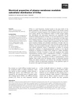
Fig. 2. Assay of o xidative a ctivity by measuring the formation of the
disulfide bridge in the peptide NRCSQGSCWN. (A) Spectrophoto-
metric method. The d isulfide bond-fo rming activity of Pf PDO w as
monitored usin g the synthetic decapeptide NRCSQGSCWN. Oxida-
tion of this dithiol peptide to the disulfide state is accompanied by a
change in tryptophan fluorescence emission intensity. (a) Control, the
same assay perfo rmed with out the protein; (b) PfPDO; (c) PfPDO
(C35S); (d) PfPDO (C146S). (B) H PLC. The disulfide bond-forming
activity of PfPDO is monitored using the synthetic decapeptide
NRCSQGSCWN and the oxidation of this dithiol peptide to the di-
sulfide state is accompanied by a change in time of retention on a
Vydac C18. Oxidative activity is expressed as a ratio between the peak
of oxidized and reduced peptide. The assay was performed at 50 °C,
with an incubation time of 210 min, at incre asing c oncentration of
PfPDO wild-type (r), PfPDO (C35S) (j); Pf PDO ( C146S) (m);
PfPDO (C35S)/(C146S) and control (d).
Fig. 3. Assay of isomerase activity of PfPDO by measuring re-activa-
tion of scrambled RNase. The recovery of RNase activity as a function
of time is presented after p reincubation wit h PDI (m); PfPDO wild-
type ( r); PfPDO (C35S) and PfPDO (C146S) and Pf PDO (C35S)/
(C146S) (j); control (d). RNase activity with RNA was measured.
3442 E. Pedone et al.(Eur. J. Biochem. 271) Ó FEBS 2004
spectra of PfPDO in the absence and presence of ATP (up
to 324 m
M
) are shown in Fig. 4 . The ellipticity of the protein
was positive between 255 and 300 nm. A signal around
279 nm can be assigned to tyrosine residues, and the major
intensity at 268 nm and 261.5 nm can be attributed to the
numerous phenylalanine residues (12 of them). After ATP
was added, the signal attributed to tryptophan and tyrosine
residues does not seem to have been affected, whereas the
signal attributed to phenylalanine residues changed consid-
erably.
Interestingly, close to the P-loop domain, there is a
phenylalanine residue at position 91. In addition, our
spectra in dicate that other a romatic r esidues are in close
proximity t o the ATP-binding domain. The CD d ata
indicate ATP binding with co-operativity and a K
d
of
230 l
M
.
The ATP binding to Pf PDO was confirmed by c ross-
linking to 8-azido-ATP after UV irradiation (Fig 5A,B).
The data show ATP b inding for PfPDO. Alcohol dehy-
drogenase (horse liver; Sigma) was used as a negative
control because it is known n ot to bind ATP, even though it
contains a putative nucleotide-binding site. The alcohol
dehydrogenase did not show any affinity for the ATP
analog, suggesting that the binding to Pf PDO was specific
under the conditions used. It has been reported that some
non-ATP-binding proteins (for example, BSA) bind
8-azido-ATP in a nonspecific way. However, in these cases,
the bound analog could not be displaced b y t he unlabeled
nucleotide [34]. In this work, photoaffinity labeling of
PfPDO with 8-azido-[
32
P]ATP[aP] was decreased by the
presence of unlabeled ATP, indicating that ATP and the
analog 8-azido-ATP recognize the same binding site.
The ATPase activity o f PfPDO w as demonstrated. T he
hydrolysis of ATP was linear for up to 30 min at every
temperature examined with the colorimetric and radioactive
assays used (see Experimental Procedures). The hydrolysis
of ATP by PfPDO required the presence of bivalent metal
ions, Mg
2+
giving the highest rate (Fig. 6 A) compared with
the activity observed in the absence of ions. When assayed in
the pH range 4.0–10.0, PfPDO catalyzed hydrolysis of ATP
with a maximum around basic values (Fig. 6B). Assays
performed in the temperature range 30) 90 °C showed that,
at 90 °C, PfPDO is still fully able to hydrolyse ATP
(Fig. 6C). The rate of spontaneous ATP hydrolysis was
followed in the same range of temperature and pH. Freshly
purified PfPDO hydrolysed ATP with a V
max
of 127.5 nmol
P
i
releasedÆmin
)1
Æmg
)1
(Mg
2+
, pH 10.0, 90 °C).
The ability of PfPDO to bind and hydrolyse ATP is
another property th at links this protein with the multifunc-
tional P DI, a s t his f eature has been observed in t he
eukaryotic protein [35].
In addition, as PfPDOexistsasadimerinthecrystal
form and PDI is a dimer in its 3D structure, we analysed the
dimerization of PfPDO by g el filtration and in the presence
of the cross-linking agent DMS. In all the conditions tested,
the presence of the dimer was never observed. It was
observed only in the presence of the cross-linking reagent
DMS. In particular, a ratio of Pf PDO to DMS of 1 : 2.5
proved to be optimal (Fig. 7).
Discussion
Insufficient information is available on protein disulfide
oxidoreductases f rom archaea to define their physiological
function(s) with any certainty. Disulfide bonds are now
known to occur in many thermophilic and intracellular
archaeal proteins, and this observation highlights the
importance of the glutaredoxin/thioredoxin system in these
micro-organisms.
Hyperthermophiles are generally capable o f growing
under extreme conditions such as low pH, h igh pressure,
and high salt concentration. Most of these organisms are
anaerobes, have extraordinarily heat-stable proteins, and
use ingenious strategies for stabilizing nucleic acids and
other macromolecules in vivo [36].
Fig. 4. Measurem ent of K
d
for ATP by CD. Near-UV CD spectra
recorded in 20 m
M
Tris/HCl (pH 7.5)/5 m
M
MgCl
2
andinthepres-
ence of in creasing c oncen trations of ATP (0–324 m
M
). The i nset shows
normalized CD variation at 260 nm vs. incr easing [ATP] concentra-
tion. CD values at 260 nm were normalized and elaborated using the
programme Microsoft Excel 2000. The curve for the determination of
the K
d
forATPwasobtainedusingtheprogram
KALEIDA GRAPH
3.0.
Fig. 5. ATP-binding capacity of PfPDO. Cross-linking of PfPDO w ith
8-azido-[
32
P]ATP[aP]: 3 lg PfPDO was incubated w ith 2 mCi 8-azido-
[
32
P]ATP[aP] for 30 min at pH 8.0 and pH 10.0 at 60° and 70 °C. To
induce cross- linking, samples were exposed for 10 min to UV irradi-
ation and then resolved by SDS/PAGE in 12% p olyacrylamide gel and
visualized by radioautography. (A) Lanes 1 and 2, pH 8.0 at 60 °Cand
70 °C; lanes 3 and 4, pH 10.0 at 60 °Cand70°C. (B) The same
procedure was used by incubating PfPDOatpH10.0at70°Cinthe
presence of increasing c oncentrations of u nlabeled A TP. Lane 1, 0 m
M
unlabeled ATP; lane 2, 50 m
M
unlabeled ATP; lane 3, 1 m
M
unlabeled
ATP.
Ó FEBS 2004 Archeal protein disulfide oxidoreductase/isomerase (Eur. J. Biochem. 271) 3443
Recently, from the resolution of t he whole genome
sequences of various hyperthermophilic archaea, it is clear
that these hyperthermophiles have proteins endowed with
thioredoxin/glutaredoxin motifs, suggesting the ubiquity of
this system in nature.
The protein from P. furiosus described here may provide
an important contribution to our understanding of the
function of these proteins in hyper thermophilic archaea a nd
bacteria. In fact, PfPDO is able to catalyse the oxidation of
dithiols, as well as the reduction and rearrangement of
disulfides. In the presence of glutathione, up to 70 °C,
PfPDO catalyses the formation of a disulfide bond between
the t wo cysteines of the peptide, an activity simil ar t o that
observed for DsbA at 25 °C [13]. At 30 °C, PfPDO is able
to catalyse the reduction of insulin disulfides in the presence
of dithiothreitol. Disulfide rearrangement was also observed
at a s imilar temperature using RNase with sc rambled
disulfides as substrate.
Using the two single mutants (C35S and C146S) and the
double mutant (C35S/C146S), we have demonstrated that
the C-terminal s ite (CPYC), which is common to a ll the
glutaredoxins, determines the reductive activity. This r esult
is in agreement with crystallographic data, which suggest a
reductive nature for t he C-unit. The lower capacity of the
N-unit to reduce disulfide bridges may be due to intrinsic
factors, such as a higher redox potential and major
conformational tension of the disulfide, but it may also
depend on external factors such as steric impediments
caused by a closed conformation of the active site in the
N-unit. As regards the oxidative activity, the two units also
display differences in their functional properties, with the
site at the C-termus always predominant, the mutant with a
nonmutagenized site at the N-terminus showing very low
activity at 50 °C. Higher temperatures, closer to the
physiological temperature at which the micro-organism
P. furiosus lives, may be necessary to obtain more kinetic
energy and allow an open conformation at the site.
Alternatively, a different substrate may be required because
of the polar nature of the amino acids close to the active site.
On the other hand, both sites are necessary for the disulfide
isomerase activity. In fact, o nly wild-type PfPDO was able
to refold scrambled RNase. This is in a greement with a
functional model of PDI in which the domains fu nction
synergistically [37,38]. The emerging m odel of PDI compri-
ses four structural domains, a, b, b¢ and a¢, plus a linker
region between b¢ and a¢ and a C-terminal acidic extension.
In this model of PDI function, individual domains with
specialized roles contribute to d ifferent activities to enable
the catalysis of complex isomerizations in substantially
folded protein s ubstrates. Mutations at the first cysteine of
the a ctive site in either the N-terminal or C-terminal
thioredoxin domain inhibits the capacity of PDI to catalyse
thiol–disulfide exchange reactions in vitro, reducing enzy-
matic activity to negligible le vels. In fact, the r edox/
isomerase activities of P DI, as in thioredoxin, are due to
the reactivity of the N-terminal Cys residue in two
Fig. 6. ATPase activity of PfPDO. The assays were performed under
standard co ndit ions (see Experimental Procedures) except for the ions
at 5 m
M
. (A) The activity assayed und er standard co ndit ions (Mg
2+
at
90 °C, pH 7.5) was 67.3 nmol P
i
releasedÆmin
)1
Æmg
)1
,whichwastaken
as 100%. Activity was assayed at different pH values [50 m
M
sodium
acetate for pH 4.0–5.5 (m); 50 m
M
sodium phosphate for pH 6.0–7.0
(j); 50 m
M
Tris/HCl for p H 7.5–8.4 ( e); 50 m
M
glycine/NaOH for
pH 9.0–10.5 (d)] (B ) and temperatures (c). The activity assayed under
standard conditions was 127.5 nmol P
i
releasedÆmin
)1
Æmg
)1
(Mg
2+
,
pH 10.0, 90 °C), which was taken as 100%. Data are means from at
least three independent experiments.
Fig. 7. Cross-linking of PfPDO with DMS. After 2 h of incubation at
room temperature in the presence of the cro ss-linking agent DMS, the
samples were loaded o n an SDS/12.5% polyacrylamide gel. Lane 1,
PfPDPfPD/DMS in a r atio 1 : 2.5; lane 2, control, Pf PDO with no
DMS; lane 3, markers of molecular mass.
3444 E. Pedone et al.(Eur. J. Biochem. 271) Ó FEBS 2004
thioredoxin-like boxes (Cys-Gly-His-Cys) within the a and
a¢ domains of the protein [39]. Although the two domains
do not possess equivalent catalytic activities or substrate-
binding affinities, they can function independently from
each other.
PfPDO resembles eukaryotic PDI, as it has two thio-
redoxin-like motifs. In PDI, the thioredoxin-like regions are
separated from each other in the primary structure, whereas
in PfPDO they are connected directly. In this work, only the
first cysteine of each redox site was mutated to investigate
the effect on the function of the protein, demonstrating that
the active site a t the C-terminus is basic for oxidative a nd
reductive activities and that the two units do not seem to be
functionally independent, c onsidering that only the wild-
type enzyme is able to refold scrambled RNase. Unlike P DI,
which is a homodimer o f two 57 kDa subunits, PfPDO
seems to be a monomer, dimerization only occurring in the
presence of the cross-linking agent DMS.
The ability o f PfPDO to b ind and hydrolyse ATP
supports its relationship to PDI [40]. In fact, an ATP-
binding site and A TPase activity related to its chaperone
role have been reported in PDI [41]. Whereas PDI binds
ATP with a K
d
of 9.66 l
M
, PfPDO binds ATP with a K
d
of
230 l
M
. PfPDO is a hyper thermostable protein, a nd the
studies of its f unctional and catalytic properties are limited
by the temperature at which its activities are studied. Such
temperatures are usually far below the physiological tem-
perature (70–103 °C) at which P. furio sus lives. The ATPase
activity does not seem to be linked to the isomerase or redox
activities, as in the presence of ATP no differences in the
activities are observed. This is in full agreement with a
report t hat t he site of phosphorylation, and thus probably
the ATPase a ctive site, lies somewhe re within the central
domain of the PDI [42], and t hat this site is far away from
the redox active sites in the sequ ence. Furthermore, the
measurements of the rates of PDI-catalysed refolding of
scrambled RNase A, in the absence or presence of ATP,
show that ATP has little or no effect on this activity.
Interestingly, comparison of the genomes of archaea and
bacteria showed the e xistence of a g roup of redox proteins
with a similar molecular mass to PfPDO. Clearly, all these
proteins also contain two active sites, although they were
often initially assigned as hypothetical thioredoxins and
glutaredoxins [43–52]. The presence o f the redox site,
CQYC, at the N-terminus of protein disulfide oxidoreduc-
tase in P. furiosus, P. abyssi,andP. horikoshii,andalsoin
the more distant S. solfataricus, further confirm the import-
ance of this site for p rotein function (Fig. 8). It is worth
noting that amino-acid residues that are probably involved
in putative ATP binding, such as Gly88, Gly97, Pro99,
Fig. 8. Comparison of the amino-acid sequences of different protein disulfide oxidoreductases. The sequences were from the following sources: Pf,
P. furiosus;Ph,P. horikoshii;Pa,P. abissi;Ss,S. solfataricus;St,S. tokodaii;Ap,Aeropyrum pernix;Ta,Thermoplasma acidophilum;Tv,Ther-
moplasma volcanium;Fa,Ferroplasma acidarmanus;Tm,Thermot oga mar itima;Aa,Aquifex aeolicus;Tt,Thermoanaerobacter tengcongensis.The
residues identical with the sequence of PfPDO in at least 90% of the sequences are indicated in bold. The underlined residue s indicate the active
sites.
Ó FEBS 2004 Archeal protein disulfide oxidoreductase/isomerase (Eur. J. Biochem. 271) 3445
Gly167 and Gly170, are well conserved, indicating their
importance. The genomes of the hyperthemophilic bacteria
Aquifex aeolicus, Thermotoga maritima and Thermoanae-
robacter tengcongensis do not encode a protein related to
bacterial DsbA and no DsbA-like protein in Archaea were
found, suggesting that PfPDO-like proteins represent a new
family characteristic of extremophiles (like DsbA in bacteria
and PDI in eukarya). It should be no ted that we found
PfPDO-like proteins only in thermophilic bacteria, i.e.
Aquifex aeolicus, Thermotoga maritima and Thermoanae-
robacter tengcongensis. A preferen tial horizontal gene
transfer has been noticed between archaea and hyperther-
mophilic bacteria, such as Aquifex and Thermotoga;infact
their proteins show greater similarity t o archaeal than to
bacterial homologs [53]. The reality of horizontal gene flow
from archaea to t hermophilic bacteria becomes even more
tangible on examination of the proteins encoded in the
genome of Thermoanaerobacter tengcongensis which con-
tains more ÔarchaealÕ genes t han appear in other bacteria.
The exclusive presence of PfPDO-like proteins in
extremophiles may suggest that they have a special role
in the adaptation t o e xtreme conditions. The P. horikoshii
genome also contains a glutaredoxin-homolog gene (88%
identity with the glutaredoxin from P. furiosus) [54]. This
protein is the first glutaredoxin-homolog protein that
directly mediates electron transfer from a thioredoxin
reductase-like flavoprotein to protein disulfide in archaea.
The redox-active sequence motifs CPYC and CQYC
suggest that P. horikoshii redox protein (PhRP) belongs
tothesamefamilyasPfPDO. PhRP has insulin-reducing
activity. Site-directed mutagenesis studies revealed that the
active site of the redox protein c orresponds to a CPYC
sequence located in the middle of the sequence, as in
PfPDO. As regards PhRP activities, the disulfide forma-
tion and its rearrangement were no t detected when
reduced or scrambled RNases were used as substrates
at 25 °C. However, the possibility that CQYC may play
some role and that PhRP has PDI-like activity in vivo at
the optimum growth temperature of P. horikoshii cannot
be excluded.
The various functions of PfPDO m ake it an interesting
model system for clarifying the long-standing debate on the
content o f cysteine residues and disulfide in thermophilic
proteins. Disulfide bonds have only rarely been found in
intracellular proteins. The pattern is consisten t with a
chemically reducing environment inside the cells and with a
PDI role in the endoplasmic r eticulum. However, recent
experiments and new calculations based on genomic data of
archaea provide striking contradictions to this pattern.
Recent results indicate that the intracellular proteins of
certain hyperthermophilic arch aea, especially some cren-
archaea such as Pyrobaculum aerop hilum and Aeropyrum
pernix, are rich in disulfide bonds [55]. This finding points to
the role of disulfide bonds in stabilizing many thermostable
proteins and suggests new chemical environments inside
these microbes.
Acknowledgements
We thank Dr Raffaele Cannio and Dr Enrico Bucci for stimulating
discussions. This work was supported by grants from MIUR (PRIN
2002).
References
1. McFarlan, S.C., Terrell, C.A. & Hogenkamp, H.P. (1992) The
purification, characterization, and primary structure of a small
redox protein from Methanobacterium thermoautotrophicum,an
archaebacterium. J. Biol. Chem. 267, 10561–10569.
2. Bhattacharyya, S., Habibi-Nazhad, B., Amegbey, G., Slupsky, C.,
Yee, A., Arrowsmith, C. & W ishart, D.S. ( 2002) Identification of a
novel archaebacterial thioredoxin: determination of function
through structure. Biochemistry 41, 4760–4770.
3. Bult, C. J., White, O. , Olsen, G.J., Zhou, L., Fleischmann, R.D.,
Sutton, G.G., Blake, J.A., FitzGerald, L.M., Clayton, R.A.,
Gocayne, J.D., Kerlavage, A.R., Dougherty, B.A., Tomb, J.F.,
Adams,M.D.,Reich,C.I.,Overbeek,R.,Kirkness,E.F.,Wein-
stock, K.G., Merrick, J.M., Glode k, A., Scott, J.L., G eoghage n,
N.S. & Venter, J.C. (1996) Complete genome sequence of the
methanogenic archaeon, Methanococcus jannaschii. Science 273,
1058–1073.
4. Lee, D.Y., Ahn, B. & Kim, K . (2000) A t hioredoxin from the
hyperthermophilic archaeon Methanococcus jannaschii has a glu-
taredoxin-like fold but thioredoxin-like activities. Biochemistry 39,
6652–6659.
5. Cave, J.W., Cho, H.S., Batchelder, A.M ., Yokota, H., Kim, R. &
Wemmer, D.E. (2001) Solution nuclear magnetic resonance
structure o f a protein disulfide o xidoreductase from Methano-
coccus jannaschii. Protein Sci. 10, 384–396.
6. Guagliardi, A., Nobile, V., Bartolucci, S. & Rossi, M. (1994) A
thioredoxin from the extreme thermophilic archaeon Sulfolobus
solfataricus. Int. J. Biochem. 26, 375–380.
7. Guagliardi, A., De Pascale, D., C annio, R., Nobile, V., Bartolucci,
S. & Rossi, M. (1995) The purification, c lon ing, a nd high level
expression of a glutaredoxin-like protein from the hyperthermo-
philic archaeon Pyrococcus furiosus. J. Biol. Chem. 270, 5748–
5755.
8. Bartolucci, S., De Pascale, D. & Rossi, M. (2001) Protein disulfide
oxidoreductase from Pyrococcus furiosus: biochemical properties.
Methods Enzymol. 334, 62–73.
9. Ren, B., Tibbelin, G., De Pascale, D., Rossi, M., Bartolucci, S. &
Ladenstein, R. (1997) Crystallization and preliminary X-ray
structure analysis of a hyperthermostable t hioltransferase f rom t he
archaeon Py rococcus furiosus. J. Struct. Biol. 119,1–5.
10. Ren, B., Tibbelin, G., De Pascale, D., Rossi, M., Bartolucci, S. &
Ladenstein, R. (1998) A protein disulfide o xidoreductase from the
archaeon Pyrococcus furiosus contains two thioredoxin fold units.
Nat. Struct. Biol. 5, 602–611.
11. Ren, B. & Ladenstein, R. (2001) Protein disulfide oxidoreductase
from Pyrococcus furiosus: structural properties. Methods Enzymol.
334, 74–88.
12. Freedman, R.B. (1998) Novel disulfide oxidoreductase in search of
a function. Nat. Struct. Biol. 5, 531–532.
13. Ruddock, L.W., Hirst, T.R. & Feedman, R.B. (1996) pH-depen-
dence of the dithiol-oxidizing activity of D sbA (a p eriplasmic
protein thiol: disulphide oxidoreductase) and protein disulphide-
isomerase: studies with a no vel simple p eptide substrate. Biochem.
J. 315, 1000–1005.
14. Sambrook, J., Fritsch, E.F. & M an iatis, T . (1989) Molecular
Cloning: A Laboratory Manual, 2nd edn. Cold Spring Harbor
Laboratory Press, Cold Spring Harbor, NY.
15. Barker, D.G. (1982) Cloning and amplified expression of the
tyrosyl-tRNA syn thetase genes of Bacillus stearothermophilus and
Escherichia coli. Eur. J. Biochem. 125, 357–360.
16. Saiki, R.K. (1990) Amplification of genomic DNA. In PCR
Protocols: A Guide to Methods and Applications (Innis, M.A.,
Gellfand, D.A., Sninski, J.J. & White, T.J., eds), pp. 13–20. Aca-
demic Press, New York.
3446 E. Pedone et al.(Eur. J. Biochem. 271) Ó FEBS 2004
17. Kunkel, T.A. (1985) Rapid and efficient site-specific mutagenesis
without phenot ypic sele ction. Proc . Natl Acad. Sci. USA 82,
488–492.
18. Sanger, F., Nicklen, S. & Coulson, A.R. (1977) DNA sequ encing
with chain-terminating inhibitors. Proc. N atl Acad. Sci. USA 76,
5653–5667.
19. Smith, P.K., K rohn, R.I., He rmanson, G.T., M allia, A.K.,
Gartner, F.H., Provenzano, M.D., Fujimoto, E.K., Goeke, N.M.,
Olson, B .J. & Klenk, D.C. (1995) Measurement of protein using
bicinchoninic acid. Anal. Biochem. 150, 76–85.
20. Rabilloud, T., Vuillard, L., Gilly, C. & Lawrence, J.J. (1994)
Silver-staining of proteins in polyacrylamide gels: a general over-
view. Cell. Mol. Biol. 40, 57–75.
21. Hollecker, M. & Creighton, T.E. (1980) Counting integral num-
bers of amino groups per polypeptide chain. FEBS Lett. 119,187–
190.
22. Davies, G .E. & Stark, G.R. (197 0) Use of dimethyl sub-
erimidate, a cross-linking reagent, in studying th e subunit
structure of oligomeric proteins. Proc.NatlAcad.Sci.USA66,
651–656.
23. Holmgren, A. (1979) Thioredoxin catalyzes the reduction of
insulin d isulfides by dithiothre itol and dihydrolipoamide . J. Bi ol.
Chem. 254, 9627–9632.
24. Lambert, N. & Freedman, R.B. (1983) Kinetics and specificity o f
homogeneous protein disu lphid e-isomerase in protein disu lphide
isomeriz atio n and in thiol-pr otei n-disulphide oxidoreduction.
Biochem. J. 213, 235–243.
25. Kunitz, M. (1946) A spectrophotometric method for the mea-
surement of ribonuclease activity. J. Biol. Chem. 164, 563–568.
26. Catz, S.D., Johnson, J.L. & Babior, B.M. (2001) Characterization
of the nucleotide-bin ding cap acity and the ATPase activity of the
PIP3-binding protein JFC1. Proc. Natl Acad. Sci. USA 28, 11230–
11235.
27. Guagliardi, A., Cerchia, L., Bartolucci, S . & Rossi, M. (1994) The
chaperonin from the archaeon Sulfolobus solfataricus promotes
correct refolding and preve nts thermal denaturation in vit ro.
Protein Sci. 3, 1436–1443.
28. Lanzetta,P.A.,Alvarez,L.J.,Reinach,P.S.&Candia,O.A.(1979)
An improved assay for nanomole amounts of inorganic phos-
phate. Anal Biochem. 100 , 95–97.
29. Guagliardi, A., Cerchia, L., Moracci, M. & Rossi, M. (2000) The
chromoso mal protein sso 7d of the crenarchaeon Sulfolobus sol-
fataricus rescues aggregated proteins in an ATP hydrolysis-
dependent manner. J. Biol. Chem. 275, 31813–31818.
30. Vuori, K., Myllyla, R., Pihlajaniemi, T. & Kivirikko, K.I. (1992)
Expression and site-directed mu tagenesis of human p ro tein dis-
ulfide isomerase in E s cherichia coli. This multifunctional poly-
peptide has two independently acting catalytic sites for the
isomerase activity. J. Biol. Chem. 267, 7211–7217.
31. Saraste, M., Sibbald, P .R. & Wittinghofer, A. (1990) T he P -loop: a
common motif in ATP- and GTP-binding proteins. Trends Bio-
chem. Sci. 15, 430–434.
32. Prodromou, C., Roe, S.M., O’Brien, R., Ladbury, J.E., Piper,
P.W. & Pearl, L.H. ( 1997) Identification and structural char-
acterization of the ATP/ADP-binding site in the Hsp90 molecular
chaperone. Cell 90, 65–75.
33. Bergerat, A., de Massy, B., Gadelle, D., Varoutas, P.C., Nicolas,
A. & Forterre, P. (1997) An atypical topoisomerase II from
Archaea with implications for meiotic recombination. Nature
(London) 386, 414–417.
34. Jakob, U., Scheibel, T., Bose, S., Reinstein, J. & Buchner, J. (1996)
Assessment of th e ATP binding prop erties of Hsp90. J. Biol.
Chem. 271, 10035–10041.
35. Quemeneur, E., Guthapfel, R. & Gueguen, P. (1994) A major
phosphoprotein of the endoplasmic r eticulum is protein disulfide
isomerase. J. Biol. Chem. 269 , 5485–5488.
36. Baross, J.A. & Holden, J.F. (1996) Overview of hyperthermophiles
and their heat-shock proteins. Adv. Prot ein Chem. 48, 1–34.
37. Freedman, R.B., Klappa, P. & Ruddock, L.W. (2002) Protein
disulfide isomerases exploit synergy between catalytic and specific
binding domains. EMBO Report 3, 136–140.
38. Kemmink, J., Darby, N .J., Dijkstra, K., Nilges, M., Creighton,
T.E. (1996) Structure determination of the N-terminal
thioredoxin-like domain of protein disulfide isomerase using
multidimensional heteronuclear
13
C/
15
N NMR spectroscopy.
Biochemistry 35, 7684–7691.
39. Darby, N.J., Kemmink, J. & Creighton, T.E. (1996) Identifying
and characterizing a structural domain of protein disulfide iso-
merase. Biochemistry 35, 10517–11052.
40. Quemeneur, E, Guthapfel, R. & Gueguen, P. (1994) A major
phophoprotein of the e n doplasmic reticulum is p ro tein disulfide
isomerase. J. Biol. Chem. 269 , 5485–5488.
41. Guthapfel, R., Gueguen, P. & Quemeneur, E. (1996) ATP binding
and hydrolysis by the multifunctional protein disulfide isomerase.
J. Biol. Chem. 271, 2663–2666.
42. Zapun, A., C reigh ton, T.E., Rowling, P.J. & Freedman, R.B.
(1992) Folding in vitro of bovine pancreatic trypsin inhibitor in the
presence of prote ins of the endoplasmic re ticulum. Prote ins 14 ,
10–15.
43. Robb, F.T ., Maeder, D.L., Brown, J.R., DiRuggiero, J., Stump,
M.D., Y eh, R.K., Weiss, R.B. & D unn, D.M. (2001) G enomic
sequence of hype rthermop hile, Pyrococcus furiosus:implications
for physio logy and enzymology. Methods E nzym ol. 330, 134–157.
44. She, Q., Singh, R.K., Confalonieri, F., Zivanovic, Y., Allard, G.,
Awayez, M .J., Ch an -Weiher, C.C., Clausen, I.G., Curtis, B .A., De
Moors, A., Erauso, G., Fletcher, C., Gordon, P.M., Heikamp-de
Jong, I., Jeffries, A.C., K ozera, C.J., Medina, N., Peng, X., Thi-
Ngoc, H.P., Redd er, P., Sc henk, M.E ., T heriault, C ., To lstrup, N .,
Charlebois, R.L., Doolittle, W.F., D uguet, M., Gaasterland, T.,
Garrett,R.A.,Ragan,M.A.,Sensen,C.W.&VanderOost,J.
(2001) The complete genome of the crenarchaeon Sulfolobus sol-
fataricus P2. Proc. Natl Acad. Sci. USA 98, 7835–7840.
45. Chinen, A., Uchiyama, I. & Kobayashi, I. (2000) Comparison
between Pyrococcus horikoshii and Pyrococcus abyssi genome
sequences reveals linkage of restriction-modification gen es with
large genome polymorphisms. Gene 259, 109–121.
46. Kawarabayasi, Y., Hino, Y., Horikawa, H., Jin-n., o, K.,
Takahashi,M.,Sekine,M.,Baba,S.,Ankai,A.,Kosugi,H.,
Hosoyama, A., Fukui, S., Nagai, Y., Nishijima, K., Otsuka, R.,
Nakazawa, H., Takamiya, M., Kato, Y., Yoshizawa, T., Tanaka,
T., Kudoh, Y., Yamazaki, J., Kushida, N., Oguchi, A., Aoki, K.,
Masuda, S., Yanagii, M., Nishimura, M., Yamagishi, A., O shima,
T. & Kikuchi, H. (2001) Complete genome sequence of an aerobic
thermoacidophilic crenarchaeon, Sulfolobus tokodai i strain7. DNA
Res. 8, 123–140.
47. Ruepp, A., Graml, W ., Santos-Martinez, M.L., Koretke, K.K.,
Volker, C., Mewes, H.W., Frishman, D., Stocker, S., Lupas, A.N.
& Baumeister, W. (2000) The genome sequence of the
thermoacidophilic scavenger Thermoplasma acidophilum. Natur e
(London) 407, 508–513.
48. Kawashima, T., Amano, N., Koike,H.,Makino,S.,Higuchi,S.,
Kawashima-Ohya, Y., Watanabe, K., Yamazaki, M., Kanehori,
K., Kawamoto, T., Nunoshiba, T., Yamamoto, Y., Aramaki, H.,
Makino, K. & Suzuki, M. (2000) Archaeal adaptation to higher
temperatures revealed by genomic sequence of Thermoplasma
volcanium. Proc.NatlAcad.Sci.USA97, 14257–14262.
49. Kawarabayasi, Y., Hino, Y., Horikawa, H., Yamazaki, S., Hai-
kawa,Y.,Jin-n.,o,K.,Takahashi,M.,Sekine,M.,Baba,S.,
Ankai, A., Kosugi, H., H os oyama, A., Fu kui, S., N agai, Y.,
Nishijima, K., Nakazawa, H., Takamiya, M., Masuda, S.,
Funahashi, T., T anaka, T., Kudoh, Y ., Yamazaki, J., K ushida,
N., Oguchi, A. & Kikuchi, H. (1999) Complete genome seque nce
Ó FEBS 2004 Archeal protein disulfide oxidoreductase/isomerase (Eur. J. Biochem. 271) 3447
of an aerobic hyper-thermoph ilic crenarchaeo n, Aeropyrum pernix
K1. DNA Res. 6, 145–152.
50. Deckert,G.,Warren,P.V.,Gaasterland,T.,Young,W.G.,Lenox,
L.,Graham,D.E.,Overbeek,R.,Snead,M.A.,Keller,M.,Aujay,
M., Huber, R., Feldman, R.A., Short, J.M., Olsen, G.J. &
Swanson, R.V. (1998) The c omplete genome of the hyper-
thermophilic bacterium Aquifex aeolicus. Nature (London) 392,
353–358.
51. Bao, Q., Tian, Y., Li, W., Xu, Z., Xuan, Z., Hu, S., Dong, W.,
Yang,J.,Chen,Y.,Xue,Y.,Xu,Y.,Lai,X.,Huang,L.,Dong,X.,
Ma, Y., L ing, L., Tan, H ., C hen, R. & Wang, J ., Yu, J. & Yang, H.
(2002) A complete seque nce of t he T. tengcongensis genome.
Genome Res. 12, 689–700.
52. Nelson, K.E., Eisen, J.A. & Fraser, C.M. (2001) Genome of
Thermotoga maritima MSB8. Methods Enzymol. 330, 169–180.
53. Makarova, K.S. & Koonin, E.V. (2003) Comparative genomics of
archaea: how much have we learned in six years, and what’s next?
Genome Biol. 4, 115–145.
54. Kashima, Y. & Ishikawa, K. (2003) A hyperthermostable novel
protein-disulfide oxidoreductase is reduced by thioredoxin reduct-
ase f rom hyperthermophilic ar chaeon Pyrococcus ho rikoshii.
Arch. Biochem. Biophysics 418, 1 79–185.
55. Mallick, P., Boutz, D.R., Eisenberg, D. & Yeates, T.O. (2002)
Genomic evidence that the intracellular proteins of archaeal
microbes contain disulfide bonds. Proc. Natl Acad. Sci. USA 99,
9679–9684.
3448 E. Pedone et al.(Eur. J. Biochem. 271) Ó FEBS 2004