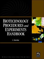
Biotechnology procedures and experiments handbook
Bạn đang xem bản rút gọn của tài liệu. Xem và tải ngay bản đầy đủ của tài liệu tại đây (5.89 MB, 711 trang )
BIOTECHNOLOGY PROCEDURES
AND
EXPERIMENTS HANDBOOK
LICENSE, DISCLAIMER OF LIABILITY, AND LIMITED WARRANTY
The CD-ROM that accompanies this book may only be used on a single PC. This license does not
permit its use on the Internet or on a network (of any kind). By purchasing or using this book/CD-
ROM package(the “Work”), you agree that this license grants permission to use the products
contained herein, but does not give you the right of ownership to any of the textual content in the
book or ownership to any of the information or products contained on the CD-ROM. Use of third
party software contained herein is limited to and subject to licensing terms for the respective
products, and permission must be obtained from the publisher or the owner of the software in
order to reproduce or network any portion of the textual material or software (in any media) that
is contained in the Work.
INFINITY SCIENCE PRESS LLC (“ISP” or “the Publisher”) and anyone involved in the creation,
writing or production of the accompanying algorithms, code, or computer programs (“the
software”) or any of the third party software contained on the CD-ROM or any of the textual
material in the book, cannot and do not warrant the performance or results that might be obtained
by using the software or contents of the book. The authors, developers, and the publisher have
used their best efforts to insure the accuracy and functionality of the textual material and
programs contained in this package; we, however, make no warranty of any kind, express or
implied, regarding the performance of these contents or programs. The Work is sold “as is”
without warranty (except for defective materials used in manufacturing the disc or due to faulty
workmanship);
The authors, developers, and the publisher of any third party software, and anyone involved in the
composition, production, and manufacturing of this work will not be liable for damages of any
kind arising out of the use of (or the inability to use) the algorithms, source code, computer
programs, or textual material contained in this publication. This includes, but is not limited to,
loss of revenue or profit, or other incidental, physical, or consequential damages arising out of the
use of this Work.
The sole remedy in the event of a claim of any kind is expressly limited to replacement of the
book and/or the CD-ROM, and only at the discretion of the Publisher.
The use of “implied warranty” and certain “exclusions” vary from state to state, and might not
apply to the purchaser of this product.
BIOTECHNOLOGY PROCEDURES
AND
EXPERIMENTS HANDBOOK
S. HARISHA, PH.D.
INFINITY SCIENCE PRESS LLC
Hingham, Massachusetts
New Delhi, India
Reprint & Revision Copyright © 2007. INFINITY SCIENCE PRESS LLC. All rights reserved.
Copyright © 2007. Laxmi Publications Pvt. Ltd.
This publication, portions of it, or any accompanying software may not be reproduced in any way, stored in a
retrieval system of any type, or transmitted by any means or media, electronic or mechanical, including, but not
limited to, photocopy, recording, Internet postings or scanning, without prior permission in writing from the
publisher.
Publisher: David F. Pallai
I
NFINITY SCIENCE PRESS LLC
11 Leavitt Street
Hingham, MA 02043
Tel. 877-266-5796 (toll free)
Fax 781-740-1677
www.infinitysciencepress.com
This book is printed on acid-free paper.
S. Harisha. Biotechnology Procedures and Experiments Handbook.
ISBN: 978-1-934015-11-7
The publisher recognizes and respects all marks used by companies, manufacturers, and developers as a
means to distinguish their products. All brand names and product names mentioned in this book are
trademarks or service marks of their respective companies. Any omission or misuse (of any kind) of
service marks or trademarks, etc. is not an attempt to infringe on the property of others.
Library of Congress Cataloging-in-Publication Data
Harisha, S. (Sharma)
[Introduction to practical biotechnology]
Biotechnology procedures and experiments handbook / S. Harisha.
p. cm. — (An introduction to biotechnology)
Originally published: An introduction to practical biotechnology. India : Laxmi Publication, 2006.
Includes index.
ISBN-13: 978-1-934015-11-7 (hardcover with cd-rom : alk. paper)
1. Biotechnology—Laboratory manuals. I. Title.
TP248.24.H37 2007
660.6—dc22
2007011159
Printed in Canada
7 8 9 5 4 3 2 1
Our titles are available for adoption, license or bulk purchase by institutions, corporations, etc. For
additional information, please contact the Customer Service Dept. at 877-266-5796 (toll free).
Requests for replacement of a defective CD-ROM must be accompanied by the original disc, your
mailing address, telephone number, date of purchase and purchase price. Please state the nature of the
problem, and send the information to I
NFINITY SCIENCE PRESS, 11 Leavitt Street, Hingham, MA 02043.
The sole obligation of INFINITY SCIENCE PRESS to the purchaser is to replace the disc, based on
defective materials or faulty workmanship, but not based on the operation or functionality of the
product.
Dedicated with profound gratitude
to my parents
and to my teachers
ch
CONTENTS
Chapter 1. General Instruction and Laboratory Methods 1
General Instruction 1
General Laboratory Methods 2
Chapter 2. Tools and Techniques in Biological Studies 9
Spectrophotometry 9
Electrophoresis 13
Column Chromatography 18
pH Meter 22
Centrifugation 24
Ultrasonic Cell Disruption 29
Conductivity Meter 32
Radioactive Tracers 32
Autoradiography 37
Photography 39
Statistics 41
Graphs 44
Computers 47
Chapter 3. Biochemistry 51
Carbohydrates 51
Exercise 1. Qualitative Tests 52
Exercise 2. Qualitative Analysis 54
Exercise 3. Estimate the Amount of Reducing Sugars 56
Exercise 4. Estimation of Reducing Sugar by Somogyi’s Method 59
Exercise 5. Estimation of Sugar by Folin-Wu Method 60
vii
viii CONTENTS
Exercise 6. Estimation of Sugar by Hagedorn-Jenson Method 61
Exercise 7. Estimation of Reducing Sugars by the Dinitro
Salicylic Acid (DNS) Method 62
Exercise 8. Determination of Blood Glucose by Hagedorn-Jenson
Method 63
Exercise 9. Determining Blood Sugar by Nelson and
Somogyi’s Method 65
Exercise 10. Determination of Blood Glucose by the O-Toluidine
Method 66
Exercise 11. Estimation of Protein by the Biuret Method 67
Exercise 12. Estimation of Protein by the FC-Method 68
Exercise 13. Protein Assay by Bradford Method 69
Exercise 14. Estimation of Protein by the Lowry Protein Assay 70
Exercise 15. Biuret Protein Assay 71
Exercise 16. Estimation of DNA by the Diphenylamine Method 72
Exercise 17. Estimation of RNA by the Orcinol Method 73
Chapter 4. Enzymology 75
Enzymes 75
Exercise 1. Demonstrating the Presence of Catalase in Pig’s Liver 83
Exercise 2. Determining the Optimum pH for Trypsin 84
Exercise 3. Extraction of Tyrosinase 85
Exercise 4. Preparation of Standard Curve 87
Exercise 5. Enzyme Concentration 89
Exercise 6. Effects of pH 90
Exercise 7. Effects of Temperature 91
Exercise 8. Computer Simulation of Enzyme Activity 92
Exercise 9. Kinetic Analysis 93
Exercise 10. Determination of K
m
and V
max
94
Exercise 11. Addition of Enzyme Inhibitors 95
Exercise 12. Protein Concentration/Enzyme Activity 97
Exercise 13. Studying the Action and Activity of Amylase
on Starch Digestion 98
Exercise 14. Determination of the Effect of pH on the
Activity of Human Salivary a-Amylase 99
CONTENTS ix
Exercise 15. Determining the Effect of Temperature on the
Activity of Human Salivary a-Amylase 101
Exercise 16. Construction of the Maltose Calibration Curve 102
Chapter 5. Electrophoresis 103
Electrophoresis 103
Exercise 1. Preparation of SDS-Polyacrylamide Gels 108
Exercise 2. Separation of Protein Standards: SDS-PAGE 110
Exercise 3. Coomassie Blue Staining of Protein Gels 111
Exercise 4. Silver Staining of Gels 112
Exercise 5. Documentation 114
Exercise 6. Western Blots 114
Sodium Dodecyl Sulfate Poly-Acrylamide Gel Electrophoresis
(SDS-PAGE) 117
Preparation of Polyacrylamide Gels 119
Agarose Gel Electrophoresis 121
Agarose Gel Electrophoresis 123
Chapter 6. Microbiology 125
Introduction 125
The Microscopy 127
Exercise 1. The Bright Field Microscope 134
Exercise 2. Introduction to the Microscope and Comparison
of Sizes and Shapes of Microorganisms 137
Exercise 3. Cell Size Measurements: Ocular and
Stage Micrometers 146
Exercise 4. Measuring Depth 147
Exercise 5. Measuring Area 148
Exercise 6. Cell Count by Hemocytometer or Measuring Volume 149
Exercise 7. Measurement of Cell Organelles 152
Exercise 8. Use of Darkfield Illumination 152
Exercise 9. The Phase Contrast Microscope 153
Exercise 10. The Inverted Phase Microscope 155
Aseptic Technique and Transfer of Microorganisms 155
Control of Microorganisms by using Physical Agents 163
x CONTENTS
Control of Microorganisms by using Disinfectants and
Antiseptics 172
Control of Microorganisms by using Antimicrobial Chemotherapy 180
Isolation of Pure Cultures from a Mixed Population 191
Bacterial Staining 201
Direct Stain and Indirect Stain 206
Gram Stain and Capsule Stain 211
Endospore Staining and Bacterial Motility 215
Enumeration of Microorganisms 221
Biochemical Test for Identification of Bacteria 233
Triple Sugar Iron Test 241
Starch Hydrolysis Test (II Method) 242
Gelatin Hydrolysis Test 244
Catalase Test 245
Oxidase Test 247
IMVIC Test 249
Extraction of Bacterial DNA 257
Medically Significant Gram–Positive Cocci (GPC) 259
Protozoans, Fungi, and Animal Parasites 263
The Fungi, Part 1–The Yeasts 264
Performance Objectives 267
The Fungi, Part 2—The Molds 269
Viruses: The Bacteriophages 274
Serology, Part 1–Direct Serologic Testing 279
Serology, Part 2–Indirect Serologic Testing 289
Chapter 7. Cell Biology and Genetics 299
Cell Cycles 299
Meiosis in Flower Buds of Allium Cepa-Acetocarmine Stain 306
Meiosis in Grasshopper Testis (Poecilocerus Pictus) 309
Mitosis in Onion Root Tip (Allium Cepa) 311
Differential Staining of Blood 313
Buccal Epithelial Smear and Barr Body 315
Vital Staining of DNA and RNA in Paramecium 316
CONTENTS xi
Induction of Polyploidy 317
Mounting of Genitalia in Drosophila Melanogaster 318
Mounting of Genitalia in the Silk Moth Bombyx Mori 319
Mounting of the Sex Comb in Drosophila Melanogaster 320
Mounting of the Mouth Parts of the Mosquito 321
Normal Human Karyotyping 322
Karyotyping 322
Black and White Film Development and Printing for Karyotype
Analysis 324
Study of Drumsticks in the Neutrophils of Females 327
Study of the Malaria Parasite 328
Vital Staining of DNA and RNA in Paramecium 330
Sex-Linked Inheritance in Drosophila Melanogaster 331
Preparation of Somatic Chromosomes from Rat Bone Marrow 333
Chromosomal Aberrations 334
Study of Phenocopy 336
Study of Mendelian Traits 337
Estimation of Number of Erythrocytes [RBC] in Human Blood 338
Estimation of Number of Leucocytes (WBC) in Human Blood 340
Culturing Techniques and Handling of Flies 342
Life Cycle of the Mosquito (Culex Pipiens) 343
Life Cycle of the Silkworm (Bombyx Mori) 344
Vital Staining of Earthworm Ovary 346
Culturing and Observation of Paramecium 347
Culturing and Staining of E.coli (Gram’s Staining) 348
Breeding Experiments in Drosophila Melanogaster 349
Preparation of Salivary Gland Chromosomes 354
Observation of Mutants in Drosophila Melanogaster 355
ABO Blood Grouping and Rh Factor in Humans 356
Determination of Blood Group and Rh Factor 357
Demonstration of the Law of Independent Assortment 358
Demonstration of Law of Segregation 360
xii CONTENTS
Chapter 8. Molecular Biology 363
The Central Dogma 363
Exercise 1. Protein Synthesis in Cell Free Systems 364
Chromosomes 366
Exercise 2. Polytene Chromosomes of Dipterans 367
Exercise 3. Salivary Gland Preparation (Squash Technique) 368
Exercise 4. Extraction of Chromatin 369
Exercise 5. Chromatin Electrophoresis 371
Exercise 6. Extraction and Electrophoresis of Histones 372
Exercise 7. Karyotype Analysis 375
Exercise 8. In Situ Hybridization 378
Exercise 9. Culturing Peripheral Blood Lymphocytes 380
Exercise 10. Microslide Preparation of Metaphases for
In-Situ Hybridization 382
Exercise 11. Staining Chromosomes (G-Banding) 384
Nucleic Acids 386
Exercise 12. Extraction of DNA from Bovine Spleen 387
Exercise 13. Purification of DNA 388
Exercise 14. Characterization of DNA 389
Exercise 15. DNA-Dische Diphenylamine Determination 390
Exercise 16. Melting Point Determination 391
Exercise 17. CsCl-Density Separation of DNA 393
Exercise 18. Phenol Extraction of rRNA (Rat liver) 394
Exercise 19. Spectrophotometric Analysis of rRNA 395
Exercise 20. Determination of Amount of RNA by the
Orcinol Method 396
Exercise 21. Sucrose Density Fractionation 397
Exercise 22. Nucleotide Composition of RNA 398
Isolation of Genomic DNA—DNA Extraction Procedure 400
Exercise 23. Isolation of Genomic DNA from Bacterial Cells 401
Exercise 24. Preparation of Genomic DNA from Bacteria 404
Exercise 25. Extraction of Genomic DNA from Plant Source 405
Exercise 26. Extraction of DNA from Goat Liver 407
Exercise 27. Isolation of Cotton Genomic DNA from Leaf Tissue 408
CONTENTS xiii
Exercise 28. Arabidopsis Thaliana DNA Isolation 411
Exercise 29. Plant DNA Extraction 412
Exercise 30. Phenol/Chloroform Extraction of DNA 414
Exercise 31. Ethanol Precipitation of DNA 414
Exercise 32. Isolation of Mitochondrial DNA 415
Exercise 33. Isolation of Chloroplast DNA 417
Exercise 34. DNA Extraction of Rhizobium (CsCl Method) 417
Exercise 35. Isolation of Plasmids 418
Exercise 36. RNA Isolation 422
Preparation of Vanadyl-Ribonucleoside Complexes
that Inhibit Ribonuclease Activity 422
Exercise 37. RNA Extraction Method for Cotton 423
Exercise 38. Isolation of RNA from Bacteroids 424
Exercise 39. Isolation of RNA from Free-Living Rhizobia 425
Estimation of DNA purity and Quantification 426
Exercise 40. Fungal DNA Isolation 427
Exercise 41. Methylene Blue DNA Staining 429
Exercise 42. Transformation 431
Blotting Techniques—Southern, Northern, Western Blotting 433
Preparing the Probe 440
Exercise 43. Southern Blotting (First Method) 444
Exercise 44. Southern Blotting (Second Method) 446
Exercise 45. Western Blotting 447
Exercise 46. Western Blot Analysis of Epitoped-tagged Proteins
using the Chemifluorescent Detection Method for
Alkaline Phosphatase-conjugated Antibodies 450
Southern Blot 452
Exercise 47. Southern Analysis of Mouse Toe/Tail DNA 453
Exercise 48. Northern Blotting 454
Restriction Digestion Methods—Restriction Enzyme Digests 457
Exercise 49. Restriction Digestion of Plasmid, Cosmid, and
Phage DNAs 460
xiv CONTENTS
Exercise 50. Manual Method of Restriction Digestion of
Human DNA 461
Exercise 51. Preparation of High-Molecular-Weight Human
DNA Restriction Fragments in Agarose Plugs 463
Exercise 52. Restriction Enzyme Digestion of DNA 465
Exercise 53. Electroelution of DNA Fragments from Agarose
into Dialysis Tubing 466
Exercise 54. Isolation of Restriction Fragments from Agarose
Gels by Collection onto DEAE Cellulose 469
Exercise 55. Ligation of Insert DNA to Vector DNA 471
PCR Methods (Polymerase Chain Reaction) 472
Exercise 56. Polymerase Chain Reaction 475
Exercise 57. DNA Amplification by the PCR Method 476
Chapter 9. Tissue Culture Techniques 477
Tissue Culture Methods 477
Plant Tissue Culture 487
Plant Tissue Culture 491
Many Dimensions of Plant Tissue Culture Research 493
What is Plant Tissue Culture? 496
Uses of Plant Tissue Culture 497
Plant Tissue Culture demonstration by Using
Somaclonal Variation to Select for Disease Resistance 498
Demonstration of Tissue Culture for Teaching 499
Preparation of Plant Tissue Culture Media 502
Plant Tissue Culture Media 505
Preparation of Protoplasts 512
Protoplast Isolation, Culture, and Fusion 514
Agrobacterium Culture and Agrobacterium—
Mediated transformation 518
Isolation of Chloroplasts from Spinach Leaves 520
Preparation of Plant DNA using CTAB 522
Suspension Culture and Production of Secondary Metabolites 525
Protocols for Plant Tissue Culture 529
Sterile Methods in Plant Tissue Culture 535
CONTENTS xv
Media for Plant Tissue Culture 538
Safety in Plant Tissue Culture 543
Preparation of Media for Animal Cell Culture 544
Aseptic Technique 546
Culture and Maintenance of Cell Lines 550
Trypsinizing and Subculturing Cells from a Monolayer 557
Cellular Biology Techniques 558
In Vitro Methods 574
Human Cell Culture Methods 577
Chapter 10. Tests 595
Starch Hydrolysis Test 595
Gelatin Hydrolysis Test 597
Catalase Test 598
Oxidase Test 599
Appendix A (Units and Measures) 603
Appendix B (Chemical Preparations) 617
Appendix C (Reagents Required for Tissue
Culture Experiments) 645
Appendix D (Chemicals Required for Microbiology
Experiments) 671
Appendix E (About the CD-ROM) 689
Index 691
ch
GENERAL INSTRUCTION
1. An observation notebook should be kept for laboratory experiments. Mistakes
should not be erased; they should be marked throughout with a single line.
The notebook should always be up-to-date and may be collected by the
instructor at any time.
2. Index: An index containing the title of each experiment and the page number
should be included at the beginning of the notebook.
3. Write everything that you do in the laboratory in your observation notebook.
The notebook should be organized by experiment only and should not be
organized as a daily log. Start each new experiment on a new page. The
top of the page should contain the title of the experiment, the date, and the
page number. The page number is important for indexing and referring to
previous experiments. Each experiment should include the following:
(i) Title/Purpose: Every experiment should have a descriptive title.
(ii) Background Information: This section should include any information that
is pertinent to the execution of the experiment or the interpretation of
the results. A simple drawing of the structure can be helpful.
(iii) Materials: This section should include any materials, i.e., solutions or
equipment, that will be needed. Composition of all buffers should be
Chapter
1
GENERAL INSTRUCTION
AND
LABORATORY METHODS
1
2 BIOTECHNOLOGY PROCEDURES AND EXPERIMENTS HANDBOOK
included, unless they are standard or included in a kit. Include all cal-
culations made in preparing solutions. Biological reagents should be
identified by their original source, for example, genotype, for strains;
concentration, source, purity, and/or restriction map for nucleic acids;
and base sequence, for oligonucleotides.
(iv) Procedure: Write down the exact procedure and flow chart before you
perform each experiment, and make sure you understand each step
before you do it.
You should include everything you do, including all volumes and
amounts; many protocols are written for general use and must be
adapted for a specific application.
Writing a procedure helps you to remember and understand what
it is about. It will also help you identify steps that may be unclear
or that need special attention.
Some procedures can be several pages long and include more
information than is necessary for a notebook. However, it is good
laboratory practice to have a separate notebook containing methods
that you use on a regular basis.
If an experiment is a repeat of an earlier experiment, you do not
have to write down each step, but can refer to the earlier experiment
by page or experiment number. If you make any changes, note the
changes and reasons why.
Flow charts are sometimes helpful for experiments that have many
parts.
Tables are also useful if an experiment includes a set of reactions
with multiple variables.
(v) Results: This section should include all raw data, including gel photo-
graphs, printouts, colony counts, graphs, autoradiographs, etc.
This section should also include your analyzed data; for example, trans-
formation efficiencies or calculations of specific activities or enzyme
activities.
(vi) Conclusions/Summary: This is one of the most important sections. You
should summarize all of your results, even if they were stated elsewhere,
and state your conclusions.
GENERAL LABORATORY METHODS
Safety Procedures
(a) Chemicals. A number of chemicals used in the laboratory are hazardous.
All manufacturers of hazardous materials are required by law to supply
GENERAL INSTRUCTION AND LABORATORY METHODS 3
the user with pertinent information on any hazards associated with their
chemicals. This information is supplied in the form of Material Safety Data
Sheets, or MSDS. This information contains the chemical name, CAS#,
health hazard data, including first aid treatment, physical data, fire and
explosion hazard data, reactivity data, spill or leak procedures, and any
special precautions needed when handling this chemical. In addition, MSDS
information can be accessed on the Web on the Biological Sciences Home
Page. You are strongly urged to make use of this information prior to using
a new chemical, and certainly in the case of any accidental exposure or
spill.
The following chemicals are particularly noteworthy:
Phenol—can cause severe burns
Acrylamide—potential neurotoxin
Ethidium bromide—carcinogen.
These chemicals are not harmful if used properly: always wear gloves
when using potentially hazardous chemicals, and never mouth-pipette
them. If you accidentally splash any of these chemicals on your skin,
immediately rinse the area thoroughly with water and inform the instructor.
Discard waste in appropriate containers.
(b) Ultraviolet Light. Exposure to ultraviolet (UV) light can cause acute eye
irritation. Since the retina cannot detect UV light, you can have serious eye
damage and not realize it until 30 minutes to 24 hours after exposure.
Therefore, always wear appropriate eye protection when using UV lamps.
(c) Electricity. The voltages used for electrophoresis are sufficient to cause
electrocution. Cover the buffer reservoirs during electrophoresis. Always
turn off the power supply and unplug the leads before removing a gel.
(d) General Housekeeping. All common areas should be kept free of clutter and
all dirty dishes, electrophoresis equipment, etc., should be dealt with
appropriately. Since you have only a limited amount of space of your own,
it is to your advantage to keep that area clean. Since you will use common
facilities, all solutions and everything stored in an incubator, refrigerator,
etc., must be labeled. In order to limit confusion, each person should use his
initials or another unique designation for labeling plates, etc. Unlabeled
material found in the refrigerators, incubators, or freezers may be discarded.
Always mark the backs of the plates with your initials, the date, and
relevant experimental data, e.g., strain numbers.
Preparation of Solutions
(a) Calculation of Molar, %, and "X" Solutions
(i) A molar solution is one in which 1 liter of solution contains the number
of grams equal to its molecular weight.
4 BIOTECHNOLOGY PROCEDURES AND EXPERIMENTS HANDBOOK
Example. To make up 100 mL of a 5M NaCl solution = 58.456 (mw of
NaCl) g × 5 moles × 0.1 liter = 29.29 g in 100 mL sol mole liter.
(ii) Percent solutions
Percentage (w/v) = weight (g) in 100 mL of solution
Percentage (v/v) = volume (mL) in 100 mL of solution.
Example. To make a 0.7% solution of agarose in TBE buffer, weigh 0.7
of agarose and bring up the volume to 100 mL with the TBE buffer.
(iii) "X" solutions. Many enzyme buffers are prepared as concentrated solutions,
e.g., 5 X or 10 X (5 or 10 times the concentration of the working
solution), and are then diluted so that the final concentration of the
buffer in the reaction is 1 X.
Example. To set up a restriction digestion in 25 mL, one would add
2.5 mL of a 10 X buffer, the other reaction components, and water for
a final volume of 25 mL.
(b) Preparation of Working Solutions from Concentrated Stock Solutions. Many buffers
in molecular biology require the same components, but often in varying
concentrations. To avoid having to make every buffer from scratch, it is
useful to prepare several concentrated stock solutions and dilute as needed.
Example. To make 100 mL of TE buffer (10 mM Tris, 1 mM EDTA), combine
1 mL of a 1 M Tris solution and 0.2 mL of 0.5 M EDTA and 98.8 mL sterile
water. The following is useful for calculating amounts of stock solution
needed:
C
i
× V
i
=C
f
× V
f
,
where C
i
= initial concentration, or concentration of stock solution
V
i
= initial volume, or amount of stock solution needed
C
f
= final concentration, or concentration of desired solution
V
f
= final volume, or volume of desired solution.
(c) Steps in Solution Preparation
(i) Refer to the laboratory manual for any specific instructions on
preparation of the particular solution and the bottle label for any specific
precautions in handling the chemical.
(ii) Weigh out the desired amount of chemical(s). Use an analytical balance
if the amount is less than 0.1 g.
(iii) Pour the chemical(s) in an appropriate size beaker with a stir bar.
(iv) Add less than the required amount of water. Prepare all solutions with
double-distilled water (in a carboy).
(v) When the chemical is dissolved, transfer to a graduated cylinder and
add the required amount of distilled water to achieve the final volume.
An exception is when preparing solutions containing agar or agarose.
Weigh the agar or agarose directly in the final vessel.
GENERAL INSTRUCTION AND LABORATORY METHODS 5
(vi) If the solution needs to be at a specific pH, check the pH meter with
fresh buffer solutions and follow the instructions for using a pH meter.
(vii) Autoclave, if possible, at 121°C for 20 minutes. Some solutions cannot
be autoclaved; for example, SDS. These should be filter-sterilized through
a 0.22-mm filter. Media for bacterial cultures must be autoclaved the
same day it is prepared, preferably within an hour or 2. Store at room
temperature and check for contamination prior to use by holding the
bottle at eye level and gently swirling it.
(viii) Solid media for bacterial plates can be prepared in advance, autoclaved,
and stored in a bottle. When needed, the agar can be melted in a micro-
wave, any additional components, e.g., antibiotics, can be added, and
the plates can then be poured.
(ix) Concentrated solutions, e.g., 1M Tris-HCl pH = 8.0, 5M NaCl, can be
used to make working stocks by adding autoclaved double-distilled
water in a sterile vessel to the appropriate amount of the concentrated
solution.
(d) Glassware. Glass and plasticware used for molecular biology must be scrup-
ulously clean.
Glassware should be rinsed with distilled water and autoclaved or baked
at 150°C for 1 hour. For experiments with RNA, glassware and solutions
are treated with diethylpyrocarbonate to inhibit RNases, which can be
resistant for autoclaving.
Plasticware, such as pipettes and culture tubes, is often supplied sterile.
Tubes made of polypropylene are turbid and resistant to many chemicals,
like phenol and chloroform; polycarbonate or polystyrene tubes are clear
and not resistant to many chemicals. Micropipette tips and microfuge tubes
should be autoclaved before use.
Disposal of Buffers and Chemicals
(i) Any uncontaminated, solidified agar or agarose should be discarded in the
trash, not in the sink, and the bottles rinsed well.
(ii) Any media that becomes contaminated should be promptly autoclaved
before discarding it. Petri dishes and other biological waste should be dis-
carded in biohazard containers, which will be autoclaved prior to disposal.
(iii) Organic reagents, e.g., phenol, should be used in a fume hood and all
organic waste should be disposed of in a labeled container, not in the
trash or the sink.
(iv) Ethidium bromide is a mutagenic substance that should be treated before
disposal and handled only with gloves. Ethidium bromide should be
disposed off in a labeled container.
6 BIOTECHNOLOGY PROCEDURES AND EXPERIMENTS HANDBOOK
Equipment
(a) General Comments. Keep the equipment in good working condition. Don't
use anything (any instrument) unless you have been instructed in its
proper use. Report any malfunction immediately. Rinse out all centrifuge
rotors after use, in particular if anything spills.
Please do not waste supplies—use only what you need. If the supply is
running low, please notify the instructor before it is completely exhausted.
Occasionally, it is necessary to borrow a reagent or equipment from another
lab; notify the instructor.
(b) Micropipettors. Most of the experiments you will conduct in the laboratory
will depend on your ability to accurately measure volumes of solutions
using micropipettors. The accuracy of your pipetting can only be as accurate
as your pipettor, and several steps should be taken to ensure that your
pipettes are accurate and maintained in good working order. Then they
should checked for accuracy following the instructions given by the
instructor. If they need to be recalibrated, do so.
There are 2 different types of pipettors, Rainin pipetmen and Oxford
benchmates. Since the pipettors will use different pipette tips, make sure
that the pipette tip you are using is designed for your pipettor.
(c) Using a pH Meter. Biological functions are very sensitive to changes in pH
and hence, buffers are used to stabilize the pH. A pH meter is an instru-
ment that measures the potential difference between a reference electrode
and a glass electrode, often combined into one combination electrode. The
reference electrode is often AgCl
2
. An accurate pH reading depends on
standardization, the degree of static charge, and the temperature of the
solution.
(d) Autoclave Operating Procedures. Place all material to be autoclaved on an
autoclavable tray. All items should have indicator tape. Separate liquids
from solids and autoclave separately. Make sure the lids on all bottles are
loose.
Make sure the chamber pressure is at zero before opening the door.
Working with DNA
(a) Storage
The following properties of reagents and conditions are important
considerations in processing and storing DNA and RNA. Heavy metals
promote phosphodiester breakage. EDTA is an excellent heavy metal
chelator.
Free radicals are formed from chemical breakdown and radiation and
they cause phosphodiester breakage. UV light at 260 nm causes a variety
GENERAL INSTRUCTION AND LABORATORY METHODS 7
of lesions, including thymine dimers and crosslinks. Biological activity
is rapidly lost. 320-nm irradiation can also cause crosslinks.
Ethidium bromide causes photo-oxidation of DNA with visible light
and molecular oxygen. Oxidation products can cause phosphodiester
breakage. If no heavy metals are present, ethanol does not damage
DNA.
5°C is one of the best temperatures for storing DNA. –20°C causes
extensive single- and double-strand breaks.
–70°C is probably excellent for long-term storage. For long-term storage
of DNA, it is best to store it in high salt (>1 M) in the presence of high
EDTA (>10 mM) at pH 8.5.
Storage of DNA in buoyant CsCl with ethidium bromide in the dark at
5°C is excellent.
(b) Purification. To remove protein from nucleic acid solutions:
(i) Treat with proteolytic enzyme, e.g., pronase, proteinase K.
(ii) Phenol Extract. The simplest method for purifying DNA is to extract
with phenol or phenol:chloroform and then chloroform. Phenol denatures
proteins and the final extraction with chloroform removes traces of
phenol.
(iii) Use CsCl/ethidium bromide density gradient centrifugation method.
(c) Quantitation
(i) Spectrophotometric. For a pure solution of DNA, the simplest method of
quantitation is reading the absorbance at 260 nm where an OD of 1 in
a 1 cm path length = 50 mg/mL for double-stranded DNA, 40 mg/mL
for single-stranded DNA and RNA and 20–33 mg/mL for oligo-
nucleotides. An absorbance ratio of 260 nm and 280 nm gives an
estimate of the purity of the solution. Pure DNA and RNA solutions
have OD
260
/OD
280
values of 1.8 and 2.0, respectively. This method is
not useful for small quantities of DNA or RNA (<1 mg/mL).
(ii) Ethidium bromide fluorescence. The amount of DNA in a solution is
proportional to the fluorescence emitted by ethidium bromide in that
solution. Dilutions of an unknown DNA in the presence of 2 mg/mL
ethidium bromide are compared to dilutions of a known amount of a
standard DNA solution spotted on an agarose gel or Saran Wrap or
electrophoresed in an agarose gel.
(d) Concentration. Precipitation with ethanol DNA and RNA solutions are
concentrated with ethanol as follows: the volume of DNA is measured and
the monovalent cation concentration is adjusted. The final concentration
should be 2–2.5 M for ammonium acetate, 0.3 M for sodium acetate, 0.2 M
for sodium chloride, and 0.8 M for lithium chloride. The ion used often
depends on the volume of DNA and the subsequent manipulations; for
8 BIOTECHNOLOGY PROCEDURES AND EXPERIMENTS HANDBOOK
example, sodium acetate inhibits Klenow, ammonium ions inhibit T4
polynucleotide kinase, and chloride ions inhibit RNA-dependent DNA
polymerases. The addition of MgCl
2
to a final concentration of 10 mM
assists in the precipitation of small DNA fragments and oligonucleotides.
Following addition of the monovalent cations, 2–2.5 volumes of ethanol are
added, mixed well, and stored on ice or at –20°C for 20 minutes to 1 hour.
The DNA is recovered by centrifugation in a microfuge for 10 minutes
(room temperature is okay). The supernatant is carefully decanted making
certain that the DNA pellet, if visible, is not discarded (often the pellet is
not visible until it is dry). To remove salts, the pellet is washed with 0.5–
1.0 mL of 70% ethanol, spun again, the supernatant is decanted, and the
pellet dried. Ammonium acetate is very soluble in ethanol and is effectively
removed by a 70% wash. Sodium acetate and sodium chloride are less
effectively removed. For fast drying, the pellet can be spun briefly in a
Speedvac, although the method is not recommended for many DNA
preparations, because DNA that has been overdried is difficult to resuspend
and also tends to denature small fragments of DNA. Isopropanol is also
used to precipitate DNA but it tends to coprecipitate salts and is harder
to evaporate since it is less volatile. However, less isopropanol is required
than ethanol to precipitate DNA, and it is sometimes used when volumes
must be kept to a minimum, e.g., in large-scale plasmid preps.
(e) Restriction Enzymes. Restriction and DNA modifying enzymes are stored at
–20°C in a non–frost-free freezer, typically in 50% glycerol. The enzymes
are stored in an insulated cooler, which will keep the enzymes at –20°C
for some period of time.
