Báo cáo khoa học: Multiple effects of DiS-C3(5) on mitochondrial structure and function pot
Bạn đang xem bản rút gọn của tài liệu. Xem và tải ngay bản đầy đủ của tài liệu tại đây (380.87 KB, 7 trang )
Multiple effects of DiS-C
3
(5) on mitochondrial structure and function
Takenori Yamamoto
1,2
, Aiko Tachikawa
1,2
, Satsuki Terauchi
1,2
, Kikuji Yamashita
3
, Masatoshi Kataoka
1
,
Hiroshi Terada
4
and Yasuo Shinohara
1,2,5
1
Institute for Genome Research,
2
Faculty of Pharmaceutical Sciences and
3
School of Dentistry, University of Tokushima, Japan;
4
Faculty of Pharmaceutical Sciences, Tokyo University of Science, Noda, Japan;
5
Single-Molecule Bioanalysis Laboratory,
National Institute of Advanced Industrial Science and Technology, Takamatsu, Japan
3,3¢-Dipropyl-2,2¢-thiadicarbocyanine iodide [DiS-C
3
(5)],
often u sed as a tracer dye t o assess t he mitochondrial mem-
brane potential, w as investigated in detail regarding its
effects on the structure and function of isolated mitochon-
dria. As reported previously, DiS-C
3
(5) had an inhibitory
effect on NADH-driven mitochondrial electron transfer.
On the contrary, in the presence of inorganic phosphate,
DiS-C
3
(5) s howed dose-dependent biphasic effects on mito-
chondria energized by succinate. At higher concentrations,
such as 50 l
M
,DiS-C
3
(5) accelerated m itochondrial oxygen
consumption. Measurements of the permeability of DiS-
C
3
(5)-treated m itochondrial membranes to poly(ethylene
glycol) and analysis of mitochondrial configuration by
transmission electron microscopy revealed that the a cceler-
ating effect of DiS-C
3
(5) on mitochondrial oxygen con-
sumption reflects t he induction of the mitochondrial
permeability transition (PT). When the mitochondrial PT
was induced by DiS-C
3
(5), release of mitochondrial cyto-
chrome c was observed, as in the c ase of t he PT induced by
Ca
2+
. O n t he contrary, at a low concentration such as 5 l
M
,
DiS-C
3
(5) showed an inhibitor y effect on the latent oxygen
consumption by mitochondria. This effect was shown to
reflect inhibition of the PT induced by a low concentration of
Ca
2+
. F urthermore, in t he absence o f inorganic phosphate,
DiS-C
3
(5) caused mitochondrial swelling. Under this
condition, DiS-C
3
(5) caused changes in the membrane status
of the mitochondria, but did not induce a release of
mitochondrial cytochrome c.
Keywords: cyanine dye; cytochrome c;DiS-C
3
(5); mito-
chondria; p ermeability transition.
The mitochondrial inner membrane is highly impermeable
even to small solutes and ions. However, under certain
conditions, such as in the presence of Ca
2+
and inorganic
phosphate (P
i
), the inner mitochondrial membrane becomes
permeable to solutes and ions up to 1500 Da. This
phenomenon is referred to as the mitochondrial permeab-
ility transition (PT), and PT is believed t o refle ct the opening
of a proteinaceous pore [1–3].
In the field of biochemistry, cyanine dyes are often
employed as an indicator dye to assess the mitochondrial
membrane potential [4,5]. In our previous studies, we
characterized the e ffects of cyanine dyes such as 2,2¢-{3-
[2-(3-butyl-4-methyl-2-thiazolin-2-ylidene)ethylidene]pro-
penylene}-bis(3-butyl-4-methyl thiazolinium iodide) [ TriS-
C
4
(5)] and 2,2¢-{3-[2-(3-hepty l-4-methyl-2-thiazolin-2-ylid-
ene) ethylidene] propenylene}-bis(3-heptyl-4-methyl thiazo-
linium i odide) [TriS-C
7
(5)], both of which have three
heterocylic groups, on mitochondrial structure and func-
tion. These cyanine dyes accelerated mitochondrial oxygen
consumption only in the presence of P
i
in the incubation
medium [6–8]. Furthermore, the accelerating effects of these
cyanine dyes on the mitochondrial oxygen consumption
were attributable mainly to the induction of the mito-
chondrial PT [9]. However, different from the classical P T
induced by Ca
2+
, that induced by these cyanine dyes was
only partially sensitive to a specific PT inhibitor, cyclosporin
A (CsA) [9,10].
On the contrary, a series of cyanine dyes used for
measurement of m itochondrial membrane potential such as
3,3¢-diethyloxadicarbocyanine were reported t o show inhib-
itory effects on c omplex I of the mitochondrial respiratory
chain [11]. Furthermore, more recently, Scorrano et al.
reported that chloromethyltetramethylrosamine (Mito-
tracker Orang
2
e
TM
, Molecular Probes, Inc., Eugene, OR,
USA), often used to monitor mitochondrial membrane
potential in situ, showed both inhibitory effects on respir-
atory complex I and PT-inducing e ffects on isolated
mitochondria [12].
These results seem to indicate that hydrophobic cations
used for measurement of mitochondrial membrane potential
have the dual effects of (i) inhibiting complex I and
(ii) inducing the mitochondrial PT, even though their
chemical struc tures are markedly different from each other.
In the present study, to examine the validity of the above
Correspondence to Y. Shinohara, Institute for Genome Research,
University of Tokushima, Kuramotocho-3, Tokushima 770-8503,
Japan. Fax: +81 8 8 633 9146
1
,
E-mail:
Abbreviations: CsA, cyclosporin A; DiS-C
3
(5), 3,3¢-dipropyl-2, 2¢-
thiadicarbocyanine iodide; PT, permeability transition; SF6847,
3,5-di-tert-butyl-4-hydroxy-benzylidene malononitrile; TEM, trans-
mission electron microscopy; TriS-C
4
(5), 2,2¢-{3-[2-(3-butyl-4-methyl-
2-thiazolin-2-ylidene)ethylidene]propenylene}-bis(3-butyl-4-methyl
thiazolinium iodide); TriS-C
7
(5), 2,2¢-{3-[2-(3-heptyl-4-methyl-2-
thiazolin-2-ylidene) ethylidene] propenylene}-bis(3-heptyl-4-methyl
thiazolinium iodide).
(Received 1 6 June 200 4, accepted 19 July 2004)
Eur. J. Biochem. 271, 3573–3579 (2004) Ó FEBS 2004 doi:10.1111/j.1432-1033.2004.04294.x
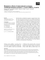
interpretation; we characterized the effects of yet another
cyanine dye, 3,3¢-dipropyl-2,2¢-thiadicarbocyanine iodide
[DiS-C
3
(5); Fig. 1], on mitochondrial structure and function.
Materials and methods
Materials
DiS-C
3
(5) and cyclosporin A (CsA) were kindly provided
by Hayashibara Biochemical Laboratories, Inc. (Okayama,
Japan) and Novartis Pharma Inc. (Tokyo), respectively.
Preparation of mitochondria
Mitochondria were isolated from the liver of normal male
Wistar rats, as described previously [13].
3,43,4
Animals were killed
by cerv ical dislocation to a void the effects o f a nesthetics on
membrane systems. All animal experiments were performed
according to the guidelines for the care and use of laboratory
animals of the University of Tokushima. Protein concentra-
tions of mitochondrial p reparations were determined b y the
Biuret method with bovine serum albumin as a standard.
Measurement of mitochondrial oxygen consumption
and swelling
For measurements of oxygen consumption and turbidity
of mitochondria, mitochondria were suspended in +P
i
medium (250 m
M
sucrose, 10 m
M
K/P
i
5
buffer, pH 7.4) to
make their final protein concentration of 0 .7 mgÆmL
)1
.
Then, they were energized by the addition of either 10 m
M
succinate (plus 0.5 lgÆmg
)1
protein rotenone) or 10 m
M
glutamate and 10 m
M
malate as respiratory substrates.
Rates o f m itochondrial oxygen consumption a t 2 5 °Cwere
measured by use of a Clark oxygen electrode (YSI
5331;Yellow Springs Instrument Co., Yellow Springs, OH,
USA)
6
. When the inhibitory effects of D iS-C
3
(5) on the
mitochondrial oxygen consumption were evaluated, the
protonophoric uncoupler 3,5-di-tert-butyl-4-hydroxy-ben-
zylidene malononitrile (SF6847) was utilized to induce
maximum oxygen consumption. Mitochondrial swelling
was monitored at 25 °C by measuring the t urbidity of the
reaction mixture at 440 nm with a Shimadzu dual-wave-
length spectrophotometer, model UV-3000.
WhentheeffectofP
i
was examined, experiments were
performed using –P
i
medium (200 m
M
sucrose, 10 m
M
KCl,
10 m
M
Tris/Cl buffer; pH 7.4) instead of +P
i
medium.
Measurement of permeability of mitochondrial
membrane to poly(ethylene glycol)
To examine the permeability of the mitochondrial mem-
brane, we measured the effects of poly(ethylene glycol)s of
various molecular sizes on the turbidity of mitochondrial
suspensions, as d escribed by Pfeiffer et al. [14]. Briefly,
mitochondria were first treated with a certain r eagent; and
then, after complete induction of swelling, 1.1 mL of 300
mOsmol solution of poly(ethylene glycol) of a given
molecular size was added. Changes i n the turbidity o f
reaction mixture were monitored at 4 40 nm.
Analysis of mitochondrial configuration by transmission
electron microscopy
Transmission electron microscopy (TEM) a nalysis of mito-
chondria under various conditions was performed, essen-
tially as described p reviously [13], using an Hitachi electron
microscope model H-800MT.
Release of mitochondrial cytochrome
c
To assess whether cytochrome c is released from mitochon-
dria, we t reated mitochondria with DiS-C
3
(5)inanoxygen
chamber at 25 °C as stated above. After certain periods of
incubation, a 500 lL aliquot of the reaction mixture was
taken i nto an E ppendorf tube, a nd the mitochond rial pellet
and supernatant were obtained by prompt centrifugation
7
at
15 000 g for 2 mins at 4 °C. After complete removal of the
supernatant, the mitochondria were resuspended in the
original volume of incubation medium. Two microliters of
mitochondrial suspension and 5 lLofsupernatantwere
subjected to SDS/PAGE and subsequent Western analysis
using a specific antibody against cytochrome c,preparedas
described previously [13].
Results
Effects of DiS-C
3
(5) on the rate of mitochondrial oxygen
consumption
DiS-C
3
(5) was reported to show inhibitory effects on the
mitochondrial NAD-linked respiratory system [15]. A s
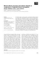
shown in Fig. 2 , we confirmed the inhibitory effect of
DiS-C
3
(5) on the glutamate/malate-driven mitochondrial
electron transfer. Under the experimental conditions used,
its concentration producing 50% inhibition (IC
50
)
8
was
about 8 l
M
. The observed inhibition of the NAD-linked
respiratory system seemed to reflect a direct effect on
complex I and was not attributable to inhibition of the
transport s ystem o f t he respiratory substrate, because
similar e ffects were also obtained when freeze/thawed
mitochondria were used (data not shown).
When succinate was added to mitochondria as the
respiratory substrate, even in the absence of DiS-C
3
(5),
slow oxygen consumption was observed, reflecting oxida-
tion of the respiratory substrate to compensate for the
leakage of H
+
across the inner membrane. Furthermore,
this slow oxygen c onsumption g radually accelerated during
the incubation period, possibly due to the i nduction of the
PT by endogenous Ca
2+
(Fig. 3 , broken line). Upon addi-
tion of DiS-C
3
(5) to the mitochondria energized by succi-
nate, two oppo site actions were observed , depending on the
concentration. The addition of DiS-C
3
(5) £ 10 l
M
caused
deceleration of mitochondrial oxygen consumption but
>20 l
M
caused acceleration. These actions of DiS-C
3
(5) on
Fig. 1. Chemical structur e of DiS-C
3
(5).
3574 T. Yamamoto et al.(Eur. J. Biochem. 271) Ó FEBS 2004
mitochondria energized by succinate were further charac-
terized, as described below in the f ollowing sections.
Characterization of mitochondrial PT induced
by DiS–C
3
(5)
In our previous studies, cyanine dyes such as TriS-C
4
(5)
were found to accelerate mitochondrial oxygen consump-
tion [6–8]. These actions of cyanine dyes were attributable
mainly to the results of induction of the mitochondrial PT
[9,10]. Thus, acceleration of mitochondrial oxygen con-
sumption by DiS-C
3
(5) was expected to be due to the
induction of mitochondrial PT. To validate this interpret-
ation, we further characterized the actions of DiS-C
3
(5)
on the mitochondrial s tructure and function and com-
pared them with those of Ca
2+
, known as a typical PT
inducer.
Like that of Ca
2+
, the addition of 50 l
M
DiS-C
3
(5) to the
mitochondrial suspension caused a massive decrease in its
turbidity, reflecting induction of mitochondrial swelling
(Fig. 4 A). In general, t he induction of mitochondrial
swelling is one of the criteria used to judge whether the
mitochondrial PT is induced. However, as reported previ-
ously, s welling can oc cur e ven under conditions where the
mitochondrial PT doe s not occ ur [13]. Thus, permeability of
the inner mitochondrial membrane was directly evaluated
by measuring the responses of preswollen mitocho ndria to
the addition of poly(ethylene glycol) of various molecular
sizes. A s shown in Fig. 4B, when m itochondria were
Fig. 3. Effects of DiS-C
3
(5) on succinate-driven mitochondrial oxygen
consumption. Effects of DiS-C
3
(5) on the oxygen consumption of
mitochondria energized by succinate were measured. Experiments
were performed as shown in the legend of Fig. 2 except f or use of
succinate ( plus 0.5 lgÆmL
)1
rotenone) as a substrate i nstead of glu -
tamate and malate. Broken line represents the oxygen consumption of
nontreated mitochondria.
Fig. 4. Effects o f DiS-C
3
(5) on the turbidity of mitochondrial suspen-
sions (A) and on permeability of mitochondrial inner membrane (B).
(A)Theeffectof50l
M
DiS-C
3
(5) on the turbidity of mitochondrial
suspensions (right trace) was compared with that of 100 l
M
Ca
2+
(left
trace). Experimental conditions are as those described in the legend for
Fig. 3, and changes in turbidity of mitochondrial suspension were
monitored a t 440 nm. ( B) Permeability of DiS-C
3
(5)-pretreated i nn er
membranes of mitochondria to poly(ethylene glycol) of various
molecular sizes was evaluated. For this, m itochondria w ere fi rst pre-
swollen by C a
2+
(left traces) or by DiS-C
3
(5) (right traces) as stated
above. Then, absorbance changes in the mitochondrial suspensions
that accompanied the addition of solution s of poly(ethylen e glycol) of
various molecular sizes w ere r ecorded at 440 nm. The vertical arrow
indicates the addition of a poly(ethylene glycol) solution. Trace ÔaÕ
represents the result obtained by the addition of medium not con-
taining poly(ethylene glycol), used as a negative control. Traces b–g
represent the results observed with the addition of solutions of
PEG600, PEG1000, PEG2000, PEG4000, PEG6 000 and P EG10000,
respectively.
Fig. 2. Inhibitory effects of DiS-C
3
(5) on NADH-driven electron
transfer. For evaluation o f the inhibitory effect o f DiS-C
3
(5) o n NADH-
driven electron transfer, mitocho ndria were suspended in +P
i
medium
at 25 °C. Then, t h ey were ene rgized by additio n of 10 m
M
glutamate
and 10 m
M
malate (glu/mal) as respiratory substrates and measured
their rates of oxygen consumption. The maximum rate of oxygen con-
sumption was induced by the addition of 50 n
M
SF6847, and this value
was utilized as the noninhibited rate o f o xygen c onsumption. I nhibitory
effects of DiS-C
3
(5) on electron t ran sfer were evaluated by measuring
the rates of oxygen consumption in the presence of both 50 n
M
SF6847
and various amounts of DiS-C
3
(5). Typical t races of oxygraphs are
shown i n (A). Dose–response curve of the effect of DiS-C
3
(5) o n t h e rate
of mitochondrial oxygen consumption is shown in (B), in which
the results are shown as mean values ± SD
12
of thre e in depe ndent runs
(bars of SD are smaller t han t he symbols).
Ó FEBS 2004 Effects of DiS-C on mitochondrial structure and function (Eur. J. Biochem. 271) 3575
preswollen w ith Ca
2+
, t he addition of poly(ethylene glycol)
having a molecular size of more than 4000 (PEG4000)
caused increased turbidity of the m itochondrial suspension,
reflecting induction of shrinkage of preswollen mitochon-
dria; whereas those smaller than 1000 did not, as reported
previously [16]. These results are thought to indicate that the
mitochondrial membrane became permeable to the mole-
cules smaller than a molecular size of 1500 by the Ca
2+
treatment. When poly(ethylene glycol) solutions were added
to the m itochondrial suspensions pretreated with DiS-C
3
(5),
massive shrinkage was not observed, even with PEG6000 or
PEG10000, indicatin g that the m embrane o f the mitochon-
dria treated with DiS-C
3
(5) became permeable to large r
molecules than Ca
2+
-treated mitochondria.
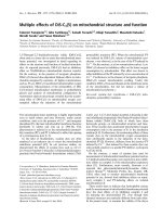
Furthermore, the results of TEM observation also
supported the changes in the permeability of the inner
mitochondrial membrane caused by DiS-C
3
(5). Compared
with the appearance of nontreated control mitochondria
(Fig. 5 A), when mitochondria were treated with Ca
2+
(Fig. 5 B), the mitochondrial inner membrane structure
disappeared s ignificantly, a s reported previously [13,17–19].
Mitochondria treated with DiS-C
3
(5) showed essentially the
same TEM features as those treated with Ca
2+
(Fig. 5C).
These r esults indicate clearly t hat acceleration of mitoch-
ondrial oxygen consumption induced by DiS-C
3
(5)at50l
M
is due to the induction of the mitochondrial PT. However,
as stated above, the membranes of mitochondria treated
with DiS-C
3
(5) became p ermeable to larger molec u les than
the Ca
2+
-treated ones. Furthermore, the increase in the
permeability of the mitochondrial membranes caused by
DiS-C
3
(5) was only partially sensitive for CsA, known as an
inhibitor of the classical PT induced by Ca
2+
(data not
shown). T hus, t he PT induced by DiS-C
3
(5) w as concluded
to be different from that indu ced by Ca
2+
.
The induction of PT is generally believed to b e associated
with the release of apoptogenic mitochondrial p roteins such
as cytochrome c [20]. Thus, we n ext examined whether
mitochondrial cytochrome c would b e released w hen mito-
chondria were treated with DiS-C
3
(5). As shown in Fig. 6,
treatment of mitochondria with 50 l
M
DiS-C
3
(5) caused a
massive release of cytochrome c,aswellaswithCa
2+
.
Inhibition of PT induction by DiS-C
3
(5) at low
concentration
As stated above, DiS-C
3
(5) at low concentrations preven-
ted progression of intrinsic oxygen consumption by
mitochondria. However, this effect of DiS-C
3
(5) did not
reflect inhibition of the mitochondrial respiratory chain, as
the addition of the protonophoric uncoupler SF6847 to
the mitochondrial suspension treated with DiS-C
3
(5) at a
low concentration caused maximum acceleration of mito-
chondrial oxygen consumption as effectively as that
observed with mitochondria not treated with DiS-C
3
(5)
(data not shown). Based on these results, we considered
that the protective effects of DiS-C
3
(5) at low concentra-
tions on the progression of intrinsic oxygen consumption
by mitochondria might reflect induction of the PT by
endogenous Ca
2+
. So next we examined the validity of
this interpretation.
First, we tested the effect of DiS-C
3
(5) o n the Ca
2+
-
induced acceleration of mitochondrial respiration and
mitochondrial swelling. As shown in Fig. 7, when D iS-
C
3
(5) was added to mitochondria pretreated with 10 l
M
Ca
2+
, it prevented not only acceleration of oxygen
consumption (Fig. 7A) but also the turbidity decrease of
mitochondrial suspensions (Fig. 7B), resulting in recovery
to the same level as found for the nontreated control
mitochondria. The protective effects of DiS-C
3
(5)atalow
concentration o n t he spontaneous induction of mitochond-
rial PT was also confirmed by observing mitochondria by
TEM (Fig. 8). The disappearance of the inner mitochond-
rial membrane structure induced by 10 l
M
Ca
2+
was
strongly suppressed by treatment of the m itochon dria with
5 l
M
DiS-C
3
(5).
Thus, we concluded that the inhibitory effect of DiS-
C
3
(5) on the spontaneous acceleration of mitochondrial
Fig. 5. TEM app earances of mitochondria
treated with D iS-C
3
(5). Mitochondria were
treated with Ca
2+
or DiS-C
3
(5) as described in
thelegendinFig.4andsubjectedtoTEM
analysis. ( A) The appearance of n ontreated
control mitochondria. (B) and (C) The
appearances o f mitochondria t reated with
100 l
M
Ca
2+
and 50 l
M
DiS-C
3
(5), respect-
ively.Barunder(C)indicates1lmforall
panels.
13
Fig. 6. Effects of DiS-C
3
(5) on the release of mitochondrial cyto-
chrome c. Release of mitoch ondr ial c yto chr ome c was examined as
describedinMaterialsandmethods. Briefly, mitochondria were first
treated w ith 50 l
M
DiS-C
3
(5), then they were precipitated by centrif-
ugation. Samples of p ellet ( P) and supernatant (S) were subj ected to
Western blotting using specific antibody against cytochrome c.Sam-
ples of nontreated mitochondria or of Ca
2+
-treated mitochondria
were also analyzed as controls. Typical results of more than three
independent experiments are s hown.
3576 T. Yamamoto et al.(Eur. J. Biochem. 271) Ó FEBS 2004
oxygen consumption could b e a ttributable to the i nhibition
of spontaneous induction of the PT by endogenous Ca
2+
.
However, it should be noted that this inhibitory effect of
DiS-C
3
(5) was only observed when the PT was i nduced by a
relatively low concentration C a
2+
such as 10 l
M
; i.e. it was
not observed at a concentration of Ca
2+
such as 50 l
M
(data not shown). Furthermore, the protective effect of
DiS-C
3
(5) on t he Ca
2+
-induced PT was not attributable to
the inhibition of C a
2+
uptake (data not shown).
Effects of DiS-C
3
(5) on mitochondria in the absence of P
i
All of the above experiments were performed in +P
i
medium con taining 10 m
M
phosphate buffer. However, the
PT-inducing effects of Ca
2+
and cyanine dyes such as
Tri-S-C
4
(5) are known to be dependent on the absence/
presence of P
i
in the incubation medium. Thus, it was of
interest to us to examine the effects of DiS-C
3
(5) on
mitochondrial structure and function in the absence of P
i
.
Where Ca
2+
had no effect on the m itochondrial oxygen
9
consumption in the absence o f P
i
,DiS-C
3
(5) moderately
accelerated mitochondrial oxygen consumption e ven in t he
absence of P
i
(Fig.9A).Furthermore,50l
M
DiS-C
3
(5)
caused massive swelling even in the absence of P
i
(Fig. 9B).
To examine whether the observed mitochondrial swelling
induced by DiS-C
3
(5) in this case was attributable to the
induction of the m itochondrial PT, we examined the
permeability o f D iS-C
3
(5)-treated mitochondrial mem-
branes to poly(ethylene glycol).
10
Unfortunately, no c lear
conclusion could be obtained, as mito chondria preswollen
by DiS-C
3
(5) did not show any clear response upon the
addition of poly(ethylene glycol) (data not shown).
However, mitochondria treated with 50 l
M
DiS-C
3
(5)
showed morphology d ifferent from that of the nontreated
control ( Fig. 10), strongly suggesting alteration of mem-
brane status. Their appearance was also apparently
different from that of mitochondria treated with Ca
2+
or DiS-C
3
(5) i n the presence of P
i
(Fig. 5 B,C). Finally, we
also tested whether the release of mitochondrial cyto-
chrome c could be induced by DiS-C
3
(5) in the absence
of P
i
. As shown in Fig. 11, when mitochondria were
incubated with 50 l
M
DiS-C
3
(5)intheabsenceofP
i
,
most of the cytochrome c was retained in these mito-
chondria as well as in the nontreated mitochondria.
Discussion
Cyanine dyes a re often used to evaluate the mitochondrial
membrane potential [4,5]. However, as reported p reviously,
some of them are also reported to show in hibitory effects on
NAD-linked electron transfer [11,12] and Ca
2+
-like
uncoupling actions [9,12]. DiS-C
3
(5) is often used for
measurements of mitochondrial membrane potential, and
its methods of interaction with mitochondria have been
Fig. 7. Inhibitory e ffects of a low concentration
DiS-C
3
(5) on the Ca
2+
-induced P T. To
examine the effects of DiS-C
3
(5) on the Ca
2+
-
induced PT, we measured its effects o n the
oxygen consumption of mitochondria (A) and
turbidity change in mitochondrial suspensions
(B) treated with 10 l
M
Ca
2+
in +P
i
medium.
Results obtained without the a ddition of Ca
2+
and DiS-C
3
(5) are shown by broken lines
(controls).
Fig. 8. TEM analysis of mitochondria treated with a l ow concentration
of DiS-C
3
(5). To examine the prote ctive effect o f DiS-C
3
(5) on the
Ca
2+
-induced PT, we also observed the electron microscopic appear-
ances of mitochondria. (A) and (B) show the appearance of mito-
chondria treated with 10 l
M
Ca
2+
andwithboth5l
M
DiS-C
3
(5) and
10 l
M
Ca
2+
, respectively. B ar under (B) indicates 1 lmforallpanels.
Fig. 9. Effects of DiS-C
3
(5) on the rate of mitochondrial oxygen con-
sumption (A) and turbidity of m itochondrial suspensions (B) in t he ab-
sence of P
i
. Experiment s were performed as described in the legends for
Figs 3and4exceptthat–P
i
medium was used inste ad of +P
i
medium.
Ó FEBS 2004 Effects of DiS-C on mitochondrial structure and function (Eur. J. Biochem. 271) 3577
studied [15,21]. H owever, characterization of its effects with
respect to PT induction had not been achieved earlier. Thus,
in the present study, we investigated in great detail the
actions of DiS-C
3
(5) on the structure and function of
isolated mitochondria.
First, we confirmed the previously reported inhibitory
effects of DiS-C
3
(5) on NAD-linked electron transfer. Th is
inhibitory effect was con sidered to reflect its direct action on
complex I. On the contrary, when DiS-C
3
(5) was added to
the mitochondria energized by succinate, both acceleration
and deceleration of oxygen consumption were observed,
depending on the concentration of the dye. At higher
concentrations such as 50 l
M
,DiS-C
3
(5) c aused acceler-
ation of mitochondrial oxygen consumption. This effect of
DiS-C
3
(5) was further characterized and concluded to be
attributable to the induction of the mitochondrial PT.
PT induced by DiS-C
3
(5) was associated with release of
cytochrome c, as was that induced by Ca
2+
. However, it
was d ifferent from the ordinary P T i nduced by Ca
2+
in the
aspects of pore size and sensitivity for CsA, known as a
specific inhibitor of the ordinary PT. P ossibly, these
differences may reflect the differe nces in the features of the
proteinaceous PT pores formed.
Cytochrome c is one of the components comprising the
respiratory chain. Thus, r elease of cytochrome c from
mitochondria would be expected to cause deceleration of
mitochondrial o xygen consumption. However, as seen with
the e ffects of 50 l
M
DiS-C
3
(5), this was not the case. Release
of cytochrome c without causing deceleration of mito-
chondrial oxygen consumption was also observed when
mitochondria were treated with Ca
2+
or valinomycin [13].
However, under these conditions, at least half of the total
cytochrome c still remained in the mitochondria. Possibly,
this cytochrome c remaining in the mitochondria was
sufficient to account for t he electron t ransfer. Further-
more, for the release of cytochrome c, permeability of
the outer mitochondrial membrane to cytochrome c must
be increased, as cytochrome c is present in the intermem-
brane s pace o f mitochondria. Several mechanisms concern-
ing the release process of cytochrome c have been proposed,
but this problem is still under debate.
Until now, there was n o detailed study on the PT-indu-
cing effects of c hemicals actually used as a t racer dye of t he
mitochondrial membrane potential except f or that on
Mitotracker Orange
TM
[12]. Possibly, induction of the PT
is one of the common a ctions of hydrophobic cations that
are u tilized as a tracer of mitochondrial membrane poten-
tial, as s imilar activities w ere observed with these c hemicals
regardless their s tructural diversity [9,12]. Further studies on
the actions of a series of hydrophobic cations will be
necessary for validation of this interpretation a nd for b etter
understanding of the features of the mitochondrial P T.
Furthermore, we observed two additional novel effects of
DiS-C
3
(5) on mitochondria: (i) inhibition of the Ca
2+
-
induced PT by a low concentration of DiS-C
3
(5) and (ii)
induction of swelling in the absence of P
i
.Withrespectto
the former feature, attention must be paid to it when this
dye is employed as a tracer for mitochondrial membrane
potential, a s it shows a protective e ffect on the induction of
PT at the concentration utilized for monitoring membrane
potentials. For the latter action, DiS-C
3
(5) caused r emark-
able swelling a nd changes in t he status of the m itochondrial
inner membrane without accompanying release o f mitoch-
ondrial c ytochrome c (Figs 9,10,11). F urther studies on the
status of the inner membrane of m itochondria treated with
DiS-C
3
(5) in the absence of P
i
may give u s insight into
the mechanisms causing configurational changes in mito-
chondria.
Until now, both CsA-sensitive and insensitive PT have
been shown to be a ssociated with the release of mitochond-
rial cytochrome c. Recently, however, we reported that
mitochondrial cytochrome c could be released even w ithout
the induction of the m itochondrial PT [13]; and this
observation was supported by another group [22]. Thus,
detailed studies on the relationship between PT induction
and release of mitochondrial cytochrome c remain to be
conducted.
In conclusion, we found DiS-C
3
(5) to show multiple
effects on the mitochondrial structure and function, effects
dependent on both its concentration and the P
i
status.
11
Acknowledgements
This work was supported by grants-in-aid for scientific research
(no. 14370746 to Y.S.) from t he Ministry of Education, Science, and
Culture of Japan, and a fellowship from Katayama Chemical
Industries, Co., Ltd (O sa ka) to T.Y.
Fig. 10. TEM appearance of mitochondria in the absence of P
i
. The
effects of DiS-C
3
(5) on the mitochondrial morphology in –P
i
medium
were also exa mined by TEM analysis. (A) and (B) s how t he appear-
ance of mitochondria incub ated in the absence an d presence of 50 l
M
DiS-C
3
(5), respective ly. Bar under ( B ) indicates 1 lm for all panels.
Fig. 11 . Effects of DiS-C
3
(5) on the allocation of mitochondrial
cytochrome c in the absence of P
i
. Release of mitochondrial cyto-
chrome c was examined as described in the legend for Fig. 6. In
addition to the samples of pellet (P) and supernatant (S) of mito-
chondria treated with 50 l
M
DiS-C
3
(5), those of nontreated mito-
chondria wer e also analyzed.
3578 T. Yamamoto et al.(Eur. J. Biochem. 271) Ó FEBS 2004
References
1. Gunter, T.E. & Pfeiffer, D.R. (1990) Mechanisms by which
mitochondria transport calcium. Am.J.Physiol.258, C755–C786.
2. Zo ratti, M . & Szabo, I. (1995) The mitochondrial pe rmeability
transition. Biochim. Biophys. Acta 12 41, 139–176.
3. Bernardi, P. ( 1999) Mitochondrial transport o f cations: c hannels,
exchangers, and permeability transition. Physiol. Rev. 79, 1127–
1155.
4. Rottenberg, H . (1979) The m easurement of membrane pote ntial
and delta pH in cells, organelles and vesicles. Methods Enzymol.
55, 547–569.
5. Waggoner, A.S. (1979) The use of cyanine dyes for the deter-
mination of membrane potentials in cells, organelles and vesi cles.
Methods En zymol. 55, 689–695.
6. Terada, H., Nagamune, H., Osaki, Y. & Yoshikawa, K. (1981)
Specific requirement for inorganic phosphate for induction of
bilayer m embrane conductan ce by the cationi c u nc oupler c arbo -
cyanine dye. Biochim. Bio phys. Acta 64 6, 488–490.
7. Terada, H . & Nagamune, H. ( 1983) A cyanine dye tri-S-C
7
(5).
Phosphate-dependent c ationic uncoupler of o xidative phosphory-
lation in mitochondria. Bioch im. Biophys. Acta 723, 7–1 5.
8. Terada, H., Nagamune, H., Morikawa, N. & Ikuno, M. (1985)
Uncoupling of oxidative phosphorylation by divalent cationic
cyanine dye. Participation of phosphate t ransporter. Biochim.
Biophys. A cta 807, 168 –176.
9. Sh inohara, Y., Bandou, S., Kora, S., Kitamura, S., Ina zumi, S . &
Terada, H. (1998) Cationic uncouplers of oxidative phosphory-
lation are inducers of mitochondrial permeability transition.
FEBS Lett. 428, 89–92.
10. Yamashita, K., Ichikawa, T., Yamamoto, T., Kataoka, M.,
Nakagawa, Y., Terada, H. & Shinohara, Y. (2003) Three-way
effect o f cyanine dye o n the structure and fun ctio n of mitochon-
dria. J. Health Sci. 49 , 448–453.
11. Conover, T.E. & Schneider, R.F. (1981) Interaction of certain
cationic dyes with the respiratory chain of rat liver mitochondria.
J. Biol. Chem. 256, 402 –408.
12. Sc orrano, L ., Petronilli, V., Colo nna, R ., Di Lisa, F. & Bernardi, P.
(1999) Chloromethyltetramethylrosamine (Mitotracker Orange)
induces the mitochondrial permeability transition and inhibits
respiratory complex I. Implications for the mechanism of cyto-
chrome c release. J. Biol. Chem. 274, 24657–24663.
13. Shinohara,Y.,Almofti,M.R.,Yamamoto,T.,Ishida,T.,Kita,F.,
Kanzaki, H., Ohnishi, M., Yamashita, K. , Shimizu, S. & Terada,
H. (2002) Permeability transition-independent release of mito-
chondrial c ytochrome c induced by valinomycin. Eur. J. Biochem.
269, 5 224–5230.
14. Pfeiffer, D.R., Gudz, T.I., Novgorodov, S.A. & Erdahl, W.L.
(1995) The peptide mastoparan is a potent facilitator of the
mitochondrial permeability transition. J. Biol. C hem. 270, 4923–
4932.
15. Okimasu, E., Akiyama, J., Shiraishi, N. & Utsumi, K. ( 1979) The
mechanism of inhibition on the endoge nous respiration of Ehrlich
ascites tumor c ells by the cyanine dye diS-C
3
(5). Physiol. Chem.
Phys. 11 , 425–433.
16. Su ltan, A. & Sokolove, P.M. (2001) Palmitic acid opens a novel
cyclosporin A-insensitive pore in the inner mitochondrial mem-
brane. Arch. Biochem. B iophys. 386, 37–5 1.
17. Beatrice, M.C., Stiers, D.L. & Pfeiffer, D.R. (1982) Increased
permeability of mitochondria during Ca
2+
release induced by
t-butyl hydroperoxide or oxalacetate. the effect of ruthenium red.
J. Bi ol. Chem. 257, 7161–7171.
18. Petronilli, V ., Cola, C., Massari, S., Colonna, R . & Bernardi, P.
(1993) Physiological e ffectors modify voltage sensing by the
cyclosporin A-sensitive permeability transition pore of mito-
chondria. J. Bio l. Chem. 268, 21939–21945.
19. Jung, D.W., Bradshaw, P.C. & Pfeiffer, D.R. (1997) Properties of
a cyclosporin-insensitive permeability transition pore i n y east
mitochondria. J. B iol. Chem. 272, 211 04–21112.
20. Scarlett, J.L. & Murphy, M.P. (1997) Release of apoptogenic
proteins from the mitochondrial intermembrane space during
the mitochondrial permeability tran sition. FEBS Lett. 41 8 , 282–
286.
21. Bammel, B.P., Bra nd, J.A ., Germon, W. & Smith, J .C.
(1986) Interaction of the extrinsic potential-sensitive molecular
probe diS-C3-(5) with pigeon heart mitochondria under equili-
brium and time-resolved conditions. Arch. Biochem. Bio phys. 244,
67–84.
22. Go gvadze, V., Robertson, J.D., Enoksson, M., Zhivotovsky, B. &
Orrenius, S. (2004) Mitochondrial cytochrome c release may occur
by volume-dependent mec hanisms not involving permeability
transition. Biochem. J. 378, 213 –217.
Ó FEBS 2004 Effects of DiS-C on mitochondrial structure and function (Eur. J. Biochem. 271) 3579