Báo cáo khoa học: Effects of cardiomyopathic mutations on the biochemical and biophysical properties of the human a-tropomyosin docx
Bạn đang xem bản rút gọn của tài liệu. Xem và tải ngay bản đầy đủ của tài liệu tại đây (396 KB, 9 trang )
Effects of cardiomyopathic mutations on the biochemical
and biophysical properties of the human a-tropomyosin
Eduardo Hilario
1
, Silvia L. F. da Silva
2
, Carlos H. I. Ramos
2
and Maria Ce
´
lia Bertolini
1
1
Instituto de Quı
´
mica, UNESP, Departamento de Bioquı
´
mica e Tecnologia Quı
´
mica, Araraquara, Sa
˜
o Paulo, Brazil;
2
Centro de Biologia Molecular Estrutural, Laborato
´
rio Nacional de Luz Sı
´
ncrotron, Campinas, Sa
˜
o Paulo, Brazil
Mutations in the protein a-tropomyosin (Tm) can cause a
disease known as familial hypertrophic cardiomyopathy. In
order to understand how such mutations lead to protein
dysfunction, three point mutations were introduced into
cDNA encoding the human skeletal tropomyosin, and the
recombinant Tms were produced at high levels in the yeast
Pichia pastoris. Two mutations (A63V a nd K70T) were
located in the N-terminal region of Tm and one (E180G) was
located close to the calcium-dependent troponin T binding
domain. The functional and structural properties of the
mutant Tms were compared to those of the wild type pro-
tein. None of the mutations altered the head-to-tail poly-
merization, although slightly higher actin binding was
observed in the mutant Tm K70T, as demonstrated in a
cosedimentation assay. The mutations also did not change
the cooperativity of the thin filament activation by increasing
the concentrations of Ca
2+
. However, in the absence of
troponin, all mutant Tms were less effective than the wild
type in regulating the actomyosin subfragment 1 Mg
2+
ATPase activity. Circular dichroism spectroscopy revealed
no differences in the secondary structure of the Tms. How-
ever, t he thermally induced unfold ing, as m onitored by
circular dichroism or differential scanning calorimetry,
demonstrated that the muta nts were less stable th an the w ild
type. These results indicate that the main effect of the
mutations is related t o t he overall stability of T m a s a whole,
and t hat the mutations have only m inor effects on the
cooperative interactions among proteins t hat c onstitute the
thin filament.
Keywords: circular dichroism; differential scanning calori-
metry; Pichia pastoris; tropomyosin.
Tropomyosins (Tms) are a family of highly conserved
proteins found in most eukaryotic cells. The striated muscle
isoform is an a-helical protein, which forms a parallel
coiled-coil dimer twisted around t he long axis of the actin
filament. Each polypeptide chain has 284 amino acid
residues, and each dimer binds to seven actin monomers
and one troponin (Tn) complex (TnC, TnI and TnT). In
striated muscle cells the Tm polymerizes in a he ad-to-tail
fashion, and together w ith the troponin c omplex, regulates
the Ca
2+
sensitivity of the actomyosin Mg
2+
ATPase
complex [ 1]. T he Tm amino acid sequence shows a seven-
residue pattern (a to g ) r epeated t hroughout the entire
sequence. Positions a and d, on the same side of the helices,
are usually occupied by apolar amino acids that allow
hydrophobic i nteractions between chains. Positions e and g
are often occupied by charged residues, and therefore
contribute to the s tabilization of the parallel coiled-coil
structure b y ionic interactions with residues a t positions e¢
and g¢ of the other helix. P ositions b, c and f are occupied by
polar or ionic residues a nd they interact with solvent o r
other proteins [1]. In addition to the heptapeptide repeat,
there are seven consecutive repetitions of approximately 40
residues each in the entire length of the chain, which
correspond to the a ctin binding sites [2].
Recombinant Tms have been produced in different host
cells and the proteins used as tools to obtain information
about the r elevant regions for functional and structural
properties. The recombinant Tm was first produced in
Escherichia coli but the protein was not N-acetylated [3],
and therefore, lacked t he functional properties that depen-
ded o n this m odification. Fully functional Tm w as produced
in E. coli by changing the primary structure of the protein
with the addition of a dipeptide or a t ripeptide a t the
N-terminal methionine [4]. Our group has successfully
shown that Pich ia pa storis and Saccharomyces cerevisiae
are capable of producing functional Tms unmodified in
their p rimary structure [5,6]. The proteins are p robably
N-acetylated, their N-terminal methionine is blocked, and
they behave iden tically to the native Tm in their functional
properties, thus making them preferable for structure–
function studies to probe amino acid mutations that have
been described in c ardiomyopathic tropomyosins.
Familial hypertrophic cardiomyopathy (FHC) is a clin-
ically and genetically heterogeneous heart disease charac-
terized b y hypertrophy and ventricular dysfunction [7]. The
incidence o f the disease is h igh [ 8], and u p to the present date
numerous mutations within the genes encoding for t he
sarcomeric cardiac proteins a-tropomyosin, troponin T, and
Correspondence to M. C . Be rtolini, Instituto d e Q uı
´
mica, UNESP,
Departamento de Bioquı
´
mica e Tecnologia Quı
´
mica, R. Professor
Francisco Degni, s/n, 14800-900, Araraqua ra, S a
˜
o Paulo, Brazil.
Fax: +55 16 222 7932, Tel.: +55 16 201 6675,
E-mail:
Abbreviations: FHC, familial h ypertrophic cardiomyopathy; S1,
myosin subfragment 1; T
m
, temperature of the midpoint of the thermal
unfolding transition; Tm, tropomyosin; Tn, troponin.
(Received 9 J uly 2004, revised 2 0 August 2004,
accepted 31 Aug ust 2004)
Eur. J. Biochem. 271, 4132–4140 (2004) Ó FEBS 2004 doi:10.1111/j.1432-1033.2004.04351.x
myosin heavy-chain have been reported. The frequency of
mutation in the a-tropomyosin gene (TPM1)islower,
accounting for approximately 5% of FHC, however,
different point mutations leading to mutant proteins have
been described i n the last few years: E62Q [9], A63V [10,11],
K70T [10], D 175N [ 12] E180G [12], E180V [13 ], L 185R [ 14].
Mutations occur mainly in two regions of the protein, one
located in the N-terminal domain a nd the other close to the
troponin-binding region of tro pomyosin.
Several studies based on t he cardiomyopathic mutations
D175N and E 180G have been reported. In vivo studies,
using t ransgenic mice as a model showed an impairment of
cardiac function by altering the s ensitivity of myofilaments
to Ca
2+
[15]. In vitro studies, w ith recombinant proteins
carrying the mutations, demonstrated small effects on the
overall stability of the protein as meas ured by circ ular
dichroism [ 16], and s howed alterations in the k inetics of
contractile force g eneration [17]. Studies with m utations
A63V and K70T reported higher muscle Ca
2+
sensitivity
both in viv o [18],and,morerecently,in vitro [19], in addition
to prominent effects on the Tm thermal stability as
monitored by circular d ichroism [19].
In the present study, w e combined the biophysical assays –
circular dichroism and differential scanning calorimetry –
and recombinant human Tm produced in P. pastoris,to
investigate the effects o f cardiomyopathic-related m utations
on the human skeletal Tm. Our data indicate that the main
effects of mutations A63V, K70T and E180G are mainly
related to the overall stability of the protein as a whole, rather
than on the position of the mutation in the polypeptide chain,
as demonstrated by the biophysical assays. Our studies h ave
provided additional contributions to the understanding of
the effects of these mutations on the clinical symptoms of
patients carrying cardiomyopathic Tms.
Experimental procedures
Construction of expression plasmids and site-directed
mutagenesis
The pPIC9 expression vector and P. pastoris strain GS115
(his4) (Invitrogen, Life Technologies) were used for Tm
production. Oligonucleotides were designed based on the
sequence of human skeletal muscle cD NA (ska-TM.1) [20].
The full length coding sequence was amplified by PCR w ith
the oligonucleotides Tm-7F (5¢-CG
GGATCCACCATGG
ATGCCATCAAG-3¢)andTm-9R(5¢-ATAAGAAT
GCG
GCCGCTTATATGGAAGTCAT-3¢). The underlined
sequences correspond to BamHI and NotI sites, r espectively.
The oligonucleotide Tm-7F contains an ACC sequence
(shown in bold) immediately upstream of the start codon
[21]. The amplified cDNA was digeste d with Bam HI and
NotI, and subcloned into t he same sites o f v ector to produce
the PIC9-WT expression plasmid.
DNA sequences encoding A63V, K70T and E180G
mutant Tms were amplified by PCR in t wo steps using
standard procedures [22]. The oligonucleotides AOX-F
(5¢-GCGACTGGTTCCAATTGAC-3¢), AOX-R (5¢-GG
TCTTCTCGTAAGTGCCC-3¢), SKTM-A63V (5¢-GAC
AAATACTCTGA
AGTACTCAAAGATGCCCAG-3¢), SK
TM-1R (5¢-CTGGGCATCTTTGAGTAC
TTCAGAGTA
TTGTC-3¢), SKTM-K70T (5¢-AAAGATGC
ACAGGAG
ACGCTGGAGCTGGCAGAG-3¢), SKTM-2R (5¢- CTCTG
CCAGCTCCAGCGTCTCCTG
TGCATCTTT-3¢), SKTM-
E180G (5 ¢-CTGGAACGTGCAG
GGGAGCGGGCTGAA
CTCTCAGAAGG-3¢) and SKTM-4R (5¢-CCTTCTGA
GAGTTCAGCCCGCTCC
CCTGCACGTTCCAG-3¢)were
used for the amplifications. To perform A63V, K70T
and E180G point mutations (underlined in the primer
sequences), two DNA fragments of each mutation were
initially amplified using, respectively, the primers AOX-F/
SKTM-A63V and AOX-R/SKTM-1R, AOX-F/SKTM-
K70T and A OX-R/SKTM-2R, and AOX-F/SKTM-
E180G and AOX-R/SKTM-4R. The entire cDNA
sequences containing the mutations were amplified with
the AOX-F and AOX-R primers, digested with BamHI and
NotI and subcloned into pPIC9 vector leading to P IC9-
A63V, PIC9-K70T, and PI C9-E180G expression plasmids.
The E. coli strain MC1061 [23] was used for plasmid
amplification. The complete c DNA se quences were con-
firmed by automatic DNA sequencing.
Production and purification of recombinant proteins
Expression plasmids were linearized with BglII, and used to
transform competent GS115 cells by electroporation. Cells
were also transformed with linearized pPIC9 plasmid not
carrying the cDNA. His
+
transformants were selected on
minimal medium agar plates containing 0.4% (w/v) yeast
nitrogen base without amino acids, 1% (w/v) ammonium
sulfate, 4 · 10
)5
% (w/v) biotin and 1% (w/v) glucose.
Production and purification of recombinant Tms was
performed as described previously [5]. After purification
the p roteins w ere analyzed by SDS/PAGE [24], and the
purified Tms were l yophilized for future a nalysis.
Purification of muscle proteins
Muscular actin was purified from acetone powder of
chicken pectoralis major and minor muscles [25]. Tn
complex was assembled [26] a fter purification of re combin-
ant TnC [27], TnT [28], and TnI [29] produced in E. coli
(1 L in 4 L flasks). Proper stoichiometry after assembling
was verified by SDS/PAGE. Chicken muscle myosin
subunit S1 was prepared from fresh hearts, according to
Margossian & L owey [30]. The myosin (S1) and troponin
concentrations were determ ined using t he following extinc-
tion coefficients (0.1% solution): E
280
¼ 0.79 for S1
(115 kDa); E
259
¼ 0.137 for TnC (18 kDa); E
280
¼ 0.623
for TnT (31 kDa) ; E
280
¼ 0.497 for TnI (21 kDa). The
tropomyosin and actin concentrations were determined [31]
using bovine serum albumin as a standard.
Functional assays
Viscosity m easurements w ere c arried out at room tempera-
ture using a Cannon–Manning semimicroviscometer (A50).
The affinity of Tm to actin in the presence of Tn was carried
out by cosedimentation in a Beckman model LE-80K
ultracentrifuge (Beckman), a nd analyzed by SDS/PAG E.
The actomyosin S1 Mg
2+
ATPase was determined in
the absence of troponin as a function of tropomyosin
concentration, and in the presence of troponin and Ca
2+
concentration varying from 10
)6
to 10
)3
M
. Inorganic
Ó FEBS 2004 Cardiomyopathic mutations on human tropomyosin (Eur. J. Biochem. 271) 4133
phosphate was determined colorimetrically according to
Heinonen & Lahti [32]. All assays were carried out
according to Monteiro et al. [4], and conditions are
described in the figur e legends.
Circular dichroism (CD)
CD measurements were recorded on a Jasco J-810 spectro-
polarimeter with the temperature controlled by a Peltier-
type Control System PFD 425S using a 10 mm path length
cuvette. The Tm c oncentration varied from 1 l
M
to 16 l
M
in 10 m
M
sodium phosphate buffer, pH 7.0, containing
200 m
M
NaCl. T he data were collected fro m 260 nm to
195 nm, and accumulated 10 times, for spectral measure-
ments, and at 222 nm for stability measurements. The
average of at least three unfolding experiments was used to
construct each curve profile. The value of T
m
,which
corresponded t o the midpoint of the thermal transition
unfolding, was determined from the derivative of the
transition curve. Curve fi tting was performed u sing
ORIGIN
(Microcal Softw are).
Differential scanning calorimetry (DSC)
The microcalorimetric study of Tm denaturation was
performed using a scanning microcalorimeter MicroCal
Ultrasensitive VP-DSC and standard software for data
acquisition and analysis. Tm concentrations were of 15 l
M
in 10 m
M
sodium phosphate buffer, pH 7.0, containing
100 m
M
NaCl and 1 m
M
dithiothreitol. Protein samples
were dialyzed against t he same buffer during 12 h and
degassed f or 30 min before loading into the calorimeter.
Runs were performed with heating/cooling rates of 30, 60
and 90 °CÆh
)1
with no observable c hange between them,
and the process was consid ered to be in equilibrium. The
unfolding was more than 95% reversible and the scan rate
independent. The data obtained were subtracted from a
baseline o f buffer against buf fer, corrected f or concentration
and fitted using
ORIGIN DSC ANA LYSIS
(MicroCal) .
Results
a-Tropomyosin production in
Pichia pastoris
We have previously demonstrated that recombinant c hicken
muscle Tm produced in the yeast P. pastoris had similar
functional properties when c ompared to the native muscle
protein [ 5]. A recombinant human Tm produced in this
organism could therefore be a g ood model for probing
amino acid mutations described in cardiomyopathic Tms.
The m utations A63V, K 70T and E180G were introduced by
PCR in the cDNA encoding the human skel etal mus cle T m,
skaTM [20], and the mutations were confirmed by DNA
sequencing. Expression plasmids carrying mutant ( PIC9-
A63V, PIC9-K70T, and PIC9-E180G) and nonmutant
(PIC9-Tm) cDNAs were used to transform yeast cells, and
recombinant c lones expressing t he proteins were utilized in a
large-scale production. Wild type and mutant T ms were
produced in yeast at high levels after methanol induction
(ranging from 20 to 30 mgÆL
)1
), and the recombinant
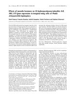
proteins purified to homogeneity. F igure 1 shows samples
of each protein after purification. Recombinant Tms
migrated with an apparent molecular mass of 36 kDa and
slightly slower migration was observed for the mutant
K70T. Mutations A63V and K70T are located at the
N-terminal region of the protein and mutation E180G is
localized near to the region where troponin interacts with
Tm (Cys190, extending to the C-terminal region). Pure
recombinant Tms containing point mutations were utilized
to evaluate the contribution of the mutant amino acids to
the Tm properties.
Functional properties of mutant tropomyosins
Recombinant Tms were assa yed by s tructural (head- to-tail
polymerization and binding to actin) and regulatory (regu-
lation of myosin S1 Mg
2+
ATPase activity) properties.
Chicken muscle p roteins [native actin and myosin (S1), and
recombinant t roponins] were used in our experiments a s
they have previously been well characterized in these assays .
Polymerization ability of T ms was analyzed by viscosity as a
function of the salt concentration. All Tms exhibited
maximal viscosity in the absence of salt and lowering
viscosity as the salt concentration increased (Fig. 2). No
difference in polymerization was observed among the
mutant Tms and between mutants and wild type Tm. In
the thin filament Tm polymerizes head-to-tail, and poly-
merization depends on the formation of a complex between
amino acid residues (at least nine) at the N-terminal end of
one Tm and residues at t he C-terminal end of a second
molecule. Mutations along the polypeptide chain, far from
the complex region in volved in the polymerization w ere
not expected to have any influence on the protein
polymerization.
Recombinant T ms were assayed by their ability to bind to
actin, in a cosedimentation assay, in the absence and in t he
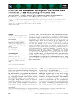
presence of troponins. In the absence of troponins, binding
of Tms to actin was very weak and only small amounts of
Tm wer e detected in gels after centrifugation (data not
shown). The addition of troponins to the reaction mixture
increased the capacity of Tms to bind to actin (Fig. 3, lanes
3, 6, 9, and 12), and only minor differences in binding
capacity among the Tms were observed. A slightly stronger
binding capacity, compared to the wild type Tm was
observed i n t he K70T mutant because no Tm was visualized
12 345
45
31
21
66
Tm
Fig. 1. Gel analysis of Tms. SDS/PAGE (12%) of pure Tms. Ten
micrograms of protein were loaded in each well. Lane 1, molecular
mass marker (kDa); lane 2, wild type Tm; l ane 3, m utant Tm A63V;
lane 4, mutant Tm K70T; and lane 5, mutant Tm E180G.
4134 E. Hilario et al.(Eur. J. Biochem. 271) Ó FEBS 2004
in the s upernatant after centrifugation ( Fig. 3, lane 8). The
fact that the actin and troponin proteins u tilized in this assay
were from chicken s hould be considered. Slight changes i n
the overall structure of the mutant Tms could not be
detected mainly due to the fact that proteins from different
organisms were utilized in the assay.
Mutant Tms were c ompared to the wild type Tm in their
ability to regulate the actomyosin S1 Mg
2+
ATPase activity.
ATPase activity was first assayed by varying the concen-
tration of Tm in the presence of constant concentration of
F-actin and myosin S1. In this condition, Tm inh ibits the
ATPase activity as its concentration increases [33]. F igure 4
shows t hat all mutant Tms were able to inhibit the ATPase
activity as the Tm concentration increased, however, they
were less effective than the wild type protein . Maximum
inhibition ( 50%) was observed at the concentration of
1.5 l
M
(a-Tm/actin r atio of 1 : 5) for the wild type Tm, a nd
2.0 l
M
(ratio of 1 : 3.5) for the mutant Tms. In addition,
comparison of m utants s howed that the E180G mutant w as
a more effective inhibititor than the K70T mutant. B ecause
the salt c oncentration used i n t his assay w as very low
(40 m
M
KCl), i t is supposed that all Tms were partially
polymerized and thus, the differences observed were due to
the mutations.
Mutant Tms were also evaluated for alterations in
Ca
2+
sensitive regulation o f actomyosin S1 Mg
2+
ATPase
activity in the presence of troponins. I n this condition, the
tropomyosin–troponin complex inhibits or activates the
actomyosin ATPase in the absence and in the presence of
calcium, respectively. All mutant T ms were able to regulate
theATPaseactivitybyCa
2+
, and the regulation was
cooperative for all Tms (Fig. 5). N o differences between
wild type and mutant Tms were observed. Maximum
activation was achieved at pCa ¼ 3.5, and the calcium
concentration where the activation was 50%, was close to
10
)4
M
(pCa ¼ 4.0) for all Tms. Both pCa v alues a re higher
than those obtained when recombinant chicken Tm was
assayed [5,6]. The difference between the present results and
those p reviously reported [5,6] may reflect the different
sources of proteins used in the present study to reconstitute
the thin filament in vitro.
Biophysical properties of mutant tropomyosins
The effect of the mutations on the overall stability of the
proteins was evaluated by circular d ichroism (CD) and
differential scanning calorimetry (DSC). The CD spectra of
Tms were typical of folded proteins, with no notable
difference among them, and were independent of concen-
tration f rom 2 l
M
to 1 6 l
M
(data n ot shown). The ellipticity
at 222 n m showed that the mutations did not cause any
severe loss of secondary structure (Table 1). The thermal-
induced unfolding of wild type Tm monitored b y CD is
shown in Fig. 6A. The actual melting temperatures were
determined from derivative plots of the melting curves of
wild type and mutant Tms (Fig . 6B). Two tra nsitions were
12345 6789 101112
MS PMS PMS P PMS
WT Ala63Val Lys70Thr Glu180Gly
Actin
Tm
Tn-T
Tn-I
Tn-C
Fig. 3. Actin-binding o f w ild type an d m utant T ms in the presence of t roponin complex. Mixtures (M), supernatants (S), and p ellets (P) of actin an d
Tm from actin-binding experiments are shown. Lanes 1–3, wild type Tm; lanes 4–6, mutant Tm A63V; lanes 7–9, mutant Tm K7 0T; and lanes
10–12, mutant E180G. Assay c onditions: 7 l
M
actin, 1 l
M
troponin and 1 l
M
Tmweremixedin150m
M
NaCl, 0.1 m
M
CaCl
2
,5m
M
MgCl
2
,
0.1 m
M
, EGTA 0.003% (w/v) sodium azide, 1 0 m
M
Tris/Cl, pH 7.0 a nd 1 m
M
dithiothreitol. The bin ding of tropomyosin-troponin to F-actin were
carriedoutat25°C, for 15 min and u ltracentrifuged at 150 000 g for 30 min, 20 °C, in a Beckman mo del O p tima L E 8 0K ultracentrifuge, Ti 90
rotor.
020406080100120
1.08
1.10
1.12
1.14
1.16
1.18
1.20
1.22
ytisocsiVm( m
2
)s/
KCl (mM)
WT
Ala63Val
Lys70Thr
Glu180Gly
Fig. 2. Effect of ionic strength on Tm polymerization. The d etermina-
tions were carried out in triplicate, and the data are shown as t he
average ± s tandard deviation. Assay conditions: Tm was dialyzed in
10 m
M
imidazole, pH 7.0, 2 m
M
dithiothreitol, and 1 mL samples
containing 0.5 mgÆmL
)1
were used i n the assays. The vi sco sit y meas-
urements were carried out at 25 ± 1 °C u sing a Cannon–Manning
semimicroviscosimeter (A50 ). (j) Wild type Tm; (d)mutantTm
A63V; (m)mutantTmK70T;(.) mutant Tm E180G.
Ó FEBS 2004 Cardiomyopathic mutations on human tropomyosin (Eur. J. Biochem. 271) 4135
identified in the thermal-induced unfolding of Tm, and the
values for the wild type and mutant T ms are shown in
Table 1. The mutants K70T and A63V were less stable than
the wild ty pe at T
m2
.
6.0 5.5 5.0 4.5 4.0 3.5 3.0
50
60
70
80
90
100
cA tivi ( yt%)
pCa (–lo
g
[Ca
2+
])
WT
Ala63Val
Lys70Thr
Glu180Gly
Fig. 5. Calcium regulation o f the actomyosin S1 M g
2+
ATPase activity
by Tm in the presence of troponin. The results are expressed as a
percentage of the actin-activated Mg
2+
ATPase of myosin S1 obtained
in th e absence o f troponin an d Tm. The results are the a verage of fo ur
independent determinations at each pCa. Assay conditions: 7 l
M
actin, 1 l
M
Tm, 1 l
M
troponin, 0.5 l
M
myosin S1 in 20 m
M
imidazole/
HCl, pH 7.0, 6.5 m
M
KCl, 1 m
M
dithiothreitol, 3 .5 m
M
MgCl
2
,
0.5 m
M
EGTA, 0 .01% (w/v) N aN
3
,1m
M
Na
2
ATP a nd CaCl
2
to give
the f ree C a
2+
concentration indicated. (j) Wild type Tm; ( d)mutant
Tm A63V; (m) mutant Tm K70T; (.) mutant Tm E180G.
0.00 0.25 0.50 0.75 1.00 1.25 1.50 1.75 2.00
50
60
70
80
90
100
gM
+2
-ATP a esa ctivity
Tropomyosin (µΜ)
WT
Ala63Val
Lys70Thr
Glu180Gly
Fig. 4. Inhibition of actomyosin S1 Mg
2+
ATPase activity by Tm.
ATPase activity was measured as a function of Tm co ncentration. The
results are the average of four independent experiments for e ach pro-
tein at each Tm concentration. Assay conditions: 7 l
M
actin, 0.5 l
M
myosin (S1) , 0–2.0 l
M
Tm in 5 m
M
imidazole/HCl, pH 7.0, 40 m
M
KCl, 0.5 m
M
dithiothreitol, 5 m
M
MgCl
2
,1m
M
Na
2
ATP. (j) W ild
type Tm ; (d)mutantTmA63V;(m) mutant Tm K70T; (.)mutant
Tm E180G.
Table 1. Circular dichroism parameters for the thermal-induced
unfolding of w ild type (WT) and mutant Tms. The values are the
mean ± standard deviation of at least three experiments. T
m1
is
the m idpoint of the thermal transition unfolding c alculated from the
derivative. T
m2
is the main transition.
Tm [Q]
222
at 37 °C (degÆcm
)2
Ædmol
)1
) T
m1
(°C) T
m2
(°C)
WT )30500 ± 800 39.9 ± 1 43.3 ± 1
A63V )29000 ± 1200 40.3 ± 1 41.6 ± 1
K70T )28500 ± 1000 38.3 ± 1 39.6 ± 1
E180G )30500 ± 1000 40.5 ± 1 42.2 ± 1
15 20 25 30 35 40 45 50 55 60 65
0
5000
10000
15000
20000
25000
30000
35000
40000
WT (-[θ]
222
)
d-[θ]
222
/dT
Temperature (
o
C)
[- θ]
2
2
2
c.ged( m
2
d.mol
1-
)
10 15 20 25 30 35 40 45 50 55 60 65 70
0
5000
10000
15000
20000
25000
30000
35000
40000
[
-
θ
]
222
.
g
e
d(
m
c
2
d
.m
o
l
1-
)
Temperature (
o
C)
WT
Ala63Val
Lys70Thr
Glu180Gly
A
B
Fig. 6. Change in ellipticity at 222 nm as a function of temperature. (A)
The change i n ellipticity of wild type (WT) Tm at 222 nm as a f unction
of temperature (s) and its derivative curve (ÆÆÆÆ). (B) Thermal-induced
unfolding of WT a nd mutant Tms monitored by the c hanges in
ellipticity at 222 nm. T he unfoldin g was more than 95% reversible f or
all proteins. Experimental conditions: the CD measurem ents were
recorded on a Jasco J-810 spectropolarimeter with the temperature
controlled by Peltier-type control system PFD 425S using a 10 mm
path length cuvette and a scan rate of 60 °CÆh
)1
. The protein con-
centration was 15 l
M
in 10 m
M
sodium phosphate buffer, pH 7.0,
containing 200 m
M
NaCl and 1 m
M
dithiothreitol.
4136 E. Hilario et al.(Eur. J. Biochem. 271) Ó FEBS 2004
Figure 7A shows the heat capacity profile for wild type
and mutant T ms measured by DSC at a scan rate of
60 °CÆh
)1
. In the experimental conditions of assay the
Cys190 residue was in the redu ced state (data not shown).
The heat capacity profile o f t he proteins showed a very
broad transition, which suggested that they unfolded in a
multistep process. The thermal-induced unfolding was
highly reversible (> 95% ), as shown by the repeatability
of the DSC endotherms upon rescanning and the recovery
of the native far-UV CD spectra upon cooling (data not
shown). The T
m
of each Tm transition is shown in T able 2,
and they w ere u sed to r ank t he proteins in order of stability:
wild type > A63V ¼ E180G > K70T. The maxima of the
transitions were not dependent on s can rate and the spectra
were essentially the same for scan rates of 30, 60 and
90 °CÆh
)1
(data not shown). Figure 7B shows the fitting of
the DSC scan for wild type Tm obtained using three
endotherms. The T
m
s of the wild type and mutant
endotherms are shown in Table 2. It is evident from the
data that the unfolding of the wild type and mutant Tms
involved more than a single t wo-state transition. There was
a g ood agreement betwe en the T
m1
and T
m2
calculated usi ng
CD an d t he corresponding values calculated using DSC
(Tables 1 and 2).
Discussion
In individuals with FHC, mutations in Tm are thought to
affect the s urface of the protein, which may compromise the
integrity of the thin filament, resulting in defects in force
transmission. In order to understand the functional conse-
quences of the m utations at a molecular level, recombinant
human Tms were produced, a nd used as model p roteins t o
study the interactions that govern the s tability of the thin
filament. Three mutations described a s c ausing cardiomyo-
pathy were introduced in the cDNA encoding the human
skeletal muscle tropomyosin. One mutation (E180G) is
located near to the troponin binding site, a nd occurs in a Tm
region highly conserved during evolution. This mutation
occurs at the e position of the heptad repeat, a nd introduces
changes in the surface charge of Tm. The two other
mutations (A63V and K70T) are located at the N-terminal
region, far from the troponin binding region and occur at
the g posi tion of t he repeat. T he K70T mutation also
introduces changes in the surface charge of Tm. All the
mutant amino acids are involved in interchain and intra-
chain interactions and therefore are important for the
stabilization of th e parallel coiled-coil struc ture.
A number of studies on the effects of cardiomyopathy
mutations in Tm are available, the D175N and E180G
being the best characterized so far. However, in all of them,
the N-terminal methionine was either unacetylated or
modified by the addition of an Ala-Ser extension in order
to compen sate for the inability of E. coli to N-a cetylate
recombinant Tm. Amino and carboxy terminal ends of Tm
are critical for p olymerization and b inding to actin. Because
Tm binds cooperatively in a head-to-tail fashion, m odifica-
tion of the amino terminus can alter the f unction of the
15 18 21 24 27 30 33 36 39 42 45 48 51 54 57 60 63
0
2000
4000
6000
8000
10000
12000
14000
16000
c( pCm/la
ol/
o
)C
WT
A63V
K70T
E180G
15 20 25 30 35 40 45 50 55 60 65
0
2000
4000
6000
8000
10000
12000
14000
16000
18000
pC( c/lam /lo
o
)C
Temperature (°C)
Temperature (°C)
A
B
Fig. 7. DSC scans. (A) Typical DSC curves f or wild type (WT) and
mutant Tms after s ubtraction of the buffer baseline and removal of the
heat capacity increment of unfolding f ollowed by normalization o f the
concentration. ( B) Typic al DS C curve fo r WT . The so lid curve r ep-
resents the observed data a nd the dashed curves represent the decon-
volution of the individual transition into three independent transitions.
See T able 2 for the thermodynamic parameters of the i ndividual
transitions. Expe rimental conditions: 15.15 l
M
of protein in 10 m
M
sodium phosphate buffer, p H 7.0 containing 100 m
M
NaCl a nd 1 m
M
dithiothreitol.
Table 2. Summary of the thermodynamic parameters determined by
DSC for the wild type (WT) and mutant Tms. T he uncertainties listed
are the standard errors of the mean a nd included the uncertainty in the
determination of protein concentrations. The values are th e mean ±
standard deviation of at least three experiments. T
m
is the midpoint o f
the thermal transition unf oldin g; DH
cal
isthecalorimetricenthalpyof
the whole transition. T
m2
is the mai n tran s itio n.
Tm
T
m
at the
maximum of
the transition
(°C)
DH
cal
(kcalÆmol
)1
Æ
°C
)1
) T
m1
(°C) T
m2
(°C) T
m3
(°C)
WT 43.5 ± 1 130 ± 10 39.0 ± 1 43.4 ± 1 50.1 ± 1
A63V 40.8 ± 1 135 ± 10 39.4 ± 1 41.0 ± 1 47.1 ± 1
K70T 38.7 ± 1 110 ± 10 38.0 ± 1 39.6 ± 1 42.4 ± 1
E180G 40.4 ± 1 120 ± 10 40.1 ± 1 42.2 ± 1 47.3 ± 1
Ó FEBS 2004 Cardiomyopathic mutations on human tropomyosin (Eur. J. Biochem. 271) 4137
protein, even though the rest of th e polypeptide chain is
identical t o t he wild type protein. The capacity of P. pastoris
to produce functionally active Tm, without modifications of
its primary sequence, provides, f or the first time, a suitable
proteintobeusedinthistypeofstudy.Recombinant
human wild type and mutant Tms were produced in the
yeast P. pastoris , and were properly N-acetylated as they
were able to polymerize and to bind to actin.
The stability of human Tm
CD and DSC experiments were used as methods for
evaluating the effect of mutations on the stability of Tms
[34]. The thermal-induced unfolding of the rabbit [35–37],
rat, and chicken [38,39] skeletal Tms have been character-
ized as a multistep process with at least two melting
transitions. Human Tm shows two melting transitions
(T
m
s), one at about 40 °C and the other at about 43 °C
during the thermal-induced u nfolding monitored by CD.
Previous investigations using CD of the thermal-induced
unfolding of skeletal Tm from other organisms also
identified t wo melting transitions: rabbit Tm has T
m
sat
43 and 51 °C [35], and rat Tm ha s T
m
sat30and44°C[38].
Chicken smooth T m has T
m
sat32and44°C as d eter-
mined b y DSC [39]. The T
m
s reported above were d ifferent
from those calculated for human Tm. The smallest differ-
ence between the first and second temperature of melting
above described is 8 °C (rabbit), w hich is much greater than
the d ifference be tween the two melting t emperatures f or
human Tm, only 3 °C.
The heat capacity profile of human Tm shows a broad
transition that is better fitted with three endotherms. This
finding agrees with the DSC results for chicken skeletal
muscle [40] and duck smooth muscle [41] Tms, which have
at least three melting transitions. The first two T
m
s
measured by DSC were similar to the two T
m
s identified
by CD during t hermal-induced unfolding. The third T
m
measured by DSC o ccurred at 50 °C, whereas the CD signal
at 222 nm showed no further change at temperatures
>46 °C. Although the CD signal at these temperatures was
low, it was greater than the signal from a random coil
structure. The CD s ignal at 222 nm was unable to monitor
the third transition, either because of lack of resolution or
because t he transition was invisible to this probe. Thus, t he
analysis of the melting profile of human Tm was e nhanced
by the use of different probes.
The mutations affect the stability of the protein
Heller et al. ([19] and references therein) identified two T
m
s
in the unfolding of chicken Tm monitored by CD and
suggested that the lower T
m
(T
m1
) reflected the stability of
the C-terminus and the higher T
m
(T
m2
) reflected t hat of t he
N-terminus. These authors showed that the mutations
A63V and K70V affected only T
m2
in the chicken Tm. In
good agreement with these data, our results showed that
none of the mutations studied here affected T
m1
, but that
the mutations on residues A63 and K70 decreased the T
m2
.
The mutation o n residue E180 did not decrease T
m1
or T
m2
but, like the other mutants, it reduced T
m3
. These results
agree w ith t he general view that FHC pathology r esults
from low stability o f the mutant Tms.
The mutations d id not affect the structure of the protein
as there was no significant alteration in the f unction or in the
amount of th e s econdary s tructure. However, the mutations
did affect the stability of the protein, and the most
destabilizing mutation was K70V, which is the most
deleterious mutation in FHC. Individuals carrying these
mutations have a high incidence of sudden death [11]. The
global T
m
for the wild type Tm is well above the normal
human body temperature (43 vs. 37 °C), which makes this
protein very s table under physiological con ditions. How-
ever, the T
m
of the mutant Tms, especially K70V, w ere
closer to the human body temperature, making them more
susceptible to partial unfolding under physiological condi-
tions and thus, affecting their normal function. These
conclusions could only be r eached because we worked with
the human Tm instead of Tms from other organisms with
different T
m
s (see above).
Although all mutations caused destabilization of the
coiled-coil, the effect of each mutation, individually, might
be due to different effects. Based on previous studies it is
known t hat T m c ontains stable coiled-coil regions inter-
rupted by domains without stable secondary structure
[42–44]. For example, Hitchcock-DeGregori et al. [45]
identified a region, from residues 166–188, that is the most
important for both function and stability of the rat T m.
This region contains the mutation E180G, which was
shown in our results to be t he least deleterious mutation in
the human Tm. O n t he other hand, Tm function was
insensitive to a deletion of a r egion from residues 4 7–88
[45], which contains the destabilizing mutations A63V and
K70T observed in our results. W hy are the A63V and
K70T the m ost destabilizing m utations? B oth m utations
are located in exon 2, a highly conserved region in striated
Tms from different organisms. In addition, mutation A63V
is close to one of the seven alanine c lusters that o ccur
periodically along tropomyosin [46]. The alanine residues
have been implicated in the wrap-around bending of Tm
on the actin helix [47], and the mutation A63V probably
allows local unfolding. T he mutation K70T changes a long
charged s ide chain to a noncharged side chain at position g
of the heptad repeat, a position involved in the stabilization
between the helices of the coiled-coil. The substitution
could cause a local change in Tm conformation and
therefore in stability.
Because the mutations did not affect the normal function
of the thin filament and the mutant Tms did not aggregate
at the high protein concentrations tested here, it could be
argued that the cause of FHC is something other than l ow
stability. However, this pathology is not detec ted in patients
until they reach a ce rtain age [48]. The low stability of the
mutants may cause a very slow loss of functionality that
accumulates over time. This hypothesis supports the fact
that the mutation that causes the greatest loss in stability
also causes FHC path ology at the youngest age [11].
Acknowledgements
We thank Dr C. Gooding, U niversity of Cambridge, UK, for the gift of
humanTmcDNA;DrS.C.Farah,InstitutodeQuı
´
mica,USP,Sa
˜
o
Paulo, for helpful discussions and f or providing the E. coli clones
carrying the plasmids pET3a-TnT, pE T3a-TnC and pET3a-TnI; Dr
J.A.Ferro,FaculdadedeCieˆ ncias Agra
´
rias e Veterina
´
rias, UNESP,
4138 E. Hilario et al.(Eur. J. Biochem. 271) Ó FEBS 2004
Jaboticabal, for d isc ussions; D r A . N hani Jr. for help in the myosin S1
preparations, and Dr R. E. Larson, Fac uldade de Medicina de Ribeira
˜
o
Preto, USP, for careful reading of the manuscript. This work was
supported by Fundac¸ a
˜
odeAmparoa
`
Pesquisa do Estado de Sa
˜
oPaulo
(FAPESP).E.H.wasagraduatefellowfromFAPESP.
References
1. Smillie, L.B. ( 1979) Structure a nd functions of tropomyosins from
muscle an d no n-muscle s ou rces. Tre nds Biochem. Sci. 4, 151–155.
2. Parry, D.A. (1975) Analysis of the primary sequence of alpha-
tropomyosin from rabbit skeletal muscle. J. Mol. Biol. 98, 519–
535.
3. Hitchcock-DeGregori, S.E. & Heald, R.W. (1987) Altered actin
and t roponin binding of amino-terminal variants of chicken s tri-
ated muscle a-tropomyosin expressed in Escherichia coli. J. Biol.
Chem. 262, 9730–9735.
4. Monteiro, P .B., Lataro, R.C., Ferro, J .A. & Reinach, F.C. (1994)
Functional a-tropomyo sin produced in Escherichia coli.A
dipeptide extension c an s ubstitute th e N-te rminal ace tyl grou p.
J. Biol. Chem. 269, 10461–10466.
5. Hilario, E., Lataro, R.C., Alegria, M.C., Lavarda, S.C., Ferro,
J.A. & B ertolini, M .C. ( 2001) Hig h-level production of functional
muscle alpha-tropomyosin in Pich ia pastoris. Biochem. Biophys.
Res. Commun. 284, 955–960.
6. Alegria, M.C., L avarda, S.C.S., Lataro, R.C., Hilario, E., F erro,
J.A. & Be rtolini, M.C. (2003) Condition affecting p rodu ction o f
functional mu scle recombinant a-tropomyosin in Saccharomyces
cerevisiae. Protein Expr. Purif. 30, 105–111.
7. Richardson, P., McKenna, W., Bristow, M., Maisch, B., Mautner,
B., O’Connell, J., Olsen, E ., T hie ne, G., Goodwin, J ., Gyarfas, I.,
Martin, I. & Nordet, P. (1996) Report of the 1995 World Health
Organization/International Society and Federation of Cardiology
Task Force on the Definition and Classification of cardiomyo-
pathies. Circulation 93, 841–842.
8. Maron, B.J., G ardin, J.M., Flack, J .M., Gidding, S .S., Kurosaki,
T.T. & Bild, D.E. ( 1995) Prevalence of hypertrophic cardiomyo-
pathy in a general population o f young adults. E chocardiographic
analysis of 4111 subjects in the CARDIA Study. C oronary Artery
Risk Developm ent in (Yo ung) Adu lts. Circulation 92, 785–789.
9. Jongbloed, R.J., Marcelis, C .L., Doevenda ns, A.P., Schmeitz-
Mulkens,J.M.,VanDockum,W.G.&Geraedts,J.P.(2003)
Variable clinical manifestation of a novel missense m utation in the
a-tropomyosin ( TPM1) g ene i n f amilial hypertrophyc cardio-
myopathy. J. Am. Coll. Cardiol. 41, 981–986.
10. Yamauchi-Takihara, K., Nakajima-Taniguchi, C., Matsui, H.,
Fujio, Y ., Kunisada, K., Nagata, S. & Kishimoto, T. (1996)
Clinical imp lications of hypertrophic cardiomyopathy asso ciated
with mutations in the a-tropomyosin gene. Heart 76, 63–65.
11. Nakajima-Taniguchi, C., M atsui, H., Nagata, S ., Kishimoto, T. &
Yamauchi-Tanihara, K. (1995) Novel missense mutation in
a-tropomyosin gene f ound in Jap anese patients with hypertrophic
cardiomyopathy. J. Mol. Cell Cardiol. 27, 2053–2058.
12. Thierfelder,L.,Watkins,H.,MacRae,C.,Lamas,R.,McKenna,
W., Vosberg, H., Seidman, J.G. & Seidman, C.E. (1994)
a-Tropomyosin an d cardiac tr oponin T mutations cause familial
hypertrophic cardiomy opathy: a disea se of t he sarcomere. Cell 77,
701–712.
13.Regitz-Zagrosek,V.,Erdmann,J.,Wellnhofer,E.,Raibe,J.&
Fleck, E. (2000) Novel mutation in t he a-tropomyosin gene and
transition from hypertrophic dilated cardiomyopathy. Circulation
102, 112–116.
14. Van Driest, S.L., Will, M.L., Atkins, D.L. & Ackerman, M.J.
(2002) A novel TPM1 mutation in a family with hype rtro phic
cardiomyopathy a nd sudden cardiac d eath in childhood. Am. J.
Cardiol. 90, 112 3–1127.
15. Evans, C.C., Pena, J.R., Phillips, R.M., Muthuchamy, M.,
Wieczorek, D.F., Solaro, R.J. & Wolska, B.M. (2000) Altered
hemodynamics in transgenic mic e harboring mutant t rop omyosin
linked to hypertrophic cardiomyopathy. Am. J. Physiol. Heart
Circ. Physiol. 279 , H2414–H2423.
16. Golitsina, N., An, Y., Greenfield, N.J., Thier felder, L., Iizuka, K.,
Seidman, J.G., Seidman, C.E., Lehrer, S.S. & Hitchcock-
DeGregori, S.E. (1997) Effects of two familial hypertrophic car-
diomyopathy-causing mutations on a-tropo myosin structure and
function. Biochemistry 36, 463 7–4642.
17. Bing, W., Redwood, C.S., Purcell, I .F., Esposito, G., Watkins , H.
& Marston, S.B. (1997) Effects of two hypertrophic cardiomyo-
pathy mutations in a-tropomyosin, Asp175Asn and E180G, on
Ca
2+
regulation of thin filament m otility. Bioche m. Biophys. Res.
Commun. 236, 7 60–764.
18. Michele, D.E., Albayya, F.P. & M etzger, J.M. (1999) D irect,
convergent hypersensitivity of calcium-activated force g eneration
produced by hypertrophic cardiomyopathy mutant alpha-tropo-
myosins in adult cardiac myocytes. Nat. Med. 5, 1413–1417.
19. Heller, M.J., Nili, M., Homsher, E. & Tobacman, L.S. (2003)
Cardiomyopathic tropomyosin mutations that increase thin fila-
ment Ca
2+
sensitivity and tropomyosinN-domainflexibility.
J. Biol. C hem. 278, 41742–41748.
20. MacLeod, A.R. & Gooding, C. (1988) Human hTM alpha g ene:
expression in muscle and nonmuscle tissue. Mol. Cell. Biol. 8,
433–440.
21. Kozak, M. (1987) An analysis of 5¢-noncoding sequences from 699
vertebrate messenger RNAs. Nucleic Acids Re s. 15, 8125–8148.
22. Ausabel, F., Brent, R., Kingston, R.E., Moore, D.D., Seidman,
J.G.,Smith,J.A.&Struhl,G.(1996)Current P rotocols in Mole-
cular Biology. John W iley and Sons, Inc, New York.
23. Casadaban, M. & Cohen, S.N. (1980) Analysis of gene control
signals by DNA fusion and cloning in Escherichia coli. J. Mol.
Biol. 138, 179–207.
24. Laemmli, U.K. (1970) Cleavage of structural proteins during
the assembly of the head of bacteriophage T4. Nature 227, 680–
685.
25. Pardee, J.D. & Spu dic h, J.A. (1982) P urification of muscle actin.
Methods Enzymol. 85, 164–181.
26. Potter, J .D. (1982) P reparation of troponin and its subunits.
Methods Enzymol. 85, 241–263.
27. Reinach, F. & Karlsson, R. (198 8) Cloning, expre ssion, and site-
directed mutagenesis o f chicken sk eletal muscle troponin C.
J. Biol. C hem. 263, 2371–2376.
28. Farah, C.S., Miyamoto, C.A., R amos, C .H., da Silva, A.C.,
Quaggio, R.B., F ujimori, K., S millie, L.B. & Reinach, F.C. (1994)
Structural and regulatory functions of the NH
2
-andCOOH-
terminal re gio ns of s keletal muscle troponin I. J. Biol. Chem. 269,
5230–5240.
29. Quaggio, R.B., Ferro, J .A., Monteiro, P.B. & Reinach, F.C.
(1993) Cl oning and ex pression of chicken skeletal muscle t roponin
IinEscherichia coli: the role of rare codons on the expression level.
Protein Sci. 2, 1053–1056.
30. Margossian, S .S. & Lowey , S. (1982) Preparation of myosin and
its subfragments from rab bit skeletal mu scle. M ethods Enzymol.
85, 55–71.
31. Hartree, E.F. (1972) Determination o f protein: a mo dification of
the L owry method that gives a linear photom etric response. Anal.
Biochem. 48, 4 22–427.
32. Heinonen, J.K. & Lahti, R.J. (1981) A n ew and convenient col-
orimetric determination of inorganic orthophosphate and its
application t o t h e assay of inorganic pyrophosphatase. Anal.
Biochem. 113, 3 13–317.
33. Lehrer, S.S. & Morris, E.P. (1982) Dual effects of t ropomyosin
and troponin-tropomyosin on a ctomyosin subfragment 1 ATPase.
J. Biol. C hem. 257, 8073–8080.
Ó FEBS 2004 Cardiomyopathic mutations on human tropomyosin (Eur. J. Biochem. 271) 4139
34. Privalov, P.L. & Potekhin, S.A. (1986) Scanning microcalorimetry
in studying temperature-induced changes in proteins. Methods
Enzymol. 131, 4–51.
35. Mani, R.S., McCubbin, W.D. & Kay, C.M. (1975) Circular
dichroism a nd fluorescence s tudies on tropo nin–tropomyosin
interactions. FEBS Lett. 52, 127–131.
36. Williams, D .L. Jr & Swenson, C.A. (1981) Tropomyosin sta bility:
Assignment of the rmally ind uced c onformational t ransitions to
separate regions of the molecule. Biochemistry 20, 385 6–3864.
37. Potekhin, S.A. & Privalov, P.L. (1982) Co-operative blocks in
tropomyosin. J. Mol. Biol. 159, 519–535.
38. Greenfield, N.J. & Hitchcock-DeGregori, S .E. (1995) The s tability
of tropomyosin, a two-stranded coiled-coil protein, is primarily a
function of the hyd rophobicity of residues at the helix–helix
interface. Biochemistry 34, 16797–16805.
39. O’Brien,R.,Sturtevant,J.M.,Wrabl,J.,Holtzer,M.E.&Holtzer,
A. (1996) A s canning calorimetric s tudy of unfolding equilibria
in hom odimeric chicken gizzard tropomyosins. Biophys. J. 70,
2403–2407.
40. Sturtevant, J.M., Holtzer, M.E. & Holtzer, A. (1991) A scanning
calorimetric stu dy of the thermally induced unfolding of various
forms of tropomyosin. Biopolymers 31, 489–495.
41. Orlov, V.N., Rostkova, E.V., Nikolaeva, O.P., Drachev, V.A.,
Gusev, N.B. & Levitsky, D.I. (1998) Thermally induced chain
exchange of smooth muscle tropomyosin dimers studied by dif-
ferential scanning calorimetry. FE BS Lett. 43 3, 241–244.
42. Paulucci, A.A., H icks, L., Machado, A ., Miranda, M.T.M.,
Kay, C.M. & Farah, C.S. ( 2002) Spec ific s equences determine the
stability and cooperativity of folding of the C-terminal half of
tropomyosin. J. Biol. Chem. 277, 39574–39584.
43. Suarez, M.C., Lehrer, S.S. & Silva, J.C. (2001) Local
heterogeneit y i n the pre ssure denaturation of the c oile d-coil
tropomyosin because of subdom ain folding un its. Biochemistry 40,
1300–1307.
44. Kwok, S.C. & Hodges, R.S. (2003) Clustering of large hydro-
phobes in the hydrophobic core of two-stranded alpha-helical
coiled-coils controls protein folding and stability. J. Biol. Ch em.
278, 35248–35254.
45. Hitchcock-De Gregori, S.E., Song, Y. & Gre enfield, N.J. (2002)
Functions of tropomyosin’s periodic repeats. Biochemistry 41,
15036–15044.
46. Brown, J.H., Kim, K.H., Jun, G., Greenfield, N.J., Dominguez,
R., Volkmann, N., Hitch cock-DeG regori, S.E. & Cohen, C.
(2001) Deciphering the de sign of the tropomyosin m olecule. Proc.
NatlAcad.Sci.USA98, 8496–8501.
47. Stewart, M. (2001) Struc tural b asis for bending tropomyosin
around actin in muscle thin filaments. Proc. Natl Acad. Sci. USA
98, 8165–8166.
48. Redwood, C.S., Moolman-Smook, J.C. & Watkins, H. (1999)
Properties of mutant contractile proteins that cause hypertrophic
cardiomyopathy. Cardiov. Res. 44, 20–36.
4140 E. Hilario et al.(Eur. J. Biochem. 271) Ó FEBS 2004