Báo cáo khoa học: 17b-Hydroxysteroid dehydrogenase type 11 is a major peroxisome proliferator-activated receptor a-regulated gene in mouse intestine pdf
Bạn đang xem bản rút gọn của tài liệu. Xem và tải ngay bản đầy đủ của tài liệu tại đây (380.01 KB, 6 trang )
17
b
-Hydroxysteroid dehydrogenase type 11
is a major peroxisome
proliferator-activated receptor a-regulated gene in mouse intestine
Kiyoto Motojima
Department of Biochemistry, Meiji Pharmaceutical University, Kiyose, Tokyo, Japan
In order to study the role of peroxisome proliferator-acti-
vated receptor a in mouse intestine, its agonist-induced
proteins were identified by peptide mass fingerprinting f ol-
lowed by Northern blot analysis using their cDNAs. One of
the most remarkably induced pro teins was identified as
17b-hydroxysterol dehydrogenase type 1 1. Its very rapid
induction by various agonists was most efficient in i ntestine
and then in liver. These findings together with recently
reported results showing the enzyme family’s wide substrate
spectrum, including not only glucocorticoids and sex ster-
oids but also bile acids, fatty acids and branched c hain amino
acids, suggest new roles for both peroxisome proliferator-
activated receptor a and 17b-hydroxysterol dehydrogenase
type 11 in lipid metabolism and/or detoxification in the
intestine.
Keywords: PPAR; intestine; hydroxysteroid dehydrogenase;
lipid metabolism.
Peroxisome proliferator-activated receptors [PPARa, b(d),
and c] are members of the nuclear hormone receptor
superfamily and function as ligand-dependent transcription
factors, playing crucial r oles in s everal processes i ncluding
energy metabolism, cellular differentiation, and i nflamma-
tion [1,2]. It is now w ell accepted that PPARa is particularly
important in lipid catabolism in the liver by upregulating
the expression of a variety of genes that encode proteins
involved in fatty acid transport [3], a-oxidation and
lipoprotein metabolism [4,5]. However, it is important to
point out that these studies have been mostly carried out
using rodent models and s trong synthetic PPARa agonists.
PPARa was originally cloned f rom a mouse cDNA
library to explain a rodent-specific response c alled per-
oxisome proliferation to a variety of synth etic c ompounds
[6]. The amount of PPARa in the mouse liver is 10 times
higher than that in human liver, and fibrates, hypolipidemic
drugs and PPARa agonists do not cause peroxisome
proliferation and a large induction of proteins involved in
lipid metabolism i n human liver [7,8]. Thus there is a
possibility that our knowledge on the ro le of PPARa in lipid
metabolism is biased against its extra-hepatic functions.
In this study, I examined the PPARa agonist-induced
proteins in the intestine, another important organ for lipid
metabolism e xpressing a fairly large amount of PPARa in
mouse and human, to obtain new insight into t he roles of
PPARa. A major change i n the intestine a t the protein level,
namely a r apid induction of 17b-hydroxycholestrol dehy-
drogenase type 11 (17b-HSD-11) by PPARa ligand, was
identified.
Materials and methods
Animals and treatment
Normal male C57BL and PPARa-null mice [9] were kept
under a 12-h light–dark c ycle and provided w ith food
and water ad libitum. Mice were fed either a control diet or
a d iet c ontaining a drug a t t he concentration (%, w/w) and
for the number of days or weeks indicated in the figure
legends. All animal procedures were approved by the Meiji
Pharmaceutical University Committee for E thics of Experi-
mentation and Animal Care.
Preparation of postnuclear fractions and SDS/PAGE
analysis
The livers and intestinal mucosa from wild (C57BL) or
PPARa-null mice fed a control or a diet containing
Wy14 643 {[4-chloro-6-(2,3-xylidino)-2-pyrimidinyl-thio]acetic
acid} purchased from Tokyo-Kasei, Tokyo, Japan) for
5 d ays were homogenized in five volumes of sucrose buffer
[0.25
M
sucrose, 1 m
M
EDTA, 0.1% (v/v) ethanol, t he
protease inhibitor mixture (Wako, Tokyo, Japan), pH 7.4]
by Po tter–Elvehjem homogenizer [10]. The homogenates
were centrifuged for 13 min at 2000 g and the supernatant,
postnuclear fraction was obtained. Total proteins in the
intestinal postnuclear fraction of wild-type mice fed a diet
containing 0.05% Wy14 643 were separated b y SDS/PAGE
(12% gel) as described previously [10].
Identification of Wy14643-induced proteins
The proteins separated by SDS/PAGE were electric-
ally transferred to a nylon membrane ( Immobilon,
Millipore, MA, USA) and stained with Coomassie blue.
The proteins of interest w ere excised fro m the membrane,
Correspondence to Department of Biochemistry, Meiji Pharmaceutical
University, 2-522-1 Noshio, Kiyose, Tokyo 204–8588, Japan.
Tel./Fax: +81 424 958474; E-mail:
Abbreviations: FABP, fatty acid binding protein; HSD, hydroxy-
steroid dehydrogenase; PMF, peptide mass fingerprinting; PPAR,
peroxisome proliferator-activated receptor.
(Received 2 0 July 2004, revised 24 A ugust 2004,
accepted 31 August 2004)
Eur. J. Biochem. 271, 4141–4146 (2004) Ó FEBS 2004 doi:10.1111/j.1432-1033.2004.04352.x
carboxymethylated and digested with endoproteinase
Lys-C. The resultant peptides were subsequently analyzed
by MALDI-TOF mass spectrometry. The spectra were used
to identify the proteins, using the
MS
-
FIT
search program
[11]. Each protein band contained two or more proteins
and the protein m ass fingerprinting alone could not identify
the proteins of interest without ambiguity.
RNA preparation and Northern blotting
Total R NA was prepared f rom the mouse tissues and
cultured cells by the acid guanidinium isothiocyanate/
phenol/chloroform extraction m ethod [12]. N orthern blot-
ting analysis was carried ou t essentially as described
previously using E xpress Hyb hybridization solution (Clon-
tech, CA, USA) [13]. The cDNAs used for probes were
described previously [3,14] or obtained by the cloning of
PCR products of cDNA synthesized from poly(A
+
)RNA
isolated from the liver of Wy14,543-fed mice. The synthe-
ticoligonucleotides used to amplify the respective cDNA
sequences were 5¢-
GGGAATTCGTTTAGGACCGGGA
ACGAGAGC-3¢ and 5¢-
CCCTCGAGCGAAATCCCTG
CAAGCACCTGT-3¢ for 17b-HSD-11 (corresponding to
nucleotide numbers 62–860 of the published sequence with
additional nucleo tides for restriction enzyme digestion
underlined; GenBank accession number AK049355);
5¢-
GGGAATTCGACGGGCGTGTGGTGTTGGTCA-
3¢ and 5 ¢-
GGCTCGAGGAAGTGGCTTATACAGCTC
CAA-3¢ for 17b-HSD-4 (corresponding to nucleotide num-
bers 43–1273 of the published sequence w ith additional
nucleotides underlined; GenBank accession number
NM008292). The PCR products were digested with EcoRI
and XhoI, cloned into a plasmid vector, and sequenced for
identification.
Cell culture and DNA transfection
Fao cells (a subclone of rat hepatoma HIIE cells) were
cultured in Ham’s F-12 medium, and CV-1 cells (monkey
kidney-derived cells) w ere c ultured i n m inimal essential
medium under the conditions des cribed previously [13].
APPARa ligand Wy14 643 was added to the medium at
a final concentration of 50 l
M
(Fao cells) or 100 l
M
(CV-1 cells). A 1.8 kb DNA fragment containing possible
enhancer sequences in the 17b-HSD-11 gene promoter was
amplified by PCR using the mouse genomic DNA and the
oligonucleotide primers. The entire fragment and digested
enhancer truncated fragment were cloned i nto the enhancer
vector pGL3 (Promega, WI, USA). Transfection was
performed i n 24-well plates with SuperFect (Qiagen, CA,
USA) using the Dual Luciferase assay system (Promega)
according to the manufacturer’s protocol [13].
Results and Discussion
SDS/PAGE analysis of the proteins whose expression
levels were regulated by PPARa and its ligand
To detect the protein bands whose expression levels were
markedly altered by a dministration of a PPAR a ligand,
postnuclear fractions of the liver and intestine were prepared
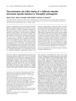
from mice fed a PPARa ligand for 5 days. The result of one-
dimensional SDS/PAGE analysis of these proteins from wild
(C57BL) or PPARa-null mice f ed a c ontrol or Wy14,6 43-
containing diet is shown i n Fig. 1. As already reported
[14,15], several p roteins including peroxisomal enzymes were
largely induced in the liver of wild mice by feeding Wy14 643
and it was difficult to assign uncharacterized new p rotein
bands on one-dimensional gels. In contrast, the number of
affected protein bands was limited in the intestine of wild
mice and we chose three protein bands as uniquely increased
in the i ntestine byWy14 643 in a PPAR a-dependent manner.
These include proteins having molecular masses of 32, 78 and
80 kDa as shown b y arrows in Fig. 1B.
Peptide mass fingerprinting (PMF) analysis
of the PPARa-regulated proteins in the intestine
Among the three proteins bands, I further selected P32 and
P78 for PMF analysis, because they s eemed to b e expressed
AB
Fig. 1. Effects of Wy14 64 3 on protein expression in the liver and intestine of control and PPARa-null mice. Wild-type (+/ +) and PPARa-null mice
(–/–) w ere fed either a control ( –) diet or one containing 0.05% Wy14 643 (+) for 5 days. T he prote ins in postnuclear fractions from th e liver and
intestine were analyzed by SDS/PAGE ( 10%, w/v) followed by staining with Coomassie brilliant blue (A). The portion s (indicated by boxes) of the
gel containing t he induced proteins (indicated by arrows) i n the intestine are shown enlarged (B).
4142 K. Motojima (Eur. J. Biochem. 271) Ó FEBS 2004
more in the intestine than in the liver and isolated from
other major protein bands on the one-dimensional g el. After
enzymatic digestion using endoproteinase Lys-C and ana-
lysis of the resulting peptides by MALDI-TOF mass
spectrometry, the masses of 12 among 29 peptides derived
from P32 were consistent with those c alculated from
the peptide sequences from 17b-HSD-11 (gi|16716597|
ref|NP_444492.1|), and the masses of 1 2 p eptides m atched
those f rom annexin IV ( gi|7304889|ref|NP_038499.1|). The
masses of 18 among 79 peptides derived from P80 were
consistent with those c alculated f rom the peptide s equences
from 17b-HSD-4 (gi|1706397|sp.|P51660|). The peptides
from P80 also contained 32 peptides from Ezrin
(gi|32363497|sp.|P26040|) a nd 18 peptides from P450
oxidoreductase (gi|6679421|ref|NP_032924.1|).
Among these proteins, 17 b-HSD-4 is known as a
peroxisomal enzyme a nd induction of several peroxisomal
enzymes in the liver has been extensively studied [16,17].
17b-HSD-4 is not a major protein in the liver, but it could be
identified a s a distinct protein b and on the one dimensional
gel of all the proteins in the post nuclear fraction probably
because of the absence o f abundant liver-specific proteins in
the intestine. Furthermore, the increase of 17b-HSD-4
caused by the PP ARa ligand in the liver as protein amount
was not remarkable when compared with that of mRNA,
and Corton et al . suggested the possibility of post-trans-
lational regulation of the protein levels in the liver [16].
It was noteworthy that t wo types of 17b-HSDs were
identified as the most remarkably increased proteins in the
intestine by PPAR a ligand in total protein mixture of the
postnuclear fraction of the mouse intestine. 17b-HSD-4 is a
multifunctional p rotein involved in not only i nactivation o f
estradiol but also successive steps of a-oxidation of long-
and b ranched-chain fatty acids in peroxisomes. In contrast,
17b-HSD-11 i s a new member of the 17 b-HSD family [18].
Mouse 17b-HSD-11 was found from a large set of full-
length cDNAs by s equence homology and functional
annotation [19] and human 17b-HSD-11 was identified in
expressed sequence tag databas es with c onserved domains
of the f amily members [20] and therefore its function has
not been fully characterize d yet [18]. 17b-HSD-11 has no
peroxisomal targeting signal at the C-terminus but a
possible hydrophobic signal sequence at the N-terminus.
The N-terminal sequence was expected to be cleavable by
signal peptidase (SignalP, ) but the
exact N -terminal sequence was detected during PMF
analysis of P32 protein as shown in Table 1. Thus
17b-HSD-11 should be a membrane protein and our
preliminary d ata using green fluorescence protein linked
to the C -terminus of the protein suggested a ssociation with
the e ndoplasmic reticulum. I n contrast, C hai et al.[20]
recently reported that the myc-tagged human 17b-HSD-11
at the N-terminus localized in the cytoplasm. The tagged
myc sequence should have abolished the function of the
Table 1. Summary of PMF analysis of P32 and P80. The residue numbers for P 32 are fro m the peptide seque nce of 1 7b-HSD-11 and those for P80
arefromthatof17b-HSD-4.
Observed m/z Theoretical MH+ Delta Residues Peptide sequence Modification
P32
766.45 766.47 )0.02 283–288 K)HRINVK
898.5 898.47 0.03 289–296 K)FDAVVGYK
936.52 936.51 0.01 151–161 K)AFLPVMMK 0Met–ox
1000.6 1000.58 0.02 63–70 K)LVLWDINK
1692.9 1692.87 0.03 140–153 K)TFEVNVLAHFWTTK
2527.6 2527.58 0.02 3–24 K)YLLDLILLLPLLIVFSIESLVK
2577.26 2577.24 0.02 83–105 K)LGAQAHPFVVDCSQREEIYSAAK
2630.44 2630.43 0.01 33–58 K)SVAGEIVLITGAGHGIGRLTAYEFAK
2951.45 2951.48 )0.03 162–189 K)NNHGHIVTVASAAGHTVVPFLLAYCSSK
2984.54 2984.5 0.04 228–254 K)NPSTNLGPTLEPEEVVEHLMHGILTEK 0Met–ox
P80
620.34 620.34 0 472–476 K)RTSEK
620.34 620.34 0 560–564 K)VRFAK
745.49 745.46 )0.05 725–730 K)LQMILK 0Met–ox
873.54 873.52 0.02 636–643 K)SVGREVVK
925.53 925.23 0 707–714 K)AFFSGRLK
970.62 970.62 0 58–65 K)VVAEIRRK
1001.61 1001.61 0 636–644 K)SVGREVVKK
1117.65 1117.62 0.03 715–724 K)ARGNIMLSQK 0Met–ox
1229.67 1229.64 0.03 579–588 K)EGNRIHFQTK
1315.73 1315.68 0.05 645–655 K)ANAVFEWHITK
152.7 1352.63 0.07 69–81 K)AVANYDVEAGEK
1597.98 1597.97 0.01 247–259 K)LRWERTLGAIVRK
1728.92 1728.97 )0.05 169–184 K)LGILGLCNTLAIEGRK
1985.99 1985.94 0.05 384–402 K)SMMNGGLAEVPGLSFNFAK 1Met–ox
1986.99 1686.94 0.05 260–275 K)RNQPMTPEAVRDNWEK 1Met–ox
2820.47 2820.4 0.07 302–330 K)VDSEGISPNRTSHAAPAATSGFVGAVGHK
2969.45 2969.42 )0.03 141–168 K)QNYGRILMTSSASGIYGNFGQANYSAAK 0Met–ox
Ó FEBS 2004 17b-HSD is a major PPAR a-regulated gene in intestine (Eur. J. Biochem. 271) 4143
N-terminal leader sequence. Table 1 summarizes assign-
ment of the peptide masses to 1 7b-HSD-11 and 17b-HSD-4.
Northern blot analysis of 17b-HSD-11 mRNA
To confirm that intestinal expression of 17b-HSD-11 is
regulated b y PPARa a nd W y14,643, we analyzed the e ffect
of the drug on t he levels of 17 b-HSD-11 and 17b-HSD-4
mRNAs i n the liver and in testine of wild-type or PPARa-
null mice (Fig. 2). Two types 17b-HSD mRNAs were
largely induced in both the liver and intestine in a P PARa-
and ligand-dependent manner. Their i nductions were more
outstanding than those o f two typical PPAR a-target gene
transcripts, liver-type fatty acid binding protein (L-FABP)
and intestine-type fatty acid binding protein (I-FABP)
mRNAs [3,21]. When compared to each other, some
preference of 17b-HSD-11 for intestine and of 17b-HSD-4
for liver was observed. Thus it was confirmed that 17b-
HSD-11 is a new protein whose expression is regulated by
PPARa and i ts ligand in t he intestine.
17b-HSD-11 mRNA was efficiently induced by various
types of PPARa activators in addition to a potent
Wy14 643 (Fig. 3 ). The time course of the induction was
very rapid not only in t he liver but also in intestine (Fig. 4)
and the rapid induction of the mRNA by Wy14 643 was
reproduced in the cultured hepatoma Fao cells (Fig. 5).
Almost a m aximal level of induction was achieved in both
tissues within a day, making a sharp contrast to the c ases of
typical PP ARa-target genes so far studied [3,13,21]. Tran-
scription of t he peroxisomal hydratase-dehydrogenase
(HD) and L-FABP genes is activated by PPARa within a
few hours and the m RNAs reach their maximal levels i n a
day in t he liver but not in intestine [13]. The levels of their
mRNAs in intestine slowly increase during a few days of
feeding a diet containing Wy14 643. This slow time course
of induction is also the case for the intestine-specific
PPARa-target gene I-FABP [21]. Thus the induction of
two 17b-HSD mRNAs by a PPAR ligand in the inte stine is
much more efficient than that of typical PPARa-target
genes.
Promoter structure of the
17
b
-HSD-11
gene and
transcriptional regulation
All the above d ata strongly suggest t hat expression o f the
17b-HSD-11 gene is directly regulated by PPARa and its
ligand. So the genome database was searched for the
Fig. 2. Influence of PPAR a and Wy14 643
on the expression levels of 17b-HSD-11 and
17b-HSD-4 mRNAs. Wild-type (+/+) and
PPARa-null mice (–/–) were fed either a con-
trol (–) d iet or one co ntaining 0.05% (w/v)
Wy14 643 for 5 days. Total RNA from indi-
vidual livers and intestines (5 lg) was analyzed
by Northern blotting using cDNAs for 17b-
HSD-11, 1 7b-HSD-4, 17b-HSD-10, liver-type
fatty acid bin ding protein (L-FABP), i ntes-
tine-type fatty acid binding protein ( I- FABP)
and ribosomal S14 p rotein (S14, loading
control).
Fig. 3. PP ARa activator s pecificity for 17b-HSD-11 mRNA induction
in the liver. Wild-type mice were fed e ither a co ntrol diet or one
containing 0 .05% (w/v) Wy14,643, 0.5% (w/v) clofibrate, 2% (w/v)
di(2-ethylhexyl)adipate (DEHA), or 2% (w/v) di(2-ethylhexyl)phtha-
late (DEHP) for 5 d ays. Total RN A isolated from individual livers was
subjected t o Northern b lot analysis using the cDNA prob es f or 17b-
HSD-11, L-F ABP, a2u-globu lin ( a2u), and apolipoprotein E ( apoE,
loading control).
4144 K. Motojima (Eur. J. Biochem. 271) Ó FEBS 2004
promoter sequence and PPAR b inding site. The mouse
17b-HSD-11 gene is located in cytoband E4 on chromo-
some 5 and the sequence has been published (accession
number, AL714024). S earching for a typical PPRE b y
TRANSSEARCH
program ( />TFSEARCHJ.html) in the region between 3 kb upstream
and 2 kb downstream of the estimated transcriptional
start site from t he 5 ¢ end of the reported cDNA [19]
showed no significantly similar motif. T he cloned p ro-
moter sequence up to )1800 bp from the transcriptional
start site did not respond to a PPARa ligand Wy14 643
in the reporter gene a ssay (data not shown). Although an
essential role of PPARa in the ligand-dependent tran-
scriptional activation of the 17b-HSD-11 gene in the
intestine became clear, whether another region of the
gene is necessary or an unknown mechanism is operating
for t he activation could not be clarified by the conven-
tional methods used in this study. The molecular
mechanism of the unusual induction of the 17b-HSD-11
gene in the intestine is clearly important and we are
trying to solve this problem in our laboratory hoping to
uncover a new role of PPARa in this organ.
Possible roles of 17b-HSD-11 in the intestine
Mouse 17b-HSD-11 may reside not in the e xtracellular
space as previously predicted but in the cell probably on the
membrane as no ted above. Suppo rting this p rospect, Chai
et al . [20] recently reported that human normal liver
parenchymal cells and epithelium of the endomerium and
small i ntestine, as well as steroidogenic cells, were immuno-
histochemically stained by anti-human 17b-HSD-11 I g.
They also suggested that 1 7b-HSD-11 was localized to
cytoplasm in the cell, but it should be associated with the
endoplasmic reticulum (see above).
17b-HSD-11 b elongs to t he short-chain d ehydrogenase/
reductase superfamily (SDR family member 8) and also
has a protein domain of glucose/ribitol dehydrogenase
(Mouse Genome Informatics, ormatics.
jax.org). Recent studies on the specificities of several
types of 17b-HSDs have revealed their wide substrate
spectrum, including not only glucocorticoids and sex
steroids but also bile acids, fatty acids and branched
chain amino acids [18,22]. Thus 17b-HSDs in the
epithelium of the intestine may metabolize potentially
toxic compounds included in the diet to protect the
organism, as s uggested by Chai et al. [20]. More work is
required to address the in vivo function of 17b-HSD-11
and physiological significance of i ts rapid and marked
induction by PPARa ligands in the intestine.
Acknowledgements
The author thanks Dr A. Iwamatsu (Central Laboratories f or Key
Technology, Kirin Brewery Co. Ltd, Yokohama, Japan) for PMF
analysis.
References
1. G elman, L. & Auwerx, J. (1999) Peroxisome proliferator-activated
receptors: mediators of a fast food impact on gene regulation.
Curr. Opin. Clin. Nutr. Metab. Care 2, 307–312.
2. Hihi, A.K., Mic halik, L. & Wahli, W. (2002 ) PPARs: t ranscrip-
tional effectors of fatty acids and their derivatives. Cell. Mol. Life
Sci. 59, 790–798.
3. Motojima, K., Passilly, P., Peters, J.M. , Gonzalez, F.J. & Latruffe,
N. (1998) Expression o f p utative fatty acid tr ansporter g enes are
regulated by p eroxisom e proliferator-activated receptor a and c
activators in a tissue- and i nd ucer-specific manner. J. Biol. Chem.
273, 16710–16714.
A
B
Fig. 4. Time course of induction of 17b-HSD-
11 and 17b-HSD-4 mRNAs i n the liv er and
intestine. (A) Wild-type mice were fed either a
control diet o r one containing 0.05%
Wy14 643 for 1–5 days as indicated. (B) W ild-
type mice were fed either a co ntrol d iet o r o ne
containing 0.5% (w/v) clofi brate (Clofib.) or
0.1% (w/v) t roglitazone (Trogli.) for 2 o r
8 weeks as indicated. Total RNA isolated
from individual livers and i ntestine was s ub-
jected to Northern blot analysis using cDNAs
asdescribedinthelegendsofFigs2and3.
Fig. 5. Time course of 17b-HSD-11 and 17 b-HSD-4 mRNA induction
by Wy14 643 in hepatoma Fao cells. Wy14 643 was added to the
medium of rat hepatoma Fao cells in the co nfluent stage at time 0 and
the cells were collected at the time indicated for total RNA isolation
and Northern b lotting analysis usin g the c DNA probes as described in
the l egends of Figs 2 and 3.
Ó FEBS 2004 17b-HSD is a major PPAR a-regulated gene in intestine (Eur. J. Biochem. 271) 4145
4. G elman, L ., Fruchart, J.C. & Auwerx, J . (1999) An update on the
mechanisms of action of the perox isome proliferator-activated
receptors (PPARs) and their r oles in inflammation and canc er.
Cell. Mol. Life Sci. 55, 932–943.
5. Staels, B., Dallongeville, J., Auwerx, J., Schoonjans, K .,
Leitersdorf, E. & Frucha rt, J.C. (1998) Mechanism of action
of fibrates o n lip id and lipoprotein metabo lism. Cir culation 98,
2088–2093.
6. Issemann, I. & Green, S. (1990) Activation of a member of the
steroid hormone receptor superfamily by peroxisome pro-
liferators. Nature 347, 645– 650.
7. Palmer, C.N., Hsu, M.H., Griffin, K .J., Ra uc y, J .L. & Johnson,
E.F. (1998) Peroxisome proliferator activated r eceptor-alpha
expression in human liver. M ol. Pharmacol. 53, 14–22.
8. H olden, P.R. & Tugwood, J.D. (1999) Peroxisome proliferator-
activated receptor alpha: r ole in rodent l ive r cancer and s peci es
differences. J. Mol. Endocrin ol. 22, 1–8.
9. Lee, S.S., Pineau, T., Drago, J., Lee, E.J., O wens, J .W., K roetz,
D.L., Fernandez-Salguero, P.M., Westphal, H. & Gonzalez, F.J.
(1995) Targeted disruption of the alpha isoform of the peroxisome
proliferator-activated receptor gene in mice results in abolishment
of t he pleiotropic effects of peroxisome p roliferators. Mol. Cell.
Biol. 15, 3012–3022.
10. Motojima, K. & Goto, S. (1994) H istidyl phosphorylation and
dephosphorylation of P 36 in rat liver extract. J. Biol. Chem. 269,
9030–9037.
11.Clauser,K.R.,Baker,P.&Burlingame,A.L.(1999)Roleof
accurate mass measu rement (+/– 10 pp m) in protein iden tification
strategies employing MS or MS/MS an d database searching. Anal.
Chem. 71, 2871–2882.
12. Chom czynski, P. & Sacchi, N . (1987) Single-step method of RNA
isolation by acid guanidinium th iocyanate-ph enol-chlorofo rm
extraction. Anal. B iochem. 162, 156–159.
13. Sato, O., Kuriki, C., Fukui, Y. & Motojima, K. (2002) Dual
promoter structure of mouse an d human fatty acid translocase/
CD36 genes a nd unique transcriptional a ctivation by peroxisome
proliferator-activated receptor a and c ligands. J. Biol. Chem. 277,
15703–15711.
14. Fukui, Y., Masui, S., Osada, S., Umesono, K. & M otojima, K.
(2000) A new thiazolidinedio ne, NC-2100, which is a weak
PPAR-c activator, exhibits potent antidiabetic effects a nd induces
uncoupling p rotein 1 i n white adipose tissue of KKAy obese mice.
Diabetes 49, 759–767.
15. Motojim a, K., Ohmori, A., T akino, Y. & Goto, S. (1993) Increase
in the amount o f elongation factor 2 in rat liver by peroxisome
proliferators. J. Biochem. (Toky o) 114, 779–785.
16. Corton, J.C., Bocos, C., Moreno, E.S., Merritt, A., Marsman,
D.S.,Sausen,P.J.,Cattley,R.C.&Gustafsson,J.A.(1996)Rat17
b-hydroxysteroid dehydrogenase type IV is a no vel peroxisome
proliferator-inducible gene. Mol. Pharmacol. 50, 1157–1166.
17. Fan, L.Q., Cattley, R.C. & Corton, J.C. (1998) Tissue-specific
induction of 1 7 b-hydroxysteroid dehydrogenase type IV by
peroxisome proliferator chemicals is dependent on the p eroxisome
proliferator-activated receptor a. J. Endocrinol. 158, 237–246.
18. Mindnic h, R., Moller, G. & Adamski, J. (2004) The role of
17b-hydroxysteroid dehydrogenases. Mol. Cell. Endocrinol. 218,
7–20.
19. Okaz aki, Y., Furuno, M., Kasukawa, T., Adachi, J., Bono, H.,
Kondo, S., Nikaido, I ., Osato, N., Saito, R., Suzuk i, H. et al.
(2002) Analysis of the m ouse transcrip tome based o n fu nctional
annotation of 60,770 full-len gth cDNAs. Nature 420, 563–573.
20. Chai, Z., Brereton, P., Suzuki, T., Sasano, H., Obeyesekere, V.,
Escher, G., S aff ery, R., Fuller, P., Enriquez, C . & K r ozowski, Z.
(2003) 17b-Hydroxysteroid dehydrogenase t ype XI localizes to
human steroidogenic cells. Endocrinology 144, 2084–2091.
21. Motojim a, K . (2000) Differential e ffects of PPARa activators o n
induction of ec topic expression of tissue-specific fatty acid binding
protein genes in the mouse liver. Int. J. Biochem. Cell. Biol. 32,
1085–1092.
22. Shafqat, N., Marschall, H .U., Filling, C., Nordling, E., Wu, X.Q.,
Bjork, L., Thyberg, J., M artensson, E., Salim, S ., Jornvall, H. &
Oppermann, U . (2003) Expanded su bstrate screenings o f human
and Drosophila type 10 17b-hydroxysteroid dehydrogenases
(HSDs) reveal mult iple specificities in bile acid and steroid hor-
mone metabolism: characterization of multifunctional 3a/7a/7b/
17b/20b/21-HSD. Biochem. J. 37 6 , 49–60.
4146 K. Motojima (Eur. J. Biochem. 271) Ó FEBS 2004