Báo cáo khoa học: Cloning, over-expression, purification and characterization of Plasmodium falciparum enolase doc
Bạn đang xem bản rút gọn của tài liệu. Xem và tải ngay bản đầy đủ của tài liệu tại đây (517.07 KB, 10 trang )
Cloning, over-expression, purification and characterization of
Plasmodium falciparum
enolase
Ipsita Pal-Bhowmick, K. Sadagopan, Hardeep K. Vora, Alfica Sehgal*, Shobhona Sharma and
Gotam K. Jarori
Department of Biological Sciences, Tata Institute of Fundamental Research, Mumbai, India
We have cloned, over-expressed a nd purified enolase from
Plasmodium falciparum strain NF54 in Escherichia coli in
active form, as an N- terminal His
6
-tagged protein. The
sequence o f the cloned enolase f rom the NF54 strain is
identical to that of s train 3D7 used in full genome sequen-
cing. The recombinant enolase (r-Pfen) could be obtained in
large quantities ( 50 mg per litre of culture) in a highly
purified form (> 95%). The purified protein gave a single
band at 50 kDa on SDS/PAGE. MALDI-TOF analysis
gave a mean ± SD mass of 51396 ± 16 Da, which is in
good agre ement w ith t h e mass calculated from the sequence.
The molecular mass of r-Pfen determined in gel-filtration
experiments was 100 k Da, indicating that P. falciparum
enolase is a homodimer. Kinetic measurements using
2-phosphoglycerate as substrate gave a specific activity
of 30 UÆmg
)1
and K
m2PGA
¼ 0.041 ± 0.004 m
M
.The
Michaelis constant for the reverse reaction (K
mPEP
)is
0.25 ± 0.03 m
M
. pH-dependent activity measurements
gave a maximum at pH 7.4–7.6 irrespective o f the direction
of catalysis. The activity of this enzyme is inhibited by Na
+
,
whereas K
+
has a slight activating effect. The cofactor
Mg
2+
has an apparent activation constant of 0.18 ±
0.02 m
M
. However, at higher co ncentrations, it has an
inhibitory effect. P olyclonal antibody raised against pure
recombinant P. falciparum enolase in rabbit showed high
specificity towards recombinant protein and is a lso a ble
to recognize enolase from the murine malarial parasite,
Plasmodium yo elii, which shares 90% id entity with the
P. falc iparum protein.
Keywords: enolase; homodimer; localization; P lasmodium
falciparum; purification.
Malaria remains one of the most infectious diseases in the
third world with abou t 500 million infections and over one
million deaths per year [1]. In the face of increasing threats
by resurgent infections and an expanding array of drug-
resistant phenotypes, the requirement of alternative pre-
ventive therapeutics is evident, especially for the most severe
form of human malaria parasite Plasmodium falciparum.
The first step in rational drug development involves
identification of macromolecular targets, which are unique
and essential for the survival of the parasite. Glycolytic
enzymes seem to be promising candidates from this
perspective, as energy production in P. falciparum depends
entirely on the glycolytic pathway as the parasite and its
mammalian host (red cells) lack a complete Krebs cycle
and active mitochondria [2,3]. The level of glycolytic flux
in parasite-infected cells is 100-fold greater than that
observed i n uninfected cel ls, and t he activity of many of the
glycolytic enzymes is higher in t he infected cells than in
uninfected ones [4]. Therefore an antimalarial that selec-
tively inhibits the parasite ATP-generating machinery
would be expected to arrest parasite development and
growth. Extensive wo rk has already been carried out with
many P. falciparum glycolytic enzymes, with aldolase,
lactate dehydrogenase and triose phosphate isomerase
showing quite promising behavior as detection tools, drug
targets and vaccine candidates [5–8]. P. falciparum enolase
(Pfen) (EC 4.2.1.11), the dehydrating glycolytic metallo-
enzyme that catalyzes the inter conversion of 2-phospho-
glyceric acid (2-PGA) and phospho enolp yruvate (PEP), h as
not yet been characterized. Enolases are highly conserved
across species [9]. In most species, i t exists a s a symmetric
homodimer [10]. However, in several bacterial species,
octameric enolases have been reported [11,12]. Conservation
is particularly pronounced for the active-site r esidues,
leading to similar kinetic properties among enolases from
diverse sources. F or activity, enolase r equires the binding of
2 m ol bivalent cations (in vivo this is usually Mg
2+
)per
subunit. Binding at site I leads to changes in the tertiary
structure of the enzyme (conformational site) whereas
binding to site II is essential for catalysis (catalytic site)
[13]. At higher concentrations, bivalent cations inhibit
activity, suggesting the existence of a third inhibitory site.
Univalent cations also influence the activity of enolases.
Most of the enolases are inhibited by Na
+
,whereasthe
effect of K
+
depends on the source of the enzyme. K
+
has
no effect on yeast enolase whereas it activates rabbit
enolases [14].
Correspondence to G. K. Jarori, Department of Biological Sciences,
Tata Institute of Fundamental Research, Homi Bhabha Road,
Colaba, Mumbai 400 005, India. Fax: +91 22 2280 4610,
Tel.: +91 22 2280 4545, E-mail:
Abbreviations:DAPI,4¢,6¢-diamidinophenylindole; PEP, phospho-
enolpyruvate; 2-PGA, 2-phosphoglyceric acid; r-Pfen, recombinant
Plasmodium falciparum enolase.
Enzyme: e nolase (EC 4.2.1.11).
*Present address: Section of Infe ctious Diseases/Internal Medicine,
Yale Unive rsity, New Haven, CT 0 6511, USA.
(Received 4 September 2004, ac cepted 22 Oc tober 2004)
Eur. J. Biochem. 271, 4845–4854 (2004) Ó FEBS 2004 doi:10.1111/j.1432-1033.2004.04450.x
There have been repo rts of antibodies to enolase detected
in high titers in Japanese and Thai P. falciparum patient sera
and use of yeast enolase for immunodiagnostic purposes
[15]. The activity of enolase in parasite-infected red blood
cells increases 15-fold [16]. The gene for P. falciparum
(strain K1) enolase (Pfen) has been cloned and characterized
[17]. However, P fen p rotein has not yet been c haracterized.
The d educed sequence o f Pfen exhibits high homology with
mammalian enolases (68–69%), but differs in containing
a plant-like pentapeptide ( EWGWS), a d ipeptide insertion,
and s ome different residues [17]. These i nclude Cys157. T he
analogous residue in Trypanosoma b rucei enolase (Cys147)
has recently b een shown to be m odified with iodoacetamide
[18,19]. Reaction with iodoacetamide also leads to partial
inactivation o f t he enzyme. It w ill be interesting t o e xamine
whether modification of Cys157 and other P. falciparum-
specific residues in the vicinity of the active site leads to
irreversible inactivation of Pfen. Comparative studies on the
structural and k inetic properties of parasitic and m amma-
lian enolases may provide clues for obtaining specific
inhibitors that can be developed as chemotherapeutic
reagents. To address questions related to the detailed
characterization of the molecular structure and kinetic
properties and to develop immunological reagents for
subcellular localization, we cloned Pfen and over-expressed
it in Escherichia coli to obtain adequate quantities of pure
recombinant P. falciparum enolase (r-Pfen). The results of
these experiments are p resented in this paper.
Materials and methods
Materials
Taq DNA polymerase, T4 DNA ligase, endonucleases
(KpnIandPstI), 4¢,6¢-diamidinophenylindole (DAPI) and
2,2¢-azinobis(3-ethylbenzo-6-thiazolinesulfonic acid) pow-
der were purchased from Roche Diagnostics Corp.
(Indianapolis, IN, USA). Mouse anti-His sera were from
Qiagen, Hilden, Germany. Horseradish peroxidase-conju-
gated anti-mouse s econdary IgG was obtained from Santa
Cruz Biotech (Santa Cruz, CA, USA), and Coomassie
Brilliant Blue R-250 was acquired from USB (Cleveland,
OH, USA). Nitrocellulose membrane, dithiothreitol,
molecular mass markers used for gel filtration and Super-
dex-75 HiLoad 16/60 (Prep grade) column were from
Amersham Pharmacia. Oligonucleotide primers, dianilino-
benzene, sodium salt of 2-PGA, rabbit muscle enolase
(b-isoform), yeast enolase, iodoacetamide, N-ethylmaleimide
and unstained high molecular mass p rotein markers for gel
electrophoresis were purchased from Sigma, St Louis, MO,
USA. Freund’s complete and incomplete adjuvants were
from Gibco-BRL, Alexa Fluor 488-conjugated anti-rabbit
IgG w as from Molecular Probes, I nc. (E ugene, OR, USA),
and vectashield-mounting medium was from Vector Labora-
tories, Inc. (Burlingame, CA, USA). Maxisorp plates for
ELISA were from Nunc, Roskilde, Denmark. All other
chemicals used in this study were of analytical grade.
PCR amplification
Sense a nd antisense primers were designed according to the
multiple cloning sites present in the pQE30 expression
vector and the published sequence of the P. falciparum
enolase gene [ 17]. The two primers w ere: PfenoEcoRIKpn I
(32-mer) 5¢-CCGGAATTCGGTACCATGGCTCATGT
AATAAC-3¢ and PfenoPstIXhoI (30-mer) 5¢-CATTCT
CGAGCTGCAGATTTAATTGTAATC-3¢.
A gametocytic cDNA library constructed from the
NF54 strain was u sed for the amplification of the enolase
gene (cDNA library used here was a gift from N. Kumar,
Johns Hopkins University, Baltimore, MD, USA).
Amplification was carried out in the s tandard Robocycler
Gradient S tratagene machine (Stratagene, La Jolla, CA,
USA) in a reaction consisting of 400 ng of each of the
primers, 100 l
M
dNTP mix, pH 8.8 buffer, 2 m
M
MgCl
2
,
50 m
M
KCl, 0.01% gelatin, 2 U Taq polymerase and
2 lL of the template library in a final volume of 20 lL.
The amplified enolase PCR product and the pQE30
plasmid vector were digested with KpnIandPstI
restriction enzymes, and these were ligated using T4
ligase. Competent XL1Blue E. coli cells were transformed
with the ligation mixture to obtain the required recomb-
inants, which were screened by PCR and plasmid DNA
preparation, and finally sequencing was performed (Mac-
rogen Inc., Seoul, South Korea) using standard protocols
[20].
Expression in
E. coli
and preparation of crude cellular
extracts
Expression was carried out in E. coli strain XL1Blue.
Cultures transformed with recombinant plasmid were
grown in Luria–Bertani medium containing 100 lgÆmL
)1
ampicillin. Cultures were induced with 0.5 m
M
isopropyl
thio-b-
D
-galactoside. Before induction, cultures were grown
at 37 °CtoanA
600
of 0.6–0.8. For analytical studies, culture
aliquots were taken at different time intervals (0, 3, 4, 5, 6 h)
after the induction and analyzed for protein production.
The cells were pelleted by centrifugation at 5 000 g for
10 min and stored at )80 °C. The cells were lyse d by
incubation in 50 m
M
sodium phosphate (10 mL per g wet
weight), pH 8.0, containing 300 m
M
NaCl, 1 mgÆmL
)1
lysozyme and 1 m
M
phenylmethanesulfonyl fluorid e for
30minoniceandsonicatedforsixcycles,15seachwith
15 s cooling between successive bursts at 5 output in a
Branson sonifier 450. The lysate was centrifuged at 45 000 g
for 3 0 min in a Beckman Ultracentrifuge (model LE-80K,
70 Ti rotor).
Affinity chromatography
His
6
-tagged r-Pfen was purified from soluble cell extract
using Ni-nitrilotriacetic acid affinity chromatography.
The binding was carried out by the batch method.
Soluble cell extract was mixe d with Ni-nitrilotriacetic acid
(pre-equilibrated with 50 m
M
sodium phosphate, pH 8 .0,
300 m
M
NaCl) slurry (8 mL per litre of culture) for 1 h
with gentle agitation. The slurry was passed through a
column and washed with 50 bed vols 50 m
M
sodium
phosphate, 4 0 m
M
imidazole, 300 m
M
NaCl, 1 m
M
phenylmethanesulfonyl fluoride, 5 m
M
2-mercaptoethanol,
pH 6.0, to remove nonspecifically bound proteins.
r-Pfen was eluted with 250 m
M
imidazole in the same
buffer.
4846 I. Pal-Bhowmick et al.(Eur. J. Biochem. 271) Ó FEBS 2004
Gel-filtration chromatography
The oligomeric st ate of r-Pfen was analyzed by gel-filtration
chromatography on a Superdex-75 Hiload-16/60 column
on an Amersham-Pharmacia Biotech (Kwai Chung,
Hong Kong), AKTA FPLC system. The column was pre-
equilibrated w ith 2 column vols buffer ( 50 m
M
sodium
phosphate, 1 50 m
M
NaCl, p H 7.4). Then 0.5 mg protein in
500 lL was applied to the column, and 2 mL fractions were
collected at a flow rate of 1 mLÆmin
)1
.Thecolumnwas
calibrated using appropriate molecular mass gel-filtration
markers.
Electrophoresis and Western blotting
Proteins were resolved on an SDS/12% polyacrylamide g el
[21] and visualized by staining with Coomassie Brilliant Blue
R-250. For Western blotting, crude cellular extracts and
purified r-Pfen separated by SDS/PAGE (12% gel) were
transferred to n itrocellulose membrane using semidry
Western transfer apparatus (Bio-Rad Laboratories, Inc.,
Hercules, C A, USA) at constant voltage (20 V) f or 35 min.
The membranes were blocked with 5% skimmed milk in
phosphate buffered saline (NaCl/P
i
;137m
M
NaCl, 2 .7 m
M
KCl, 10.0 m
M
Na
2
HPO
4
,1.8m
M
KH
2
PO
4
, pH 7.4) con-
taining 0.05% Tween 20 for 1 h. The blots were treated
with the mouse anti-His s erum and hors eradish peroxidase-
conjugated anti-mouse secondary IgG, respectively
(1 : 1000 dilution for both). The immunoblots w ere d evel-
oped using dianilinobenzene substrate.
Protein measurements and enzyme assay
Protein concentrations were determined by the Bradford
method using Bio-Rad protein assay dye reagent with
BSA as s tandard [22]. All kinetic m easurements w ere made
at 20 ± 1 °C. Enolase activity was measured i n the forward
(formation of PEP from 2-PGA) a nd reverse (formation of
2-PGA from P EP) direction by monitoring the increase o r
decrease respectively in PEP absorbance at 240 nm in a
continuous spectrophotometric assay on a Perkin-Elmer
lambda 40 spectrophotometer. T he change in PEP concen-
tration was determined using an absorption coefficient
(e
240nm
) ¼ 1400
M
)1
Æcm
)1
. As the absorption coefficient of
PEP varie s with pH a nd concentration of Mg
2+
,in
experiments where pH or Mg
2+
were varied, appropriate
values of molar absorptivity for PEP were used [23].
Typically, 540 lL of assay mixture containing 1.5 m
M
2-PGA (for the forward reaction) or 1.1 m
M
PEP (for the
reverse reaction) and 1.5 m
M
MgCl
2
in 50 m
M
Tris/HCl,
pH 7.4, was used. One unit of enzyme was defined as the
amount of enzyme that converts 1 lmol substrate (2-PGA
or PEP) into product (PEP or 2-PGA) in 1 min at 20 °C.
Kinetic parameters were determined from [substrate] vs.
velocity curves by fitting the data to t he Michae lis–Menten
equation using the
SIGMAPLOT
software.
MALDI-TOF analysis
For determination of the exact molecular mass of the
expressed recombinant protein, MALDI-TOF mass spectra
were recorded in linear mo de on Tof-Spec 2E ( Micromass,
Manchester, UK), fitted with a 337-nm laser. Protein
[5 pmol in 0.5 lL 40% acetonitrile/0.1% trifluoroacetic
acid (v/v)] was mixed with an equal volume of matrix
[saturated solution of sinapinic acid in 40% acetonitrile/
0.1% trifluoroacetic acid (v/v) i n water] and applied to the
MALDI t arget plate. This was allowed to dry at room
temperature to form cocrystals of p rotein and matrix. BSA
was used as an external mass standard. S ingle and double
charged peaks arising from BSA were used for calibration.
The operating parameters were: operating voltage, 20 kV;
sampling rate, 500 MHz; sensitivity, 50 mV. Typically
20–25 scans were averaged to obtain the spectrum.
Primary sequences and 3D structure modeling
The enolase sequences were aligned using
CLUSTAL W
for
homology comparisons [24]. The 3D structures of r-Pfen
and rabbit muscle enolases were modeled according to the
known 3D structure of T. brucei enolase (PDB:1OEP)
published previously, u sing the
SWISS
-
MODEL
server [25] and
structures were viewed wi th
VIEWERPRO
5.0 (Accelerys, S an
Diego, CA, USA).
Reaction with thiol-modifying reagents
r-Pfen or rabbit muscle enolase (0.1 l
M
) was placed in
buffer (1 m
M
KH
2
PO
4
,5m
M
MgCl
2,
0.1 m
M
dithiothreitol
and 50 m
M
triethanolamine/HCl, pH 8.0) and incubated
for 30 min at 37 °C. Different amounts of iodoacetamide or
N-ethylmaleimide were added to the enzyme samples and
allowed to react at 37 °C. Enzyme activity was assayed a t
different time intervals.
Generation of antiserum and ELISA
Standard protocols were followed to r aise rabbit polyclo nal
antiserum [ 26]. B riefly, 500 lg r -Pfen w as emulsified w ith
Freund’s complete adjuvant and injected into a 2-month-
old N ew Zealand White rabbit. Two boosts of 100 lgeach
of the r -Pfen e mulsified with incomplete Freund’s adjuvant
were given at an interval of 21 days. Ten days after the
second booster, the r abbit serum was collected. All animal
experiments were carried out as per the Guidelines of the
Committee for the purpose of control and supervision of
experiments on animals (CPCSEA), Animal Welfare
Division, Government of India. The specific immunization
experimental protocol was examined and cleared by the
Institutional Animal Ethics Committee.
For ELISA, t he r -Pfen, r abbit muscle and yeast enolases
were coated ( 100 lLof0.6 l
M
per well) on a Maxisorp plate
overnight at 4 °C. Unbound antigen was removed by
washing with NaCl/P
i
. The wells were blocked with 5%
skimmed milk in NaCl/P
i
containing 0.05% Tween 20
(NaCl/P
i
/Tween) for 1 h at 37 °C. This was washed twice
with NaCl/P
i
/Tween. Antibodies raised in rabbit were
diluted (2000–128 000-fold), and 100 lL of this was added
to each well. Each dilution was coated in duplicates. This
wasallowedtobindtotheantigensfor2 hat37 °Candthen
washed 6–7 times with NaCl/P
i
/Tween. T o this, goat anti-
rabbit secondary I gGs c onjugated with horseradish peroxi-
dase (1 : 2000 dilution; 100 lL p er well) in NaCl/P
i
/Tween
containing 0.01% BSA was added a nd al lowed t o i ncubate
Ó FEBS 2004 Characterization of P. falciparum enolase (Eur. J. Biochem. 271) 4847
for 45 m in at 37 °C. This was thoroughly washed with
NaCl/P
i
/Tween (7 –8 times). Then 1 00 lLof1mgÆmL
)1
2,2¢-azinobis(3-ethylbenzo-6-thiazolinesulfonic acid), pre-
paredin20m
M
citrate/80 m
M
Na
2
HPO
4
, pH 4.3, contain-
ing 1 lLÆmL
)1
30% H
2
O
2
,wasaddedtoeachwelland
incubated f or 10 min in t he dark. T he absorbance was read
at 405 nm on an EL808 Ultra Microplate reader (Biotek
Instruments Inc., Winooski, V T, USA).
Indirect immunofluorescence assay
An imm unofluorescence assay w as performed o n the blood
smears obtained from Plasmodium yoelii-infected mouse as
described previously [27]. Briefly, the smears were fixed for
30 s using chilled methanol and treated with preimmune
(control) or anti-(r-Pfen) serum at a dilution of 1 : 50 at
room temperature for 1 h. This was then stained for 45 min
with Alexa Fluor 488-conjugated an ti-rabbit IgG. Parasite
nuclei were stained with DAPI at a final concentration of
1 lgÆmL
)1
. The necessary washes were given after each
antibody incubation step, and slides were mounted under
glasscoverslipsin5lL vectashield mounting medium.
Slides were examined using a Nikon fluorescence micro-
scope.
Results and Discussion
Clone sequence and recombinant protein purification
Native enolase from P. falciparum strain K1 [17] and
strain 3D7 (NCBI: NP_700629) are predicted to contain
446 amino acids. The PCR amplification of the enolase
gene from the gametocyte cDNA library of the NF54
strain of E. coli resulted in a f ragment of t he expected size
of 1.4 kb. This fragment was cloned in pQE30 vector, and
E. coli cells were transformed w ith the recombinant
plasmid as described above (Materials and methods).
The cloned gene was subjected to DNA sequencing, and
the full amino-acid sequence of the recombinant protein
was deduced. The amino-acid sequence was found to be
identical with the 3D7 strain. However, these two strains
differ from the K1 strain at position 131 in having an
alanine residue in place of a proline. Figure 1 shows a
comparison of amino-acid sequences of enolases from
P. falc iparum strains NF54 (this work), K1 [17] and
P. yoelii (NCBI: AA1892).
The pQE30 vector is specifically designed for the over-
expression of heterologous proteins in E. coli. It allows the
expression of the recombinant protein and results in the
addition of a short noncleavable His tag sequence at its
N-terminus. Cloning resulted in incorporation of an a ddi-
tional 18 (MRGSHHHHHHGSACELGT-) and seven
(-LQPSLIS) residues to the N-terminus and C-terminus,
respectively, of Pfen. This would yield a r-Pfen protein of
mass 51 389.73 Da in contrast with 48 677 Da for the
native enzyme.
For purification of r-Pfen, typically 1 L culture was
grown at 37 °C, yielding 2 g wet cell pellet. Cells were
lysed, and the extract was subjected to centrifugation to
obtain soluble supernatant and pellet fractions. Both
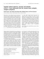
fractions contained r -Pfen (Fig. 2A, lanes 1 and 2). As the
soluble fraction contained a decent amount of r-Pfen,
recombinant protein was purified from this fraction by
affinity chromatography using an agarose/Ni-nitrilotriacetic
acid column as described in Materials and methods. As
expected, most of the enolase bound to the resin, and a wash
with 40 m
M
imidazole removed nonspecifically bound
proteins (lanes 3 and 4 of Fig. 2A). Finally pure enolase
P. falciparum NF54 MAHVITRINAR EILDSRGNPTVEVDLETNLGIFRAAVPSGASTGIYEALEL 51
P. falciparum K1 MAHVITRINAR EILDSRGNPTVEVDLETNLGIFRAAVPSGASTGIYEALEL 51
P. yoelii MLVKYWLASYFMIINPKNYEHIFYSRGNPTVEVDLETTLGIFRAAVPSGASTGIYEALEL 60
:* : **.: .*: *************.**********************
P. falciparum NF54 RDNDKSRYLGKGVQKAIKNINEIIAPKLIGMNCTEQKKIDNLMVEELDGSKNEWGWSKSK 111
P. falciparum K1 RDNDKSRYLGKGVQKAIKNINEIIAPKLIGMNCTEQKKIDNLMVEELDGSKNEWGWSKSK 111
P. yoelii RDNDKSRYLGKGVQQAIKNINEIIAPKLIGLDCREQKKIDNMMVQELDGSKTEWGWSKSK 120
**************:***************::* *******:**:******.********
P. falciparum NF54 LGANAILAISMAVCRAGAAANKVSLYKYLAQLAGKKSDQMVLPVPCLNVINGGSHAGNKL 171
P. falciparum K1 LGANAILAISMAVCRAGAAPNKVSLYKYLAQLAGKKSDQMVLPVPCLNVINGGSHAGNKL 171
P. yoelii LGANAILAISMAICRAGAAANKTSLYKYVAQLAGKNTEKMILPVPCLNVINGGSHAGNKL 180
************:******.**.*****:******::::*:*******************
P. falciparum NF54 SFQEFMIVPVGAPSFKEALRYGAEVYHTLKSEIKKKYGIDATNVGDEGGFAPNILNANEA 231
P. falciparum K1 SFQEFMIVPVGAPSFKEALRYGAEVYHTLKSEIKKKYGIDATNVGDEGGFAPNILNANEA 231
P. yoelii SFQEFMIVPVGAPSFKEAMRYGAEVYHTLKSEIKKKYGIDATNVGDEGGFAPNILNAHEA 240
******************:**************************************:**
P. falciparum NF54 LDLLVTAIKSAGYEGKVKIAMDVAASEFYNSENKTYDLDFKTPNNDKSLVKTGAQLVDLY 291
P. falciparum K1 LDLLVTAIKSAGYEGKVKIAMDVAASEFYNSENKTYDLDFKTPNNDKSLVKTGAQLVDLY 291
P. yoelii LDLLVASIKKAGYENKVKIAMDVAASEFYNSETKTYDLDFKTPNNDKSLVKTGQELVDLY 300
*****::**.****.*****************.******************** :*****
P. falciparum NF54 IDLVKKYPIVSIEDPFDQDDWENYAKLTAAIGKDVQIVGDDLLVTNPTRITKALEKNACN 351
P. falciparum K1 IDLVKKYPIVSIEDPFDQDDWENYAKLTAAIGKDVQIVGDDLLVTNPTRITKALEKNACN 351
P. yoelii IELVKKYPIISIEDPFDQDDWENYAKLTEAIGKDVQIVGDDLLVTNPTRIEKALEKKACN 360
*:*******:****************** ********************* *****:***
P. falciparum NF54 ALLLKVNQIGSITEAIEACLLSQKNNWGVMVSHRSGETEDVFIADLVVALRTGQIKTGAP 411
P. falciparum K1 ALLLKVNQIGSITEAIEACLLSQKNNWGVMVSHRSGETEDVFIADLVVALRTGQIKTGAP 411
P. yoelii ALLLKVNQIGSITEAIEACLLSQKNNWGVMVSHRSGETEDVFIADLVVALRTGQIKTGAP 420
************************************************************
P. falciparum NF54 CRSERNAKYNQLLRIEESLGNNAVFAGEKFRLQLN 446
P. falciparum K1 CRSERNAKYNQLLRIEESLGNNAVFAGEKFRLQLN 446
P. yoelii CRSERNAKYNQLFRIEESLGANGSFAGDKFRLQLN 455
************:******* *. ***:*******
Fig. 1. Amino-acid sequence alignment of
enolases fr om P. falciparum strain NF54 with
P. falciparum strain K1 [17] and P. yoelli
(NCBI:AA18892) u sing
CLUSTAL W
[24].
Enolase from s train N F54 diff ers fro m that of
strain K1 in having a P131A mutation (shown
in bold).
4848 I. Pal-Bhowmick et al.(Eur. J. Biochem. 271) Ó FEBS 2004
protein was eluted with 250 m
M
imidazole. The eluted
protein showed a single band at the expected molecular
mass ( 50 kDa) on SDS/PAGE (Fig. 2A, lane 5). The
identity of the protein was f urther established b y Western
blotting using anti-His serum (Fig. 2B). About 50 mg active
r-Pfen was purified from 1 L E. coli culture.
The m olecular m ass of the recombinant protein was also
analyzed by MS. The MALDI-TOF spectrum of purified
r-Pfen contained three peaks at m/z 25707, 51 383.04 and
102 782. The peak at m/z 51 383.04 can be a ttributed to a
singly charged monomeric species of r-Pfen, which is in
good agreement with the calculated average mass of
51 389.73 Da. The peak at m/z 25 707 represents a doubly
charged monomeric species, and the one at m/z 102 782 is
attributed to the presence of a singly charged dimeric species
of r-Pfen.
The r-Pfen sequence gave a theoretical absorption
coefficient (e
280
) of 41400
M
)1
Æcm
)1
. The concentration of
purified r-Pfen determined by Bradford assay using BSA
as standard was in good agreement with that obtained
by measuring A
280
and using the theoretical absorption
coefficient.
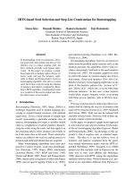
Oligomeric state of r-Pfen
The oligomeric state of r-Pfen was examined by gel-filtration
chromatography. Figure 3 shows an elution profile of
0.5 mg r -P fen in 500 lL50m
M
sodium phosphate/150 m
M
NaCl, pH 7.4, o n a Superdex-75 column. The column was
calibrated u sing appropriate molecular mass markers. The
apparent molecular mass determined for native r-Pfen was
100 kDa. Purified r-Pfen when a nalyzed on SDS/PAGE
showed a single band at 50 kDa (Fig. 2A, lane 5),
indicating that it forms a homodimer in the native state. It is
also interesting to note that, in the MALDI-TOF spectrum,
a peak was observed at m/z 102 782 corresponding to a
singly charged dimeric form of r-Pfen . Enolases from most
organisms form dimers of 40–50-kDa subunits [10,12],
exception for octameric enolases from thermophilic [12] and
sulfate-reducing bacteria [28]. The oligomeric state of none
of the apicomplexan enolases has been reported so far.
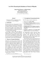
Kinetic characterization
Purified r-Pfen was assayed f or enolase activity b y measur-
ing either t he conversion of 2-PGA into PEP (forward
reaction) or PEP into 2-PGA (reverse reaction). The enzyme
had a specific activity of 30 ± 3 UÆ(mg protein)
)1
in the
forward direction and 10 ± 2 UÆmg
)1
in the reverse
direction. For the determination of K
m
, initial reaction
rates were measured at several different concentrations of
2-PGA (Fig. 4A) and PEP (Fig. 4B). Data were fitted to the
40 50 60 70 80 90
0
10
20
30
Elution Volume (ml)
OD
280
(mAU)
Fig. 3. Gel-filtration chromatogram of r-Pfen. Protein (0.5 mg in
500 lL) was r un on a Superdex-75 column precalibrated using
appropriate molecular mass markers ( chymotrypsin ogen A, 25 kDa;
ovalbumin, 43 kDa; BSA, 67 kDa; yeast enolase, 93 kDa; alcohol
dehydrogenase, 150 kDa). Blue Dextran 2000 was used to measure the
void volume. The molecular mass obtained for r-Pfen from this
experimentwas98±5kDa.
205
116
97
M 1 2 3 4 5
1 2 3 4 5
66
45
29
kDa
50 kDa
Fig. 2. Analysis of proteins from transformed E. coli XL1 B lue c ells over-expressing r-Pfen. Cells were induced with 0.5 m
M
isopropyl thio-b-
D
-
galactoside for 6 h and harvested. (A) Analysis on SDS/PAGE (12% gel). Lane M, Molecular mass markers; lanes 1 and 2, insoluble and soluble
fractions, respect ively, of the E. coli extract; lane 3, flow through after binding of the r-Pfen supernatant fraction to Ni-nitrilotriacetic a cid; lane 4,
40 m
M
imidazole wash of the protein bound to Ni-nitrilotriacetic acid resin; lane 5, elution of r-Pfen with 250 m
M
imidazole. ( B) Immunoblot of
cells over-expressing r-Pfen prob ed with 1 : 1000 anti-His serum. Th e arrow shows the position of r-Pfe n.
Ó FEBS 2004 Characterization of P. falciparum enolase (Eur. J. Biochem. 271) 4849
Michaelis–Menten equation {v ¼ V
max
[S]/(K
m
+[S])}
using
SIGMAPLOT
software. The best nonlinear fit gave
K
m2PGA
¼ 0.041 ± 0.004 m
M
and K
mPEP
¼ 0.2 5 ±
0.03 m
M
. These values for K
m2PGA
and K
mPEP
are similar
to those r eported f or mammalian , yeast and other enolases
[18]. The variation of r-Pfen activity as a function of pH was
also analysed. F igure 4 C,D shows plots of enzyme activity
vs. pH when 2-PGA or PEP was used as substrate. Maximal
r-Pfen activity is observed in the range pH 7 .4–7.6 irres-
pective of the substrate used. Most mammalian enolases
have their activity maxima in the range pH 6.8–7.1, whereas
the plant ones are around pH 8.0 [10].
The effect of univalent cations on the activity of r-Pfen
was also investigated. Figure 5A shows the variation in
r-Pfen activity with increasing concentrations of NaCl and
KCl. NaCl inh ibits the enzyme w ith 50% inhibition around
0.3–0.4
M
. This inhibitory e ffect of Na
+
is very similar to
that observed for mammalian enolases [14]. In contrast,
KCl showed a s light activating effect on r-Pfen. T he activity
of all three rabbit isozymes (aa, bb and cc) a re significantly
stimulated (40–100%) by KCl at lower concentrations
(< 400 m
M
), whereas in t he higher concentration range the
activation e ffect i s lost [14]. KCl has a mild activating eff ect
on yeast enolase at concentrations < 200 m
M
, but strongly
inhibits activity at higher concentrations [14]. This kinetic
response of r-Pfen to various concentrations of KCl is at
variance to those of mammalian and yeast enolases.
Figure 5B shows the effect of increasing concentrations of
Mg
2+
on the activity of r-Pfen, rabbit and yeast enolases. In
the low concentration range, Mg
2+
acts as an activating
cofactor for all the enolases. D ata from the low c oncentra-
tion range (£ 1m
M
) were fitted to the Michaelis–Menten
equation to derive the apparent activation coefficient. The
activation constant derived for r-Pfen from the data
presented here is 0.18 ± 0.02 m
M
. Higher concentrations
of Mg
2+
have an inhibitory effect on r-Pfen activity. The
maximal inhibition observed for r-Pfen is much less
(< 40%) than that observed for the yeast and rabbit
muscle enzymes (60–70%) (Fig. 5B). Previous kinetic stud-
ies have suggested the presence of three bivalent cation-
binding sites o n enolase, with the first two h igh-affinity sites
involved in activation and a third low-affinity site
involved in inhibition [13]. In the crystal structure, two
Mg
2+
-binding sites h ave been detected. These are believed
to be involved in assembly of the active site and catalysis
[29,30]. Recently, a third bivalent cation-binding site has
been identified i n the structure of T. brucei enolase. It has
been suggested that binding of Mg
2+
at this site may be
0.00.20.40.60.81.01.21.41.6
0
10
20
30
40
6.6 6.8 7.0 7.2 7.4 7.6 7.8 8.0 8.2
6
8
10
12
14
16
18
20
Activity (milliUnits)
p
H
5.56.06.57.07.58.08.5 9.0
0
5
10
15
20
25
pH
Activity (milliUnits)
[PEP](mM)
Activity (milliUnits)
0.0 0.2 0.4 0.6 0.8 1.0 1.2 1.4 1.6
0
10
20
30
40
50
Activity (milliUnits)
[2-PGA](mM)
A
D
B
C
Fig. 4. Kinetic c haracterization of r -Pfen. (A) Plot o f [2-PGA] vs. a ct ivity; and (B) plot of [PE P] vs. activity for the determ ination of K
m
.A5lL
sample of enzyme containing 1.5 and 3.0 lg of r-Pfen , respect ively, were u sed for the 2-PGA and PEP assay, respectively. Experimental data were
fitted according to the Michaelis–Menten equation using
SIGMAPLOT
. The best fit gave K
m2PGA
¼ 0.041 ± 0.004 m
M
and K
mPEP
¼
0.25 ± 0 .03 m
M
. pH was plotted against activity using (C) 2-PGA and (D) PEP as substrates. A 5 lL sample of enzyme containing 0.5 lg r-Pfen
was used f o r the 2 -PGA assay and 2.5 lg was used for the PEP assay.
4850 I. Pal-Bhowmick et al.(Eur. J. Biochem. 271) Ó FEBS 2004
responsible for the observed inhibition at high metal ion
concentrations [19].
Homology-based structure modeling
Enolase is highly conserved across species. The overall
structure of enolase comprises an eightfold a/b barrel
domain preceded b y a n N-terminal a + b domain [19]. A
highly conserved catalytic site is located between the two
domains. It w ill be interesting to model t he parasite enzyme
on the basis of the known enolase structure and examine the
structural differences between Pfen and t he mammalian
enzyme in the vicinity of the conserved a ctive site. Such an
exercise may lead to identification of parasite-specific
residue(s), which may be amenable to specific chemical
modifications and hence selective i nactivation. We modeled
the 3D structure of r-Pfen and rabbit muscle enzymes on the
basis of T. brucei enolase (PDB: 1OEP) which is 60%
homologous to Pfen. Figure 6 shows t he active-site regions
of these enzymes along with some of the residues in the
vicinity. In a recent study on the T. brucei enzyme, it was
shown that modification of C ys241 and Cys147 with
iodoacetamide leads to partial inactivation o f the Trypano-
soma enzyme [18,19]. This inactivation was attributed to the
perturbation caused to active-site structure by the addition
of a c arboxamidomethyl group to Cys147 and/or Cys241.
Analogous positions in Pfen are occupied by Ala251 and
Cys157. Ala148 replaces Cys157 in Pfen in rabbit muscle
enolase. It will be interesting t o examine the effect of thiol-
modifying regents on r-Pfen. It is expected that similar to
Cys147 in T. brucei, Cys157 in Pfen will be carboxamido-
methylated, causing partial inactivation. As the rabbit
enzyme does not have a similar Cys, it may not be affected.
To determine whether Cys157 is accessible to chemical
modification, which may lead to inactivation (similar to
T. brucei [19]), we treated the enzyme with iodoacetamide
(Fig. 6D). There was no e ffect on the activity of r-Pfen even
after 2 h of treatment with 10 m
M
iodoacetamide. As
expected, the addition of iodoacetamide to rabbit muscle
enolase also did not have any effect on the activity.
Although Cys157 occupies a position similar to Cys147 in
T. brucei (Fig. 6A,B), the microenvironment in the two
cases may be quite different. It is likely that either the
Cys157 is not accessible to iodoacetamide o r the carboxam-
idomethyl group fits into the cavity ar ound the Cys without
any perturbation of the arrangement of the active-site
residues. T he latter possibility would s ugge st that the use of
larger thiol-modifying reagents (e.g. N-ethylmaleimide)
might lead to inactivation. In the c ase of T. brucei enolase,
complete inactivation by N-ethylmaleimide has been
observed [19]. The addition of N-ethylma leimide t o r -Pfen
did lead to partial inactivation of the enzyme (Fig. 6D).
However, similar inactivation w as also observed for rabbit
enolase, which does not have analogous Cys157 near the
active site (Fig. 6 C), suggesting that N-ethylmaleimide-
induced inactivation is probably due to modification of
other Cys residues in the protein. Although these prelim-
inary attempts have not succeeded in achieving species-
specific inactivation, efforts will be made to design
substrate-based active-site-directed affinity reagent(s) for
selective inactivation of the parasite enzyme.
Reactivity and specificity of anti-(r-Pfen) evaluated
by ELISA
Antibodies raised in rabbit after two boosts of r-Pfen
protein showed quite high titer and reactivity with r-Pfen.
Reactivity was observed e ven at a dilution of > 64 000
(Fig. 7A). I n comparison, when equimolar quantities of
rabbit muscle and yeast enolases were u sed as antigens,
almost no significant reactivity was observed beyond an
antiserum dilution of 1 : 16 000. To rule out the possibility
that this antiserum may contain a significant fraction of
antibodies directed against t he His
6
tag of r-Pfen, we used
an unrelated His
6
-tagged protein (rOS-F, a recombinant
odorant-binding protein from Drosophila) as control. No
significant cross-reactivity was observed against this pr otein
(data not shown). Although there is 61–68% homology
A
B
Fig. 5. Effect of univalent and bivalent cations on r-Pfen activity.
(A) E ffect of NaCl (d)andKCl(s). Data are plotted as percentage
activity vs. [salt]. A 540 lL volume of assay mixture containing 1.1 m
M
PEP and 1.5 m
M
MgCl
2
in 50 m
M
Tris/HCl, pH 7.4, was used. A 5 lL
volume of enz yme solution containing 2.5 lg enolase protein was used
for each assay. (B) A comparison of the effect of MgCl
2
on the activity
of r-Pfen (d), yeast enolase (s) and rabbit muscle enolase (.). The
assay mixture consisted of 1.1 m
M
PEP in 50 m
M
Tris/HCl, pH 7 .4.
The r esidual activity i n the absence of Mg
2+
is due to contaminating
bivalent cation s i n the assay m ixture. For compariso n, d ata for each
enzyme were norm alized taking highest observed activity a s 100%.
Ó FEBS 2004 Characterization of P. falciparum enolase (Eur. J. Biochem. 271) 4851
among yeast, rabbit and P. falciparum enolases, the poly-
clonal antibodies raised here exhibit considerably higher
specificity for r-Pfen.
We further assessed the specificity of the antiserum by
performing an indirect immunofluorescence assay on b lood
smears obtained from P. yoelii -infected mice. The gene
sequences of enolase from murine malarial parasite, P. yoelii
and P. falciparum, exhibit 90% i dentity a nd 94% s imilarity
in their amino-acid sequences (Fig. 1). On the basis of such a
large sequence homology, it is expected that polyclonal
antibodies raised against r-Pfen would c ross-react with the
P. yoelii enolase protein. A s s hown in Fig. 7B, the i mmune
serum reacted with the parasite-infected mouse red blood
cells and not with unin fected red blood cells. T he parasite-
infected cells can be identified by using DAP I staining. As
uninfected r ed cells do not have a nucleus, they do not pick
up DAPI. DAPI-positive cells (parasite-infected) are the
only ones stained by anti-(r-Pfen). All the erythrocytic stages
of the parasite (rings, trophozoites a nd schizonts) reacted to
anti-(r-Pfen). A control immunofluorescence assay experi-
ment was also performed using preimmune rabbit serum.
As expected, no staining of the parasite-infected cells was
observed (Fig. 7C). These experiments also demonstrate
that anti-(r-Pfen) sera did not have any cross-reactivity
towards the mammalian red blood cell enolase protein.
Conclusions
We have cloned and developed an over-expression system for
P. falc iparum enolase. This has allowed us to obtain decent
amounts of pure protein (50–60 mg per litre of culture). The
measured physicochemical parameters (molecular mass and
absorption coefficient at 280 nm) for the expressed protein
are in good agreement with those predicted on the basis of the
cloned sequence. The presence of a 50-kDa band on SDS/
PAGE for purified r-Pfen and 100 kDa on gel-filtration
A
B
010020 40 60 80
20
40
60
80
100
120
Time (min)
% Activity
D
C
Fig. 6. Comparison of the active-site regions of (A) T. brucei (PDB code 1OEP), (B) P. falciparum and (C) rabbit muscle enolase. P. falciparum and
rabbit muscle (P25704; ENOB_rabbit) enolases were modeled using the T. brucei X-ray crystallographic structure. Residues involved in substrate
and metal binding are shown in green and magenta, respectively. (D) Effect of iodoacetamide (open symbols) and N-ethylmaleimide (filled symbols)
on r-Pfen (circles) and ra bbit muscle enolase (squares). E nolase (20 lg) was i ncubate d with 10 m
M
iodoacetamide or 8 m
M
N-ethylmaleimide.
Enzyme activity was assayed at vari o us time points.
4852 I. Pal-Bhowmick et al.(Eur. J. Biochem. 271) Ó FEBS 2004
chromatography suggests that, in its native state, r-Pfen
forms an active homodimer similar to the enolases from
several other sources [10,12]. This is further supported by the
presence of a peak at m/z 102 782 in the MALDI spectrum.
Kinetic measurements showed substrate affinity to be similar
to that of mammalian enolases. r -Pfen differs from rabbit
enolases in its extent of inhibition caused by high Mg
2+
concentration (Fig. 5B) and inability of K
+
to activate it
significantly (Fig. 5A) [14]. A lthough e nolases from rabbit
muscle and P. falciparum exhibit a high degree of sequence
homology (67–69%), antibodies raised against r-Pfen in
rabbit are quite specific, as evident from ELISA (Fig. 7A)
and the fact that they fail to react with mammalian enolases
(Fig. 7 B). This recombinant protein is highly immunogenic,
as only two booster doses were sufficient to give titers of
> 1 : 6 4 000 for specific reactivity with the antigen. This
polyclonal antibody is being used to investigate subcellular
localization of enolase at different stages in the life cycle of the
parasite. The availability of large quantities of r-Pfen will also
facilitate structural investigations on this apicomplexan
glycolytic enzy me.
Acknowledgements
We are grateful to D r Nirbhay Kumar of Johns H opkins University,
Baltimore, MD, USA for the gift of k Orient P. falciparum strain NF54
gametocyte asexual s tage library. We thank Mr Prateek Gupta and
Mr Yogesh Gupta for help with some of the experiments.
References
1. Engers, H.D. & Godal, T. (1998) Malaria vaccine development:
current status. Par asitol. Today 14, 56–64.
2. Oelshlegel, F.J. Jr & Brewer, G.J. (1975) Parasitism and the red
cell. In The Red Cell (Surgenor, D.M., ed.), Vol. 2, pp. 1264.
Academic Pres s, San D iego.
3. Trager, W. (1986) Meta bolism: Energy Sources, Respiration. In
Living Together: the Biology of Animal Parasitism, pp. 147–169.
Plenum Press, New York.
4. Roth,E.F.Jr,Raventos-Suarez, C., Perkins, M. & Nagel, R.L.
(1982) Glutathione s tability and oxidative stress i n P. falciparum
infection in vitro; responses of no rmal and G 6PD-de ficient ce lls.
Biochem. Biop hys. Res. Commun. 109, 355–362.
5. Certa, U., Ghersa, P., Dobel, H ., Matile, H., Kocher, H.P.,
Shrivastava, I.K., Shaw, A.R. & Perrin, L.H. (1988) Aldolase
activity of a Plasmodium falciparum protein with protective
properties. Science 240, 1036–1038.
6. Gomez, M.S., Piper, R.C., Hunsaker, L.A., Royer, R.E., Deck,
L.M., Makler, M.T. & Vander Jagt, D.L. ( 1997) Substrate and
cofactor specificity and selective inhibition of lactate dehydro-
genase from the malarial parasite P. falciparum. Mol. Biochem.
Parasitol. 90 , 235–246.
7. Piper,R.,Lebras,J.,Wentworth,L.,Hunt-coole,A.,Houze,S.&
Makler, M . (1999) Immunocapture diagnostic a ssays for malaria
using Plasmodium lactate dehydrogen ase . Am. J. Trop. Med. Hyg.
60, 1 09–118.
8. Velank ar, S.S., Ray, S.S ., Gokhale, R.S., Suma, S., Balaram, H.,
Balaram, P. & Murthy, M.R.N. ( 1997) Triosephosphate iso-
merase from Plasmodium falciparum: the crystal structure provides
insights into antim alarial drug design. St ructure 5, 7 51–761.
9. Panc holi, V. (2001) Multifunctional a-enolase: its role in diseases.
Cell. Mol. Life Sci. 58, 9 02–920.
10. Wold, F. (1971) Enolase. In The Enzymes (Boyer, P.D., ed.), Vol.
5, pp. 499–538. Academic Pre ss, New Y ork.
11. Barnes, L.D. & Stellwagen, E. (1973) Enolase from the thermo-
phile Thermus X-1. Bioc hem istry 12, 1559–1565.
12. Brown, C.K., Kuhlman, P.L., Mattingly, S., Slates, K., Calie, P.J.
& Farrar, W.W. (1998) A mo del of quaternary s tructure of
enolases, based on structural and evolutionary analysis of
octameric enolase from Bacillus subtilis. J. Protein Chem. 17, 855–
866.
13. Faller, L.D., Baroudy, B.M., Jonson, A.M. & Ewall, R.X. (1977)
Magnesium ion requirements for y e ast enolase activity. Biochem-
istry 16 , 3864–3869.
14. Kornblat, M .J. & Klugerman, A. (198 9) Characterization of the
enolase isozyme s of rabbit brain: kinetic diffe rences between
mammalian and yeast e no lases. Biochem. Ce ll Biol. 67 , 103–107.
0.0
0.4
0.8
1.2
1.6
2.0
2,000 4,000 8,000 16,000 32,000 64,000 128,000
a
b
c
Antiserum (fold dilution)
A
405
nm
a:
p
re-immune b: DAPI
a: anti-r-pfen b: DAPI
Fig. 7. Specificity of polyclonal antibodies raised against r-Pfen in
rabbit. (A) ELISA reactivity of anti-(r-Pfen) with (a) r-Pfen, (b) rabbit
muscle enolase and (c) yeast enolase, measured as A
405
and plotted
against increasing dilutions of antibody. (B) Immunofluorescence
assay with P. yoelii-infectedmouseredbloodcellstreatedwith(a)
anti-(r-Pfen) seru m (1 : 50 dilution) and (b) DAPI (1 lgÆmL
)1
).
(C) P. yoelii-infected cells were treated with (a) preimmune sera
(1 : 5 0 dilution) and (b) DAPI (1 lgÆmL
)1
).
Ó FEBS 2004 Characterization of P. falciparum enolase (Eur. J. Biochem. 271) 4853
15. Sato, K., Kano, S., Matsumoto, Y., Glanarongran, R., Krudsood,
S., L ooareesuwan, S., A ikawa, M. & Suzuki, M. (1998)
Serological e valuation of malaria patient in Thailand, w ith parti-
cular reference to the reactivity against a 47kD antigenic
polypeptide of Pl asmodiu m fa lcip arum. TokaiJ.Exp.Clin.Med.
23, 9 7–98.
16. Roth, E.F. J r, Calvin, M.C., Max-Audit, I., Rosa, J. & Rosa, R.
(1988) The enzymes of the glycolytic pathway in erythrocytes
infected with Plasmodium falc iparum malaria p arasites. Blood 72,
1922–1925.
17. Read, M., Hicks, K.E., Sims, P.F.G. & Hyde, J.E. (1994)
Molecular characterization of the enolase gene from the hum an
malaria parasite Plasmodium falciparum. Eur. J. Biochem. 220,
513–520.
18. Hannaert, V., Albert, M A., Rigden, D.J., d a Silva, Giotto, M.T.,
Thiemann,O.,Garratt,R.C.,VanRoy,J.,Opperdoes,F.R.&
Michels, P.A.M. (2003) Kinetic characterization, structure mod-
elling studies and crystallization of Trypanosoma brucei enolase.
Eur. J. Biochem. 270, 3 205–3213.
19. da Silva. Giotto, M.T., Hannaert, V., Vertommen, D., de
Navarro, M.V.A.S., Rider, M.H., Michels, P.A.M., Garratt, R.C.
& Rigden, D.J. (2003) The crystal structure of Trypanosoma brucei
enolase: visualization o f the inhibitory metal binding site III and
potential as target for selective, irreversible inhibition. J. Mol. Biol.
331, 6 53–665.
20. Samb rook, J., Fritsch, E.F. & Maniatis, T. (1989) Molecular
Cloning: a Laboratory Manual, 2nd edn. Cold Spring Harbor
Laboratory Press, Cold Spring Harbor, NY.
21. Laemmli, U.K. (1970) C leavage of structural proteins during
assembly of the h ead of t he bac teriophage T4. Nature ( London)
227, 6 80–685.
22. Bradford, M.M. (1976) A rapid and sensitive method for
the quantitation of microgram quantities of protein utilizing
the principle of prote in-dye binding. Anal. Biochem. 72 , 248–254.
23. Wold, F. & Ballou, C .E. (1957) Studies on t he enzyme enolase I .
Equilibrium studies. J. Biol. C hem. 227, 301 –312.
24. Th ompson, J.D., Higgins, D.G. & Gibson, T.J. (1994) CL USTAL
W: improving the sensitivity of progressive multiple sequence
alignment through sequen ce weighting, positions-sp ecific gap
penalties and wei ght matr ix choice. Nucleic Acids Res. 22, 4673–
4680.
25. Guex, N. & Peitsc h, M.C. (1997) S WISS-MODEL and the Swi ss
Pdb viewer: an environment for comparative protein modeling.
Electrophoresis 18 , 2714–2723.
26.Harlow,E.D.&Lane,D.(1988)Antibodies: A Laboratory
Manual. Cold Spring Harbor Laboratory Press, Cold Spring
Harbor, NY.
27.Goswami,A.,Singh,S.,Redkar,V.D.&Sharma,S.(1997)
Characterization of P0, a ribosomal phosphoprotein of Plasmo-
dium fal ciparum. J. Bio l. Chem. 272, 1213 8–12143.
28. Kitamura,M.,Takayama,Y.,Kojima,S.,Kohno,K.,Ogata,H.,
Higuchi, Y. & Inoue, H. (2004) Cloning and e xpression of the
enolase gene from Desulfovibrio v ulgaris (Miyazaki F ). Biochim.
Biophys. A cta 1676, 172–181.
29. Wed ekind, J.E., Poyner, R.R., Reed, G.H. & R ayment, I. ( 1994)
Chelation of serine 39 to Mg
2+
latches a gate at the active-site o f
enolase: structu re of the bis (Mg
2+
) complex of yeast enolase and
the intermediate analog phosphoacetohydroxamate at 2.1 A
˚
resolution. Biochemistry 33 , 9333–9342.
30. Kuhnel, K . & Luisi, B.F. (2001) Crystal structure o f t he E s cher-
ichia coli RNA degradosome component enolase. J. Mol. Biol.
313, 5 83–592.
4854 I. Pal-Bhowmick et al.(Eur. J. Biochem. 271) Ó FEBS 2004