Báo cáo khoa học: Mercury(II) binding to metallothioneins Variables governing the formation and structural features of the mammalian Hg-MT species pptx
Bạn đang xem bản rút gọn của tài liệu. Xem và tải ngay bản đầy đủ của tài liệu tại đây (529.83 KB, 9 trang )
Mercury(II) binding to metallothioneins
Variables governing the formation and structural features of the mammalian
Hg-MT species
A
`
ngels Leiva-Presa, Merce
`
Capdevila and Pilar Gonza
`
lez-Duarte
Departament de Quı
´
mica, Facultat de Cie
`
ncies, Universitat Auto
`
noma de Barcelona, Spain
With the a im of extending our knowledge on the reaction
pathways of Zn-metallothionein (M T) and apo-MT species
in the presence of Hg(II), we monitored the titration of
Zn
7
-MT, Zn
4
-aMT and Zn
3
-bMT proteins, at pH 7 and 3,
with either HgCl
2
or Hg(ClO
4
)
2
by CD and UV-vis spectr-
oscopy. Detailed analysis of the optical data revealed that
standard variables, such as the pH of the solution, the
binding ability of the counter-ion (chloride or perchlorate),
and t he time elapsed b etween subsequent additions of Hg(II)
to the protein, play a determinant role in the stoichiometry,
stereochemistry and degree of folding o f the Hg-MT species.
Despite the fact that the effect of these variables is unques-
tionable, it is difficult to generalize. Overall, it can be c on-
cluded that the reaction conditions [pH, time elapsed
between subsequent additions of Hg(II) to the p rotein] affect
the structural properties more substantially than the s toi-
chiometry of the Hg-MT species, and that the role of the
counter-ion becomes particularly apparent on the structure
of overloaded Hg-MT.
Keywords: mercury(II) binding; mercury-metallothionein;
metallothionein; a-metallothionein; b-metallothionein.
Mercury t hiolates provide representative examples of the
structural diversity shown by the extensive family of metal
thiolates [1–4]. The most striking features of mercury
thiolates in the solid phase are the different structures
obtained when Hg(II) is co-ordinated to very similar
thiolate ligands [5,6] and the distinctive behavior of Hg(II)
towards a particular thiolate compared with that of Zn(II)
or Cd(II) [7], which has been referred to a s the zinc family
paradox [3]. Moreover, correlations between solid-state and
solution complexes cannot be easily established. Overall, the
diverse co-ordination preferences of Hg(II) ions (mainly
tetrahedral, trigonal-planar and digonal) and their coexist-
ence in polynuclear complex species, the various ligation
modes of the thiolate ligands (i.e. terminal, l
2
-bridging or
l
3
-bridging) and the possibility of secondary Hg(II)–sulfur
interactions [8] make it difficult to anticipate the structure of
a particular mercury thiolate complex [1,3,9]. This results
from the interplay of not only the above factors, but a lso the
reaction conditions. Of these, the presence of additional co-
ordinating species, such as halide ions, make the bonding
situation for mercury even less straightforward than in the
case of homoleptic mercury thiolates [10,11].
The biological chemistry of mercury is dominated by
co-ordination t o cysteine thiolate groups in agreement with
the preference of this metal ion for the soft sulfur ligands.
The high binding constants for binding of Hg(II) to cysteine
residues account for the irreversible replacement of essential
metals (Zn, Cu) in cysteine-containing metalloproteins and
thus for the high toxicity of mercury to living systems.
Within the same context of the highly favored thermo-
dynamically Hg-S bond, resistance to Hg(II) toxicity in
several bacteria is based on an ensemble of proteins
designated as Mer, most of which bind Hg(II) i ons through
cysteine residues ([3] and references therein). In mammals,
detoxification of mercury by metallothioneins (MTs) occurs
via cysteine complexation a nd sequestration [12]. A major
feature of this very large family of ubiquitous low molecular
mass proteins is their extremely high content of cysteine
residues, the binding of which to metal centers determines
the 3D structure of the protein [13]. Consideration of the
high flexibility and multidentate ligand nature of t he peptide
chain in MTs together with the intrinsic complexity of
mercury thiolate complexes suggests that elucidation of the
stoichiometry and co-ordination geometries of mercury in
solution Hg-MT species may be rather i ntricate.
To date, optical spectroscopy (UV-vis a nd CD) has
played a major role in the study of the mercury-binding
properties of mammalian MTs, for which several Hg-MT
stoichiometries have been reported [14]. Thus, a detailed
analysis of the electronic spectra o f Hg(II)-reconstituted M T
led Vas
ˇ
a
´
k et al. [15] to propose that Hg(II) in Hg
7
-MT is
co-ordinated at sites w ith tetrahedrally related geometry.
Subsequent studies by Johnson & A rmitage [16] of the UV
spectral data obtained in the titration of C d(II)
7
-MT with
Hg(II) showed that Hg(II) initially occupies tetrahedral sites
but, above a Hg/MT stoichiometry of four, there is a shift
to linear co-ordination. However, on the basis of X-ray
Correspondence to M. Capdevila, Departament de Quı
´
mica, Facultat
de Cie
`
ncies, Universitat Auto
`
noma de Barcelona, E-08193 Bellaterra,
Barcelona, Spain. Fax: + 34 935813 101, Tel.: + 34 935813 323,
E-mail:
Abbreviations: MT, metallothionein; TDPAC, time differential per-
turbed angular correlation of c-r ays; UV-vis, ultraviolet-visible elec-
tronic absorption; t, stabilization time allowed for the co-ordination of
Hg(II) to the protein; X, counter-ion of the Hg(II) salt added as
titrating agent.
(Received 19 July 2004, revised 21 October 2004,
accepted 25 October 2004)
Eur. J. Biochem. 271, 4872–4880 (2004) Ó FEBS 2004 doi:10.1111/j.1432-1033.2004.04456.x
absorption studies conducted on some of the species
observed in the titration of either apo-MT or Zn
7
-MT with
Hg(II), monitored by optical spectroscopy, Lu & Stillman
[17] proposed a d istorted tetrahedral co-ordination for
Hg(II) in Hg
7
-MT with two short (2.33 A
˚
)andtwolong
(3.4 A
˚
) Hg-S distances [18]. Previous extended X-ray
absorption fine structure (EXAFS) results for Hg
7
-MT
were consistent with a Hg-S bond length of 2.42 A
˚
and
suggested that Hg(II) was in a three-co-ordinate thiolate
environment [19].
Although the protective role of MTs a gainst Hg(II)
toxicity provides particular interest for the study of the
Hg(II)-MT system, most existing results are difficult to
reconcile. With t he aim of finding new strategies for this
study, w e now report o n the effect of two variables, the
reaction time and the presence of chloride ions, on
the stoichiometry, stereochemistry and degree of folding
of the Hg(II)-MT species formed by either the binding of
Hg(II) to apo-MT or Zn/Hg replacement in Zn
7
-MT.
Materials and methods
Protein preparation and characterization
Fermentator-scale cultures, purification of the glutathione-
S-transferase-MT fusion p roteins, and recovery and ana-
lysis of the recombinant mouse Zn
7
-MT1, Zn
4
-aMT1 and
Zn
3
-bMT1 domains were performed a s p reviously described
[20,21]. The Zn
7
-MT, Zn
4
-aMT and Zn
3
-bMT species were
obtained in both Tris/HCl and Tris/HClO
4
buffer (50 m
M
,
pH 7) [22]. The protein concentration was 0.1 m
M
in the
six solutions, which were diluted to a final concentration of
10 l
M
(MT) or 20 l
M
(aMT and bMT fragments) with
MilliQ-purified and Ar-degassed water before being titrated
with Hg
2+
solutions at 25 °C.
The apoproteins were prepared by acidification of the
recombinant material with 10 m
M
HCl or HClO
4
, respect-
ively, until pH 3. At pH values lower than 3.5 the Zn
7
-MT,
Zn
4
-aMT and Zn
3
-bMT species are entirely devoid of
metal, according to their respective CD spectra. In contrast,
Hg(II) remains bound to SCys at this pH.
Metal solutions
Glassware and solutions used in metal ion-binding studies
were prepared as described [20]. A Riedel-de Hae
¨
natomic
absorption spectrometry Hg
2+
standard of 1000 p.p.m. was
used as the HgCl
2
solution. The Hg(ClO
4
)
2
solution was
prepared from the corresponding salt in MilliQ-purified
water, and the Hg(II) concentration was quantified by
atomic absorption spectrometry using a Perkin–Elmer 2100
atomic absorption spectrometer. In both cases the Hg(II)
concentration of the tit rating agents was in the 1–10 m
M
range.
Metal ion-binding reactions
Metal-binding experiments were carried out by sequentially
adding molar-ratio a liquots of concentrated Hg(II) stock
solutions to single solutions of either the holo proteins or
apoproteins and followed spectropolarimetrically (CD) and
spectrophotometrically (UV-vis). Two sets o f titrations,
which differ in the time elapsed between subsequent
additions of Hg(II) to the protein, were carried out. In
one set, the standard titration procedure [ 22] was followed,
whereas in the other consecutive additions of Hg(II) were
made every 24 h. The electronic absorption and CD
measurements were performed and corrected as already
described [22].
All m anipulations involving the protein and metal ion
solutions were performed in Ar atmosphere, and the
titrations were carried out at least in duplicate t o ensure
the reproducibility of each point.
The pH (7 or 3) for all experiments remained constant
throughout. A t pH 7, t he acidity of t he Hg(II) solutions
required the addition of appropriate buffer solutions of
Tris/HCl or Tris/HClO
4
(50 or 70 m
M
at pH 7), but no
buffering was required for the titrations carried out at pH 3.
Results and Discussion
In view of the well-known complexity of Hg(II)–thiolate
systems, the difficulties we encountered in analyzing the
results obtained th rough preliminary titrations of the
Zn-MT proteins with Hg(II) were not a surprise. They
indicate that the nature of the counter-ion (X) and the time
elapsed between subsequent additions of the Hg(II) solution
(t) have a significant effect on the stoichiometry, stereo-
chemistry and degree of folding of the species formed. Thus,
to understand the reaction pathways followed by Zn-MT
and apo-MT species in the presence of Hg(II), the e ffect of
each of the previous variables was analyzed separately. T o
this end, the titration of Zn
7
-MT, Zn
4
-aMT and Zn
3
-bMT
proteins, at pH 7 and 3, with either HgCl
2
or Hg(ClO
4
)
2
were spectroscopically monitored.
The CD and UV-vis spectroscopic techniques used in this
work are currently used to study metal-binding features of
MT as they provide i nformation on the c o-ordinative
features of the predominant metal-MT species present in
solution at each titration point and on the number of s pecies
formed during the titration. Furthermore, titration of the
separate fragments provides information on the depend-
ence/independence relationship between the t wo constitu-
tive domains of the whole MT protein [21,23].
With regard to the two pH values, titrations at pH 7 a llow
the subsequent substitution of Zn(II) and thus formation of
heterometallic Zn,Hg-MT species, and titrations at acidic
pH values provide information on the binding of Hg(II) to
the corresponding apo-MT form [23]. In a ddition, compar-
ison of the two sets of data gives an indication of the role of
Zn(II) in the Hg(II)-containing species formed at physiolo-
gical pH. The use of two different Hg(II) salts allowed
analysis of the possible role of the physiologically relevant
chloride anion, which has a strong tendency to co-ordinate
and b ridge Hg(II) ions, i n the degree of folding and 3D
structure of the Hg-MT species. The perchlorate anion is
well known for its low co-ordinating ability towards metal
centers.
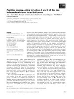
As regards the time variable, the spectroscopic changes
observed in the titrations of Zn
7
-MT, Zn
4
-aMT and
Zn
3
-bMT with Hg(II), after different times were allowed
for the reaction between the MT protein and the added
Hg(II) ions, were indicative of a strong dependence of the
Hg-MT system on this variable (Fig. 1). Thus, titrations
Ó FEBS 2004 Variables governing the binding features of Hg-MT (Eur. J. Biochem. 271) 4873
with HgCl
2
were carried o ut at two different times, t ¼ 0h
and t ¼ 24 h, whereas those with Hg(ClO
4
)
2
were only
performed at t ¼ 24 h. The t ¼ 0 h label denotes that the
titration was performed under k inetic control c onditions,
which means that, for each addition, the protein sample was
allowed to react with the metal ion u ntil subsequent CD
spectra were essentially coincident [22]. However, for most
samples, if the CD spectrum was recorded again after 24 h,
it showed significant differences from that recorded at t ¼
0 h. For this reason, titrations labeled t ¼ 24 h denote those
carried out under thermodynamic control conditions, where
each molar-ratio aliquot of Hg(II) was added every 24 h, as
longer time intervals showed no further changes in the
spectroscopic features.
Overall, evaluation of all the variables in the Hg-MT
system required the performance and analysis o f 18
titrations and the corresponding duplicates. The detailed
and comparative analysis of the set of CD, UV-vis and
difference electronic absorption spectra recorded for e ach
titration (provided as Supplementary Material) provides
information on the species formed by the Zn-MT
peptides in the p resence of Hg(II) under t he d ifferent
experimental conditions assayed and has allowed us to
propose the reaction pathways (Schemes 1–3) for Zn/Hg
replacement in Zn-MT species ( pH 7) and for the
binding of Hg(II) to apo-MT (pH 3) that are discussed
below.
Mercury content in the Hg(II)-MT species at each
titration point has traditionally been established b y assu-
ming that, in solution, only one species is present, the
metal c ontent of which coincides with the number of
Hg(II) equivalents (eq) added . To validate the previous
assumptions as well as to quantify the Zn content in t he
Zn,Hg-MT species observed at pH 7 (Schemes 1A, 2A and
3A), we unsuccessfully devoted m uch e ffort to obtaining
ESI-MS data. Thus, information on the Zn(II) content was
retrieved from CD data and it is mainly of a qualitative
nature.
Reaction of recombinant mouse Zn
7
-MT with Hg(II)
Analysis of the CD, UV-vis and UV-vis difference spectra
obtained in the titration of Zn
7
-MT with Hg(II) at pH 7
(Fig. 2, S 1 and S2) and pH 3 (Figs S3–S5) for each set
of X and t values led to the reaction pathways shown
in Scheme 1.
Comparative analysis of the three sets of data indicates
that the stoichiometry of the species formed along the three
titrations at pH 7 depends on neither the stabilization time,
t, nor the nature o f th e counter-ion. The unique exceptions
Fig. 1. Evolution w ith time of t he CD spectra corresponding to the
addition of the tenth Hg(II) to Zn
7
-MT at pH 7.
Scheme 1. Proposed reaction pathways for Hg(II) binding to recombinant Zn
7
-MT at pH 7 (A) and at pH 3 (B), under th ermodynamic (t ¼ 24 h)
or kinetic (t ¼ 0 h) control conditions, using HgCl
2
or Hg(ClO
4
)
2
as titrating agents. The and „ symbols denote similarity and difference,
respectively, between the structure of two species compared.
4874 A
`
. Leiva-Presa et al.(Eur. J. Biochem. 271) Ó FEBS 2004
to this rule are : (a) Z n,Hg
2
-MT, observed as an i ntermediate
species only at t ¼ 24 h; (b) the stoichiometries of the fully
loaded species, Hg
15
-MT and Hg
16
-MT. Conversely, the
chirality of the species is highly dependent on the previous
variables, t ¼ 24 h and X ¼ Cl
–
affording t he most chiral
species, as s hown by the intensity of t he CD bands of the
Hg(II)-MT species formed under these conditions (Fig. 2).
Similarly, t and X have a significant effect on the structure
of the Hg-MT aggregates, with a Hg to MT ratio equal or
higher than 7, as evidenced by the comparison of the CD
spectra of isostoichiometric species obtained under different
conditions. The contribution of the counter-ion to the 3D
structure of the Hg-MT aggregates is demonstrated by the
outstanding example of Hg
11
-MT, which becomes one of
the most chiral species if formed in the presence of Cl
–
under
both kinetic and thermodynamic control conditions
(Fig. 3 ).
Another relevant feature is the formation of hetero-
metallic Zn,Hg
5
-MT and Zn,Hg
7
-MT, both present in the
three titrations. T he former shows a very specific CD
fingerprint. The significance of the latter lies in the Hg(II)
stoichiometry, as previous studies proposed formation o f
homometallic Hg
7
-MT species [17,24]. Under the experi-
mental conditions used, the evolution of the CD spectra is
fully consistent with the presence of heterometallic
Zn,Hg
7
-MT as an intermediate species between Zn,Hg
5
-
MT and Hg
9
-MT. Overall, the information obtained using
the optical techniques allows Zn,Hg
5
-MT a nd Hg
11
-MT to
be considere d the hallmark species formed in the Zn/Hg
replacement in Zn
7
-MT.
ABC
Fig. 2. (A) CD, (B) absorp tion UV-vis, and (C) differe nce abso rption UV-vis s pectra obtained by s ubtracting the successive spectra of (B), corresponding
to the titration of recombinant mouse Zn
7
-MT1 with HgCl
2
at pH 7 and t ¼ 24 h. The Hg(II) to MT m olar ratios are indicated within each fram e.
Ó FEBS 2004 Variables governing the binding features of Hg-MT (Eur. J. Biochem. 271) 4875
Data obtained a t pH 3 show a strong influence of
t and X on the stoichiometry and structure of the species
formed, as shown i n Scheme 1B, and thus, the three
reaction pathways followed at this pH are remarkably
different. Notwithstanding this, there is a minor effect of
t and X at the b eginning and end o f the titration. Thus,
the addition of the first 4–6 of Hg(II) to apo-MT gives
rise to Hg-MT species of comparable stoichiometry and
structure, i.e. Hg
4
-MT and Hg
5)6
-MT,andalsothe
presence of an excess of Hg(II) cation leads invariably to
Hg
18
-MT. Furthermore, within the previous range [ from
4–6 to 18 Hg(II)], subsequent additions of Hg(II) led to
low-chirality Hg-MT species under all conditions. T he
only exception is Hg
13
-MT, formed at t ¼ 0handX¼
Cl
–
, which shows a well-defined CD fingerprint, also
indicative of a highly chiral species. Concerning the role
of the counter-ion, the differences observed in the CD
spectra of overloaded Hg-MT species, such as Hg
10
-MT
and Hg
18
-MT, formed at t ¼ 24 h, provide evidence for
the interaction of the chloride anion with Hg(II), as
already found at pH 7.
Fig. 3. Role of the chloride anion in the d egree of folding of Hg-MT
species observed by comparing the CD spectra of the Hg
11
-MT species
obtained in the titration of Zn
7
-MT with either HgCl
2
(in black) or
Hg(ClO
4
)
2
(in grey), both at pH 7 and t ¼ 24 h.
Scheme 2. Proposed reaction pathways f or Hg(II) binding to recombinant Zn
4
-aMT at pH 7 (A) and a t pH 3 (B), under thermodynamic (t ¼ 24 h )
or kinetic (t ¼ 0 h) control conditions, using HgCl
2
or Hg(ClO
4
)
2
as titrating agents. The and „ symbols denote similarity and difference,
respectively, between the structure of two species compared.
Fig. 4. CD spectra o f (A) the Z n
2
Hg
4
-aMT (in b lack) and Hg
5
-aMT (in
grey), and Zn,Hg
4
-aMT (in black) and Zn,Hg
5
-aMT (in grey) species,
respectively, obtained in the titrations of Zn
4
-aMT with HgCl
2
(solid
lines) or Hg(ClO
4
)
2
(dashed lin es), both at pH 7 and t ¼ 24 h and (B) the
Hg
11
-aMT spe cies obtained in the titrations of Zn
4
-aMT with HgCl
2
(in
black) or Hg(ClO
4
)
2
(in grey), both a t pH 3 and t ¼ 24 h.
4876 A
`
. Leiva-Presa et al.(Eur. J. Biochem. 271) Ó FEBS 2004
Reaction of recombinant mouse Zn
4
-aMT with Hg(II)
Consideration of the optical spectroscopic data obtained in
the titrations of Zn
4
-aMT with Hg(II) at pH 7 (Figs S6–S8)
and pH 3 (Figs S9–S11) allows the proposal of the reaction
pathways shown in Scheme 2.
Analogously to Zn
7
-MT, the stoichiometry of the
Hg-aMT species formed at pH 7 (Scheme 2A) along the
three titrations does not depend on t and X. Notwith-
standing this, the Hg
7
-aMT species is absent in the
presence of Cl
–
at t ¼ 24 h, and t he species containing the
highest H g(II) content, Hg
11
-aMT, is only obtained if
t ¼ 0h and X¼Cl
–
. Conversely, t he structure a nd
chirality of t he various Hg-aMT species are significantly
influenced by t and X, as evidenced by their CD spectra.
Thus, the species with a Hg to aMT molar ratio higher
than 6–7 became mo re chiral if formed in the presence of
Cl
–
, a mong which, tho se formed a t t ¼ 0 h show the
highest degree of chirality. Exceptionally, only the
Zn,Hg
4
-aMT species are comparable with respect to their
chirality and structure under the three sets of experimental
conditions.
Interestingly, concerning the Zn,Hg
4
-aMT species, the
244(+) nm CD band recorded after t he addition of 4 H g(II)
to Zn
4
-aMT under all sets of conditions not only gives a
clear indication of the presence of Zn(II) in the aggregate,
but its intensity also suggests that the highest Zn(II) content
is found when X ¼ ClO
4
–
(Fig. 4A). A similar analysis
reveals the presence of Zn(II) in the Hg
5
-aMT species
formed with X ¼ ClO
4
–
but its absence for X ¼ Cl
–
.
Chelex-100 treatment [23] of a n aliquot of the correspond-
ing sample and subsequent analysis of the Zn and Hg
content by inductively coupled plasma atomic emission
spectroscopy and i nductively coupled plasma mass spectro-
metry a llowed us to unequivocally establish the Zn
2
Hg
4
-
aMT and Hg
5
-aMT stoichiometrie s for the species formed
at t ¼ 24 h and X ¼ Cl
–
. Overall, all previous data indicate
that the replacement o f Zn(II) by Hg(II) in Zn
4
-aMT
proceeds more efficiently in the presence of Cl
–
than in the
presence of ClO
4
–
.
At pH 3 (Scheme 2B) neither t nor X has a substantial
effect on the stoichiome try of the s pecies formed during the
titrations, except for the formation of two additional
species, Hg
3
-aMT and Hg
7
-aMT, at t ¼ 24 h and X ¼
ClO
4
–
. Conversely, the nature of the counter-ion strongly
affects the chirality of the species. This effect is remarkable
for those species with a H g(II) stoichiometry equal to o r
higher than 6, X ¼ Cl
–
and t ¼ 24 h. In contrast, the
Hg-aMT species formed in the presence of ClO
4
–
show a
very low degree of folding, indicating that Cl
–
ions strongly
participate in the acquisition of the 3D structure of the Hg-
aMT species (Fig. 4B).
Reaction of recombinant mouse Zn
3
-bMT with Hg(II)
The spectroscopic data obtained in the titrations of
Zn
3
-bMT with Hg(II) at pH 7 (Figures S 12–S14) and pH
3 ( Figures S15–S17) are consistent with the reaction
pathways shown in Scheme 3. Comparison of the three
sets of data recorded at pH 7 (Scheme 3A) reveals that the
Hg:bMT stoichiometry of the species does not depend on
the nature of the counter-ion. Conversely, the stabilization
time determines the Hg-bMTstoichiometryofmostofthe
species formed and becomes particularly evident as the
Scheme 3. Proposed reaction pathways for Hg(II) binding to recombinant Zn
3
-bMT at pH 7 (A) and at pH 3 (B), under thermodynamic ( t ¼ 24 h)
or kinetic (t ¼ 0 h) control conditions, using HgCl
2
or Hg(ClO
4
)
2
as titrating agents. The and „ symbols denote similarity and difference,
respectively, between the structure of two species compared.
Ó FEBS 2004 Variables governing the binding features of Hg-MT (Eur. J. Biochem. 271) 4877
nuclearity of the species increases. Notwithstanding this,
saturation occurs in all cases for 10 Hg(II). On the other
hand, CD data indicate that the degree of chirality an d the
structure of the species formed up to Zn,Hg
3)4
-bMT
depend on t and X, the most chiral species being those
obtained at t ¼ 24 h and X ¼ Cl
–
. As opposed to that
observed for the aMT fragment, the CD spectra reveal that
the presence of Cl
–
favors the Zn(II) ions remaining bound
to the bMT protein in the first stages of the titration.
Titrations carried out at pH 3 (Scheme 3B) reveal that
the stoichiometries of the Hg-bMT species become
dependent on t and X after t he forma tion of Hg
7
-bMT.
Comparison of the three sets of CD data indicates that the
degree of chirality of the Hg-bMT species is generally
independent of t. However, the chirality o f the species
obtained in the presence of Cl
–
is much higher than that
achieved when X ¼ ClO
4
–
,exceptfortheHg
3
-bMT
species, with a very low chirality in both cases, a nd the
Hg
7
-bMT species, which show comparable chirality for
X ¼ Cl
–
and ClO
4
–
(Fig. 5). Comparison of the CD
fingerprints of the H g-bMT species formed a long the three
titrations sh ows t hat their 3D structure is s trongly
dependent on t an d X, except for Hg
3
-bMT, which is
poorly structured under all conditions.
Co-ordination environments around Hg(II) in Hg-MT
species
The c omplexity of the Hg(II)-MT system, which is mainly
the result of its Hg-thiolate nature, makes it difficult to
obtain information on the co-ordination geometry around
Hg(II) in the Hg(II)-MT aggregates from optical techniques
(CD and/or UV-vis spectra) by simple treatment of the
data. There are several reasons: (a) the presence of different
chromophores in the same species including Zn and/or Hg
as metal ions and SCys and/or Cl
–
as ligands; (b) the
absence of w ell-established relationships between most of
the previous chromophores and the corresponding absorp-
tion wavelengths [3]; (c) the overlapping of the absorption
bands corresponding to different chromophores, as shown
by the spectral envelopes in the difference UV-vis spectra.
Despite this, analysis of the difference UV-vis data, which
discloses the effect of each Hg(II) addition, can give an
insight into the evolution of the co-ordination geometry
about Hg(II) in the M T species for med by either Zn/Hg
replacement in Zn
7
-MT or the addition of Hg to apo-MT.
By following this approach, comparison of the difference
UV-vis spectra obtained in the titrations of Zn
7
-MT,
Zn
4
-aMT and Zn
3
-bMT with HgCl
2
at pH 7 and t ¼
24 h (Fig. 2, S6 and S12) indicates a parallel evolution of the
co-ordination geometry about Hg(II) in the three peptides.
These spectra evo lve according to the following pattern: (a)
the addition of the first 7 Hg(II) eq to Zn
7
-MT, or the first 4
Hg(II) eq to any of t he aMT and bMT fragments, causes
initially the appearance of an asymmetric broad band
Fig. 5. CD spectra of the H g
7
-bMT species obtained in the titrations
of Zn
3
-bMT at pH 3 with HgCl
2
at t = 24 h (solid black line) or
t = 0 (solid grey line), or with Hg(ClO
4
)
2
at t = 24 h (dashed g rey
line).
AB
Scheme 4. An insight into t he evolution of the coordination geometries about Hg(II) in the Hg- MT species formed during the titrations of Zn
7
-MT,
Zn
4
-aMT and Zn
3
-bMT with HgCl
2
at t ¼ 24 h and pH 7 (A) or pH 3 (B). The different coloured are as have been d educ ed from the d ifference UV-
vis spectra. Preliminary TDPAC me asurements on the Hg-MT spec ies within a square enable co rrelation of e ac h area w ith an spec ific coordination
geometry about Hg(II).
4878 A
`
. Leiva-Presa et al.(Eur. J. Biochem. 271) Ó FEBS 2004
(230–340 nm), which eventually transforms into two new
broad overlapping bands with absorption maxima at 230
and 320 nm; (b) the next Hg(II) eq added to the three
peptides gives rise to a negative broad band with absorption
minima at 260 and 310 nm, together with a positive
absorption with a maximum intensity in the range 220–
230 nm; (c) further Hg(II) additions to Hg
11
-MT, Hg
6
-aMT
and H g
5
-bMT cause the former envelope to turn into a
positive broad band with an absorption maximum at
250 nm with a shoulder at 310 nm; (d) this profile
collapses in the last steps of the titrations to give rise to very
weak absorptions along the whole wavelength range. This
common evolution of the three titrations gives force to
different scenarios (denoted differently in Scheme 4A),
which may be consistent with the presence of three different
sets of co-ordination environments around Hg(II) in MT.
Although the UV-vis difference spectra also suggest the
existence of d ifferent scenarios in the binding of Hg(II) to
Zn
7
-MT, Zn
4
-aMT and Zn
3
-bMT at pH 3 a nd t ¼ 24 h
(Figures S3, S9 and S15), their evolution for the three
peptides (Scheme 4B) does not show such good parallelism
as that found at pH 7. Thus, at the beginning and end of the
three titrations, the spectral e nvelopes compare well and
suggest two different scenarios. The former includes all the
species formed up to Hg
5
-MT, Hg
4
-aMT and Hg
4
-bMT,
and consists of a positive very intense band with a maximum
at 220 nm and a shoulder at 290 nm. The second
scenario, which includes the species with the highest Hg(II)
to MT ratios, is c haracterized by very low absorptions along
the whole wavelength range. In addition, a broad band with
amaximumat 250 nm and a shoulder at 310 nm
denotes a t hird common feature apparent in different
intermediate stages of t he three titrations. However, o nly
MT and the aMT peptides give rise to a fourth common
profile showing negative a bsorptions at 260 and 310 nm
together with a positive absorption within the range 220–
230 nm.
The evolution of the difference UV-vis spectra at pH 7
(Scheme 4A) and pH 3 (Scheme 4B) is consistent with
preliminary time differential perturbed angular correlation
of c-rays (TDPAC) measurements (A
`
. Leiva-Presa, M.
Capdevila, P. Gonza
`
lez-Duarte & W. Tro
¨
ger, unpublished
results) on several Hg-MT species. These results not only
corroborate the proposals made from the d ifference UV-vis
spectra but also suggest the specific co-ordination environ-
ments a bout Hg(II) associated with each scenario. The
correlation between optical and TDPAC data is summarized
in Scheme 4, where the influence of the pH on the co-ordi-
nation geometry about Hg(II) becomes apparent. One main
difference is the predominance of tetrahedral geometry at pH
7 and digonal geometry at pH 3, both coexisting with other
co-ordination geometries at increasing Hg to MT molar
ratios. Interestingly, TDPAC measurements disclose two
types of linear co-ordination environments about mercury:
[Hg(SCys)
2
] and [Hg(SCys)Cl]. Further TDPAC studies,
now in progress, should provide definitive data on the
co-ordinative features of the Hg-MT species.
Concluding remarks
The above results document the strong influence of standard
variables (pH of the s olution, reaction time, a nd binding
ability of the counter-ions) on the nature and structural
features of the H g(II)-MT s pecies obtained by Zn/Hg
replacement in recombinant Zn
7
-MT, Zn
4
-aMT and
Zn
3
-bMT. Table 1 shows that this dependence is d iverse
and thus difficult to generalize. However, it can be
concluded that t he reaction conditions (pH, t) a ffect the
structural properties more substantially than the stoichiom-
etry of the Hg-MT species, and that the effect of the
counter-ion (X) is particularly apparent on the structure of
overloaded Hg-MT. Specific findings of this work are: (a)
the high number of Hg-MT species observed (Schemes 1–
3); (b) the fo rmation of heterometallic Zn,Hg-MT aggre-
gates, which include species such as Zn,Hg
7
-MT and
Zn,Hg
4
-aMT, where the Hg(II) content equals that tradi-
tionally expected for bivalent metal ions; (c) the nonadditive
behavior of the a and b fragments with respect to the whole
MT. Moreover, the stoichiometry found for the Zn
2
Hg
4
-
aMT species indicates that the binding of one Hg(II) cation
to MT does not require the displacement of one Zn(II) from
the protein. N o such findings have previously been r eported.
Earlier reports including CD and UV-vis data for the
titration of native apo-MT2 and Zn
7
-MT2 with Hg(II) at
pH 7 proposed formation of t he same set of species, Hg
7
-
MT, Hg
11
-MT and Hg
20
-MT, along both titrations, the
latter being replaced by Hg
18
-MT in the titration of apo-
MT2 at pH 2. Similarly, the titration of both apo-MT2 and
Zn
4
-aMT2 at pH 7 resulted in formation of Hg
4
-aMT and
Hg
11
-aMT exclusively [14,17]. Possibly, the different source
of the protein and the different experimental conditions
used account for the discrepancy between these results and
those reported i n t his work. Overall, the optical spectral
data sets observed for Hg(II) binding to either Zn-MT or
apo-MT confirm the requirement for accurate control o f the
experimental conditions.
Particularly relevant is the time variable, which has been
scarcely considered in previous metal-M T binding studies.
On the one hand, it has often been considered that metal
displacement reactions in MT are kinetically facile and are
generally complete within a few seconds [25]. Moreover, the
kinetic lability and consequently continuous breaking and
reforming of the metal-sulfur bonds are well documented
for t he group 12 metal thiolates in solution [26]. On the
other hand, the mechanism involved in the binding of
Table 1. Influence of the reaction time (t) and binding ability of the
counter-ions (X) on the nature and structural features of th e set of
Hg(II)-MT species formed during the corresponding titration. Variables
in bold deno te that the y have a stron g influence on most of the H g-MT
species formed. Variables underlined affect only a minority of the
species. Voids den ote that no general conc lusions can be drawn. The
effect of t he p H can be deduced by co mparing the data of the same
protein at the two pH values.
Set of
Hg-MT
species
Set of
Hg-aMT
species
Set of
Hg-bMT
species
pH 7 pH 3 pH 7 pH 3 pH 7 pH 3
Stoichiometry
t, X t, X t, X t, X t, X
Chirality t, X t, XX t, X
t, X
Structure t, X t, X t, X t, X t, X
Ó FEBS 2004 Variables governing the binding features of Hg-MT (Eur. J. Biochem. 271) 4879
Hg(II) to MTs, which would determine its reaction rate, is
unreported. Remarkably, our results show that not only
do the reaction pathways at t ¼ 0handt ¼ 24 h differ
considerably, but also that the CD features of a particular
species formed along the titration at t ¼ 0 h do not evolve
with time to those found for the isostoichiometric species at
t ¼ 24 h.
Acknowledgements
This work was supported by a grant from the Spanish Ministerio de
Ciencia y Tecnologı
´
a (BQU2001-1976 ). Dr Sı
´
lvia Atrian, who kindly
provided us with the recombinant p roteins used in this work,
acknowledges the Spanish Ministerio de Ciencia y Tecnologı
´
a for
financial support (BIO2003-03892 ). We also acknowledge the Servei
d’Ana
`
lisi Quı
´
mica, Universitat Auto
`
noma de Barcelona (CD, UV-vis)
and the Serveis Cientı
´
fico-Te
`
cnics, Universitat de Barcelona (inductively
coupled plasma-atomic emission spectroscopy and inductively coupled
plasma mass spectrometry) for allocating instrument time.
References
1. Dance, I.G. (1986) The structural chemistry of metal thiolate
complexes. Polyhedron 5, 1037–1104.
2. Dance, I.G., Fisher, K. & Lee, G. (1992) Metal-thiolate com-
pounds. structure and dynamics of metal-thiolate and metal-
sulfide-thiolate compounds. In Metallothioneins (Stillman, M.J.,
Shaw, C.F. I II & Suzuki, K.T., eds), pp. 284–345. VCH Publish-
ers, New York.
3. Wright, J.G., Natan, M.J., MacDonell, F.M., Ralston, D.M. &
O’Halloran, T.V. (1990) Mercury (II)-thiolate chemistry and the
mechanism of the heavy metal biosensor MerR. Progr. Inorg.
Chem. 38, 323–412.
4. Blower,P.J.&Dilworth,J.R.(1987)Thiolato-complexesofthe
transition metals. Coord. Chem. Rev. 76, 121–185.
5. Kunchur, N.R. (1964) Polymer structure of mercury tert-butyl
mercaptide. Nature (London) 204, 468.
6. Bradley, D.C. & Kunchur, N.R. (1965) Structures of mercury
mercaptides. II. X-ray structural analysis of mercury ethylmer-
captide. Can. J. Chem. 43, 2786–2792.
7. Casals, I., Gonza
´
lez-Duarte, P., Sola, J., Miravitlles, C. & Molins,
E. (1988 ) Mercury-sulfur stretching frequencies, s ynthesis
and x-ray crystal structure of caten a -l-(3-dimethylammonio-1-
propanethiolato) dichloromercury (II). Polyhedron 7, 2509–2514.
8. Grdenic
´
, G. (1965) The structural chemistry of merc ury. Quart.
Rev. Chem. Soc. 19, 303–328.
9. Govindaswamy, N., Moy, J., Millar, M. & Koch, S.A. (1992) A
distorted mercury [Hg(SR)
4
]
2–
complex with alkanethiolate lig-
ands: the fictile c oordination sph ere of monomeric [Hg(SR)
x
]
complexes. Inorg. Chem. 26, 5343–5344.
10. Biscarini, P., Foresti, E. & Pradella, G . (1984) Organothiometallic
compounds. Crystal structure a nd spectroscopic prope rties of
(isopropylthio)mercury (II) chloride. J. Chem. Soc. Dalton Trans.
953–957.
11. Alsina, T., Clegg, W., Fraser, K.A. & Sola, J. (1992)
[Hg
7
(SC
6
H
11
)
12
Br
2
], a novel mercury cage molecule with bridging
and t erminal thiolate ligands and with te rminal and l
6
bromide.
J. Chem. Soc. Chem. Commun. 1010–1011.
12. Satoh, M., Nishimura, N., Kanayama, Y., Naganuma, A.,
Suzuki, T. & Tohyama, C. (1997) Enhanced renal toxicity by
inorganic mercury in metallothionein-null m ice. J. Pharmacol.
Exp. Ther. 283, 1529–1533.
13. Gonza
´
lez-Duarte, P. (2003) Met allothioneins. In Comprehensive
Coordination Chemistry II (McCleverty, J.A. & Meyer, T.J., eds),
Vol. 8, pp. 213–228. Elsevier-Pergamon, Amsterdam.
14. Stillman, M.J. (1995) Metallothioneins. Coord. Chem. Rev. 144,
461–511.
15. Vas
ˇ
a
´
k, M., Ka
¨
gi, J.H.R. & Hill, H.A.O. ( 1981) Zinc (II), cadmium
(II), and mercury (II) thiolate transitions in metallothionein. Bio-
chemistry 20, 2852–2856.
16. Jonhson, B.A. & Armitage, I.M. (1987) Equilibrium and kinetic
analysis of the interaction of m ercury (II) w ith cadmium (II)
metallothionein. Inorg. Chem. 26, 3139–3144.
17. Lu, W. & Stillman, M.J. (1993) Mercury thiolate cluster in
metallothionein. Analysis of circular dichroism spectra of
complexes formed between a-metallothionein, apometallothion-
ein, zinc metallothionein and cadmium metallothionein and H g
2+
.
J. Am. Chem. Soc. 115, 3291–3299.
18. Jiang, D.T., Heald, S.M., Sham, T.K. & Stillman, M.J. (1994)
Structures of the cadmium, mercury and zinc th iolate cluster in
metallothionein. XAFS study of Zn
7
-MT, Cd
7
-MT, Hg
7
-MT and
Hg
18
-MT formed form rabbit liver metallothionein2. J. Am.
Chem. Soc. 116, 11004–11013.
19. Charnock, J.M., Garner, C.D., Abrahams, I.L., Arber, J.M.,
Hasnain, S.S., H enehan, C. & Vas
ˇ
a
´
k, M. (1989) EXAFS studies of
metallothionein. Physica B: Condensed Matter 158, 93–94.
20. Capdevila, M., Cols, N., Romero-Isart, N., Gonza
`
lez-Dua rte , R. ,
Atrian, S. & Gonza
`
lez-Duarte, P. (1997) Recombinant synthesis
of mo use Zn
3
-b and Zn
4
-a metallothionein 1 domains and char-
acterization of their cadmium (II) binding capacity. Cell. Mol. Li fe
Sci. 53, 681–688.
21. Cols, N., Romero-Isart, N., Capdevila, M., Oliva, B., Gonza
`
lez-
Duarte, P., G onza
`
lez-Duarte,R.&Atrian,S.(1997)Bindingof
excess cadmium (II) to Cd
7
-metallothionein from recombinant
mouse Zn
7
-metallothionein 1. UV-VIS absorption and circular
dichroism studies and theoretical location approach by surface
accessibility analysis. J. Inorg. Biochem. 68, 157–166.
22. Bofill, R., Palacios, O., Capdevila, M., Cols, N., Gonza
´
lez-Duarte,
R., A trian, S. & Gonza
´
lez-Duarte, P. (1999) A new ins ight into the
Ag
+
and Cu
+
binding sites in the metallothionein b domain.
J. Inorg. Biochem. 73, 57–64.
23. Bofill,R.,Capdevila,M.,Cols,N.,Atrian,S.&Gonza
´
lez-Duarte,
P. (2001) Zinc (II) is required for the in vivo and in vitro fold ing of
mouse copper metallothionein in two domains. J. Biol. Inorg.
Chem. 6, 405–417.
24. Bernhard, W., Go od, M., Vas
ˇ
a
´
k, M. & K a
¨
gi, J.H.R. (1983)
Spectroscopic stud ies an d c haracteriza tion o f meta llothioneins
containing mercury, lead and bismuth. Inorg. Chim. Acta 79 , 154 –
155.
25. Li, H . & Otvos, J.D. (1996)
111
Cd NMR studies of the domain
specificity of Ag
+
and Cu
+
binding to m etallothionein. Bio-
chemistry 35, 13929–13936.
26. Clark-Baldwin, K., Tierney, D.L., Govindaswamy, N., Gruff,
E.S.,Kim,C.,Berg,J.,Koch,S.A.&Penner-Hahn,J.E.(1998)
The limitations of X-ray abso rption spectroscopy f or determi ning
the structure o f zinc sites in proteins. When is a t etrathiolate no t a
tetrathiolate? J. Am. Chem. Soc. 120, 8401–8409.
Supplementary material
The following material is available from http://www.
blackwellpublishing.com/products/journals/suppmat/EJB/
EJB4456/EJB4456sm.htm
Figs. S1–S17.
4880 A
`
. Leiva-Presa et al.(Eur. J. Biochem. 271) Ó FEBS 2004