Báo cáo khoa học: EspB from enterohaemorrhagic Escherichia coli is a natively partially folded protein pptx
Bạn đang xem bản rút gọn của tài liệu. Xem và tải ngay bản đầy đủ của tài liệu tại đây (299.37 KB, 13 trang )
EspB from enterohaemorrhagic Escherichia coli
is a natively partially folded protein
Daizo Hamada
1
, Tomoaki Kato
1,2
, Takahisa Ikegami
3
, Kayo N. Suzuki
1
, Makoto Hayashi
2
,
Yoshikatsu Murooka
2
, Takeshi Honda
4
and Itaru Yanagihara
1
1 Department of Developmental Infectious Diseases, Research Institute, Osaka Medical Center for Maternal and Child Health, Japan
2 Department of Biotechnology, Graduate School of Engineering, Osaka University, Japan
3 Laboratory of Structural Proteomics, Institute for Protein Research, Osaka University, Japan
4 Department of Bacterial Infections, Research Institute for Microbial Diseases, Osaka University, Japan
Several bacteria, including enterohaemorrhagic and
enteropathogenic Escherichia coli (EHEC and EPEC,
respectively), express type III secretion systems [1]
consisting of various proteins encoded at the genetic
locus of enterocyte effacement [2–5]. To date, type III
secretion systems have been identified in more than
20 pathogenic bacterial species [6]. Type III secretion
systems are multiprotein complexes that span the
bacterial and host membranes, permitting the direct
delivery of effector proteins, such as the EPEC pro-
teins [7], Tir [8–10], EspF [11,12], EspG [13] and
Orf19 [14]. In the case of EHEC and EPEC, such
complexes are formed by proteins including EspA,
EspB and EspD [15,16]. Thus, the type III system
regulates effector secretion and delivery into host
cells.
Keywords
circular dichroism; natively partially folded
proteins; nuclear magnetic resonance;
fluorescence quenching; multiangle laser
light scattering
Correspondence
I. Yanagihara, Department of Developmental
Infectious Diseases, Research Institute,
Osaka Medical Center for Maternal and
Child Health, 840 Murodo, Izumi, Osaka
594-1011, Japan
Fax: +81 725 57 3021
Tel: +81 725 56 1220 (ext. 5302)
E-mail:
(Received 20 August 2004, revised 17
November 2004, accepted 2 December
2004)
doi:10.1111/j.1742-4658.2004.04513.x
The structural properties of EspB, a virulence factor of the Escherichia coli
O157 type III secretion system, were characterized. Far-UV and near-UV
CD spectra, recorded between pH 1.0 and pH 7.0, show that the protein
assumes a-helical structures and that some tyrosine tertiary contacts may
exist. All tyrosine side-chains are exposed to water, as determined by acryl-
amide fluorescence quenching spectroscopy. An increase in the fluorescence
intensity of 8-anilinonaphthalene-1-sulfonate was observed at pH 2.0 in the
presence of EspB, whereas no such increase in fluorescence was observed at
pH 7.0. These data suggest the formation of a molten globule state at
pH 2.0. Destabilization of EspB at low pH was shown by urea-unfolding
transitions, monitored by far-UV CD spectroscopy. The result from a sedi-
mentation equilibrium study indicated that EspB assumes a monomeric
form at pH 7.0, although its Stokes radius (estimated by multiangle laser
light scattering) was twice as large as expected for a monomeric globular
structure of EspB. These data suggest that EspB, at pH 7.0, assumes a
relatively expanded conformation. The chemical shift patterns of EspB
15
N-
1
H heteronuclear single quantum correlation spectra at pH 2.0 and 7.0
are qualitatively similar to that of urea-unfolded EspB. Taken together, the
properties of EspB reported here provide evidence that EspB is a natively
partially folded protein, but with less exposed hydrophobic surface than
traditional molten globules. This structural feature of EspB may be advan-
tageous when EspB interacts with various biomolecules during the bacterial
infection of host cells.
Abbreviations
ANS, 8-anilinonaphthalene-1-sulfonate; EHEC, enterohaemorrhagic Escherichia coli; EPEC, enteropathogenic Escherichia coli; HSQC,
heteronuclear single quantum correlation; LB, Luria–Bertani.
756 FEBS Journal 272 (2005) 756–768 ª 2005 FEBS
EspB is an E. coli type III system protein that inter-
acts with various biomolecules. For example, EspB
binds to EspD, forming a pore complex of 3–5 nm
diameter in the host cell membrane [17]. The N-ter-
minal region of EspB also binds to host cell a-catenin
and inhibits F-actin accumulation at adherence sites
[18]. It has been recently shown that a1-antitrypsin, a
host cellular protein, binds to and interferes with the
function of EspB [19]. Moreover, EspB may bind to
the external end of the filamentous apparatus formed
by EspA proteins [20]. The filamentous apparatus is
characteristic of type III secretion systems [21,22]. Fila-
mentous EspA may form a conduit for translocation
of bacterial effector proteins into host cells [23]. It has
been suggested that EspA filaments attach to host cells
via EspB ⁄ D pore complexes and that the pore complex
also interacts specifically with the host protein, a-cate-
nin [16]. However, other studies have demonstrated
that EspB is not required for the interaction of EspA
with host cells [20].
Although the precise functions of EspB during bac-
terial infection are still somewhat ambiguous, the
information discussed above indicates that EspB is a
multifunctional protein with the potential to interact
with various biological molecules. Knowledge of the
conformational properties of EspB may clarify the role
of EspB in bacterial attachment, but no information
about the structural properties of EspB is currently
available.
In this study, we characterized the conformational
properties of EspB in solution by using several spectro-
scopic and hydrodynamic techniques, including CD,
8-anilinonaphthalene-1-sulfonate (ANS) binding, ultra-
centrifugation, multiangle laser light scattering and
heteronuclear NMR. The results of our analyses allow
us to understand the conformational property of EspB
and predict its role in bacterial infection to the host cell.
Results
CD
The secondary structure of EspB, predicted from its
amino acid sequence by using the PredictProtein server
[24–26], indicates that the protein is predominantly
a-helical (Fig. 1). As stated in the Experimental proce-
dures, the recombinant EspB was purified from both
soluble and insoluble fractions of cell lysates. At
pH 7.0 and at a temperature of 20 °C, recombinant
EspB prepared from the insoluble fraction showed a
far-UV CD spectrum equivalent to EspB prepared
from the soluble fraction. This suggests that both
purification procedures adequately yielded the native
conformation of EspB. The CD spectra are typical for
the presence of a-helices (Fig. 2). However, the a-heli-
cal content estimated from far-UV CD data is % 23%,
Fig. 1. Secondary structure prediction of EspB derived from its
amino acid sequence. H and E refer to a-helical and b-strand struc-
tures, respectively. The data were obtained by using the Predict-
Protein server [24,25].
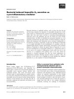
Fig. 2. CD spectra of EspB. (A) Far-UV and (B) near-UV CD spectra
of recombinant EspB purified from the insoluble fraction at pH 2.0
(dashed lines) and 7.0 (solid lines), and from the soluble fraction at
pH 7.0 (s). (C) The dependence of the ellipticity, at 222 nm, on pH.
D. Hamada et al. EspB is a natively partially folded protein
FEBS Journal 272 (2005) 756–768 ª 2005 FEBS 757
which is substantially less than the predicted amount
(76.3%; Table 1).
The near-UV CD spectrum of EspB at pH 7.0 and
20 °C shows a minimum at around 280 nm. It is in the
near-UV spectrum that aromatic residues display opti-
cal activity. EspB contains three tyrosines at positions
66, 75 and 212, and no tryptophans. Therefore, the
shape of the near-UV CD spectrum of EspB implies
the presence of some tertiary contacts involving at
least one of the tyrosines, although the intensity of
each peak is not very high.
To gain further insight into the conformational prop-
erties of EspB, we recorded the far-UV CD spectrum
of recombinant EspB prepared by different protocols
between pH 1.0 and pH 7.0 (Fig. 2C). Interestingly,
irrespective of the preparation procedures and pH con-
ditions, these far-UV CD spectra are almost identical.
Therefore, the amount of secondary structure seems to
be virtually same at each pH value (Fig. 2 and
Table 1). On the other hand, the near-UV CD spec-
trum at pH 2.0 showed a less intense signal at 280 nm
relative to the spectrum at pH 7.0, suggesting destabili-
zation of tertiary interactions upon decreasing pH.
Owing to the small difference observed here between
the recombinant proteins prepared by the different
procedures, we mostly used the EspB prepared from
insoluble fraction because the purification yield was
much higher.
Quenching of protein tyrosine fluorescence
by acrylamide
A fluorescence spectrum of intrinsic tryptophan and
tyrosine residues in proteins can be a good conforma-
tional probe. In particular, the fluorescence quenching
effect by small chemicals such as acrylamide provides
information on the solvent-exposure of aromatic side-
chains in proteins.
As mentioned above, EspB contains only three tyro-
sines and no tryptophan. The quenching effect of acryl-
amide on the fluorescence of these EspB tyrosine
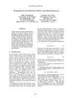
side-chains at pH 7.0 was analyzed. Interestingly, a
plot of F
0
⁄ F
obs
vs. [Q] (Stern–Volmer plot [27]), where
F
0
and F
obs
are the fluorescence intensities in the
absence and presence of quencher, respectively, and [Q]
is the concentration of quencher, shows a positive devi-
ation from linearity at high acrylamide concentrations
(Fig. 3). Therefore, the quenching behavior does not
follow the simple Stern–Volmer equation (F
0
⁄ F
obs
¼
1+K
sv
[Q]). This finding suggests that the tyrosine
residues in EspB behave as independent fluorophores,
each having their own K
sv
value. Additional informa-
tion was obtained by analyzing the data using the
following modified Stern–Volmer equation [28]:
Table 1. Secondary structure content of EspB at various pH values.
Values were calculated using the data from Fig. 2A in conjunction
with the
CDPRO package [70,71]. Predicted values are calculated
from the results of secondary structure prediction (Fig. 1) using the
PHDsec algorithm available at the PredictProtein server (http://
cubic.bioc.columbia.edu/predictprotein/) [24,25].
Condition a-Helix (%) b-Sheet (%) Others (%)
pH 1.0 26.2 ± 1.8 20.5 ± 1.1 53.4 ± 0.9
pH 2.0 22.8 ± 1.5 22.9 ± 1.7 53.5 ± 0.8
pH 3.0 27.5 ± 1.9 20.0 ± 1.7 52.5 ± 1.0
pH 4.0 31.1 ± 0.5 16.9 ± 0.9 52.0 ± 1.0
pH 5.0 26.6 ± 2.1 20.8 ± 1.9 52.5 ± 0.4
pH 6.0 23.5 ± 1.4 22.8 ± 1.8 53.3 ± 0.8
pH 7.0 23.1 ± 1.1 22.9 ± 1.2 53.1 ± 0.9
Predicted values 76.3 4.2 19.5
Fig. 3. Fluorescence quenching of intrinsic tyrosine. (A) Stern–Volmer
plot. (B) Modified Stern–Volmer plot (Eqn 1).
EspB is a natively partially folded protein D. Hamada et al.
758 FEBS Journal 272 (2005) 756–768 ª 2005 FEBS
F
0
=ðF
0
À F
obs
Þ¼1=ðf
a
K
sv
½QÞ þ 1=f
a
ð1Þ
where f
a
is the fraction of accessible tyrosines. The plot
of F
0
⁄ (F
0
– F
obs
) vs. 1 ⁄ [Q] (Fig. 3) shows a linear cor-
relation between F
0
⁄ (F
0
– F
obs
) and 1 ⁄ [Q]. The values
for K
sv
and f
a
are calculated as 32.7 ± 1.5Æm
)1
and
1.05 ± 0.01, respectively. An f
a
value close to 1 sug-
gests that the three tyrosine residues are likely to be
solvent-exposed at neutral pH.
ANS binding
ANS binds to solvent-accessible hydrophobic surfaces
and, when bound, an increase in ANS fluorescence
intensity near 500 nm occurs. This property of ANS is
often used to detect the presence of partially folded
protein intermediates, e.g. molten globules [29]. Molten
globule is originally defined as the partially folded state
of protein that assumes a significant amount of native-
like secondary structures but disrupted in tertiary struc-
tures [30–38]. We used ANS fluorescence spectroscopy
to probe the hydrophobic surface accessibility of EspB.
At pH 4 and 7, the ANS fluorescence is low
(Fig. 4), suggesting that hydrophobic surfaces are not
exposed to solvent. On the other hand, ANS fluores-
cence increases as the pH is decreased to 2.0 (Fig. 4).
This observation suggests that hydrophobic surfaces
are solvent-exposed at more acidic pH values. Under
the same conditions, the protein assumes an a-helical
conformation according to the far-UV CD spectrum at
pH 2.0 (Fig. 2). The results obtained by CD and ANS
fluorescence suggest the formation of a typical molten
globule structure for EspB at acidic pH 2.0.
Below pH 2.0, the ANS fluorescence decreased. For
these experiments, the pH of the solution was adjusted
by the addition of HCl. The decreased fluorescence
intensity may be caused by the quenching effect of
chloride ions on ANS fluorescence, rather than reflect-
ing additional conformational changes in EspB.
Urea unfolding
Urea-induced unfolding transitions of EspB were
monitored by far-UV CD spectroscopy. Plots of [h]
at 222 nm vs. urea concentration show co-operative
unfolding transitions throughout the pH range of
1.0–7.3 (Fig. 5). Between pH 3.0 and 7.3, unfolding
Fig. 4. 8-Anilinonaphthalene-1-sulfonate (ANS) fluorescence at
500 nm as a function of pH. Data were taken at 20 °C in the pres-
ence of 0.1 mgÆmL
)1
EspB. Circles represent the raw data. The line
is drawn only for visual assistance and is not a mathematical fit.
Fig. 5. Urea unfolding of EspB at various pH values and at 20 °C.
(A) The far-UV CD spectra obtained in the presence and absence
of urea. The numbers refer to the concentration of added urea.
(B) The urea-unfolding transition curves obtained at pH 2.0 (s),
pH 5.4 (h) and pH 7.3 (n). Continuous lines are theoretical curves.
The dotted and dashed lines correspond to the baselines for
unfolded and folded states.
D. Hamada et al. EspB is a natively partially folded protein
FEBS Journal 272 (2005) 756–768 ª 2005 FEBS 759
transitions occur between 2.5 and 4.5 m urea, but shift
to lower urea concentrations of 1.0–3.5 m at pH 2.0.
The urea-induced unfolding curves (Fig. 5) were
analyzed assuming a linear relationship between DG
and urea concentration and assuming a two-state fold-
ing mechanism, although there is no direct evidence
that the transitions are two-state in nature. The
derived parameters, DG
water
and m, are summarized in
Table 2.
Compared to the conformational state at pH 2.0,
those at higher pH values are stabilized by % 3–10 kJÆ
mol
)1
. However, their m-values, which probably cor-
relate with changes in the solvent-exposed surface
area associated with unfolding (DASA), are similar
regardless of pH. In our experiments, DASA largely
reflects structural changes in the folded species. There-
fore, given the m-values, EspB, at pH 2.0, which pos-
sesses molten globule-like properties, has a similar
accessible surface area as EspB conformations existing
at higher pH. Therefore, EspB at around neutral pH
should be a less compact structure than typical glob-
ular proteins.
Hydrodynamic property of EspB
The hydrodymanic property of EspB has been ana-
lyzed by multiangle dynamic scattering and ultracen-
trifugation.
Multiangle laser light scattering experiments for
EspB at pH 2.0 and pH 7.0 revealed the presence of
a single species with a Stokes radius of 3.7 and
3.1 nm, respectively (Fig. 6). A similar value was
obtained at pH 4.0 and pH 6.0 (3.4 and 3.5 nm,
respectively). This size is larger than the expected
value for a globular protein of 32 kDa molecular
mass, and corresponds to the value of globular pro-
teins, of % 70 kDa. From its amino acid sequence,
the molecular mass of the recombinant EspB is calcu-
Table 2. Values of DG
water
and m for urea-induced unfolding of
EspB.
pH
(kJÆmol
)1
)
DG
water
(kJÆmol
)1
ÆM
)1
) m
2.0 6.5 ± 2.1 3.9 ± 1.1
3.0 13.2 ± 1.7 3.9 ± 0.5
4.1 14.7 ± 1.9 4.1 ± 0.5
5.4 16.5 ± 1.6 3.8 ± 3.4
6.6 11.5 ± 2.3 3.4 ± 0.6
7.3 9.5 ± 1.3 3.0 ± 0.4
Fig. 6. Hydrodynamic property of EspB at
20 °C. Multiangle laser light scattering of
EspB at pH 2.0 (A) and pH 7.0 (B). (C) Sedi-
mentation equilibrium of EspB at pH 7.0.
The data were analyzed assuming a single
species in solution. In the lower panel, raw
data are shown by circles, and the line is
the theoretical curve. The upper panel
shows the difference between raw data and
theoretical values.
EspB is a natively partially folded protein D. Hamada et al.
760 FEBS Journal 272 (2005) 756–768 ª 2005 FEBS
lated to be 32 kDa. Thus, if EspB assumes a rigid
globular conformation at these conditions, the protein
should assume a dimeric structure.
The result of the sedimentation equilibrium study at
pH 7.0 also indicated the presence of only a single spe-
cies (Fig. 6C). The molecular mass estimated from this
experiment is, however, 34 kDa, which is similar to the
expected value for the monomeric EspB.
From these results, we concluded that EspB at
pH 7.0 assumes a relatively expanded monomeric con-
formation whose Stokes radius is approximately twice
as large as expected for a globular protein with a
molecular weight similar to that of EspB.
Importantly, the Stokes radius of EspB estimated
from light scattering was almost independent of pro-
tein concentration or pH value. This suggests that only
a single monomeric species is present in each protein
solution at pH 2.0–7.0.
Heteronuclear NMR spectroscopy
To further probe the structural properties of EspB, we
recorded its
15
N-
1
H heteronuclear single quantum cor-
relation (HSQC) spectra at pH 2.0 in the absence of
urea and at pH 7.0 in the presence and absence of
urea.
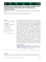
At pH 2.0, in the absence of urea, the
15
N-
1
H
HSQC spectrum shows little chemical shift dispersion
(Fig. 7). Although the resolution is poor owing to the
overlapping of peaks, the number of peaks that cor-
respond to the main-chain
1
H-
15
N crosspeaks was
estimated to be % 120. These peaks are relatively
sharp and may reflect the amino acid residues that
rapidly fluctuate with a timescale of nanosecond
order. The recombinant EspB used in this study
contains 333 amino acid residues. Thus, % 64% of
main-chain
1
H-
15
N crosspeaks are missing. A similar
phenomenon is often found for the molten globule
state, reflecting the slow fluctuation of a particular
region of protein molecules with a timescale of micro-
second to millisecond order. This is highly consistent
with our other spectroscopic studies, which show that
the protein, at pH 2.0, is in partially folded confor-
mation, similar to that of molten globules. Interest-
ingly, the NMR spectrum of EspB at pH 7.0 in the
absence of urea also shows little chemical shift disper-
sion, with % 110 possible main-chain
1
H-
15
N cros-
speaks. Thus, about 67% of main-chain
1
H-
15
N
crosspeaks are probably slowly fluctuating. Both of
the aforementioned NMR spectra are similar to the
spectrum obtained for urea-unfolded EspB at pH 7.0.
In this case, the number of peaks that correspond to
main-chain
1
H-
15
N crosspeaks slightly increased to
about 140. This implies the significant overlapping of
crosspeaks or the presence of some residual struc-
tures, even in the presence of 8.0 m urea.
The result of little chemical shift dispersions with the
small number of observable crosspeaks in the
15
N-
1
H
HSQC spectrum of EspB at pH 7.0 in the absence of
urea is inconsistent with the previous data obtained by
CD and fluorescence spectroscopies showing the pres-
ence of well-ordered conformations. This discrepancy
suggests that, at neutral pH, EspB assumes a natively
partially folded conformation without exposed hydro-
phobic clusters accessible to ANS molecules.
Fig. 7.
15
N-
1
H Heteronuclear single quantum correlation (HSQC)
spectra of EspB taken at 15 °C. (A) pH 7.0 in the absence of urea.
(B) pH 2.0 in the absence of urea. (C) pH 7.0 in the presence of
8.0
M urea.
D. Hamada et al. EspB is a natively partially folded protein
FEBS Journal 272 (2005) 756–768 ª 2005 FEBS 761
Discussion
Conformational properties of EspB
We analyzed the conformational properties of EspB by
using three spectroscopic techniques. The spectral
results suggest that EspB assumes intrinsically partially
folded conformations [39–44] under various conditions.
The different conformational states of EspB, found
under different pH conditions, are summarized in
Table 3. The shapes of the far-UV CD spectra suggest
the presence of a substantial amount of secondary
structure for EspB throughout the pH range of 1.0–
7.0. On the other hand, ANS fluorescence spectroscopy
indicates that EspB shows a conformational transition
involving the exposure of hydrophobic clusters when
the pH of the protein solution is decreased to 2.0.
Thus, at pH 2.0, the structure of EspB is consistent
with the traditional definition of a molten globule, i.e.
a compact partially folded state with a significant
amount of native-like secondary structures, but disrup-
ted in tertiary contacts [30–38].
Importantly, all EspB NMR spectra had chemical
shift signals that were less dispersed than those of
globular proteins, even at pH 7.0 in the absence of de-
naturant. The NMR spectra reported here are qualita-
tively similar to those observed for proteins that are
unstructured when in the presence of denaturants. On
the other hand, when analyzed by far-UV CD spectro-
scopy, EspB showed the presence of secondary
structures. EspB is therefore in a partially folded
conformation, even at near-physiological conditions.
However, ANS fluorescence spectroscopy suggests the
presence of a negligible amount of exposed hydropho-
bic surface for EspB at pH 7.0. This is a very unusual
result because partially folded proteins generally have
exposed hydrophobic clusters that are detected by
increases of ANS fluorescence intensity. One possible
explanation for the discrepancy could be that the
hydrophobic clusters, found for the EspB molten glob-
ule at pH 2.0, are disrupted in the structure found at
pH 7.0. A similar situation is, indeed, often found
for a-helical polypeptides in alcohol ⁄ water solvents
[45–51]. However, this explanation can be ruled out as
EspB is more stable at higher pH, which would prob-
ably be inconsistent with the loss of intramolecular
hydrophobic contacts. Thus, most EspB hydrophobic
clusters should be buried at neutral pH. Variations in
the conformational and thermodynamic properties of
molten globules have been characterized. For example,
the thermal unfolding experiments on the molten glob-
ule state of a-lactalbumin shows a gradual transition,
which suggests less organized hydrophobic contacts
[34]. However, the cytochrome c molten globule state
is highly ordered and the thermal unfolding transition
of this species is co-operative with a clear enthalpy
change upon unfolding [52,53]. This indicates that
some organized hydrophobic contacts exist in the mol-
ten globule state of cytochrome c. Furthermore, the
presence of tertiary contacts in the molten globule
states are shown by apomyoglobin and cytochrome c
[54,55], and EspB also showed the presence of weak,
but distinctive, peaks in the near-UV CD spectrum.
Therefore, EspB, at neutral pH, may have the charac-
ter of a highly ordered molten globule [56] with dis-
tinct and ordered regions probably stabilized by the
interactions between hydrophobic clusters. On the
other hand, the NMR data also indicate the presence
of highly fluctuating regions in EspB. As the data from
ultracentrifugation and laser light scattering suggest
that EspB assumes an expanded monomeric form,
EspB may assume a partially folded structure with
well-ordered regions and highly fluctuating regions
under near-physiological conditions.
Uversky et al. [41] proposed that natively unfolded
proteins tend to have a low mean hydrophobicity
and a relatively high net charge, and provided the
following expression of inequality, <H><(<R>
+1.151) ⁄ 2.785, between the hydrophobicity value
<H> and the mean net charge <R> for this class
of proteins. According to the amino acid sequence
of EspB, its <H> and <R> values are 0.478 and
0.013, respectively. These values actually do not satisfy
the above criteria.
Several www servers, which predict the disordered
regions in a protein from its amino acid sequence, are
currently available. We used GlobPlot (http://globplot.
embl.de/cgiDict.py), DisEMBL ( />cgiDict.py) and PONDRÒ ()
[57–60]. In the case of DisEMBL, some disordered
regions are predicted and, according to Remark-465
definition, residues at 12–27, 124–145, 157–188, 246–
257 and 303–312 are disordered. On the other hand,
GlobPlot, when using the Russel ⁄ Linding definition,
does not show a high probability for EspB to be largely
Table 3. The conformational properties of EspB at pH 2.0 and 7.0,
at a temperature of 20 °C. ANS, 8-anilinonaphthalene-1-sulfonate;
HSQC, heteronuclear single quantum correlation.
pH
Far-UV
CD
Near-UV
CD
Hydrophobic
exposure
by ANS
15
N-
1
H
HSQC
Urea
unfolding
7.0 Folded Folded Less exposed Unfolded Co-operative
2.0 Folded Partially
folded
Highly exposed Unfolded Co-operative
EspB is a natively partially folded protein D. Hamada et al.
762 FEBS Journal 272 (2005) 756–768 ª 2005 FEBS
disordered. The results from PONDRÒ suggest that
the amino acid residues at 1–53, 128–230 and 247–287
may be disordered. This is relatively consistent with the
prediction of DisEMBL. If the prediction from
PONDR is correct, only the amino acid sequences at
residues 54–127 and 288–325 of EspB, i.e. one-third of
the EspB sequence, assume ordered conformations.
This amount may overestimate the disordered regions
as the CD spectum indicates that about 50% of the
EspB sequence should be, at least, partially folded.
Of various approaches, only PONDR and DisEM-
BL indicated that EspB may be natively partially
folded. Incidentially, both prediction methods are
based on artificial neural networks, whereas GlobPlot
or the category shown by Uversky et al. [41] rely on
the physicochemical propensities of amino acids to
favor the disordered or globular structures. These
results may not be surprising as, in contrast to natively
unfolded proteins in general, EspB has a relatively
ordered conformation. Importantly, human a-lactalbu-
min, in the absence of Ca
2+
[34], adopts a typical mol-
ten globule structure at neutral pH. However, none of
the algorithims predict such a property of human
a-lactalbumin. Thus, the prediction of natively parti-
ally folded protein from its amino acid sequence
should still be difficult compared with the prediction
of natively unfolded proteins.
Implications for the function of EspB
It is well established that proteins fold to their unique
native conformations, as determined by their amino
acid sequences [61]. However, it is also clear that some
proteins are unable to maintain well-defined structures,
even under physiological conditions [39–44]. These
proteins are often called natively unfolded or intrinsic-
ally disordered proteins and assume either partially
folded or completely unfolded conformations in an
aqueous environment at neutral pH and, ideally, under
near-physiological conditions.
Our results clearly indicate that the structural char-
acteristics of EspB are those of a natively partially
folded protein. The far-UV CD spectra of IpaC, a
homolog of EspB from Shigella flexneri, revealed an
absence of significant amounts of secondary structure
at neutral pH [62]. Thus, the intrinsically less organ-
ized conformations of EspB and IpaC may be a com-
mon property for this class of proteins.
Importantly, some proteins that are natively unfol-
ded show dramatic conformational changes into well-
ordered structures when bound to their target
molecules [40–44]. Therefore, it will be important to
characterize the conformational state of EspB when
bound to its target molecules, e.g. EspA, EspD, a-cate-
nin and a1-antitrypsin.
Using the genomic sequence of E. coli, Dunker and
co-workers predicted that 8% of all proteins will have
intrinsically disordered segments of greater than 50 res-
idues in length [62]. Interestingly, the same predictions
indicated that this percentage increases to 41% for
Drosophila melanogaster proteins. Thus, intrinsically
structural protein disorder is probably a common
occurrence in vivo. It is unclear why structural disorder
would confer a physiological advantage to the function
of a protein function. Several possible reasons have
been proposed to answer this question [39–44]. For
example, if a protein is highly flexible, its association
with various targets of different molecular dimensions
and binding surfaces would be facilitated as different
conformations might be assumed. Indeed, EspB prob-
ably associates with various biological molecules, e.g.
EspA [20], EspD [17], a-catenin [18] and a1-antitrypsin
[19]. The molecular weights, physicochemical proper-
ties and functions of EspA, EspD, a-catenin and
a1-antitrypsin differ significantly. It is probable that
different areas of EspB bind different target molecules.
However, while EHEC and EPEC invade a variety of
animals with target molecules of varying amino acid
sequences, EspB should still specifically recognize
isoforms of the target molecules at the same binding
surfaces. Therefore, the conformational flexibility of a
virulence factor should provide a mechanism that
enables bacteria to infect various host species via the
same infection system.
Interestingly, exogenously added IpaC, an EspB
homolog from S. flexneri, enhanced the invasion activ-
ity of this bacterium into host cells [63]. As discussed
above, IpaC assumes an almost fully unstructured con-
formation near physiological conditions in vitro [64].
Such a property may also facilitate the penetration of
this molecule into host cells. Thus, the conformational
flexibility of EspB may also be advantageous for effi-
cient penetration into host cell membranes. This idea
is consistent with the concept that partial unfolding
may be required for the insertion of protein toxins into
host membranes [65].
Experimental procedures
Expression and purification of EspB
The cDNA, encoding EspB, was amplified from an EHEC
E. coli O157:H7 cosmid library (RIMD 0509890, Sakai
strain) [66,67] by PCR and cloned into a pT7 vector
(Novagen). The full-length espB gene was subcloned into
the expression vector pET28a (Novagen, Madison, WI,
D. Hamada et al. EspB is a natively partially folded protein
FEBS Journal 272 (2005) 756–768 ª 2005 FEBS 763
USA). The recombinant EspB has 20 amino acids, MGSS
HHHHHHSSGLVPRGSH, added at the N terminus of the
original sequence.
Recombinant EspB was expressed in E. coli BL21(DE3)
transformed with the afore mentioned plasmid. Cultures of
Luria–Bertani (LB) broth, supplemented with 50 lgÆmL
)1
of
kanamycin, were inoculated with colonies and grown over-
night at 37 °C with shaking. Then, a portion of each culture
was diluted 100-fold into 1 L of fresh LB medium and incu-
bated at 37 °C with shaking. Protein expression was induced
by the addition of isopropyl thio-b-d-galactoside (at a final
concentration of 1 mm) when cultures reached an attenuance
(D)of% 0.6 at 600 nm. For the expression of protein uni-
formly labeled with
15
N, M9 medium supplemented with
15
NH
4
Cl (Nippon Sanso Co., Kanagawa, Japan) was used
instead of LB medium. After 4 h of further shaking at
37 °C, the cells were harvested by centrifugation (10 min,
10 000 g,4°C) and placed on ice. Protein was expressed as
both soluble and insoluble fractions when the cells were dis-
rupted by 20 mm sodium phosphate, pH 7.0, containing
0.1% (v ⁄ v) Triton X-100. However, the solubility was quite
low. On the other hand, EspB cannot be extracted into the
soluble fraction when the cells are disrupted by 20 mm
sodium phosphate, pH 7.0. The purifications were therefore
performed from either the soluble fraction obtained by dis-
ruption of the cells in the presence of Triton X-100 or from
the insoluble fraction obtained by cell disruption in the
absence of detergent. For preparation from the soluble frac-
tion, the cells were suspended with 20 mm sodium phos-
phate, pH 7.0, containing 0.1% (v ⁄ v) Triton X-100 and the
solution was separated by centrifugation (15 000 g, 10 min,
4 °C). The solution was loaded onto Chelating Sepharose
Fast Flow (Amersham Biosciences, Corp., Piscataway, NJ,
USA) supplemented with NiCl
2
in 20 mm sodium phos-
phate, pH 7.0, washed with the same buffer and eluted using
a 0–1.0 m imidazole gradient. The eluted protein was further
purified with size-exclusion chromatography (S-300; 20 mm
sodium phosphate, pH 7.0; Amersham Biosciences, Corp.).
For preparation from the insoluble fraction, the cells were
suspended in 20 mm sodium phosphate, pH 7.0, and lysed
by sonication. The solution was centrifuged (15 000 g,
10 min, 4 °C) to separate the soluble and pellet fractions.
The protein was extracted from the pellet by the addition of
20 mm sodium phosphate, pH 7.0, containing 8.0 m urea.
This solution was clarified by centrifugation and diluted
100-fold by dropwise addition into 20 mm sodium phos-
phate, pH 7.0, at 4 °C. The solution was then purified by
Chelating Sepharose Fast Flow supplemented with NiCl
2
and further purified by size-exclusion chromatography
(S-300) as in the case of preparation from the soluble frac-
tions. The purification yields from soluble and insoluble
fractions were 15 and 30 mg from 1 L of culture in LB
medium, respectively. As judged by SDS ⁄ PAGE, the purity
of recombinant EspB prepared from the insoluble fraction is
relatively higher than that of EspB purified from the soluble
fraction. According to the CD spectrum, both purifications
yielded the same conformational state of EspB (see text for
details). Owing to the higher yields of purification,
15
N pro-
tein was prepared from insoluble fractions.
The protein concentration was determined by absorption
at 276 nm with the extinction coefficient of 4350 mÆcm
)1
calculated from amino acid composition [68]. The protein
solution was stored at )20 °C.
CD spectroscopy
CD spectra were measured by using a J-600 spectropola-
rimeter (Jasco, Tokyo, Japan). The temperature was held at
20 °C by using a thermostatically controlled cell holder in
conjunction with a circulating waterbath. For far-UV and
near-UV CD spectra, cells of 1 mm and 1 cm path length
were used, respectively. Protein concentrations were 0.1 and
1mgÆmL
)1
for far-UV and near-UV CD measurements,
respectively. The data are expressed as molar residue ellip-
ticity [69], [h], with [h] ¼ 100 h
obs
(cl)
)1
. The value, h
obs
,is
the observed intensity, c is the concentration in residue
moles per litre, and l is the path length in cm. A secondary
structure prediction was made by using the amino acid
sequence of EspB in conjunction with the program package
cdpro, in which selcon3, cdsstr and continll programs
are included [70,71]. The reported values are the average of
results from the above three programs.
Fluorescence spectroscopy
The fluorescence spectra of intrinsic EspB tyrosines and
ANS were measured by using a FP-777 fluorimeter (Jasco).
For tyrosine fluorescence, the excitation wavelength was
280 nm and the fluorescence emission was 300–350 nm. The
protein concentration was 0.1 mgÆmL
)1
. For ANS fluores-
cence, the excitation wavelength was 350 nm and the emis-
sion was measured between 400 and 650 nm. The protein
concentration was 0.1 mgÆmL
)1
and the ANS concentration
was 5 lm. The temperature was maintained at 20 °C with a
peltier-type thermostatically controlled cell holder.
Fluorescence quenching of EspB tyrosines was measured
in the presence of various concentrations of acrylamide,
with spectra acquired as described above.
Urea-induced unfolding measurements
Urea-unfolding curves were plotted with [h] at 222 nm vs.
the urea concentration. The data were analyzed assuming
a two-state unfolding mechanism and assuming that the
change in free energy of unfolding (DG), is linearly depend-
ent on urea concentration:
DG ¼ DG
water
À m½ureað2Þ
Here, DG
water
corresponds to DG of unfolding in the
absence of urea; m is a measure of the co-operativity
EspB is a natively partially folded protein D. Hamada et al.
764 FEBS Journal 272 (2005) 756–768 ª 2005 FEBS
of the unfolding transition; and [urea] is the urea concen-
tration.
The fractions of unfolded (f
U
) and folded (f
F
) species at
various urea concentrations can be expressed as:
f
U
¼ 1=½1 þ expðÀDGR
À1
T
À1
Þ ð3Þ
and as:
f
F
¼ 1 À f
U
ð4Þ
where R is the gas constant and T is the temperature in
Kelvin.
The theoretical value of [h] at 222 nm ([h]
222
), observed
in the presence of various concentrations of urea, can be
expressed as:
½h
222
¼½h
F
f
F
þ½h
U
f
U
ð5Þ
Here, [h ]
F
and [h]
U
are the [h]
222
of the folded and unfolded
species, respectively.
The values for DG
water
and m were obtained by nonlinear
curve fitting to the transition curves, according to
Eqns (2–5), by using the program igorpro (WaveMetrics
Inc., Lake Oswego, OR, USA). The linear dependences of
[h]
F
and [h]
U
on urea concentrations were also considered
in the fitting analysis. The same baselines for folded and
unfolded species were used for the fitting of data obtained
at pH 2.0–7.0.
Multiangle laser light scattering
Multiangle laser light scattering data were obtained by
using a dynapro Molecular Sizing Instrument (Protein
Solutions Inc., Milton Keynes, UK) at 20 °C. Various con-
centrations of protein solution at pH 2.0, 4.0, 6.0 and 7.0
(400 lL) were passed through 0.22 lm of centrifugal filter
unit, ultrafree-MC from Millipore (Billerica, MA, USA),
and further centrifuged at 20 000 g for 10 min. Only the
clear solution at the top of a tube (100 lL) was used for
the light scattering analysis.
Ultracentrifugation
Sedimentation equilibrium experiments were performed by
using a Beckman Optima XL-I analytical ultracentrifuge
(Fullerton, CA, USA) at 11 300 g,20°C. The protein con-
centration was 3 mgÆmL
)1
.
NMR spectroscopy
2D
15
N-
1
H HSQC spectra were recorded at 15 °C on either
a 500 or an 800 MHz spectrometer (Brucker DRX500 or
DRX800, respectively, Brucker Biospin GmbH, Karlsruhe,
Germany), each equipped with a triple axis gradient and a
triple-resonance probe. Protein concentrations were 1–2 mm
in buffered solution containing 10%
2
H
2
O. For DRX500
experiments, the number of complex points and spectral
widths were 1024, 12019 Hz (
1
H, F
2
) and 64, 1168 Hz
(
15
N, F
1
), and for those using the DRX800 spectrometer,
the parameters were 1024, 12821 Hz (
1
H, F
2
) and 64,
1866 Hz (
15
N, F
1
). The
1
H carrier was set at 4.7 p.p.m.,
and the
15
N carrier at 120 p.p.m. The
15
N-
1
H HSQC
experiments included the WATERGATE and Water-flip-
back techniques. The data were processed by using nmrpipe
[72] and visualized by using sparky (TD Goddard & DG
Kneller, University of California, San Francisco, CA, USA;
/>Acknowledgements
The authors acknowledge Prof. Yuji Goto for access
to the CD spectropolarimeter, Prof. Atsushi Nakagawa
for access to light scattering, and Miyo Sakai for per-
forming ultracentrifugation. This work was supported,
in part, by grants-in-aid for scientific research from the
Japan Ministry of Education, Science, Culture and
Sports, and JSPS Research Fellowships for Young Sci-
entists (to D.H.).
References
1 Galan JE & Collmer A (1999) Type III secretion
machines: bacterial devices for protein delivery into host
cells. Science 284, 1322–1328.
2 McDaniel TK, Jarvis KG, Donnenberg MS & Kaper
JB (1995) A genetic locus of enterocyte effacement con-
served among diverse enterobactrial pathogens. Proc
Natl Acad Sci USA 92, 1664–1668.
3 McDaniel TK & Kaper JB (1997) A cloned pathogeni-
city island from enteropathogenic Escherichia coli con-
fers the attaching and effacing phenotype on K-12
E. coli. Mol Microbiol 23, 399–407.
4 Perna NT, Mayhew GF, Po
´
sfai G, Eliott SJ, Donnenberg
MS, Kaper JB & Blattner FR (1998) Molecular evolution
of a pathogenicity island from enterohaemorrhagic
Escherichia coli O157:H7. Infect Immun 66, 3810–3817.
5 Zhu C, Agin TS, Elliott SJ, Johnson LA, Thate TE,
Kaper JB & Boedeker EC (2001) Complete nucleotide
sequence and analysis of the locus of enterocyte efface-
ment from rabbit diarrheagenic Escherichia coli
RDEC-1. Infect Immun 69 , 2107–2115.
6 Shuch R & Maurelli AT (2000) The Type III secretion
pathway. Dictating the outcome of bacterial–host inter-
actions. In Virulence Mechanisms of Bacterial Pathogens,
3rd edn (Brogden KA, Roth JA, Stanton TB, Bolin
CA, Minion FC & Wannemuehler MJ, eds), pp. 203–
233. ASM Press, American Society for Microbiology,
Washington, DC, USA.
7 Nougayre
`
de J-P, Fernandes PJ & Donnenberg MS
(2003) Adhesion of enteropathogenic Escherichia coli to
host cells. Cell Microbiol 5, 359–372.
D. Hamada et al. EspB is a natively partially folded protein
FEBS Journal 272 (2005) 756–768 ª 2005 FEBS 765
8 Kenny B, DeVinney R, Stein M, Reinscheid DJ, Frey
EA & Finlay BB (1997) Enteropathogenic E. coli
(EPEC) transfers its receptor for intimate adherence
into mammalian cells. Cell 91, 511–520.
9 Vlademir VC, Takahashi A, Yanagihara I, Akeda Y,
Imura K, Kodama T, Kono G, Sato Y & Honda T
(2001) Talin, a host cell protein, interacts directly with
the translocated intimin receptor, Tir, of enteropatho-
genic Escherichia coli, and is essential for pedestal
formation. Cell Microbiol 3, 745–751.
10 Vlademir VC, Takahashi A, Yanagihara I, Akeda Y,
Imura K, Kodama T, Kono G, Sato Y, Iida T &
Honda T (2002) Cortactin is necessary for F-actin
accumulation in pedestal structure induced by entero-
pathogenic Escherichia coli infection. Infect Immun 70,
2206–2209.
11 Crane JK, McNamara BP & Donnenberg MS (2001)
Role of EspF in host cell death induced by enteropatho-
genic Escherichia coli. Cell Microbiol 3 , 197–211.
12 McNamara BP & Donnenberg MS (1998) A novel pro-
line-rich protein, EspF, is secreted from enteropatho-
genic Escherichia coli via the type III export pathway.
FEMS Microbiol Lett 166, 71–78.
13 Elliott SJ, Krejany EO, Mellies JL, Robins-Browne
RM, Sasakawa C & Kaper JB (2001) EspG, a novel
type III system-secreted protein from enteropathogenic
Escherichia coli with similarities to VirA of Shigella
flexneri. Infect Immun 69, 4027–4033.
14 Kenny B & Jepson M (2000) Targeting of an entero-
pathogenic Escherichia coli (EPEC) effector protein to
host mitochondria. Cell Microbiol 2, 579–590.
15 Clarke SC, Haigh RD, Freestone PP & Williams PH
(2003) Virulence of enteropathogenic Escherichia coli,a
global pathogen. Clin Microbiol Rev 16 , 65–78.
16 Roe AJ, Hoey DE & Gally DL (2003) Regulation,
secretion and activity of type III-secreted proteins of
enterohaemorrhagic Escherichia coli O157. Biochem Soc
Trans 31, 98–103.
17 Ide T, Laarmann S, Greune L, Schillers H, Oberleithner
H & Schmidt MA (2001) Characterization of transloca-
tion pores inserted into plasma membranes by type III-
secreted Esp proteins of enteropathogenic Escherichia
coli. Cell Microbiol 3, 669–679.
18 Kodama T, Akeda Y, Kono G, Takahashi A, Imura K,
Iida T & Honda T (2002) The EspB protein of entero-
haemorrhagic Escherichia coli interacts directly with
a-catenin. Cell Microbiol 4, 213–222.
19 Knappstein S, Ide T, Schmidt MA & Heusipp G (2004)
a1-Antitrypsin binds to and interferes with functionality
of EspB from atypical and typical enteropathogenic
Escherichia coli strains. Infect Immun 72, 4344–4350.
20 Hartland E, Daniell SJ, Dalahay RM, Neves BC, Wallis
T, Shaw RK, Hale C, Knutton S & Frankel G (2000)
The type III protein translocation system of entero-
pathogenic Escherichia coli involves EspA–EspB protein
interactions. Mol Microbiol 35, 1483–1492.
21 Daniell SJ, Kocsis E, Morris E, Knutton S, Booy FP &
Frankel G (2003) 3D structure of EspA filaments from
enteropathogenic Escherichia coli. Mol Microbiol 49,
301–308.
22 Sekiya K, Ohishi M, Ogino T, Tamano K, Sasakawa C
& Abe A (2001) Supermolecular structure of the entero-
pathogenic Escherichia coli type III secretion system and
its direct interaction with the EspA-sheath-like struc-
ture. Proc Natl Acad Sci USA 98, 11638–11643.
23 Knutton S, Rosenshine I, Pallen MJ, Nisan I, Neves
BC, Bain C, Wolff C, Dougan G & Frankel G (1998) A
novel EspA-associated surface organelle of enteropatho-
genic Escherichia coli involved in protein translocation
into epithelial cells. EMBO J 17, 2166–2176.
24 Rost B & Sander C (1993) Prediction of protein struc-
ture at better than 70% accuracy. J Mol Biol 232, 584–
599.
25 Rost B & Sander C (1994) Combining evolutionary
information and neural networks to predict protein
secondary structure. Proteins 19, 55–72.
26 Rost B, Sander C & Schneider R (1994) PHD – an
Automatic Mail Server for Protein Secondary Structure
Prediction. CABIOS 10, 53–60.
27 Lehrer SS & Leavis PC (1978) Solute quenching of pro-
tein fluorescence. Methods Enzymol 49, 222–236.
28 Lehrer SS (1971) Solute perturbation of protein fluores-
cence. The quenching of the tryptophan fluorescence of
model compounds and lysozyme by iodide ion. Bio-
chemistry 10, 3254–3263.
29 Semisotnov GV, Rodionova NA, Kutyshenko VP,
Ebert B, Blanck J & Ptitsyn OB (1987) Sequential
mechanism of refolding of carbonic anhydrase B. FEBS
Lett 224, 9–13.
30 Arai M & Kuwajima K (2000) Role of the molten
globule state in protein folding. Adv Protein Chem 53,
209–282.
31 Creighton TE (1997) How important is the molten glob-
ule for correct protein folding? Trends Biochem Sci 22,
6–10.
32 Kuwajima K (1989) The molten globule state as a clue
for understanding the folding and cooperativity of glob-
ular-protein structure. Proteins 6, 87–103.
33 Kuwajima K (1992) Protein folding in vitro. Curr Opin
Biotechnol 3, 462–467.
34 Kuwajima K (1996) The molten globule state of a-lact-
albumin. FASEB J 10, 102–109.
35 Ohgushi M & Wada A (1983) ‘Molten-globule state’: a
compact form of globular proteins with mobile side-
chains. FEBS Lett 164, 21–24.
36 Ohgushi M & Wada A (1984) Liquid-like state of side
chains at the intermediate stage of protein denaturation.
Adv Biophys 18, 75–90.
EspB is a natively partially folded protein D. Hamada et al.
766 FEBS Journal 272 (2005) 756–768 ª 2005 FEBS
37 Ptitsyn OB (1995) How the molten globule became.
Trends Biochem Sci 20, 376–379.
38 Ptitsyn OB (1995) Molten globule and protein folding.
Adv Protein Chem 47, 83–229.
39 Dunker AK, Lawson JD, Brown CJ, Williams RM,
Romero P, Oh JS, Oldfield CJ, Campen AM, Ratliff
CM, Hipps KW et al. (2001) Intrinsically disordered
protein. J Mol Graph Model 19, 26–59.
40 Dyson HJ & Wright PE (2002) Coupling of folding and
binding for unstructured proteins. Curr Opin Struct Biol
12, 54–60.
41 Uversky VN, Gilespie JR & Ink AL (2000) Why are
‘natively unfolded’ proteins unstructred under physio-
logic conditions? Proteins 41, 415–427.
42 Uversky VN (2002) Natively unfolded proteins: a point
where biology waits for physics. Protein Sci 11, 739–
756.
43 Uversky VN (2002) What does it mean to be natively
unfolded? Eur J Biochem 269, 2–12.
44 Wright PE & Dyson HJ (1999) Intrinsically unstruc-
tured proteins: re-assessing the protein structure–func-
tion paradigm. J Mol Biol 293, 321–331.
45 Hamada D, Kuroda Y, Tanaka T & Goto Y (1995)
High helical propensity of the peptide fragments derived
from b-lactoglobulin, a predominantly b-sheet protein.
J Mol Biol 254, 737–746.
46 Hirota-Nakaoka N & Goto Y (1999) Alcohol-induced
denaturation of b-lactoglobulin: a close correlation to
the alcohol-induced a-helix formation of melittin.
Bioorg Med Chem 7, 67–73.
47 Hirota N, Mizuno K & Goto Y (1997) Cooperative
a-helix formation of b-lactoglobulin and melittin
induced by hexafluoroisopropanol. Protein Sci 6,
416–421.
48 Hirota N, Mizuno K & Goto Y (1998) Group additive
contributions to the alcohol-induced a-helix formation
of melittin: implication for the mechanism of the alco-
hol effects on proteins. J Mol Biol 275, 365–378.
49 Hoshino M, Hagihara Y, Hamada D, Kataoka M &
Goto Y (1997) Trifluoroethanol-induced conformational
transition of hen egg-white lysozyme studied by small-
angle X-ray scattering. FEBS Lett 416, 72–76.
50 Khan F, Khan RH & Muzammil S (2000) Alcohol-
induced versus anion-induced states of a-chymotrypsi-
nogen A at low pH. Biochim Biophys Acta 1481,
229–236.
51 Kumar S, Modig K & Halle B (2003) Trifluoroethanol-
induced b fi a transition in b-lactoglobulin: hydration
and cosolvent binding studied by
2
H,
17
O, and
19
F mag-
netic relaxation dispersion. Biochemistry 42, 13708–
13716.
52 Hagihara Y, Tan Y & Goto Y (1994) Comparison of
the conformational stability of the molten globule and
native states of horse cytochrome c. J Mol Biol 237,
336–348.
53 Hamada D, Kidokoro S, Fukada H, Takahashi K &
Goto Y (1994) Salt-induced formation of the molten
globule state of cytochrome c studied by isothermal
titration calorimetry. Proc Natl Acad Sci USA 91,
10325–10329.
54 Bertagna AM & Barrick D (2004) Nonspecific hydro-
phobic interactions stabilize an equilibrium interemedi-
ate of apomyoglobin at a key position within the
AGH region. Proc Natl Acad Sci USA 101, 12514–
12519.
55 Marmorino JL, Lehti M & Pielak G (1998) Native ter-
tiary structure in an A-state. J Mol Biol 275, 379–388.
56 Redfield C, Smith RA & Dobson CM (1994) Structural
characterization of a highly-ordered ‘molten globule’ at
low pH. Nature Struct Biol 1, 23–29.
57 Li X, Romero P, Rani M, Dunker AK & Obradovic Z
(1999) Predicting protein disorder for N-, C-, and inter-
nal regions. Genome Informatics 10, 30–40.
58 Romero P, Obradovic Z, Li X, Garner E, Brown C &
Dunker AK (2001) Sequence complexity of disordered
protein. Proteins: Struct Funct Gen 42, 38–48.
59 Romero P, Obradovic Z & Dunker AK (1997) Sequence
data analysis for long disordered regions prediction in
the calcineurin family. Genome Informatics 8, 110–124.
60 Romero P, Obradovic Z, Kissinger CR, Villafranca JE
& Dunker AK (1997) Identifying disordered regions in
proteins from amino acid sequences. Proc I.E.E.E.
International Conference on Neural Networks 1, 90–95.
61 Anfinsen CB (1973) Principles that govern the folding
of protein chains. Science 181, 223–230.
62 Dunker AK, Obradovic Z, Romeo P, Garner EC &
Brown CJ (2000) Intrinsic protein disorder in complete
genomes. Genome Inform Series Workshop Genome
Inform 11, 161–171.
63 Kueltzo LA, Osiecki J, Barker J, Picking WL, Ersoy B,
Picking WD & Middaugh R (2003) Structure–function
analysis of invasion plasmid antigen C (IpaC) from
Shigella flexneri. J Biol Chem 278, 2792–2797.
64 Tran N, Serfis AB, Osiecki JC, Pocking WL, Coye L,
Davis R & Picking WD (2000) Interaction of Shigella
flexneri IpaC with model membranes correlates with
effects on cultured cells. Infect Immun 68, 3710–3715.
65 van der Goot FG, Lakey JH & Pattus F (1992) The
molten globule intermediate for protein insertion or
translocation through membranes. Trends Cell Biol 2,
343–348.
66 Hayashi T, Makino K, Ohnishi M, Kurokawa K, Ishii
K, Yokoyama K, Han CG, Ohtsubo E, Nakayama K,
Murata T et al. (2001) Complete genome sequence of
enterohaemorrhagic Escherichia coli O157:H7 and
genomic comparison with a laboratory strain K-12.
DNA Res 8, 11–22.
67 Perna NT, Plunkett G, III, Burland V, Mau B, Glasner
JD, Rose DJ, Mayhew GF, Evans PS, Gregor J,
Kirkpatrick HA et al. (2001) Genome sequence of
D. Hamada et al. EspB is a natively partially folded protein
FEBS Journal 272 (2005) 756–768 ª 2005 FEBS 767
enterohaemorrhagic Escherichia coli O157:H7. Nature
409, 529–533.
68 Gill SC & von Hippel PH (1989) Calculation of protein
extinction coefficients from amino acid sequence data.
Anal Biochem 182, 319–326.
69 Hamada D, Kuroda Y, Kataoka M, Aimoto S,
Yoshimura T & Goto Y (1996) Role of heme axial
ligands in the conformational stability of the native and
molten globule states of horse cytochrome c. J Mol Biol
256, 172–186.
70 Sreerama N & Woody RW (2000) Estimation of protein
secondary structure from circular dichroism spectra:
comparison of CONTIN, SELCON, and CDSSTR
methods with an expanded reference set. Anal Biochem
287, 252–260.
71 Sreerama N, Venyaminov SY & Woody RW (2000)
Estimation of protein secondary structure from circular
dichroism spectra: inclusion of denatured proteins with
native proteins in the analysis. Anal Biochem 287, 243–
251.
72 Delaglio F, Grzesiek S, Vuister G, Zhu G, Pfeifer J &
Bax A (1995) NMRPipe: a multidimensional spectral
processing system based on UNIX pipes. J Biomol
NMR 6, 277–293.
EspB is a natively partially folded protein D. Hamada et al.
768 FEBS Journal 272 (2005) 756–768 ª 2005 FEBS