Báo cáo khoa học: Entamoeba histolytica TATA-box binding protein binds to different TATA variants in vitro ppt
Bạn đang xem bản rút gọn của tài liệu. Xem và tải ngay bản đầy đủ của tài liệu tại đây (401.45 KB, 13 trang )
Entamoeba histolytica TATA-box binding protein binds
to different TATA variants in vitro
Guadalupe de Dios-Bravo
1,2
, Juan Pedro Luna-Arias
3
, Ana Marı
´a
Rivero
´
n
4
, Jose
´
J Olivares-Trejo
5
,
Ce
´
sar Lo
´
pez-Camarillo
2
and Esther Orozco
5
1 Programa de Biomedicina Molecular, Escuela Nacional de Medicina y Homeopatı
´
a del Instituto Polite
´
cnico Nacional, Me
´
xico
2 Programa de Ciencias Geno
´
micas, Universidad de la Ciudad de Me
´
xico, Me
´
xico
3 Departamento de Biologı
´
a Celular, Centro de Investigacio
´
n y de Estudios Avanzados, Me
´
xico
4 Departamento de Biologı
´
a Molecular, Centro Nacional de Investigacio
´
n Cientı
´
fica (CNIC), Habana, Cuba
5 Departamento de Patologia Experimental, Centro de Investigacio
´
n y de Estudios Avanzados, Me
´
xico
Entamoeba histolytica is the protozoan responsible for
human amoebiasis. E. histolytica strains have distinct
capacity to damage cultured cells and human tissues
[1–4]. Expression of many molecules and cellular func-
tions involved in E. histolytica pathogenicity such as
lectins [5,6], adherence molecules [7], proteases [8,9]
and amoebapores [10] correlates with its virulence.
Variability in virulence exhibited by E. histolytica
strains might be controlled in part by transcription of
these and other virulence genes.
Transcription factors cooperate with other proteins
to regulate gene expression. First, the preinitiation
complex (PIC) is positioned around the transcription
initiation site and then, PIC interacts with other
proteins bound to upstream motifs to facilitate the
RNA polymerase II function. The absence or the pres-
ence of some nuclear factors interacting with PIC may
inhibit or promote gene expression to modulate cellu-
lar functions [11–13]. Mechanisms, molecules and
DNA sequences controlling the spatial and temporal
Keywords
Entamoeba histolytica; K
D
; promiscuous
DNA-binding activity; TATA-binding protein;
TATA variants
Correspondence
Esther Orozco, Departamento de Patologı
´
a
Experimental, Centro de Investigacio
´
nyde
Estudios Avanzados, IPN. C. P. 07360,
Me
´
xico, D. F.
Fax: +52 55 57477108
Tel: +52 55 50613800 ext 5642
E-mail:
(Received 23 June 2004, revised 8 December
2004, accepted 11 January 2005)
doi:10.1111/j.1742-4658.2005.04566.x
The ability of Entamoeba histolytica TATA binding protein (EhTBP) to
interact with different TATA boxes in gene promoters may be one of the
key factors to perform an efficient transcription in this human parasite. In
this paper we used several TATA variants to study the in vitro EhTBP
DNA-binding activity and to determine the TATA-EhTBP dissociation
constants. The presence of EhTBP in complexes formed by nuclear extracts
(NE) and the TATTTAAA oligonucleotide, which corresponds to the
canonical TATA box for E. histolytica, was demonstrated by gel-shift
assays. In these experiments a single NE-TATTTAAA oligonucleotide
complex was detected. Complex was retarded by anti-EhTBP Igs in super-
shift experiments and antibodies also recognized the cross-linked complex
in Western blot assays. Recombinant EhTBP formed specific complexes
with TATA variants found in E. histolytica gene promoters and other
TATA variants generated by mutation of TATTTAAA sequence. The dis-
sociation constants of recombinant EhTBP for TATA variants ranged
between 1.04 (±0.39) · 10
)11
and 1.60 (±0.37) · 10
)10
m. TATTTAAA
and TAT_ _AAA motifs presented the lowest K
D
values. Intriguingly, the
recombinant EhTBP affinity for TATA variants is stronger than other
TBPs reported. In addition, EhTBP is more promiscuous than human and
yeast TBPs, probably due to modifications in amino acids involved in
TBP-DNA binding.
Abbreviations
EhTBP, Entamoeba histolytica TATA-box binding protein; EMSA, electrophoretic mobility shift assays; rEhTBP, recombinant Entamoeba
histolytica TATA-box binding protein; NE, nuclear extracts.
1354 FEBS Journal 272 (2005) 1354–1366 ª 2005 FEBS
transcription patterns during growth, differentiation
and development have been widely studied [14,15].
In eukaryotes, general transcription factors such as
TFIID ⁄ TFIIB, TFIIA, TFIIE, TFIIF ⁄ RNA poly-
merase II and TFIIH are assembled on the core pro-
moter before transcription begins [16,17].
The TATA binding protein (TBP) is the first fac-
tor that binds DNA to recruit proteins on PIC and
initiate gene transcription [18,19]. Mammalian TBPs
can productively bind to a large number of diverse
TATA elements. An exhaustive statistical genomic
survey documented that the TATA box is an A ⁄ T-
rich 8 bp segment, often flanked by G⁄ C-rich
sequences [20].
Certain E. histolytica genes are activated or down
regulated during liver abscesses production by tro-
phozoites [9] and during epithelia colonization and
invasion. However, we ignore which transcription
factors modulate these events and others related to
the parasite survival such as trophozoites differenti-
ation into cysts. Few transcription factors have been
detected and cloned in E. histolytica. URE3-BP,
EhEBP1 and EhEBP2 proteins regulate the hgl5 gene
expression [21,22] and an EhC ⁄ EBP-like protein is
involved in EhPgp1 gene activation [23,24]. Addition-
ally, Ehtbp [25] and Ehp53 [26] have been character-
ized as the orthologous of the mammalian tbp and
p53 genes, respectively. The E. histolytica TATA-
binding protein (EhTBP) is the only member of the
basal transcription machinery cloned and character-
ized in this parasite.
The EhTBP functional DNA-binding domain has
55% homology with human TBP, whereas the
EhTBP N-terminal domain is rich in hydrophobic
residues and quite different from mammalian TBPs
[25]. EhTBP C-terminus displays a predicted protein
structure fitting to the crystallized human TBP
[27,28], but its biological activity has been poorly
studied.
E. histolytica gene promoters have the TATTTAAA
sequence, which is considered as the canonical TATA
box for EhTBP [29]. However, EhTBP binding to this
sequence has not been fully demonstrated. In addition,
TATTTAAA variants have been found surrounding
the core promoter, suggesting that these motifs could
also act as TATA boxes [30,31], but EhTBP affinity
for these sequences is also unknown. In this paper, we
studied the in vitro EhTBP binding affinity for differ-
ent TATA sequences found in E. histolytica gene pro-
moters and others designed by us producing mutations
in the TATTTAAA sequence. We also calculated the
K
D
of recombinant E. histolytica TBP (rEhTBP) for
several TATA variants.
Results
E. histolytica nuclear extracts (NE) and TATTT-
AAA(1) oligonucleotide form specific complexes
Bruchhaus et al. [29] using NE in EMSA and doing
in silico analysis proposed that the TATTTAAA
sequence is the consensus TATA box for E. histolytica.
On the other hand, Luna-Arias et al. [25] showed the
homology of EhTBP with human TBP. However,
the presence of EhTBP in complexes formed with
E. histolytica NE and TATTTAAA(1) oligonucleotide
has not been directly demonstrated yet. We first investi-
gated the presence of EhTBP in the complex formed by
E. histolytica NE and TATTTAAA(1) oligonucleotide
by supershift, cross-linking and Western blot assays.
When incubated with fresh NE, TATTTAAA(1) oligo-
nucleotide migration was retarded, forming a single
band (Fig. 1A, lane 1). The NE-TATTTAAA(1) com-
plex was specifically competed by TATTTAAA(1) cold
oligonucleotide (Fig. 1A, lane 2), whereas it remained
when double-stranded poly(dG-dC) or TtTTTttt(7)
oligonucleotide were used as unspecific competitors
(Fig. 1A, lanes 3 and 4, respectively). The presence of
EhTBP in this complex was evidenced in supershift
assays by anti-rEhTBP Igs. Two bands appeared when
1 lL of antibodies was added to the mixture (Fig. 1B,
lane 3). The lower band comigrated with that formed
by NE and TATTTAAA(1) oligonucleotide, whereas
the other band migrated slower, due to the partial
supershift produced by the antibody. When 5 lL
of anti-rEhTBP Igs were added to the mixture, the
complex was completely disrupted (Fig. 1B, lane 4), as
it has been reported for other supershift experiments
[32]. Anti-E. histolytica actin antibodies had no effect
on the complex formed (Fig. 1C, lane 2).
In cross-linking assays, using a UV-irradiated mix-
ture of E. histolytica NE and TATTTAAA(1) oligonu-
cleotide, we distinguished a radioactive DNA–protein
band of 50 kDa (Fig. 1D, lane 5). This band may be
formed by the radioactive probe (11 kDa) bound to
endogenous EhTBP (26 kDa) and other protein cross-
linked to the complex. As expected, the 50 kDa radio-
active band was competed by TATTTAAA(1) cold
oligonucleotide (Fig. 1D, lane 6), but it remained in
the presence of the poly (dG-dC) unspecific competitor
(Fig. 1D lane 7). No complexes were detected in lanes
with either nonirradiated or irradiated free probe, or
with the nonirradiated oligonucleotide-NE mixture
(Fig. 1D, lanes 2–4, respectively). In Western blot
assays of UV cross-linked DNA–protein complexes,
anti-rEhTBP Igs recognized the radioactive 50 kDa
band. This confirms that EhTBP is part of the complex
G. de Dios-Bravo et al. Promiscuous E. histolytica TBP
FEBS Journal 272 (2005) 1354–1366 ª 2005 FEBS 1355
probably associated to another % 13 kDa unknown
protein (Fig. 1E, lanes 5–7). The antibodies also recog-
nized the same band in the lane where TATTTAAA(1)
cold oligonucleotide was used as specific competitor
(Fig. 1E, lane 6). As expected, in this lane the complex
was formed by the irradiated cold oligonucleotide and
NE mixture. Unbound 26 kDa EhTBP comigrated in
the gel with the 25 kDa marker (Fig. 1E, lanes 4–7).
Data from these experiments altogether demonstrated
the presence of EhTBP in NE-TATTTAAA(1) oligo-
nucleotide complexes.
Recombinant EhTBP binds to TATTTAAA(1)
oligonucleotide
The rEhTBP was expressed in bacteria as a His
6
-
tagged 30 kDa polypeptide. rEhTBP was purified by
affinity chromatography and its integrity and identity
were verified by Coomassie blue stained gels
(SDS ⁄ PAGE) (Fig. 2A) and Western blot assays using
anti-rEhTBP Igs (Fig. 2B). In EMSA, purified rEhTBP
formed a single band with TATTTAAA(1) probe
(Fig. 2C, lane 2). The complex was competed by cold
TATTTAAA(1) oligonucleotide, whereas it remained
in the presence of poly (dG-dC) unspecific competitor
(Fig. 2C, lanes 3 and 4). To discard endogenous
TATA binding activity in bacterial extracts, we tested
by EMSA the capacity of induced and noninduced
bacterial extracts to form complexes with TATTT
AAA(1) probe. Results showed that complex was only
formed with extracts of induced bacteria expressing
rEhTBP (Fig. 2D, lane 3) whereas noninduced bacteria
did not form any complexes with TATTTAAA(1)
oligonucleotide (Fig. 2D, lane 2).
DNA-binding activity of purified rEhTBP
for TATA variants
In humans and other organisms, variants of the canon-
ical TATA box have been reported to be functional
[33,34]. On the other hand, the TATTTAAA sequence
and several variants are found in many E. histolytica
gene promoters at ) 20 to )40 bp upstream the tran-
scription initiation site [30], although other TATA
variants have been experimentally found at longer
distances in Ehtbp and EhRabB genes (our unpublished
data). We studied the binding activity of rEhTBP for
different TATA sequences present in gene promoters
(oligonucleotides TATTTAAA(1), TAT_ _AAA(4),
TAT_ _AAg(5) and TATTaAAA(6)), and for mutated
versions of TATTTAAA(1) probe [oligonucleotides
TAgTgAAA(2) and TATTggAA(3)] (Table 1). We
ABCD E
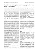
Fig. 1. Binding of nuclear extracts to TATTTAAA(1) oligonucleotide. (A) NE (25 lg) and [
32
P]ATP[cP] end-labeled TATTTAAA(1) oligonucleotide
(10 000 c.p.m., 157 p
M) were incubated for 15 min at 4 °C for EMSA as described in Experimental procedures. Lane 1, no competitor; lane
2, 300-fold molar excess of unlabeled TATTTAAA(1) oligonucleotide as specific competitor (sc); as unspecific competitors we added 300-fold
molar excess of: lane 3, poly(dG-dC) and, lane 4, oligo(dT)
18
. (B) Supershift gel assay using purified anti-rEhTBP Igs. EMSA were performed
as above, except that before adding the labeled oligonucleotide, the mixture was preincubated with: lane 1, no NE; lane 2, no antibody; lane
3, 1 lL of purified anti-rEhTBP Igs; lane 4, 5 lL of anti-rEhTBP Igs. (C) Supershift gel assay performed as in B, but using anti-E. histolytica
actin Igs. Lane 1, no antibody; lane 2, 5 lL of anti-E. histolytica actin Ig. (D) UV-cross-linking assay of NE (60 lg) and TATTTAAA(1) (50 000
c.p.m., 785 p
M). Mixtures for EMSA were UV irradiated at 320 nm for 10 min at 4 °C, analyzed by 12% SDS ⁄ PAGE and radioactivity was
determined as described in Experimental procedures. Lane 1, molecular mass markers; lane 2, nonirradiated free probe; lane 3, irradiated
free probe; lane 4 , nonirradiated NE-oligonucleotide mixture; lane 5, irradiated NE-oligonucleotide mixture; lane 6, irradiated NE-oligonucleo-
tide mixture containing 300-fold molar excess of unlabeled TATTTAAA(1) oligonucleotide as specific competitor (sc); lane 7, irradiated
NE-oligonucleotide mixture containing 300-fold molar excess of poly (dG-dC) as unspecific competitor (uc). (E) Western blot assay of UV
cross-linked DNA–protein complexes shown in D, using anti-rEhTBP Igs.
Promiscuous E. histolytica TBP G. de Dios-Bravo et al.
1356 FEBS Journal 272 (2005) 1354–1366 ª 2005 FEBS
introduced g’s in the third, fifth and sixth positions of
the TATTTAAA(1) sequence, because these positions
have been reported as important for DNA-binding
activity for human and yeast TBPs [33,34].
DNA-binding activity of rEhTBP for distinct TATA
oligonucleotides was evaluated by EMSA using
433 nm (over-saturating concentration) of purified
rEhTBP and 10 000 c.p.m. (157 pm) of the probes.
Figure 3 displays experiments showing that rEhTBP
specifically binds to all oligonucleotides tested. Com-
plexes formed by rEhTBP and TATA box variants
were fully competed by the same probe and by TAT-
TTAAA(1) oligonucleotide (Fig. 3). In these assays,
two complexes were observed with TAT_ _AAA(4)
and TAT_ _AAg(5) probes, which were specifically
competed by TATTTAAA(1) oligonucleotide and by
the same probe. The presence of two complexes in
some experiments could be due to conformational
Table 1. Positions of TATTTAAA (1) sequence and putative TATA variants in E. histolytica gene promoters.
TATA variants Gene promoter First nucleotide location
b
Reference
(12 3456 78)
a
5’-T A T T T A A A-3’ (1) EhPgp5
Ehactin
(-31)
c
(-30)
d
[47]
( />5’-T A g T g A A A-3’ (2) Not found
5’-T A T T ggA A-3’ (3) Not found
5’-T A T __A A A-3’ (4) Ehtbp (-109)
c
(Unpublished)
Ehtub1 (-27)
d
( />EhRabB (-44)
c
(Unpublished)
5’-T A T __AAg-3’ (5) Ehenol (-50)
d
( />5’-T A T T a A A A-3’ (6) Ehpfo (-31)
c
[48]
a
Numbers show the base composition in TATA variants.
b
Nucleotide position is referred to the experimentally
c
and in silico
d
determined
transcription initiation sites. Putative TATA boxes are defined as TATA sequences upstream of the ATTCA ⁄ G, ATCA or ACGC consensus
transcription initiation sites.
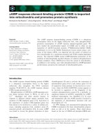
AB C D
Fig. 2. Immunodetection of rEhTBP, and EMSA of TATTTAAA(1) and rEhTBP. rEhTBP was produced by IPTG induced bacteria transformed
with the full length Ehtbp gene cloned in pRSET A and purified through nickel NTA-agarose columns as described in Experimental proce-
dures. (A) Coomassie blue stained gel (12% SDS ⁄ PAGE) of purified rEhTBP under native conditions. Lane 1, molecular mass markers; lane
2, purified rEhTBP. (B) Western blot assay of purified rEhTBP using anti-rEhTBP Igs. Lane 1, molecular weight markers; lane 2, stripe
sequentially incubated with anti-rEhTBP Igs and peroxidase-coupled goat anti-rabbit secondary Igs; lane 3, as in lane 2 but anti-rEhTBP Igs
were omitted. (C) EMSA of purified rEhTBP with TATTTAAA(1) oligonucleotide as described in Experimental procedures. Lane 1, free probe;
lane 2, no competitor; lane 3, 300-fold molar excess of unlabeled TATTTAAA(1) probe as specific competitor (sc); lane 4, 300-fold molar
excess of unspecific competitor (uc). (D) EMSA using 15 lg of bacterial extracts. Lane 1, free probe; lane 2, non induced bacteria (nib) carry-
ing pRSET A-Ehtbp plasmid; lane 3, induced bacteria (ib) expressing rEhTBP.
G. de Dios-Bravo et al. Promiscuous E. histolytica TBP
FEBS Journal 272 (2005) 1354–1366 ª 2005 FEBS 1357
changes of the DNA–protein complex, which may
affect its electrophoretic migration. To discard the pos-
sibility that rEhTBP could bind to any AT rich
sequence, we performed a shift assay with rEhTBP
and [
32
P]ATP[cP] end-labeled double stranded
TtTTTttt(7), TATaTAtA(8) or TtTTaAAA(9) oligonu-
cleotides. rEhTBP did not bind to these sequences
(data not shown), indicating that rEhTBP is not
merely an AT-rich DNA binding protein without dis-
crimination capacity. Obviously, these oligonucleotides
did not compete the complex formed with TATTT-
AAA(1) and rEhTBP (Fig. 3F). We also verified that
in our experiments, rEhTBP was indeed bound to dou-
ble-stranded oligonucleotides and not to free labeled
single-stranded probes. Labeled probes were passed
through a hydroxyapatite column and c.p.m. were
counted in the unbound and eluted fractions. In all
cases, more than 99% of the radioactivity was found
bound to the hydroxyapatite column and it was eluted
with 0.4 m phosphate buffer. Figure 3G shows the elu-
tion profile for TATTTAAA(1) oligonucleotide as a
representative experiment. All together these results
showed that rEhTBP has an in vitro binding capacity
for distinct TATA elements.
Quantification of rEhTBP DNA-binding activity
for different TATA oligonucleotides
Binding activity of rEhTBP for TATA variants was
quantified (as described in Experimental procedures) at
A
FG
BCD
E
Fig. 3. rEhTBP specifically binds to TATTTAAA(1) oligonucleotide and TATA variants. (A–E) Purified rEhTBP (433 nM) was incubated with
[
32
P]ATP[cP] end-labeled TATA variants (10 000 c.p.m., 157 pM) for EMSA as described in Experimental procedures. Lane 1, free probe; lane
2, no competitor; lane 3, competition with 300-fold molar excess of the same TATA variant as specific competitor (sc); lane 4, competition
with 300-fold molar excess of TATTTAAA(1) oligonucleotide; lane 5, competition with 300-fold molar excess of unspecific competitor (uc).
The TATA oligonucleotide used in each case is shown below each gel. (F) Control binding assay of rEhTBP (433 n
M) with 157 pM (10 000
c.p.m.) of TATTTAAA(1) probe. Lane 1, TATTTAAA(1) free probe; lane 2, purified rEhTBP incubated with TATTTAAA(1) probe; lane 3, purified
rEhTBP preincubated with 300-fold molar excess of unlabeled poly (dG-dC) before adding the labeled TATTTAAA(1) probe; lane 4, unlabeled
TTTTTTTT(7) oligonucleotide; lane 5, unlabeled TATATATA(8) oligonucleotide, and lane 6 TTTTAAAA(9) oligonucleotide were used as unspe-
cific competitors. (G) Elution profile of labeled TATTTAAA (1) probe passed through a hydroxyapatite column as described in Experimental
procedures. Fraction 1, unbound single stranded DNA (SS); fractions 2–6, washes with 2.5 mL of 0.12
M phosphate buffer pH 6.8 (W); frac-
tions 7–11, elution with 2.5 mL of 0.4
M phosphate buffer pH 6.8 (DS). Volume of each fraction was 0.5 mL. Radioactivity was represented
as percentage of the total radioactivity (30 000 c.p.m.) loaded into the column.
Promiscuous E. histolytica TBP G. de Dios-Bravo et al.
1358 FEBS Journal 272 (2005) 1354–1366 ª 2005 FEBS
distinct total rEhTBP concentrations (Fig. 4A–F).
Experimental variations for each gel were normalized
using the total radioactivity in each lane (complex
formed plus free oligonucleotide at the bottom of the
gel).
Quantification of DNA–protein complexes in each
EMSA experiment was performed to calculate K
D
val-
ues of the rEhTBP and each TATA oligonucleotide
using the method described by Coleman and Pugh [35]
(see Experimental procedures). First, we estimated the
c.p.m. present in the shifted protein-TATA oligonucleo-
tide (S
x
) for each rEhTBP concentration tested (x).
Then, the natural logarithms of S
x
(ln S
x
) values were
plotted against a given rEhTBP concentration (x). As
c.p.m. is a discrete variable, we used ln S
x
(Eqn 2) to
warrant normal distribution of the residual error E in
the regression analysis [36,37] in order to obtain more
precise and representative data. Thus, these experimen-
tal points were fitted as a polynomial function of x
(Eqn 2) (Fig. 5). In all cases we obtained a second
degree polynomial function describing the relationship
between ln S
x
and x (Table 2). The summary of coeffi-
cients, variances, and results of the statistical Student’s
t-tests obtained are also presented in Table 2.
Once we had defined the mathematical relationship
between ln S
x
and x for each experiment, we deter-
mined the average amount of radioactivity (S
f
) present
in the rEhTBP–TATA oligonucleotide complexes when
the reaction reached the titration end point. This was
done first using Eqn 3 to calculate the x-value at which
the mathematical function has a maximum (x
max
).
For oligonucleotides TATTTAAA(1), TAgTgAAA(2),
TATTggAA(3), TAT_ _AAA(4), and TAT_ _AAg(5),
the x
max
values were 280, 287, 281, 232 and 270 nm,
which corresponded to S
f
values of 324 ± 6, 1038 ±
20, 702 ± 34, 335 ± 10, 431 ± 9 c.p.m., respectively.
In the case of TATTaAAA(6) oligonucleotide, which
formed two specific DNA–protein complexes, the x
max
values were 261 and 307 nm for the slower and faster
bands, respectively, which corresponded to 190 ± 5
and 325 ± 4 c.p.m.
The next step was to obtain F-values using Eqn 1 as
described in Experimental procedures and plot it ver-
sus the rEhTBP ⁄ TATA molar ratio. F-values also fit-
ted to a polynomial function of the molar ratio of
rEhTBP ⁄ TATA oligonucleotide. Figure 6A shows an
example of the F experimental points obtained for
TAT_ _AAg(5) oligonucleotide. Results for all oligo-
nucleotides showed a second degree polynomial func-
tion (Table 2). The statistical test gave similar results
to those obtained for Eqn 2. Then, we obtained the
rEhTBP ⁄ TATA oligonucleotide molar ratios at which
F corresponds to 1 (maximum value). rEhTBP ⁄ TATA
oligonucleotide molar ratios were 1458, 1959, 1599,
2006 and 2030 for TATTTAAA(1), TAgTgAAA(2),
TATTggAA(3), TAT_ _AAA(4), and TAT_ _AAg(5)
ABC
DEF
Fig. 4. Affinity quantification of rEhTBP-TATA variant complexes as a function of the rEhTBP concentration by EMSA. (A–F) EMSA of [
32
P]
ATP[cP] end-labeled TATA variants (10 000 c.p.m., 157 p
M) incubated with different rEhTBP concentrations as described in Experimental pro-
cedures: lane 1, 0 n
M; lane 2, 50 nM; lane 3, 97 nM; lane 4, 145 nM; lane 5, 193 nM; lane 6, 242 nM; lane 7, 290 nM, and lane 8, 338 n M.
Arrows show the complexes analyzed. The TATA oligonucleotide used in each case is shown below each gel.
G. de Dios-Bravo et al. Promiscuous E. histolytica TBP
FEBS Journal 272 (2005) 1354–1366 ª 2005 FEBS 1359
Fig. 5. Graphical representation of data obtained in quantification of rEhTBP-TATA variant complexes. (A–F) ln of S
x
(the radioactivity present
in DNA–protein complexes) versus x (rEhTBP concentrations). Data were obtained from EMSA experiments as shown in Fig. 4. F-1 and F-2
correspond to the slower and faster DNA–protein complexes formed with TATTaAAA(6) oligonucleotide, respectively. Dots represent experi-
mental data. Continuous line is the graph predicted by the second degree polynomial function (Eqn 2 in Experimental procedures). The TATA
oligonucleotide used in each case is shown below each graph.
Table 2. Mathematical relationships between ln S
x
vs. x and ln F vs. rEhTBP ⁄ TATA molar ratio. a, Coefficients of the equation ln Sx ¼ a
0
+
a
1
x+a
2
x
2
+E) N(0,1) and ln F ¼ a
0
+a
1
(rEhTBP ⁄ TATA) + a
2
(rEhTBP ⁄ TATA)2 + E ) N(0,1). Sa is the standard deviation of a
n
coefficients.
I, slower DNA-protein complex; II, faster DNA-protein complex.
ln S
x
vs. x ln F vs. rEHTBP ⁄ TATA molar ratio
a
0
a
1
a
2
S
2
a
0
S
2
a
1
S
2
a
2
a
0
a
1
a
2
S
2
a
0
S
2
a
1
S
2
a
2
1 5.85 7.66 x 10
)3
)1.34 · 10
)5
1.20 · 10
)1
1.48 · 10
)6
6.01 · 10
)12
)1.10 1.12 · 10
)3
)2.86 · 10
)7
1.20 · 10
)1
3.16 · 10
)8
2.76 · 10
)15
2 4.37 1.56 · 10
)2
)2.77 · 10
)5
8.24 · 10
)1
1.25 · 10
)5
6.31 · 10
)11
)2.19 2.74 · 10
)5
)8.56 · 10
)7
8.24 · 10
)1
3.87 · 10
)7
6.03 · 10
)14
3 5.04 7.91 · 10
)3
)1.16 · 10
)5
1.68 · 10
)1
2.55 · 10
)6
1.29 · 10
)11
)1.34 1.24 · 10
)3
)2.87 · 10
)7
1.68 · 10
)1
6.29 · 10
)8
7.80 · 10
)15
4 5.48 4.34 · 10
)3
)8.03 · 10
)6
1.96 · 10
)1
2.98 · 10
)6
1.50 · 10
)11
)5.87 5.78 · 10
)4
)1.42 · 10
)7
1.96 · 10
)1
5.30 · 10
)8
4.72 · 10
)15
5 3.39 1.42 · 10
)2
)2.73 · 10
)5
2.44 · 10
)1
3.71 · 10
)6
1.87 · 10
)11
)1.86 2.23 · 10
)3
)6.71 · 10
)7
2.44 · 10
)1
9.13 · 10
)8
1.13 · 10
)14
6I 4.61 7.62 · 10
)3
)1.24 · 10
)5
6.54 · 10
)2
9.94 · 10
)7
5.01 · 10
)12
)1.17 1.46 · 10
)3
)4.57 · 10
)7
6.54 · 10
)2
3.67 · 10
)8
6.82 · 10
)15
6II 4.76 7.28 · 10
)3
)1.30 · 10
)5
1.16 · 10
)1
1.76 · 10
)6
8.85 · 10
)12
)1.02 1.40 · 10
)3
)4.80 · 10
)7
1.16 · 10
)1
6.48 · 10
)8
1.20 · 10
)14
Promiscuous E. histolytica TBP G. de Dios-Bravo et al.
1360 FEBS Journal 272 (2005) 1354–1366 ª 2005 FEBS
oligonucleotides, respectively. For the complexes
formed with TATTaAAA(6) oligonucleotide, the val-
ues were 1664 and 1599 for the slower and faster
bands, respectively. The reciprocal of all these values
were then used to calculate the total active rEhTBP
concentrations (P
T
) (see Experimental procedures) as
reported [35].
K
D
values of rEhTBP for TATA variants
The reciprocal of F-values gave the following linear
function: (1 ⁄ F) ¼ 1+K
D
(1 ⁄ P). The slope of this lin-
ear function corresponds to K
D
. Therefore, the data
(1 ⁄ F) and (1 ⁄ P) were fitted using the robust linear
regression method [36,37], which should give an equa-
tion of the type (1 ⁄ F) ¼ c
0
+ c
1
(1 ⁄ P) if the variables
were linearly related. An example of the fitness between
these variables using the robust linear regression
method is presented for TAT_ _AAg(5) oligonucleo-
tide (Fig. 6B). These calculations were performed for
all TATA oligonucleotides.
K
D
values and their standard deviations are shown
in Table 3. K
D
values of rEhTBP for TATA variants
ranged between 1.04 (± 0.39) · 10
)11
and 1.60
(± 0.37) · 10
)10
m, which corresponded to oligonucleo-
tides TAT_ _AAA(4) and TAgTgAAA(2), respectively.
TATTTAAA(1) and TAT_ _AAA(4) oligonucleotides
had the lowest K
D
values that did not significantly dif-
fer each other (Table 3). Additionally, oligonucleotides
TATTggAA(3) and the two complexes formed with
TATTaAAA(6) gave similar K
D
values. The next
larger value corresponded to TAT_ _AAg(5), and the
largest to TAgTgAAA(2). Therefore, we could order
the oligonucleotides according to their TBP affinity as
follows: TATTTAAA(1) ¼ TAT_ _AAA(4) > TATTgg
AA(3) ¼ TATTaAAA(6) > TAT_ _AAg(5) > TAgTg
AAA(2).
Discussion
In this paper we studied the rEhTBP affinity for sev-
eral TATA variants present in E. histolytica gene pro-
moters and TATA box versions designed by us
(Table 1). Our data showed that the promiscuity of rE-
hTBP for TATA variants is higher than those reported
for Homo sapiens, Saccharomyces cerevisiae and Ara-
bidopsis thaliana TBPs [33,38]. Therefore, in addition
to TATTTAAA(1) sequence, we showed here that
TAT_ _AAA(4), TAT_ _AAg(5) and TATTaAAA(6)
are, at least in vitro, EhTBP binding motifs. In addi-
tion, rEhTBP can also bind in vitro to TAgTgAAA(2)
and TATTggAA(3) oligonucleotides that are mutated
versions of TATTTAAA(1) sequence. Thus, based on
our in vitro experiments, the E. histolytica TATA
box could be proposed as 5¢-(1: T)(2: A)(3: T ⁄ G)(4:
T ⁄ G ⁄ A)(5: T ⁄ G ⁄ A)(6: A ⁄ G)(7: A) (8: A)-3¢ (numbers
indicate the nucleotide position in TATA box). In vitro
transcription assays are needed to accurately establish
Fig. 6. Relationships between ln F and rEhTBP ⁄ TAT_ _AAg(5)
molar ratio, and 1 ⁄ F and 1 ⁄ P. (A) Experimental data obtained from
relationships between ln F and rEhTBP ⁄ TAT_ _AAg(5) molar ratio.
(B) Graphical representation of 1 ⁄ F and 1 ⁄ P for the rEhTBP ⁄
TAT_ _AAg(5) complexes. The dots represent experimentally
obtained data. The continuous line is the graph predicted by the
second degree polynomial function (A) and linear function for K
D
estimation (B).
Table 3. Dissociation constants of rEhTBP for TATA variants.
Oligonucleotide (K
D
± SD) M
5’-TATTTAAA-3’ (1) 1.96 (± 0.58) · 10
-11
5’-TAgTgAAA-3’ (2) 1.60 (± 0.37) · 10
-10
5’-TATTggAA-3’ (3) 3.18 (± 1.16) · 10
-11
5’-TAT_ _AAA-3’ (4) 1.04 (± 0.39) · 10
-11
5’-TAT_ _AAg-3’ (5) 8.26 (± 2.20) · 10
-11
5’-TATTaAAA -3’ (6) 4.28 (± 0.47) · 10
-11 a
3.94 (± 0.44) · 10
-11 b
a
Upper DNA-protein complex;
b
lower DNA-protein complex; SD,
Standard deviation.
G. de Dios-Bravo et al. Promiscuous E. histolytica TBP
FEBS Journal 272 (2005) 1354–1366 ª 2005 FEBS 1361
the quantitative value of each base in different position
and in vivo experiments will demonstrate the function
of these TATA elements in the cell.
Based on a systematic X-ray crystallographic study
of the A. thaliana TBP isoform 2, Patikoglou et al. [38]
defined the TATA sequence as an eight bp variable
motif formed by 5¢-T ) c>a¼ g ⁄ A ) t ⁄ T ) a ¼
c ⁄ A ) t ⁄ T ) a ⁄ A ) g>c¼ t ⁄ A ¼ T>g>c⁄ G ¼
A>c¼ t-3¢. Recently, in S. cerevisiae, Basehoar et al.
[39] identified the TATA box element as TAT
A(A ⁄ T)A(A ⁄ T)(A ⁄ G). A. thaliana TBP isoform 2
recognizes 10 variants of the adenovirus major late pro-
moter TATA element that in many organisms is
located at )25 to )40 bp from the transcription initi-
ation site. However, some S. cerevisiae gene promoters
have the TATA box at )40 to )120 bp [13,40] and
the Ehtbp gene presents the TAT_ _ AAA sequence
located at )109 bp from the transcription initiation site
that, accordingly to our unpublished data, might func-
tion as TATA element. Additionally, recent results
gave also evidence that the EhRabB gene promoter
TATA box maps at )44 bp (Rodrı
´
guez, M.A., personal
communication) (Table 1).
In about 20 E. histolytica genes, the transcription ini-
tiation site is known [29] and from these data the TAT
TTAAA sequence has been proposed as the canonical
EhTBP binding motif. However, this is the first
published report experimentally demonstrating that
rEhTBP binds to TATTTAAA and other related
sequences. Here, we experimentally showed that
EhTBP is in the complex formed in vitro by the consen-
sus TATTTAAA(1) oligonucleotide and NE (Fig. 1)
and that rEhTBP specifically binds to this DNA
sequence (Fig. 2). However, E. histolytica genes contain
different TATA elements (Table 1) that could be used
by TFIID transcription factor. We also showed that
rEhTBP forms specific complexes with all TATA vari-
ants tested, although with different affinity, showing a
more relaxed DNA-binding specificity of EhTBP than
those described for other systems [33,34,38,41].
DNA-binding activities of H. sapiens [34,38], A. thali-
ana [38] and S. cerevisiae [33,34] TBPs are severely affec-
ted when TATA oligonucleotides contain g’s in first,
second, third, fourth, fifth and sixth positions. In con-
trast, EhTBP formed complexes with TAgTgAAA(2)
and TATTggAA(3) oligonucleotides used here (Fig. 3),
indicating that g’s in these positions do not affect
EhTBP DNA-binding activity, and showing that at least
in in vitro assays, EhTBP is even more promiscuous than
other TBPs studied. In vivo studies are needed to define
whether this also occurs in the trophozoites.
The dip in data at higher titration point in curves of
Figs 5 and 6 can be explained by the dimerization [35]
or oligomerization [42] of TBP molecules at high TBP
concentration. These multimers have no ability to bind
DNA [35,42]. We cannot discard multiple TBP binding
events.
K
D
values of rEhTBP for TATA variants ranged
from 10
)11
to 10
)10
m (Table 3). These results indica-
ted that: (1) the rEhTBP has similar affinity for TAT
TTAAA(1) and TAT_ _AAA(4) oligonucleotides (2)
variations of the nucleotide at position 5 slightly
reduce the affinity of rEhTBP for TATA variant
in relation to the TATTTAAA oligonucleotide;
(3) TAT_ _AAg(5) and TAgTgAAA(2) oligonucleo-
tides are bound by rEhTBP with less affinity than
those TATA variants designed by us.
An alignment of EhTBP with the H. sapiens,
A. thaliana and S. cerevisiae TBPs showed that EhTBP
has the residues reported as involved in TATA box
binding in the same positions than other TBPs [27,38].
Thirty of them are identical (Fig. 7, open arrowheads)
and of the remaining seven residues, five are conserved
and two are nonconserved changes (Fig. 7, filled
arrowheads). Interestingly, EhTBP presents a T in
position 192, which corresponds to V203 in yeast, to
V161 in A. thaliana and to V301 in human TBPs
(Fig. 7, arrow). Strubin and Struhl [34] substituted the
V203 of yeast TBP by T and the resultant mutant TBP
showed an increased DNA-binding activity for the
TGTAAA element of the his3 gene promoter. Thus,
the presence of T192 in EhTBP sequence could influ-
ence its DNA-binding specificity for TATA variants.
However, this is still to be experimentally demonstra-
ted. The promiscuous DNA-binding activity of EhTBP
may have conferred an evolutionary advantage to
E. histolytica, because certain mutations in the TATA
box would not affect gene expression.
Experimental procedures
E. histolytica cultures
Trophozoites of E. histolytica clone A (strain HM1:IMSS)
[1] were axenically cultured in TYI-S-33 medium at 37 °C
and harvested during exponential growth phase [43].
Electrophoretic mobility shift assays (EMSA),
competitions and supershift gel assays using
NE and rEhTBP
Aliquots of 25 lg of NE, obtained as described [23], or 30–
300 ng (50–500 nm) of purified rEhTBP [25] were used for
EMSA. NE or rEhTBP were incubated for 15 min at 4 °C
with poly(dG-dC) (1 lgÆlL
)1
) in binding buffer containing
12 mm Hepes pH 7.9, 60 mm KCl, 10% (v ⁄ v) glycerol and
Promiscuous E. histolytica TBP G. de Dios-Bravo et al.
1362 FEBS Journal 272 (2005) 1354–1366 ª 2005 FEBS
1mm each dithiothreitol, EDTA, spermidine and MgCl
2
.
Then, [
32
P]ATP[cP] end-labeled double-stranded oligo-
nucleotides (10 000 c.p.m., 157 pm): 5¢-TATTTAAA-3¢(1),
5¢-TAgTgAAA-3¢(2), 5¢-TATTggAA-3¢(3), 5¢-TAT_ _AAA-
3¢(4), 5¢-TAT_ _AAg-3¢(5), 5¢-TATTaAAA-3¢(6) (Table 1),
5¢-TtTTTttt-3¢(7), 5¢-TATaTAtA-3¢(8) or 5¢-TtTTaAAA-
3¢(9) were added to the mixture. Incubation continued for
other 10 min at 4 °C. Mutations introduced in TATTT
AAA box are marked in small letters and deletions are in
dashes in the oligonucleotide sequences. In all cases, flank-
ing bases were added to obtain 18 bp length oligonucleo-
tides. Numbers in parenthesis after the sequences identify
each oligonucleotide. For supershift gel assays, before add-
ing oligonucleotides, the mixture was preincubated for
15 min at 4 °C with 1 or 5 lL of purified rabbit anti-
rEhTBP Igs [25] or with 5 lL of anti-E. histolytica actin Igs
(kindly given by Manuel Herna
´
ndez, CINVESTAV, IPN,
Me
´
xico). In competition experiments, 300-fold molar excess
of tested oligonucleotide, or the TATTTAAA(1) oligo-
nucleotide, or the unspecific competitors poly(dG-dC),
TtTTTttt(7), TATaTAtA(8) or TtTTaAAA(9) were incuba-
ted with the mixture 10 min before the probe was added.
Samples were electrophoresed on 6% nondenaturing
polyacrylamide gels (PAGE) in 0.5X TBE. Gels were
vacuum-dried and radioactive complexes were detected in a
Phosphor Imager apparatus (Bio-Rad). Shifted radioactive
bands were quantified by densitometry using the Quantity
One software version 4 (Bio-Rad). All experiments reported
here were carried out at least three times by duplicate with
reproducible results. To discard the presence of single-stran-
ded oligonucleotides, labeled probes (30 000 c.p.m.) were
passed through a hydroxyapatite (Bio-Rad) column (1 cm
in length · 0.7 cm in diameter) at 4 °C. We collected
0.5 mL fractions. The flowthrough contained the unbound
material, which corresponds to single stranded DNA. The
column was washed with 2.5 mL of 0.12 m phosphate buf-
fer pH 6.8 and double-stranded DNA was eluted with
2.5 mL of 0.4 m phosphate buffer pH 6.8 [44]. Finally,
radioactivity in each fraction was measured in a Beckman
LS 6500 liquid scintillation counter.
Cross-linking and Western blot assays
Protein concentration of NE was measured by the Bradford
method [45]. For cross-linking assays, 60 lg of proteins
were incubated with radioactive TATTTAAA(1) oligonucle-
otide (50 000 c.p.m., 785 pm) and UV irradiated at 320 nm
directly on a transilluminator apparatus (UVP Inc., San
Gabriel, CA, USA) for 10 min at 4 °C. Complexes formed
after cross-linking assays were resolved through 12%
SDS ⁄ PAGE. Gels were scanned in a Phosphor Imager
apparatus and were transferred to nitrocellulose membranes
for Western blot assays [46]. Membranes were blocked with
0.05% (v ⁄ v) Tween 20 and 5% (w ⁄ v) nonfat milk in
NaCl ⁄ P
i
for 2 h at room temperature and incubated over-
night at 4 °C with purified anti-rEhTBP Igs (1 : 1000). Im-
munoreactivity was detected with peroxidase-labeled goat
anti-rabbit Igs (Zymed, San Francisco, CA, USA)
(1 : 2000) and chemiluminescence method, using ECL Plus
Kit (Amersham, Piscataway, NJ, USA).
Expression and purification of rEhTBP and
anti-rEhTBP Igs generation
The full-length Ehtbp gene, cloned in pRSET A [25] was
expressed in Escherichia coli BL21(DE3)pLysS strain
(Invitrogen, Carlsbad, CA, USA) as a His
6
-tagged 30 kDa
polypeptide. Proteins from IPTG (1 mm) induced bacteria
were separated by 12% SDS ⁄ PAGE and gels were
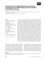
Fig. 7. Predicted amino acid residues
involved in EhTBP binding to DNA.
Conserved C-terminal domain sequences of
TBPs from Saccharomycces cerevisiae
(ScTBP) (P13393), Arabidopsis thaliana
(ArathTBP2) (P28148), Homo sapiens (hTBP)
(P20226) and E. histolytica (EhTBP) (P52653)
were aligned using the
CLUSTAL W program.
Black boxes indicate identical amino acids in
at least two sequences and grey boxes the
amino acid conserved changes. Unfilled and
filled arrowheads indicate the 37 amino acid
residues involved in DNA binding activity.
Filled arrowheads correspond to the seven
amino acid residues involved in DNA binding
activity that are changed in EhTBP
sequence. Arrow denotes the amino acid
change in EhTBP position 192.
G. de Dios-Bravo et al. Promiscuous E. histolytica TBP
FEBS Journal 272 (2005) 1354–1366 ª 2005 FEBS 1363
Coomassie blue stained. rEhTBP was purified by nickel-
agarose affinity columns as described by the manufacturer
(Qiagen, Standford, CA, USA). Then, 150 lg of purified
rEhTBP were subcutaneously inoculated three times in
rabbits each 15 days. One week after last immunization,
rabbits were bled. Antibodies were twice precipitated from
serum with 60% (w ⁄ v) (NH
4
)
2
SO
4
, dialyzed using NaCl ⁄ P
i
buffer and immunoadsorbed against nitrocellulose-immobi-
lized rEhTBP before using them for supershift and Western
blot assays.
Quantification of DNA–protein complexes
Complexes formed by distinct probes with rEhTPB were
quantified by densitometry measuring pixels in bands by
the quantity one software and normalized against free
probe to correct the total c.p.m. loaded in each lane of the
gel.
Determination of dissociation constants (K
D
)
of DNA–protein complexes
The K
D
for EhTBP and each TATA oligonucleotide was
determined by EMSA as described [35] with some modifica-
tions. Complexes formation with rEhTBP and radiolabeled
oligonucleotides was measured as a function of protein con-
centration. The fraction (F) of DNA probe bound by pro-
teins was calculated using the Eqn 1
F ¼ðS
x
À S
0
Þ=ðS
f
À S
0
ÞðEqn 1Þ
where, S
x
is the radioactivity present in the shifted protein-
DNA complex at protein concentration x. S
0
is the corres-
ponding radioactivity present when x ¼ 0 (i.e. the absence of
protein). S
f
is the average amount of radioactivity present
when F becomes independent of x (i.e. when the reaction rea-
ches the titration end point).
To accurately determine S
f
values for each EMSA experi-
ment we plotted the S
x
values for each rEhTBP concentra-
tion tested (x). Then, we determined the polynomial
function best describing the curve behavior as:
ln S
x
¼ a
0
þ a
1
x þ a
2
x
2
þÁÁÁþa
n
x
n
þ E À Nð0; 1ÞðEqn 2Þ
where ln S
x
is the natural logarithm of S
x
,a
n
is the numer-
ical coefficient, n is the equation degree and E is the resid-
ual error in the regression analysis [36,37]. As c.p.m. is a
discrete variable, we used ln S
x
(Eqn 2) to warrant normal
distribution of the residual error E in the regression analy-
sis [36,37]. The fitness analysis was performed by least
square regression analysis. The degree of Eqn 2 that we
determined in our experiments was n ¼ 2, that was the
power of x that corresponded to the last coefficient differ-
ing significantly from zero. We estimated the statistical Stu-
dent’s t-test (t) for each a
n
as t
n
¼ (a
n
)0) ⁄ Sa
j,
where j
ranges from 0 to n, and Sa
n
is the standard deviation of the
coefficient a
n
. We took the difference (a
n
) 0) as significant
only if the Student’s t-test probability P(t) was lower than
0.05.
Once we estimated the coefficients of Eqn 2 for each
experiment, we determined the rEhTBP concentration x
max
at which the curve reached a maximum by deriving (d) Eqn
2 and solving it for zero value (Eqn 3).
dðln S
x
Þ=dðxÞ¼0 ðEqn 3Þ
Then, we used x
max
value in Eqn 2 to obtain the S
f
value.
Finally, F was calculated with Eqn 1 for each rEhTBP con-
centration x.
Estimation of the molar ratio of rEhTBP ⁄ TATA
oligonucleotide when F ¼ 1
Following the method described by Coleman and Pugh
[35], we estimated the fraction of active rEhTBP that binds
to TATA oligonucleotides (active unbound plus active
bound rEhTBP), assuming a binding stoichiometry of 1.
Therefore, F was plotted as a function of the molar ratio of
total rEhTBP to TATA oligonucleotides. As for Eqn 2, we
fitted ln F as a polynomial function of the molar ratio of
rEhTBP ⁄ TATA oligonucleotide. The fitness was also done
by the least square method and the maximum of the curve
was determined as before. When F ¼ 1 (the saturating
point), the reciprocal of the x intercept was multiplied by
total rEhTBP concentration to get the total fraction of act-
ive rEhTBP in the mixture. We called it (x ⁄ TATA)
max
,
which according to Coleman and Pugh [35] it corresponds
to F ¼ 1.
Confidence intervals (CI) of polynomial function
predictions
Confidence intervals were estimated using Yp ± CI, in
which Yp is the predicted value by the function for a
particular x, and CI is defined by Eqn 4
CI ¼½1=tð0:975; glÞf½X
0
R
À1
Xr
2
1=2
ðEqn 4Þ
where t(0.975, gl) is the Student’s t-test for 0.975 percentile,
and gl the degrees of freedom; X¢, is the vector of the val-
ues raised to the transposed X vector, R
)1
is the inverse of
the regression matrix, and r
2
is the residual variance.
K
D
calculation
The K
D
of protein–DNA complexes was calculated from
Eqn 5 [35]
F ¼ P=ðK
D
þ PÞðEqn 5Þ
where, P is the uncomplexed active rEhTBP. P is related to
total active rEhTBP concentration P
T
by Eqn 6
Promiscuous E. histolytica TBP G. de Dios-Bravo et al.
1364 FEBS Journal 272 (2005) 1354–1366 ª 2005 FEBS
P
T
¼ P þ PD (Eqn 6Þ
where, PD is the rEhTBP concentration in the protein–
DNA complex. The apparent association equilibrium con-
stant K
a
is the reciprocal of K
D
.
As Eqn 5 is a hyperbolic function, then 1 ⁄ F should fit to a
linear function of 1 ⁄ P and therefore K
D
is the slope of this
line. These two variables were fitted by means of a robust
regression method [36,37] that avoided the deleterious effect
of data outliers on K
D
values. This fitness does not assume
normal distribution of residual error. Calculations of coeffi-
cients and variances were done by programming iterative
algorithms which used least square estimates as initial values.
Acknowledgements
This work was supported by CONACYT (Me
´
xico)
and by the European Community. We are grateful to
Mr Alfredo Padilla-Barberi for his excellent technical
assistance in the artwork.
References
1 Orozco E, Sua
´
rez ME & Sa
´
nchez T (1985) Differences
in adhesion, phagocytosis and virulence of clones from
Entamoeba histolytica, strain HM1: IMSS. Int J Parasi-
tol 15, 655–660.
2 De Menezes Feitosa L, Salgado LM, Rodrı
´
guez MA,
Vargas MA & Orozco E (1997) Phenotype variability
and genetic polymorphism in Entamoeba histolytica clo-
nal populations. Arch Med Res 28, 27–29.
3 Padilla-Vaca F, Ankri S, Bracha R, Koole LA & Mirel-
man D (1999) Down regulation of Entamoeba histolytica
virulence by monoxenic cultivation with Escherichia coli
O55 is related to a decrease in expression of the light
(35-kilodalton) subunit of the Gal ⁄ GalNAc lectin. Infect
Immun 67, 2096–2102.
4 Bruchhaus I, Roeder T, Lotter H, Schwerdtfeger M &
Tannich E (2002) Differential gene expression in Enta-
moeba histolytica isolated from amoebic liver abscess.
Mol Microbiol 44, 1063–1072.
5 Petri WA Jr, Smith RD, Schlesinger PH, Murphy CF &
Ravdin JI (1987) Isolation of the galactose-binding lec-
tin that mediates the in vitro adherence of Entamoeba
histolytica. J Clin Invest 80, 1238–1244.
6 Tannich E, Ebert F & Horstman RD (1991) Primary
structure of the 170-kDa surface lectin of pathogenic
Entamoeba histolytica. Proc Natl Acad Sc USA 88,
1849–1853.
7 Garcı
´
a-Rivera G, Rodrı
´
guez MA, Oca
´
diz R, Martı
´
nez-
Lo
´
pez MC, Arroyo R, Gonza
´
lez-Robles A & Orozco E
(1999) Entamoeba histolytica: a novel cysteine protease
and an adhesin form the 112 kDa surface protein. Mol
Microbiol 33, 556–568.
8 Que X & Reed SL (2000) Cysteine proteinases and the
pathogenesis of amebiasis. Clin Microbiol Rev 13, 196–
206.
9 Bruchhaus I, Loftus BJ, Hall N & Tannich E (2003)
The intestinal protozoan parasite Entamoeba histolytica
contains 20 cysteine protease genes, of which only a
small subset is expressed during in vitro cultivation.
Eukaryot Cell 2, 501–509.
10 Zhai Y & Saier MH Jr (2000) The amoebapore super-
family. Biochem Biophys Acta 1469, 87–99.
11 Hahn S (1998) The role of TAFs in RNA polymerase II
transcription. Cell 95, 579–582.
12 Berk AJ (1999) Activation of RNA polymerase II tran-
scription. Curr Opin Cell Biol 11, 330–335.
13 Smale ST & Kadonaga JT (2003) The RNA polymerase
II core promoter. Annu Rev Biochem 72, 449–479.
14 Lee TI & Young RA (2000) Transcription of eukaryotic
protein-coding genes. Annu Rev Genet 34, 77–137.
15 Levine M & Tjian R (2003) Transcription regulation
and animal diversity. Nature 424, 147–151.
16 Burley SK & Roeder RG (1996) Biochemistry and
structural biology of transcription factor IID (TFIID).
Annu Rev Biochem 65, 769–799.
17 Hochheimer A & Tjian R (2003) Diversified transcrip-
tion initiation complexes expand promoter selectivity
and tissue-specific gene expression. Genes Dev 17 , 1309–
1320.
18 Hernandez N (1993) TBP, a universal eukaryotic tran-
scription factor? Genes Dev 7, 1291–1308.
19 Imbalzano AN, Zaret KS & Kingston RE (1994) Facili-
tated binding of TATA-binding protein to nucleosomal
DNA. J Biol Chem 18, 8280–8286.
20 Bucher P (1990) Weight matrix descriptions of four
eukaryotic RNA polymerase II promoter elements
derived from 502 unrelated promoter sequences. J Mol
Biol 212, 563–568.
21 Gilchrist CA, Holm CF, Hughes MA, Schaenman JM,
Mann BJ & Petri WA Jr (2001) Identification and char-
acterization of an Entamoeba histolytica upstream regu-
latory element 3 sequence-specific DNA-binding protein
containing EF-hand motifs. J Biol Chem 276, 11838–
11843.
22 Schaenman JM, Gilchrist CA, Mann BJ & Petri WA Jr
(2001) Identification of two Entamoeba histolytica
sequence-specific URE4 enhancer-binding proteins with
homology to the RNA-binding motif RRM. J Biol
Chem 276, 1602–1609.
23 Go
´
mez C, Pe
´
rez DG, Lo
´
pez-Bayghen E & Orozco E
(1998) Transcriptional analysis of the EhPgp1 promoter
of Entamoeba histolytica multidrug-resistant mutant.
J Biol Chem 273, 7277–7284.
24 Marchat LA, Go
´
mez C, Pe
´
rez DG, Paz F, Mendoza L
& Orozco E (2002) Two CCAAT ⁄ enhancer binding
protein sites are cis-activator elements of the Entamoeba
G. de Dios-Bravo et al. Promiscuous E. histolytica TBP
FEBS Journal 272 (2005) 1354–1366 ª 2005 FEBS 1365
histolytica EhPgp1 (mdr-like) gene expression. Cell
Microbiol 4, 725–737.
25 Luna-Arias JP, Herna
´
ndez-Rivas R, de Dios-Bravo G,
Garcı
´
a J, Mendoza L & Orozco E (1999) The TATA-box
binding protein of Entamoeba histolytica: cloning of the
gene and location of the protein by immunofluorescence
and confocal microscopy. Microbiol 145, 33–40.
26 Mendoza L, Orozco E, Rodrı
´
guez MA, Garcı
´
a-Rivera
G, Sa
´
nchez T, Garcı
´
a E & Gariglio P (2003) Ehp53, an
Entamoeba histolytica protein, ancestor of the mamma-
lian tumour suppressor p53. Microbiol 149, 885–893.
27 Nikolov DB, Chen H, Halay ED, Hoffmann A, Roeder
RG & Burley SK (1996) Crystal structure of a human
TATA box-binding protein ⁄ TATA element complex.
Proc Natl Acad Sci USA 93, 4862–4867.
28 De Dios-Bravo G, Lo
´
pez C, Luna-Arias JP & Orozco E
(2000) DNA binding activity and predicted tertiary
structure of the TATA binding protein of Entamoeba
histolytica. Arch Med Res 31, S299–S300.
29 Bruchhaus I, Leippe M, Lioutas C & Tannich E (1993)
Unusual gene organization in the protozoan parasite
Entamoeba histolytica. DNA Cell Biol 12, 925–933.
30 Purdy JE, Pho LT, Mann BJ & Petri WA Jr (1996)
Upstream regulatory elements controlling expression of
the Entamoeba histolytica lectin. Mol Biochem Parasitol
78, 91–103.
31 Singh U, Gilchrist CA, Schaenman JM, Rogers JB,
Hockensmith JW, Mann BJ & Petri WA Jr (2002) Con-
text-dependent roles of the Entamoeba histolytica core
promoter element GAAC in transcriptional activation
and protein complex assembly. Mol Biochem Parasitol
120, 107–116.
32 Bidder M, Shao JS, Charlton-Kachigian N, Loewy AP,
Semenkovich CF & Towler DA (2002) Osteopontin
transcription in aortic vascular smooth muscle cells is
controlled by glucose-regulated upstream stimulatory
factor and activator protein-1 activities. J Biol Chem
277, 44485–44496.
33 Wobbe CR & Struhl K (1990) Yeast and human
TATA-binding proteins have nearly identical DNA
sequence requirements for transcription in vitro. Mol
Cell Biol 10, 3859–3867.
34 Strubin M & Struhl K (1992) Yeast and human TFIID
with altered DNA-binding specificity for TATA ele-
ments. Cell 68, 721–730.
35 Coleman RA & Pugh BF (1995) Evidence for func-
tional binding and stable sliding of the TATA binding
protein on nonspecific DNA. J Biol Chem 270,
13850–13859.
36 Atkinson AC (1985) The algebra of deletion. In Plots,
Transformations and Regression: an Introduction to
Graphical Methods of Diagnostic Regression Analysis
(Copas, JB, Dawid, AP, Eagleson, GK, Pierce, DA &
Silverman, BW, eds), pp. 13–15. Oxford University
Press, Oxford, UK.
37 Lopez-Canovas L, Biscay R, Noa MD, PerezPerez G,
Herrera JA, Orozco E & Riveron AM (1998) Compari-
son of DNA migration in two CHEF chambers of dif-
ferent sizes. J Chromat 806, 187–197.
38 Patikoglou GA, Kim JL, Sun L, Yang SH, Kodadek T
& Burley SK (1999) TATA element recognition by the
TATA box-binding protein has been conserved through-
out evolution. Genes Dev 13, 3217–3230.
39 Basehoar AD, Zanton SA & Pugh F (2004) Identifica-
tion and distinct regulation of yeast TATA box-contain-
ing genes. Cell 116, 699–700.
40 Chen W & Struhl K (1988) Saturation mutagenesis of a
yeast his3 ‘TATA element’: genetic evidence for a speci-
fic TATA-binding protein. Proc Natl Acad Sci USA 85,
2691–2695.
41 Hoopes BC, LeBlanc JF & Hawley DK (1992) Kinetic
analysis of yeast TFIID-TATA box complex formation
suggests a multi-step pathway. J Biol Chem 16, 11539–
11547.
42 Perez-Howard GM, Weil PA & Beechem JM (1995)
Yeast TATA binding protein interaction with DNA:
Fluorescence determination of oligomeric state,
equilibrium binding, on-rate, and dissociation kinetics.
Biochemistry 34, 8005–8017.
43 Diamond LS, Harlow R & Cunnick C (1978) A new
medium for the axenic cultivation of Entamoeba histoly-
tica and other Entamoeba. Trans R Soc Trop Med Hyg
72, 431–432.
44 Richard EJ (1994) Separation of double- and
single-stranded nucleic acids using hydroxylapaytite
chromatography. In Current Protocols in Molecular
Biology (Ausubel, FM, Brent, R, Kingston, RE, Moore,
DD, Sheidman, JG, Smith, JA & Struhl, K, eds),
pp. 2.13.1–2.13.3. John Wiley & Sons, Inc., Chichester,
UK.
45 Bradford MM (1976) A rapid and sensitive method for
the quantitation of microgram quantities of protein util-
izing the principle of protein-dye binding. Anal Biochem
72, 248–254.
46 Towbin H, Staehelin T & Gordon J (1979) Electro-
phoretic transfer of proteins from polyacrylamide gels to
nitrocellulose sheets: procedure and some applications.
Proc Natl Acad Sci USA 76, 4350–4356.
47 Pe
´
rez DG, Go
´
mez C, Lo
´
pez-Bayghen E, Tannich E &
Orozco E (1998) Transcriptional analysis of the EhPgp5
promoter of Entamoeba histolytica multidrug-resistant
mutant. J Biol Chem 273, 7285–7292.
48 Rodrı
´
guez MA, Hidalgo ME, Sa
´
nchez T & Orozco E
(1996) Cloning and characterization of the Entamoeba
histolytica pyruvate: ferredoxin oxidoreductase gene.
Mol Biochem Parasitol 78, 273–277.
Promiscuous E. histolytica TBP G. de Dios-Bravo et al.
1366 FEBS Journal 272 (2005) 1354–1366 ª 2005 FEBS