Báo cáo khoa học: Differential effects of histone deacetylase inhibitors on phorbol ester- and TGF-b1 induced murine tissue inhibitor of metalloproteinases-1 gene expression docx
Bạn đang xem bản rút gọn của tài liệu. Xem và tải ngay bản đầy đủ của tài liệu tại đây (409.78 KB, 15 trang )
Differential effects of histone deacetylase inhibitors on
phorbol ester- and TGF-b1 induced murine tissue inhibitor
of metalloproteinases-1 gene expression
David A. Young*, Olivia Billingham, Clara L. Sampieri, Dylan R. Edwards and Ian M. Clark
School of Biological Sciences, University of East Anglia, Norwich, UK
Remodelling of the extracellular matrix (ECM) is an
essential physiological process in, e.g. development,
wound healing and angiogenesis. Aberrant ECM turn-
over is also associated with a number of pathological
processes such as joint destruction in the arthritides,
tumour metastasis, and fibrosis [1]. Central to the
turnover of ECM is the matrix metalloproteinase
(MMP) family; these number 23 enzymes in man
which, between them, have the capability of degrading
the majority of ECM proteins [2]. Four tissue inhibi-
tors of metalloproteinases (TIMPs) safeguard ECM
integrity by virtue of their ability to inhibit the
MMPs [3]. The TIMPs display a high degree of func-
tional overlap, but show dramatic differences in their
Keywords
acetylation; c-Jun;TGFb; TIMP; trichostatin A
Correspondence
I. M. Clark, School of Biological Sciences,
University of East Anglia, Norwich, NR4
7TJ, UK
Tel: +44 1603 592760
Fax: +44 1603 592250
E-mail:
*Present address
Department of Rheumatology, University of
Newcastle- upon-Tyne, NE2 4HH, UK
(Received 16 November 2004, revised 12
January 2005, accepted 21 February 2005)
doi:10.1111/j.1742-4658.2005.04622.x
Expression of the tissue inhibitor of metalloproteinases-1 (Timp-1) gene can
be induced by either phorbol myristate acetate (PMA) or transforming
growth factor b1 (TGF-b1), although the signalling pathways involved are
not clearly defined. Canonically, histone deacetylase inhibitors (HDACi)
such as trichostatin A (TSA) or sodium butyrate (NaB) increase total
cellular histone acetylation and activate expression of susceptible genes.
Remarkably, PMA and TGF-b1 stimulation of Timp-1 show a differential
response to TSA or NaB. TSA or NaB potentiate PMA-induced Timp-1
expression but repress TGF-b1-induced Timp-1 expression. The repression
of TGF-b1-induced Timp-1 by TSA was maximal at 5 ngÆmL
)1
, while for
the superinduction of PMA-induced Timp-1 expression, the maximal dose
is > 500 ngÆmL
)1
TSA. A further HDACi, valproic acid, did not block
TGF-b1-induced Timp-1 expression, demonstrating that different HDACs
impact on the induction of Timp-1. For either PMA or TGF-b1 to induce
Timp-1 expression, new protein synthesis is required, and the induction of
AP-1 factors closely precedes that of Timp-1. The effects of the HDACi
can be reiterated in transient transfection using Timp-1 promoter con-
structs. Mutation or deletion of the AP-1 motif ()59 ⁄ )53) in the Timp-1
promoter diminishes PMA-induction of reporter constructs, however, the
further addition of TSA still superinduces the reporter. In c-Jun– ⁄ – cells,
PMA still stimulates Timp-1 expression, but TSA superinduction is lost.
Transfection of a series of Timp-1 promoter constructs identified three
regions through which TSA superinduces PMA-induced Timp-1 and we
have demonstrated specific protein binding to two of these regions which
contain either an avian erythroblastosis virus E26 (v-ets) oncogene homo-
logue (Ets) or Sp1 binding motif.
Abbreviations
AP-1, activating protein-1; EMSA, electrophoretic mobility-shift assay; Ets, avian erythroblastosis virus E26 (v-ets) oncogene homologue;
HAT, histone acetyltransferase; HDAC, histone deacetylase; HDACi, histone deacetylase inhibitor; MMP, matrix metalloproteinase;
NaB, sodium butyrate; PMA, phorbol myristate acetate; TIMP, tissue inhibitor of metalloproteinases; TGF, transforming growth factor;
TSA, trichostatin A; VPA, valproic acid.
1912 FEBS Journal 272 (2005) 1912–1926 ª 2005 FEBS
patterns of expression both during development and in
response to stimuli. Timp-1 gene expression can be
induced by a variety of stimuli including phorbol esters
(PMA), serum, transforming growth factor b (TGFb),
retinoids and interleukin-6 family members; where it
has been assessed, induction is at the level of transcrip-
tion [4]. The expression of Timp-1 has been implicated
in many disease processes including tumour progres-
sion, fibrosis, cardiovascular disease and arthritis [3].
Modulation of Timp-1 may therefore have therapeutic
potential in several pathologies [5], and an understand-
ing of the mechanisms impacting upon Timp-1 gene
expression is paramount.
Dissection of the Timp-1 gene promoter has revealed
several important cis-acting sequences. An AP-1 site
located at )59 ⁄ )53 in the murine gene is important in
both basal and inducible Timp-1 gene expression, with
a neighbouring avian erythroblastosis virus E26 (v-ets)
oncogene homologue (Ets)-binding site playing a more
minor role [6–8]. Many other regions of the gene pro-
moter and first intron have been shown to be import-
ant, e.g. a hypoxic response element has been mapped
to )26 ⁄ )23 [9]; an Sp1 motif confers at least part of the
repressive nature of the first intron of the Timp-1 gene
by binding Sp1 and Sp3, along with an Ets-related
factor [10].
Recently, our laboratories have examined the induc-
tion of Timp-1 gene expression by TGF-b1, and shown
that this is independent of the Smad pathway [11].
This induction is dependent on the promoter proximal
AP-1 site, requires at least c-fos, c-Jun and JunD, and
is sensitive to extracellular signal-regulated kinase
(ERK) and p38 mitogen-acivated protein kinase (p38
MAPK) inhibitors. Phorbol esters (e.g. phorbol myri-
state acetate, PMA) are also robust inducers of Timp-1
gene expression. Compared to the induction of Timp-1
by TGFb, the PMA induction occurs with more rapid
kinetics, is less sensitive to p38 MAPK inhibitors, and
less dependent on c-fos. Hence, it appears that whilst
there may be some overlap in the signalling pathways
used by TGFb vs. PMA to impact on the Timp-1 gene,
there are also some pathways exclusive to each factor
([11]; D. A. Young, D. R. Edwards and I. M. Clark,
unpublished observation).
The packaging of eukaryotic DNA into chromatin
plays an important role in regulating gene expression.
The DNA is wound round a histone octamer consist-
ing of two molecules each of histones H2A, H2B, H3
and H4 to form a nucleosome. This unit is repeated at
approximately 200 bp intervals with histone H1 associ-
ating with the intervening DNA. Nucleosomes are gen-
erally repressive to transcription, hindering access of
the transcriptional apparatus [12]. However, two major
mechanisms exist that modulate chromatin structure to
allow transcriptional activity: first, ATP-dependent
nucleosome remodellers such as the Swi⁄ Snf complex
[13,14] and second, the enzymatic modification of
histones, via acetylation, methylation and phosphory-
lation [15–18].
Acetylation by histone acetyltransferases (HATs)
occurs on specific lysine residues on the N-terminal tails
of histone H2A, H2B, H3 and H4. This neutralization
of positive charge leads to a loosening of the his-
tone:DNA structure, allowing access of the trans-
criptional machinery; furthermore, the acetyl groups
may associate with and recruit factors containing
bromo-domains [12]. Many transcriptional activators or
coactivators have (or recruit) HAT activity, giving a
mechanism whereby acetylation can be targeted to
specific gene promoters [15,16]. Conversely, histone
deacetylases (HDACs) have also been characterized.
Hypoacetylation of histones associates with transcrip-
tional silence, and several transcriptional repressors and
corepressors have been identified which have (or recruit)
HDAC activity [17,19]. Non-histone substrates of HATs
have also been described, e.g. p53, E2F, NF-jB, Sp3
and c-Jun; these latter two transcription factors are
known to be important in Timp-1 expression [20,21].
Trichostatin A (TSA), sodium butyrate (NaB) and
valproic acid (VPA) are HDAC inhibitors (HDACi)
[22–24]. Addition of these reagents to cells should there-
fore block histone deacetylation and result in increased
acetylation of susceptible genes. The prediction would
be that this would lead to an increase in gene expression.
Here, we demonstrate for the first time that HDACi
impact upon Timp-1 gene expression. Furthermore, the
response of the gene to HDACi is dependent upon the
stimulus – either PMA or TGF-b1 – used to induce
Timp-1 expression. Dose–response curves and the use of
the more HDAC specific HDACi, VPA, shows that at
least two targets of HDACi exist which affect down-
stream Timp-1 expression. Both TGF-b1 and PMA are
known to act via the AP-1 motif to induce the Timp-1
gene. We show that for HDACi to superinduce PMA-
induced Timp-1, the AP-1 factor c-Jun is essential; how-
ever, the HDACi acts through both an Ets and GC-box
(Sp factor binding) motif in the Timp-1 promoter itself.
Results
The effects of HDAC inhibitors on Timp-1 gene
expression
As outlined above, the prediction is that HDAC inhibi-
tors should induce expression of susceptible genes.
Figure 1A,B shows that both TSA and NaB
D. A. Young et al. HDAC inhibitors and Timp-1 expression
FEBS Journal 272 (2005) 1912–1926 ª 2005 FEBS 1913
superinduce PMA-induced Timp-1 expression measured
by qRT-PCR in C3H10T1 ⁄ 2 cells, but potently repress
TGF-b1 induction of the gene. These effects appear spe-
cific for Timp-1, as no equivalent alteration in Timp-2
or -3 expression is seen (data not shown). These unex-
pected data suggest a differential involvement of acetyl-
ation in the control of Timp-1 gene expression by PMA
vs. TGFb1. Figure 1C shows a comparison between
two cell lines, C3H10T1 ⁄ 2 and Swiss3T3, stimulated
with TGF-b1 or PMA, in the presence or absence of
TSA to confirm the original observations were not a
cell-type dependent phenomenon. The fold induction by
TSA above the PMA-induced level is higher in
C3H10T1 ⁄ 2 than Swiss 3T3 as the response to PMA
alone is greater in the latter cell line. The effect of NaB
on induced Timp-1 expression in Swiss 3T3 cells also
mirrors that seen in C3H10T1 ⁄ 2 (data not shown).
Using an anti-(acetyl-lysine) Ig, TSA could be seen to
cause an increase in acetylation of total histones from
C3H10T1 ⁄ 2 nuclear cell extracts (Fig. 1C; lower panel).
PMA- vs. TGF-b1 induction of Timp-1 display
differential sensitivity to HDACi
Representative dose–response curves of the effect of
HDACi’s on either PMA- or TGF-b1-induced Timp-1
gene expression are shown in Fig. 2A–C. For PMA
induction of Timp-1 expression, TSA superinduces
this expression with an optimum dose of 250 > 1000
ngÆmL
)1
(0.8–3.3 lm, depending upon experiment),
whilst for NaB the optimum dose is 5 mm. Another
known HDACi, VPA, had no effect on PMA-induced
Timp-1 expression until a concentration greater than
2mm was added. In contrast to this, TGF-b1-induced
Timp-1 expression is more sensitive to HDACi, with an
optimum dose of TSA being less than 50 ngÆmL
)1
(165 nm) (and in a further experiment 5 ngÆmL
)1
still
potently inhibited TGF-b1 induction of Timp-1) and of
NaB less than 1 mm. VPA had no effect on TGF-b1-
induced Timp-1 expression, but was shown to be
functional as, in the same samples, even the lowest
concentration of VPA (0.5 mm) repressed TGFb1-
induced ADAM12 expression (Fig. 2C, inset panel).
A
B
C
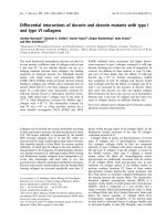
Fig. 1. Histone deacetylase inhibitors have differential effects on
PMA- vs. TGF-b1-induced Timp-1 expression. (A and B) C3H10T1 ⁄ 2
murine fibroblasts were serum starved for 24 h, then stimulated
with 10
)7
M PMA or 2 ngÆmL
)1
TGF-b1 for 6 h in the presence or
absence of (A) 250 ngÆmL
)1
TSA or (B) 1 mM NaB. Total RNA was
isolated and subjected to real-time qRT-PCR using a specific primer
set for the Timp-1 gene [49]; data were normalized to the 18S
rRNA housekeeping gene. Data are plotted as mean + SEM (A)
n ¼ 7(B)n ¼ 3. (C) C3H10T1 ⁄ 2 and Swiss-3T3 fibroblast cells
were serum starved for 24 h, then stimulated with 10
)7
M PMA
or 4 ngÆmL
)1
TGF-b1 for 6 h in the presence or absence of
500 ngÆmL
)1
TSA. Isolated total RNA subjected to qRT-PCR and
normalized as described above. Nuclear extracts (10 lg) from the
C3H10T1 ⁄ 2 cells were western blotted with an anti-(acetyl–lysine)
Ig to monitor acetylation (histone band assigned by molecular mass
and abundance).
HDAC inhibitors and Timp-1 expression D. A. Young et al.
1914 FEBS Journal 272 (2005) 1912–1926 ª 2005 FEBS
These data show that different HDACs are involved in
the TGFb vs. PMA induction of Timp-1. Two specific
inhibitors of the Sir2 family of deacetylases (class 3
HDACs), sirtinol and nicotinamide, had no effect on
induced or basal Timp-1 expression (data not shown)
showing that class 3 HDACs are not involved.
Induction of the Timp-1 gene by PMA or TGF-b1
requires new protein synthesis
In order to assess the possibility that the effects of
HDACi are secondary, acting to modulate the
expression of an intermediate, the requirement for
new protein synthesis in the induction of Timp-1 was
assessed. Figure 3 shows that addition of the protein
synthesis inhibitor, emetine, completely abrogates both
PMA- and TGF-b1-induction of the Timp-1 gene. In the
presence of emetine, the HDACi have no further effect
(data not shown). The action of HDACi could therefore
be either on the Timp-1 gene itself, or on the expression
of a protein(s) required for induction of the Timp-1 gene.
Time course of TSA action upon PMA- and
TGF-b1-induced Timp-1 expression
The time course of induction of Timp-1 gene expression
by PMA and TGF-b1 measured by qRT-PCR is identi-
cal to our previous northern blot data with PMA giving
a more rapid but transient induction and TGF-b1 indu-
cing a slower more sustained stimulation of the gene
[25]. The effect of TSA on both PMA- and TGF-b1-
induced Timp-1 expression is evident as early as 3 h
after addition of reagents (Fig. 4A). This represents the
earliest time point that induction of the gene by PMA
or TGF-b1 is measurable by qRT-PCR. The magnitude
of TSA superinduction of PMA-induced Timp-1 increa-
ses to 12 h, and remains at 24 h, even when the PMA-
induced levels have returned to baseline. TSA continues
to repress TGF-b1-induced Timp-1 expression for as
long as the TGF-b1 induction is measurable (> 24 h).
Induction of c-fos by PMA, TGF-b1 and TSA
immediately precedes that of Timp-1
As both PMA and TGF-b1 require new protein synthe-
sis to induce Timp-1 expression, we examined the
Fig. 2. Histone deacetylase inhibitors display different dose-
responses on PMA- vs. TGF-b1-induced Timp-1 expression.
C3H10T1 ⁄ 2 murine fibroblasts were serum starved for 24 h, then
stimulated with 10
)7
M PMA or 2 ngÆmL
)1
TGF-b1 for 6 h in the
presence or absence of (A) 0–1000 ngÆmL
)1
TSA; (B) 0–10 mM
NaB, or (C) 0–8 mM VPA. Total RNA was isolated and subjected to
real-time qRT-PCR for Timp-1; data were normalized to the 18S
rRNA housekeeping gene. Data is representative of at least two
independent experiments in all cases.
Fig. 3. The induction of Timp-1 by PMA or TGF-b1 is protein synthe-
sis dependent. C3H10T1 ⁄ 2 murine fibroblasts were serum starved
for 24 h, then stimulated with 10
)7
M PMA or 2 ngÆmL
)1
TGF-b1for
6 h in the presence or absence of the protein synthesis inhibitor
emetine at 10 l gÆmL
)1
. Total RNA was isolated and subjected to
real-time qRT-PCR for Timp-1; data were normalized to the 18S
rRNA housekeeping gene. Data is plotted mean + SEM, n ¼ 3.
D. A. Young et al. HDAC inhibitors and Timp-1 expression
FEBS Journal 272 (2005) 1912–1926 ª 2005 FEBS 1915
expression of the AP-1 family member c-fos, over the
same time course experiment (Fig. 4A). Previously, we
have shown that c-fos overexpression induces Timp-1
expression [11]; c-fos is a known immediate early gene
induced by PMA. We initially confirmed this using the
protein synthesis inhibitor cycloheximide, which as
B
A
C
Fig. 4. Time-course of trichostatin A action
on PMA- vs. TGF-b1-induced Timp-1 and
c-fos expression and AP-1 binding.
C3H10T1 ⁄ 2 murine fibroblasts were serum
starved for 24 h, then stimulated with 10
)7
M
PMA or 2 ngÆmL
)1
TGF-b1 in the presence or
absence of 250 ngÆmL
)1
TSA. (A) Total RNA
was isolated at timepoints 1, 3, 6, 12 and
24 h and subjected to real-time qRT-PCR for
Timp-1 and c-fos; data were normalized to
the 18S rRNA housekeeping gene. Data
plotted is representative of three independ-
ent time course experiments. (B) Nuclear
extracts were isolated at 1 and 3 h after
stimulation and subjected to EMSA using the
Timp-1 AP-1 motif as probe. (C) Isolated
nuclear extracts (2 lg) from cells stimulated
with PMA or PMA and TSA for 3 h were
incubated in the presence or absence of
antibodies against either acetyl–lysine and ⁄ or
c-fos and subjected to EMSA.
HDAC inhibitors and Timp-1 expression D. A. Young et al.
1916 FEBS Journal 272 (2005) 1912–1926 ª 2005 FEBS
expected, did not block the induction of c-fos mRNA by
PMA. The kinetics of c-fos induction closely precedes
that of Timp-1.At1h,c-fos was dramatically induced
by PMA. Further, this induction was superinduced by
TSA; by 3 h the PMA induction was lost, but TSA in
the presence of PMA still superinduced c-fos expression.
Compared to PMA, TGF-b1 induction of c-fos was
delayed, only becoming apparent by 3 h; this induction
remained at 6 h but was lost by 12 h. Similar to Timp-1
expression, TSA blocked TGF-b1 induced c-fos at all
time points where TGF-b1 alone induced c-fos expres-
sion. Interestingly, TSA alone induced c-fos but only
after 12 h of stimulation.
The Timp-1 gene contains an AP-1 motif at
)59 ⁄ )53 bp relative to the transcription start site [6].
Nuclear proteins were isolated from cells stimulated
for either 1 or 3 h with TGF-b1 or PMA, with or
without TSA. In an electrophoretic mobility-shift assay
(EMSA), AP-1 factors could be seen to bind a 30 bp
sequence encompassing the Timp-1 AP-1 motif and
this binding was induced by PMA (at both 1 and 3 h)
or TGF-b1 stimulation (at 3 h) (Fig. 4B). As with the
induction of c-fos mRNA, AP-1 protein binding activ-
ity was greater with PMA than TGF-b1, and the
induction by PMA occurred earlier than that by TGF-
b1. Stimulation with TSA alone or in the presence of
PMA or TGF-b1 appeared to have little effect on the
overall amount of AP-1 protein binding the Timp-1
AP-1 sequence; this was in marked contrast to that
seen for c-fos mRNA levels.
The specificity of the AP-1 binding activity was con-
firmed using DNA-binding competition studies. The
AP-1 complex could be competed by an excess of
‘wild-type’ DNA, but not by an equivalent DNA frag-
ment containing a mutation in the AP-1 binding
sequence (DAP-1; Fig. 4C). The AP-1 complex was not
seen when the DAP-1 DNA was used as the radio-
labelled probe (data not shown). Further, the presence
of c-fos in the AP-1 complex was confirmed by super-
shift analysis using an anti-(c-fos) Ig (Fig. 4C). As the
total binding of the AP-1 complex did not significantly
alter on the addition of TSA, and c-Jun, another AP-1
member, is a potential target for acetylation, we per-
formed supershift analysis using an antibody raised
against acetylated lysine. When added to nuclear
extracts, a low mobility ‘supershifted’ complex was evi-
dent in all extracts treated with TSA. However, the
binding intensity of this complex did not alter upon
various stimulations and the appearance of the super-
shift did not coincide with the loss of another band
(Fig. 4C). Further analysis confirmed this complex did
not appear to contain c-Jun or other AP-1 factors [i.e.
antibodies to these factors do not alter the supershift
seen with the anti-(acetyl-lysine) Ig, data not shown]
and the lack of competition for this band by the excess
of cold AP-1 oligonucleotide (Fig. 4C) strongly sug-
gests that it is not related to AP-1 factor acetylation.
TSA superinduction of PMA-induced Timp-1
requires c-Jun
To establish unequivocally the role of specific AP-1
family members upon Timp-1 expression, qRT-PCR
was performed on RNA from c-fos, c-Jun or junD defi-
cient cells (– ⁄ –) stimulated with PMA or TGF-b1 with
or without TSA; Swiss-3T3 cells were used as a
control. Surprisingly, Swiss-3T3, c-fos– ⁄ – and junD – ⁄ –
cells had the same Timp-1 expression profile as each
other and as that seen previously for C3H10T1 ⁄ 2 cells
(Fig. 5A). The fold induction by PMA or TGF-b1 was
remarkably similar between the different cell types and
for each of those three cell lines, TSA superinduced
PMA-induced Timp-1 expression and repressed TGF-
b1-induced Timp-1 expression. This shows that neither
c-fos nor junD are essential for the observed affects of
TSA on Timp-1 expression. However, although the
expression of Timp-1 in c-Jun– ⁄ – cells was induced by
either PMA or TGF-b1, TSA was unable to super-
induce the PMA-induced Timp-1 expression whilst
retaining the ability to repress the TGF-b1 induced
Timp-1 expression. In fact, in c-Jun– ⁄ – cells, much like
for the TGF-b1 response, TSA repressed PMA-
induced Timp-1, indicating that in the absence of c-Jun
(or a c-Jun-regulated factor), the default pathway for
the effect of TSA on induced Timp-1 expression is
repressive.
Many AP-1 members are differentially regulated
in response to PMA or TGF-b1 with or without
TSA
As even in the absence of c-fos, c-Jun or junD mouse
fibroblast cells are able to induce Timp-1 in response
to PMA or TGF-b1, and yet the AP-1 motif in the
Timp-1 promoter is important for such a response, the
expression profile by qRT-PCR of all the Fos and Jun
family members was determined in C3H10T1 ⁄ 2 cells
stimulated for 1 h (Fig. 5B). PMA significantly
induced fosB, fra-1, fra-2, junB and c-Jun; of these
only junB was further induced by TSA, while PMA-
induced fra-2 was repressed by TSA. TGF-b1 induced
levels of fosB, fra-2 and junB, all of which were then
repressed by TSA. ATF2 and junD in general showed
little regulation by TSA, PMA or TGF-b1. It is there-
fore possible that in AP-1 deficient cells, the lack of a
specific factor may be compensated for by the presence
D. A. Young et al. HDAC inhibitors and Timp-1 expression
FEBS Journal 272 (2005) 1912–1926 ª 2005 FEBS 1917
of an additional, functionally overlapping, family
member. However, the function of c-Jun, in mediating
the superinduction of PMA-induced Timp-1 expres-
sion, appears unique.
The effects of HDAC inhibitors on Timp-1 gene
expression can be reiterated in transient transfec-
tion of Timp-1 promoter-containing plasmids
Figure 6A shows that the effect of TSA on both
PMA- and TGF-b1-induced Timp-1 expression can be
reiterated in a )95 ⁄ +47 Timp-1 promoter-reporter
construct (in the pGL2 vector), transiently transfected
into C3H10T1 ⁄ 2 cells. Deletion mutation to )50 ⁄ +47,
removing the promoter-proximal AP-1 site, shown
previously to be important in induction of the Timp-1
gene, shows that TGF-b1-induction is lost (and hence,
TSA can no longer repress this); however, some PMA
induction of the deleted construct remains, and this is
superinduced by TSA (Fig. 6B). These data were con-
firmed using point mutation of the AP-1 site in a
)223 ⁄ +47 Timp-1 promoter construct (in the pGL3
vector); here, some PMA- and TGF-b1-induction of
the mutant construct remains. TSA superinduces
PMA-induced expression of the wild-type and mutant
AP-1 constructs, but TSA no longer represses the
residual TGF-b1-induction (Fig. 6C,D). This suggests
that the effect of TSA on TGF-b1-induced Timp-1
expression is mediated through the promoter proximal
AP-1 site, whilst the effect of TSA on PMA-induced
Fig. 5. TSA superinduction of PMA-induced
Timp-1 requires c-Jun but other AP-1
members may compensate for the loss of
c-fos. (A) Swiss-3T3, c-fos– ⁄ –, c-Jun– ⁄ –and
junD– ⁄ – mouse fibroblast cells were serum
starved for 24 h, then stimulated with 10
)7
M
PMA or 4 ngÆmL
)1
TGF-b1 in the presence or
absence of 500 ngÆmL
)1
TSA. Total RNA was
isolated after 6 h and subjected to real-time
qRT-PCR for Timp-1. Data were normalized
to the 18S rRNA housekeeping gene. Data is
representative of two independent experi-
ments with each experiment performed in
triplicate and data is plotted as mean + SEM.
(B) C3H10T1 ⁄ 2 murine fibroblasts were
serum starved for 24 h, then stimulated with
10
)7
M PMA or 2 ngÆmL
)1
TGF-b1 in the
presence or absence of 250 ngÆmL
)1
trichostatin A (TSA) for 1 h before the
isolation of total RNA. qRT-PCR for AP-1
members fosB, fra-1, fra-2, ATF2, junB, junD
and c-Jun is shown, plotted as fold-control
levels for direct comparison. C, control; P,
PMA (10
)7
M) and T, TGF-b1(4ngÆmL
)1
).
Data is representative of three independent
experiments.
HDAC inhibitors and Timp-1 expression D. A. Young et al.
1918 FEBS Journal 272 (2005) 1912–1926 ª 2005 FEBS
Timp-1 expression is independent of the Timp-1 AP-1
motif. It should be noted that TSA alone induces
expression from promoter constructs made in pGL3,
and similarly induces the empty pGL3-basic vector
(data not shown). The more robust expression levels
from this pGL3 compared to pGL2 (which is not
induced by TSA) make the data more reliable despite
this difference, and the effects of TSA upon PMA-
and TGF-b1-stimulated promoter-reporter expression
remain clear.
TSA superinduction of PMA-induced Timp-1
is lost by mutation of a GC-box or Ets binding
motif within the Timp-1 promoter
In order to establish if promoter elements downstream
of the AP-1 site can mediate the TSA superinduction
of PMA-induced Timp-1, a series of insertion mutants
were prepared in )223 ⁄ +47. In this set of constructs,
the AP-1 site at )59 ⁄ )53 remains intact, but down-
stream of this, blocks of five bases are replaced with
adenosine; these mutants overlap by two bases, giving
a set of 20 mutant constructs. Figure 7A shows that
only mutants m4, m16 and m20 lose the superinduc-
tion of PMA-induced expression by TSA. Mutant m4
alters a canonical Ets binding site, shown previously to
be important for basal expression of the Timp-1 gene;
mutant 16 alters a canonical Sp1 binding site (GC-
box); mutant 20 does not alter any known consensus
for transcription factor binding.
EMSA was used to determine the protein factors
binding to the ‘wild-type’ m4, m16 and m20 or the
mutated sequences. Specific factors binding to both the
‘wild-type’ m4 and m16 sequences could be seen
(Fig. 7B,C), however, the binding of these factors did
not change upon TSA stimulation (data not shown).
The m4 sequence (Fig. 7B) bound several complexes,
although competition analysis with the ‘wild-type’ and
mutant m4 revealed only one complex to be specific.
The identity of this, presumably Ets family member,
remains to be determined. The pattern of three bands
(a, b and c) bound to the ‘wild-type’ m16 probe in
Fig. 7C is identical to that described in the literature as
deriving from the binding of both Sp1 and Sp3 trans-
Fig. 6. The impact of TSA on PMA- vs. TGF-b1-induced Timp-1 gene expression is reiterated on Timp-1 promoter-reporter constructs and
the effect of TSA on PMA-induced Timp-1 is independent of the promoter proximal AP-1 site. C3H10T1 ⁄ 2 murine fibroblasts were transi-
ently transfected with (A) )95 ⁄ +47, (B) )50 ⁄ +47 Timp-1 promoter constructs in pGL2, (C) )223 ⁄ +47 or (D) )223 ⁄ +47 DAP-1 Timp-1 promo-
ter constructs in pGL3. Following serum starvation for 24 h, cells were stimulated with 10
)7
M PMA or 2 ngÆmL
)1
TGF-b1 for 6 h in the
presence or absence of 250 ngÆmL
)1
trichostatin A (TSA) prior to harvest for luciferase assay. Experiments were performed three times in
triplicate. Results are plotted as mean + SEM.
D. A. Young et al. HDAC inhibitors and Timp-1 expression
FEBS Journal 272 (2005) 1912–1926 ª 2005 FEBS 1919
cription factors [26]. The ‘wild-type’ dsDNA (50-fold)
competed for all three Sp bands while the mutant m16
at the same concentration did not. An anti-Sp1 Ig
resulted in the loss of band a, and partially blocked b
with the subsequent appearance of a supershifted com-
plex; anti-Sp3 Ig blocks formation of bands b and c.
This suggests that band a contains Sp1, band b con-
tains both Sp1 and Sp3, and band c contains Sp3.
Discussion
HDACs usually act as transcriptional repressors, there-
fore HDAC inhibitors should induce expression of
susceptible genes, and this is the typical experimental
finding. However, in yeast, deletion of the HDAC
Rpd3 down-regulates a subset of genes, and many of
these are also repressed by treatment with TSA [27].
There are also many individual instances of HDAC
inhibitors acting as repressors of gene expression, e.g.
TSA and NaB cause a reduction in mRNA levels for
the cdk1 gene [28]; TSA represses b-casein expression
in mammary epithelial cells [29]; TSA represses cyclin
B1 and A [30]; TSA inhibits MMTV transcription [31].
One could postulate that these effects are indirect, with
TSA leading to the induction of a factor (or factors)
involved in the repression of a downstream target;
alternatively, a direct effect on either the acetylation of a
transcription factor, or recruitment of repressive factors
to acetyl–histones via bromodomain interactions could
be envisaged. In support of the latter notion the down-
B
A
C
Fig. 7. The impact of TSA on PMA-induced
Timp-1 gene expression is mediated via
three sites in the proximal promoter. (A)
C3H10T1 ⁄ 2 murine fibroblasts were transi-
ently transfected with a )223 ⁄ +47 Timp-1
promoter construct in pGL3 and 20mutant
constructs as shown. Following serum
starvation for 24 h, cells were stimulated
with 10
)7
M PMA in the presence (solid bars)
or absence (open bars) of 500 ngÆmL
)1
trichostatin A (TSA) prior to harvest for luci-
ferase assay. Data is representative of three
independent experiments, each performed in
triplicate and results are plotted as mean +
SEM. On the Timp-1 sequence, AP-1, Ets
and Sp1 binding motifs are shown in bold,
position of each of the 20 (m1 to m20) d(A)
5
mutation are shown underlined. (B) PMA
(10
)7
M) + TSA (500 ngÆmL
)1
) stimulated
nuclear were incubated with a ‘wild-type’ m4
DNA probe (Table 2) and subjected to EMSA.
A 50-fold excess of self and mutant m4 DNA
was used to define binding specificity. (C)
Nuclear extracts (as in B) were incubated
with a ‘wild-type’ m16 DNA probe (Table 2)
and subjected to EMSA A 50-fold excess of
self and mutant m16 DNA confirmed binding
specificity. Sp factor binding was confirmed
by incubation of extracts with either 2 lg
of an anti-Sp1 Ig or anti-Sp3 Ig prior to
electrophoresis.
HDAC inhibitors and Timp-1 expression D. A. Young et al.
1920 FEBS Journal 272 (2005) 1912–1926 ª 2005 FEBS
regulation of cyclin A and B1 upon TSA treatment is
via diminished activity of NF-Y-associated HAT and
is mediated through CCAAT motifs; further, the
diminished HAT activity is mediated by phosphoryla-
tion of hGCN5 [30]. Moreover, the inhibition of
MMTV transcription by TSA does not depend on
changes in chromatin remodelling or increased histone
acetylation, but is mediated via the TATA-box region
[31].
Both TGF-b1- and PMA-induction of Timp-1
require new protein synthesis. As both events have at
least some dependency on a promoter proximal AP-1
site, this could reflect in part the synthesis of Fos and
Jun family members. Immediate early gene induction
correlates with a nucleosomal response whereby gene-
associated nucleosomes are subject to both phosphory-
lation and acetylation on histone H3 and acetylation
on histone H4 [16]. Whilst it is reported that TSA does
not activate c-fos or c-Jun expression in C3H10T1 ⁄ 2
cells [32], TSA can clearly modulate these genes in the
additional presence of TGF-b1 or PMA (Figs 4A and
5B). Moreover, TSA alone did induce c-fos expression
in our experiments after 12 h of stimulation. As the
kinetics of c-fos induction so clearly preceded that of
Timp-1, we analysed the expression of Timp-1 in c-fos
deficient mouse fibroblasts (Fig. 5A). Surprisingly,
mouse cells lacking c-fos, c-Jun or junD showed no
alteration in Timp-1 induction by PMA or TGF-b1.
This could be due to compensation for the lack of the
factor by another AP-1 family member and our data
demonstrate that expression of many AP-1 members
are up-regulated in response to PMA or TGF-b1.
However, Timp-1 expression in PMA-stimulated
c-Jun– ⁄ – cells was not superinduced in response to
TSA, and was in fact repressed, resembling the situ-
ation with TGF-b1 induction. c-Jun is known to be
acetylated at Lys271 by the transcriptional coactivator
p300 upon its interaction with the adenoviral protein
E1A [21]. Overexpression of c-Jun in C3H10T1 ⁄ 2 cells
induced Timp-1 reporter expression by twofold, but
mutation of Lys271 fi Arg had no effect on this
induction, even in the presence of PMA and ⁄ or TSA
(data not shown).
Phosphorylation of c-Jun by the mitogen-activated
protein kinase (MAPK) JNK on Ser63 and Ser73, as
well as on Thr91 or Thr93, or both, increases its trans-
activating potential and DNA-binding activity by
mediating its dissociation from an inhibitory complex
containing HDAC3, a class 1 HDAC [33–35]. HDAC3
associates with class 2 HDACs and c-Jun in repressor
complexes such as those containing the corepressors
N-CoR and SMRT [34,36]. Phosphorylation by JNK
causes a reduction in c-Jun ubiquitination and
subsequent protein stabilization [37,38]. The proteo-
some inhibitor lactacystin inhibited the PMA induction
of Timp-1 and no induction was seen in the additional
presence of TSA (data not shown). We propose that
lactacystin prevents the degradation of ubiquitinated
c-Jun thus leading to its accumulation and preventing
its activation by JNK and subsequent downstream acti-
vation events that would lead to Timp-1 up-regulation.
A possible explanation for the lack of a TSA super-
induction of PMA-induced Timp-1 in c-Jun– ⁄ – cells is
that the expression of one or more HDACs is c-Jun-
dependent. To test this, we monitored the expression
of HDACs 1–11 in response to TGF-b1 or PMA, with
or without TSA, between C3H10T1 ⁄ 2, Swiss-3T3 and
c-Jun– ⁄ – cells by RT-PCR. The expression of class 1
HDACs is reported to be ubiquitous while class 2
HDAC appear more tissue-specific [39]. All three cell
lines expressed the majority of HDACs, with only
expression of HDAC9 and )10 being undetectable and
HDAC8 was up-regulated in c-Jun– ⁄ – cells compared
to C3H10T1 ⁄ 2 or Swiss-3T3 (data not shown). Only
HDAC7 and HDAC11 were regulated differentially by
any of the stimuli, and this was identical between all
three cell lines. HDAC7 was, in all cell lines, repressed
by the presence of TSA while HDAC11 expression was
interestingly induced only by the combination of TGF-
b1 and TSA (data not shown). Hence, none of the
HDACs exhibit c-Jun dependent expression.
Although the induction by PMA alone was partially
abrogated upon mutation or deletion of the )59 ⁄ )53
AP-1 motif of Timp-1, TSA was still able to superin-
duce reporter expression in the presence of PMA
(Fig. 7). This suggests that the impact of c-Jun on the
superinduction of Timp-1 is not directly on the Timp-1
gene, but via a c-Jun-dependent intermediate. It should
be noted that whilse transiently transfected plasmid
DNA is not integrated into the host cell chromosomes,
there is evidence that it can be assembled into a chro-
matin-like structure [40,41]. If this is true in the current
system, then data from transient transfection experi-
ments could still be interpreted at the level of histone
or factor acetylation.
Using a series of 20 overlapping mutant promoter
constructs we demonstrated three mutants, m4, m16
and m20, were no longer able to superinduce reporter
expression above PMA alone in the additional presence
of TSA (though variation in absolute levels of induc-
tion is seen across the mutant constructs). Further, spe-
cific binding of Sp1 and Sp3 to wild-type m16 and a
putative Ets factor to wild-type m4 were identified
(Fig. 7C,D). It is surprising that other mutants that
overlap the consensus sequences for these transcription
factors do not impact upon the effect of TSA. The
D. A. Young et al. HDAC inhibitors and Timp-1 expression
FEBS Journal 272 (2005) 1912–1926 ª 2005 FEBS 1921
basal and cobalt-induced expression of Timp-1 is
known to be partially dependent upon Sp1, although
this is via sequences upstream of the )59 ⁄ )53 AP-1
motif or within intron 1 [10,42]. Sp3 transcription
factor is reported to either activate or repress gene
expression in target genes [43]. It has been shown that
Sp3 can be acetylated potentially via p300 [20]. For the
TGFbRII gene at least, this acetylation acts as a switch
to turn Sp3 from a transcriptional repressor into an
activator [44]. We were unable to detect whether Sp3 or
Sp1 in our C3H10T1 ⁄ 2 cell system were acetylated
using the anti-(acetyl–lysine) Ig (data not shown).
Unlike many genes, Timp-1 induction by TGF- b1is
largely independent of the Smad signalling pathway in
C3H10T1 ⁄ 2 cells and is instead AP-1-dependent [11].
It has been shown previously that the AP-1 site at
)59 ⁄ )53 can act in concert with the neighbouring Ets
site at )45 ⁄ )41 (mutant m4) [45]. TGF-b1 causes the
acetylation of Ets1 and this is proposed to contribute
to the ability of Ets1 overexpression to abrogate TGF-
b1 induction of the Timp-1 gene [46]. HDACi would
probably increase the level of Ets1 acetylation by
TGF-b1, though the functional outcome of this is
unclear. The consequences of any potential PMA-
mediated acetylation of Ets1 are also unknown, but of
future interest, as mutation of the Ets binding motif
(m4) results in both a loss of protein binding and the
TSA superinduction of Timp-1.
Eleven NAD-independent HDACs have been des-
cribed in human and mouse, although few have been
characterized in detail. These have been subdivided
recently into three groups based upon phylogenetic
analysis [47]. Class 1 HDACs are structurally related
to yeast scRPD3, and contain HDAC1, )2, )3 and
)8, while class 2 HDACs, containing HDAC4, )5, )6,
)7, )9 and )10 are similar to yeast scHDA1.
HDAC11 alone represents class 4 HDACs and
HDAC11 related proteins have been described in all
eukaryotic organisms other than fungi. As described,
HDACs often act in complexes with other proteins
and cofactors and different HDACs are often present
in the same complexes. One common feature of class 2
HDACs is that they appear to be able to homo- or
heterodimerize, leading to speculation that duplication
of the catalytic domain as seen in HDAC6 may have
occurred as a way of ensuring self association [47].
There are no inhibitors specific to a single HDAC
available currently and most HDACs are believed to
be equally sensitive to TSA and NaB, although
HDAC6 is a possible exception [39]. VPA has been
shown to have some selectivity against different
HDAC classes. Only at concentrations > 1 mm is
VPA reported to inhibit class 2 subclass 1 HDACs (at
least HDAC4, )5 and )7) [24,48]. VPA was unable to
inhibit the class 2 subclass 2 HDACs (6 and 10) even
at concentrations up to 20 mm [48]. From the NaB
and TSA dose-curves it is clear that HDACi effects on
PMA and TGF-b1-induced Timp-1 are via the inhibi-
tion of different HDACs. Therefore TSA or NaB do
not inhibit all HDACs equally. As a concentration of
>2 mm VPA is required to stimulate PMA-induced
Timp-1 expression further, it is probable that VPA is
inhibiting a class 2 subclass I enzyme in this case. Even
at the highest concentration used (8 mm) VPA did not
affect TGF-b1 induced Timp-1 expression, although it
did block TGF-b1 induced ADAM12 expression in a
dose-dependent manner (Fig. 2 and inset). This would
imply that a class 2 subclass 2 enzyme (i.e. HDAC6 or
10) is involved in the induction of Timp-1 by TGF-b1.
A caveat to this is the HDAC inhibition profiles of all
these HDACi are incomplete and are generally based
upon semipurified protein fractions and in vitro assays.
Finally, it is interesting to note that the HDACi
do not have an obvious effect on basal expression of
Timp-1, only on induced gene expression.
In conclusion, this is to our knowledge, the only
described instance of HDAC inhibitors having oppos-
ite effects on the same gene, depending upon the initial
stimulus used to induce expression. We have shown
that different HDACs are involved in the response of
the Timp-1 gene to PMA compared to TGF-b1. More-
over, c-Jun mediates the effect of HDAC inhibitors on
PMA-induced Timp-1, though not via the promoter
proximal AP-1 site. We have also identified cis-acting
promoter elements essential for the effect of HDAC
inhibitors on PMA-induced Timp-1 expression.
Experimental procedures
Cell culture
Murine C3H10T1 ⁄ 2 fibroblasts and Swiss-3T3 cells were
routinely cultured in Minimal Essential Medium (MEM)
with Earle’s salts and l -glutamine (2 mm) (Invitrogen,
Paisley, UK) containing 10% foetal bovine serum (FBS,
Invitrogen), 1% nonessential amino acids, 100 IUÆmL
)1
penicillin, 100 lgÆmL
)1
streptomycin and 20 unitsÆmL
)1
nystatin. AP-1 knockout cells (c-Jun– ⁄ –, c-fos– ⁄ – a kind gift
from E. Wagner (Research Institute of Molecular Pathology,
University of Vienna, Austria) and P. Angel (Department of
Signal Transduction and Growth Control, Deutsches Krebs-
forschungszentrum (DKFZ), University of Heidelberg,
Germany) and junD– ⁄ –, a kind gift from M. Yaniv and
J. Weitzman (Pasteur Institute, Paris, France) were cultured
as above, but in Dulbecco’s MEM. Serum-free conditions
used identical medium without FBS. For assays, cells were
HDAC inhibitors and Timp-1 expression D. A. Young et al.
1922 FEBS Journal 272 (2005) 1912–1926 ª 2005 FEBS
grown to confluence, then serum-starved for 24 h prior to
the addition of TGF-b1 (R & D Systems, Abingdon, Oxon,
UK; 2 or 4 ngÆmL
)1
) or phorbol 12-myristate-13-acetate
(PMA, Sigma, Poole, UK; 10
)7
m) in the absence or pres-
ence of HDAC inhibitors (trichostatin A, TSA; sodium
butyrate, NaB; valproic acid, VPA; Calbiochem, Notting-
ham, UK) at the concentrations described. Experiments were
repeated 2–4 times to ensure the pattern of response was
reproducible and representative data are shown throughout.
RT-PCR
RNA was isolated from monolayer cultures using Trizol
reagent (Invitrogen). One microgram of total RNA was
reverse transcribed using 2 lg random hexamers (Amersham
Biosciences, Chalfont St Giles, Bucks, UK) and 200 U of
Superscript II reverse transcriptase (Invitrogen), according
to the supplier’s instructions. Quantitative RT-PCR (qRT-
PCR) was performed using the Applied Biosystems, War-
rington, UK) ABI Prism 7700 sequence detection system
(TaqManÒ) as described [49]. Table 1 contains sequences of
TaqMan primers and probes for AP-1 family members.
Nuclear extracts and electrophoretic mobility-
shift assays
Nuclear extracts were prepared essentially as described pre-
viously [11] except where indicated cells were stimulated
with TSA (500 ngÆmL
)1
) and TSA (500 ngÆmL
)1
) was inclu-
ded in the all solutions for nuclear extract preparation.
Electrophoretic mobility-shift assays (EMSA) were
performed essentially as previously described [10,11]. Oligo-
nucleotides were synthesized by MWG-Biotech (London,
UK) or Sigma-Genosys (Haverhill, UK) (Table 2). Double-
stranded probes were labelled with [
32
P]ATP[cP] using T4
polynucleotide kinase. Nuclear extracts (2 lg), 0.5 lgof
poly(dIdC.dIdC), and radiolabelled probe (20 000 c.p.m.)
were incubated in 1· binding buffer (10 mm Tris ⁄ HCl,
pH 7.5, 50 mm NaCl, 0.5 mm dithiothreitol, 5 mm MgCl
2
,
and 5% glycerol) with or without competitor DNA for
20 min at 4 °C in a total volume of 10 lLat4°C. For
antibody supershift analyses, 2 lg of the appropriate anti-
body [anti-(c-fos) Ig, sc-52-Gx (Santa Cruz Biotechnology,
Inc., from Autogen Bioclear, Calne, UK), anti-(acetyl–
lysine) Ig, clone 4G12 (Upstate Ltd., Milton Keynes, UK)]
was incubated with nuclear extract for 20 min at 4 °C prior
to the addition of the DNA probe. Samples were separated
on a 5% polyacrylamide gel in 0.5· TBE (45 mm Tris ⁄ HCl,
45 mm boric acid, 1 mm EDTA). Gels were prerun at
10mA for 1 h at 4 °C and run at 4 mA for 3–6 h at 4 °C.
Gels were dried and autoradiographed.
Plasmid construction and transient transfection
Constructs using Timp-1 promoter driving luciferase
expression were in pGL2-basic or pGL3-basic (Promega,
Table 1. Mouse AP-1 gene qRT-PCR primer and probe sets.
Gene Sequence
c-fos Forward Primer 5¢-CCTGCCCCTTCTCAACGA-3¢
Reverse Primer 5¢-CTCCACGTTGCTGATGCTCTT-3¢
Probe 5¢-CCCAAGCCATCCTTGGAGCCAGT-3¢
c-Jun Forward Primer 5¢-GAAGTGACGGACCGTTCTATGAC-3¢
Reverse Primer 5¢-GGAGGAACGAGGCGTTGAG-3¢
Probe 5¢-AAGATGGAAACGACCTTCTACGACGATGC-3¢
junB Forward Primer 5¢-GGAGCAGGAGGGCTTTGC-3¢
Reverse Primer 5¢-GGCGTCACGTGGTTCATCT-3¢
Probe 5¢-ACGGTTTTGTCAAAGCCCTGGACGAC-3¢
junD Forward Primer 5¢-CGCAAGCTGGAGCGTATCTC-3¢
Reverse Primer 5¢-GACGCCAGCTCGGTGTTCT-3¢
Probe 5¢-CGCCTGGAGGAGAAAGTCAAGACCCTC-3¢
ATF2 Forward Primer 5¢-CAGCCACCTCCACTACAGAAACT-3¢
Reverse Primer 5¢-TTCTTCGACGGCCACTTGTAT-3¢
Probe 5¢-TCTCCAGCTCACACAACTCCTCAGACCC-3¢
fosB Forward Primer 5¢-GCTCCCCTATCCTCGATATTTGA-3¢
Reverse Primer 5¢-CAGAACTCGTCTTTGGGACTGA-3¢
Probe 5¢-TTCCCACTATCCCACTCCATCCAATTCC-3¢
fra-1 Forward Primer 5¢-TGAACCGGAAGCACTGCATA-3¢
Reverse Primer 5¢-GTGAAAACCAGACTCGGAGTAAAAG-3¢
Probe 5¢-CACGCTCATGACCACACCCTCTCTGAC-3¢
fra-2 Forward Primer 5¢-CATCACTCCCGGCACTTCA-3¢
Reverse Primer 5¢-CGACGAAGGCGACTCCTG-3¢
Probe 5¢-TTGTCTTCACCTACCCCAATGTCCTGGA-3¢
D. A. Young et al. HDAC inhibitors and Timp-1 expression
FEBS Journal 272 (2005) 1912–1926 ª 2005 FEBS 1923
Southampton, UK); point mutations altered the wild-type
AP-1 site (5¢-TGAGTAA-3 ¢) to a nonfunctional mutant
AP-1 site (5¢-GgAGTgA-3¢) as described previously [11].
Twenty independent overlapping mutants were all gener-
ated in pGL2-223 ⁄ +47 [11], the Timp-1 promoter regions
were HindIII isolated and subcloned into pGL3-basic
(Fig. 7A). All mutagenesis was performed using the Quik-
Change method (Stratagene, Amsterdam, the Netherlands).
All mutations were verified by DNA sequencing.
Cells were seeded in six-well or 24-well plates at a density
of 8850 cellsÆcm
)2
and grown overnight in medium contain-
ing 10% (v ⁄ v) FBS at 37 °C in a 5% (v ⁄ v) CO
2
atmosphere. Cells were transfected overnight in serum-
containing medium with 1 lg per well (six-well plates) or
0.21 lg per well (24-well plates) reporter plasmid using
FuGene6 (Roche, Lewes, UK) according to the manufac-
turers’ instructions. The following day, cells were washed in
Hank’s Balanced Salts Solution (HBSS) and incubated in
serum-free medium overnight. Cells were then stimulated
with PMA (10
)7
m) or TGF-b1(2or4ngÆmL
)1
) in the
presence or absence of HDAC inhibitors, prior to harvest.
Harvest and assay for luciferase were according to manu-
facturer’s instructions (Roche).
Acknowledgements
D.A.Y. was funded by the Dunhill Medical Trust.
C.L.S. is funded by the Consejo Nacional de Ciencia y
Tecnologia, Mexico. D.R.E. would like to acknow-
ledge the support of the European Union Framework
6 Cancerdegradome project (LSHC-CT-2003–503297).
References
1 Egeblad M & Werb Z (2002) New functions for the
matrix metalloproteinases in cancer progression. Nat
Rev Cancer 2, 161–174.
2 Brinckerhoff CE & Matrisian LM (2002) Matrix metal-
loproteinases: a tail of a frog that became a prince. Nat
Rev Mol Cell Biol 3, 207–214.
3 Baker AH, Edwards DR & Murphy G (2002) Metallo-
proteinase inhibitors: biological actions and therapeutic
opportunities,. J Cell Sci 115, 3719–3727.
4 Edwards DR, Leco KJ, Leco PA, Lim MS, Phillips
BW, Raja J & Sharma R (2000) Regulation of TIMP
gene expression. In Tissue inhibitors of metalloprotei-
nases in development and disease (Hawkes SP, Edwards
DR & Khokha R, eds), pp. 13–24. Harwood Academic
Press, Lausanne, Switzerland.
5 Brand K (2002) Cancer gene therapy with tissue inhibi-
tors of metalloproteinases (TIMPs). Curr Gene Ther 2,
255–271.
6 Edwards DR, Rocheleau H, Sharma RR, Wills AJ,
Cowie A, Hassell JA & Heath JK (1992) Involvement of
AP1 and PEA3 binding sites in the regulation of murine
tissue inhibitor of metalloproteinases-1 (TIMP-1)
transcription. Biochim Biophys Acta 1171, 41–55.
7 Clark IM, Rowan AD, Edwards DR, Bech-Hansen T,
Mann DA, Bahr MJ & Cawston TE (1997) Transcrip-
tional activity of the human tissue inhibitor of metallo-
proteinases 1 (TIMP-1) gene in fibroblasts involves
elements in the promoter. Exon 1 Intron 1, Biochem J
324, 611–617.
8 Phillips BW, Sharma R, Leco PA & Edwards DR
(1999) A sequence-selective single-strand DNA-binding
protein regulates basal transcription of the murine tissue
inhibitor of metalloproteinases-1 (Timp-1) gene. J Biol
Chem 274, 22197–22207.
9 Norman JT, Clark IM & Garcia PL (2000) Hypoxia
promotes fibrogenesis in human renal fibroblasts.
Kidney Int 58, 2351–2366.
10 Dean G, Young DA, Edwards DR & Clark IM (2000)
The human tissue inhibitor of metalloproteinases
(TIMP)-1 gene contains repressive elements within the
promoter and intron 1. J Biol Chem 275, 32664–32671.
11 Hall MC, Young DA, Waters JG, Rowan AD, Chantry
A, Edwards DR & Clark IM (2003) The comparative
role of activator protein 1 and Smad factors in the regu-
lation of Timp-1 and MMP-1 gene expression by trans-
forming growth factor-beta 1. J Biol Chem 278, 10304–
10313.
12 Wolffe AP & Guschin D (2000) Review: chromatin
structural features and targets that regulate transcrip-
tion,. J Struct Biol 129, 102–122.
13 Narlikar GJ, Fan HY & Kingston RE (2002) Coopera-
tion between complexes that regulate chromatin struc-
ture and transcription. Cell 108, 475–487.
14 Sudarsanam P & Winston F (2000) The Swi ⁄ Snf family
nucleosome-remodeling complexes and transcriptional
control. Trends Genet 16, 345–351.
15 Kouzarides T (2000) Acetylation: a regulatory modifica-
tion to rival phosphorylation? Embo J 19, 1176–1179.
16 Clayton AL, Rose S, Barratt MJ & Mahadevan LC
(2000) Phosphoacetylation of histone H3 on c-fos- and
c-Jun-associated nucleosomes upon gene activation.
Embo J 19, 3714–3726.
17 Ng HH & Bird A (2000) Histone deacetylases: silencers
for hire. Trends Biochem Sci 25, 121–126.
Table 2. EMSA oligonucleotides. In sequences, upper strands only
depicted. Mutated sequences are shown in lowercase.
Name Sequence
Timp-1 AP-1 5¢-CGGTGGGTGGATGAGTAATGCGTCCAGG-3¢
Timp-1 DAP-1 5¢-CGGTGGGTGGAgGAGTgATGCGTCCAGG-3¢
‘wild-type’ m4 5¢-AATGCGTCCAGGAAGCCTGGAGGCAGTGAT-3¢
m4 5¢-AATGCGTCCAGGAAaaaTGGAGGCAGTGAT-3¢
‘wild-type’ m16 5¢-GCCAACTCCGCCCTTCGCATGGACATTTAT-3¢
m16 5¢-GCCAACTCCGCCaaaaaCATGGACATTTAT-3¢
HDAC inhibitors and Timp-1 expression D. A. Young et al.
1924 FEBS Journal 272 (2005) 1912–1926 ª 2005 FEBS
18 Berger SL (2002) Histone modifications in transcrip-
tional regulation. Curr Opin Genet Dev 12, 142–148.
19 Gao L, Cueto MA, Asselbergs F & Atadja P (2002)
Cloning and functional characterization of HDAC11, a
novel member of the human histone deacetylase family.
J Biol Chem 277, 25748–25755.
20 Braun H, Koop R, Ertmer A, Nacht S & Suske G
(2001) Transcription factor Sp3 is regulated by acetyla-
tion. Nucleic Acids Res 29, 4994–5000.
21 Vries RG, Prudenziati M, Zwartjes C, Verlaan M,
Kalkhoven E & Zantema A (2001) A specific lysine in
c-Jun is required for transcriptional repression by
E1A and is acetylated by p300. EMBO J 20, 6095–
6103.
22 Yoshida M, Kijima M, Akita M & Beppu T (1990)
Potent and specific inhibition of mammalian histone
deacetylase both in vivo and in vitro by trichostatin A.
J Biol Chem 265, 17174–17179.
23 Kruh J (1982) Effects of sodium butyrate, a new phar-
macological agent, on cells in culture. Mol Cell Biochem
42, 65–82.
24 Gottlicher M, Minucci S, Zhu P, Kramer OH, Schimpf
A, Giavara S, Sleeman JP, Lo Coco F, Nervi C, Pelicci
PG & Heinzel T (2001) Valproic acid defines a novel
class of HDAC inhibitors inducing differentiation of
transformed cells. EMBO J 20, 6969–6978.
25 Leco KJ, Khokha R, Pavloff N, Hawkes SP & Edwards
DR (1994) Tissue inhibitor of metalloproteinases-3
(TIMP-3) is an extracellular matrix-associated protein
with a distinctive pattern of expression in mouse cells
and tissues. J Biol Chem 269, 9352–9360.
26 Qin H, Sun Y & Benveniste EN (1999) The transcrip-
tion factors Sp1, Sp3, and AP-2 are required for consti-
tutive matrix metalloproteinase-2 gene expression in
astroglioma cells. J Biol Chem 274, 29130–29137.
27 Bernstein BE, Tong JK & Schreiber SL (2000) Genome-
wide studies of histone deacetylase function in yeast.
Proc Natl Acad Sci USA 97, 13708–13713.
28 Saunders N, Dicker A, Popa C, Jones S & Dahler A
(1999) Histone deacetylase inhibitors as potential anti-
skin cancer agents. Cancer Res 59, 399–404.
29 Pujuguet P, Radisky D, Levy D, Lacza C & Bissell MJ
(2001) Trichostatin A inhibits beta-casein expression in
mammary epithelial cells. J Cell Biochem 83, 660–670.
30 Nair AR, Boersma LJ, Schiltz L, Chaudhry MA, Mus-
chel RJ & Chaudry A (2001) Paradoxical effects of tri-
chostatin A: inhibition of NF-Y-associated histone
acetyltransferase activity, phosphorylation of hGCN5
and downregulation of cyclin A B1 Mrna. Cancer Lett
166, 55–64.
31 Mulholland NM, Soeth E & Smith CL (2003) Inhibi-
tion of MMTV transcription by HDAC inhibitors
occurs independent of changes in chromatin remodel-
ing and increased histone acetylation. Oncogene 22,
4807–4818.
32 Thomson S, Clayton AL & Mahadevan LC (2001) Inde-
pendent dynamic regulation of histone phosphorylation
and acetylation during immediate-early gene induction.
Mol Cell 8, 1231–1241.
33 Morton S, Davis RJ, McLaren A & Cohen P (2003)
A reinvestigation of the multisite phosphorylation of
the transcription factor c-Jun. EMBO J 22, 3876–
3886.
34 Weiss C, Schneider S, Wagner EF, Zhang X, Seto E &
Bohmann D (2003) JNK phosphorylation relieves
HDAC3-dependent suppression of the transcriptional
activity of c-Jun. EMBO J 22, 3686–3695.
35 Papavassiliou AG, Chavrier C & Bohmann D (1992)
Phosphorylation state and DNA-binding activity of
c-Jun depend on the intracellular concentration of
binding sites. Proc Natl Acad Sci USA 89, 11562–
11565.
36 Fischle W, Dequiedt F, Hendzel MJ, Guenther MG,
Lazar MA, Voelter W & Verdin E (2002) Enzymatic
activity associated with class II HDACs is dependent on
a multiprotein complex containing HDAC3 and
SMRT ⁄ N-CoR. Mol Cell 9, 45–57.
37 Musti AM, Treier M & Bohmann D (1997) Reduced
ubiquitin-dependent degradation of c-Jun after phos-
phorylation by MAP kinases. Science 275, 400–402.
38 Wertz IE, O’Rourke KM, Zhang Z, Dornan D, Arnott
D, Deshaies RJ & Dixit VM (2004) Human de-etio-
lated-1 regulates c-Jun by assembling a CUL4A ubiqui-
tin ligase. Science 303, 1371–1374.
39 de Ruijter AJ, van Gennip AH, Caron HN, Kemp S &
van Kuilenburg AB (2003) Histone deacetylases
(HDACs): characterization of the classical HDAC
family. Biochem J 370, 737–749.
40 Reeves R, Gorman CM & Howard B (1985) Minichro-
mosome assembly of non-integrated plasmid DNA
transfected into mammalian cells. Nucleic Acids Res 13,
3599–3615.
41 Jeong S & Stein A (1994) Micrococcal nuclease diges-
tion of nuclei reveals extended nucleosome ladders hav-
ing anomalous DNA lengths for chromatin assembled
on non-replicating plasmids in transfected cells. Nucleic
Acids Res 22, 370–375.
42 Lee M, Song SU, Ryu JK & Suh JK (2004) Sp1-depen-
dent regulation of the tissue inhibitor of metalloprotei-
nases-1 promoter. J Cell Biochem 91, 1260–1268.
43 Majello B, De Luca P & Lania L (1997) Sp3 is a bifunc-
tional transcription regulator with modular independent
activation and repression domains. J Biol Chem 272,
4021–4026.
44 Ammanamanchi S, Freeman JW & Brattain MG (2003)
Acetylated sp3 is a transcriptional activator. J Biol
Chem 278, 35775–35780.
45 Logan SK, Garabedian MJ, Campbell CE & Werb Z
(1996) Synergistic transcriptional activation of the tissue
inhibitor of metalloproteinases-1 promoter via
D. A. Young et al. HDAC inhibitors and Timp-1 expression
FEBS Journal 272 (2005) 1912–1926 ª 2005 FEBS 1925
functional interaction of AP-1 and Ets-1 transcription
factors. J Biol Chem 271, 774–782.
46 Czuwara-Ladykowska J, Sementchenko VI, Watson DK
& Trojanowska M (2002) Ets1 is an effector of the
transforming growth factor beta (TGF-beta) signaling
pathway and an antagonist of the profibrotic effects of
TGF-beta. J Biol Chem 277, 20399–20408.
47 Gregoretti IV, Lee YM & Goodson HV (2004) Molecu-
lar evolution of the histone deacetylase family: func-
tional implications of phylogenetic analysis. J Mol Biol
338, 17–31.
48 Gurvich N, Tsygankova OM, Meinkoth JL & Klein PS
(2004) Histone deacetylase is a target of valproic acid-
mediated cellular differentiation. Cancer Res 64, 1079–
1086.
49 Young DA, Phillips BW, Lundy C, Nuttall RK, Hogan
A, Schultz GA, Leco KJ, Clark IM & Edwards DR
(2002) Identification of an initiator-like element essential
for the expression of the tissue inhibitor of metallopro-
teinases-4 (Timp-4) gene. Biochem J 364, 89–99.
1926 FEBS Journal 272 (2005) 1912–1926 ª 2005 FEBS
HDAC inhibitors and Timp-1 expression D. A. Young et al.