Báo cáo khoa học: ERS1 encodes a functional homologue of the human lysosomal cystine transporter pptx
Bạn đang xem bản rút gọn của tài liệu. Xem và tải ngay bản đầy đủ của tài liệu tại đây (653.44 KB, 15 trang )
ERS1 encodes a functional homologue of the human
lysosomal cystine transporter
Xiao-Dong Gao
1
, Ji Wang
2
, Sabine Keppler-Ross
2
and Neta Dean
2
1 Research Center for Glycoscience, National Institute of Advanced Industrial Science and Technology, Tsukuba, Japan
2 Department of Biochemistry and Cell Biology, Institute for Cell and Developmental Biology, State University of New York, USA
Cystinosis is a lysosomal storage disease whose hall-
mark is the accumulation of cystine in the lysosome
[1,2]. During the normal degradation of proteins in the
lysosome, cystine, the disulfide-linked form of cysteine,
is not reduced in the oxidating environment of the
lysosome but must be transported to the cytoplasm
where it is reduced. Cystine is very insoluble and a
defect in its efflux causes it to form crystals. Cystine is
normally transported from the lysosome to the cyto-
plasm by cystinosin, a lysosomal membrane protein
encoded by the CTNS gene. Cystinosin is a proton
symporter, coupling cystine transport with protons
generated by the vacuolar (H
+
)-ATPase [3]. Thus cys-
tine transport removes both cystine to the reducing
environment of the cytoplasm, along with protons
whose luminal accumulation may have an overly acidi-
fying effect on the lysosome.
A number of different mutations have been charac-
terized from cystinosis patients, almost all of which
map to the CTNS gene [3]. Several forms of this auto-
somal recessive disease occur, which differ in both
severity and age of onset. The most severe form is
infantile nephropathic cystinosis. These individuals are
normal at birth, but within several months develop
progressively severe nephratic disorders that culminate
in renal failure by the age of 10 years. In this form of
the disease, small molecules fail to be re-absorbed in
the renal tubules, resulting in excessive urinary loss of
vital components. This generalized renal tubule dis-
order, known as Fanconi syndrome, leads to growth
Keywords
cystinosin; ERS1; GTR1; MEH1; vacuole
Correspondence
N. Dean, Department of Biochemistry and
Cell Biology, Institute for Cell and
Developmental Biology, State University of
New York, Stony Brook, New York 11794-
5215, USA
Fax: +1 631 632 8575
Tel: +1 631 632 9309
E-mail:
(Received 5 November 2004, revised
11 March 2005, accepted 18 March 2005)
doi:10.1111/j.1742-4658.2005.04670.x
Cystinosis is a lysosomal storage disease caused by an accumulation of
insoluble cystine in the lumen of the lysosome. CTNS encodes the lyso-
somal cystine transporter, mutations in which manifest as a range of
disorders and are the most common cause of inherited renal Fanconi
syndrome. Cystinosin, the CTNS product, is highly conserved among mam-
mals. Here we show that the yeast Ers1 protein and cystinosin are func-
tional orthologues, despite sharing only limited sequence homology. Ers1 is
a vacuolar protein whose loss of function results in growth sensitivity to
hygromycin B. This phenotype can be complemented by the human CTNS
gene but not by mutant ctns alleles that were previously identified in cysti-
nosis patients. A genetic screen for multicopy suppressors of an ers1D yeast
strain identified a novel gene, MEH1, which is implicated in regulating
Ers1 function. Meh1 localizes to the vacuolar membrane and loss of
MEH1 results in a defect in vacuolar acidification, suggesting that the vac-
uolar environment is critical for normal ERS1 function. This genetic sys-
tem has also led us to identify Gtr1 as an Meh1 interacting protein. Like
Meh1 and Ers1, Gtr1 associates with vacuolar membranes in an Meh1-
dependent manner. These results demonstrate the utility of yeast as a
model system for the study of CTNS and vacuolar function.
Abbreviations
ER, Endoplasmic reticulum; GFP, green fluorescent protein; HA, haemagglutinin; hygB, hygromycin B.
FEBS Journal 272 (2005) 2497–2511 ª 2005 FEBS 2497
retardation, hypothyroidism, photophobias, and neuro-
logical dysfunctions if untreated. These individuals
appear to be null mutants with a complete loss of
CTNS function. A less severe form is juvenile cystino-
sis. In addition to having much milder renal problems,
these individuals also suffer ocular disorders, such as
light sensitivity or retinal blindness caused by cystine
crystal deposits in the cornea of the eyes. This ocular,
non-nephropathic form of cystinosis characterizes the
mildest form of the disease in individuals whose onset
occurs as adults. The mutations in these individuals
suggests that these milder forms represent partial loss-
of-function CTNS mutations [4]. The only proven
therapeutic agent that exists for the treatment of cysti-
nosis is cysteamine, a membrane permeable reagent
that reduces cystine to produce cysteine and cysteam-
ine:cystine, whose exit from the lysosome occurs via a
lysine transporter [5,6]. Cysteamine is limited in its
applications because its efficacy as a therapeutic
requires early functional diagnosis and because it is
difficult to administer [7,8].
The human CTNS gene product is highly conserved
amongst all mammals but shows more limited similar-
ity to the Saccaromyces cerevisiae Ers1protein (28%
identical ⁄ 46% similar). ERS1 was isolated as a gene
that when over expressed can suppress the secretion of
resident endoplasmic reticulum (ER) proteins in erd1D
mutants [9]. Yeast strains lacking ERD1 display a
number of pleiotropic Golgi defects, including the
secretion of ER proteins that are normally retrieved
from the Golgi and the underglycosylation of proteins
normally modified in the Golgi [10]. Neither the pre-
cise function of ERD1 nor the mechanism of ERS1-
mediated suppression of erd1D is known.
Although ERS1 is related in sequence to CTNS,
there are some notable differences in their gene prod-
ucts. Both genes encode membrane proteins that are
predicted to contain seven membrane-spanning
domains, reminiscent of G-protein-coupled receptors.
However, cystinosin differs from Ers1p in that it con-
tains an extended N-terminal domain of 121 amino
acids predicted to face the lumen [11]. Furthermore,
unlike CTNS mutants, loss of ERS1 in yeast leads to
no detectable growth phenotypes. These differences in
both protein sequence and mutant phenotype have
raised the question of whether or not these two pro-
teins perform similar functions. Here we show that
CTNS and ERS1 are indeed orthologous genes. The
Ers1 protein localizes to the endosomes and yeast
vacuole, an organelle that is functionally equivalent to
the mammalian lysosome. Although ers1D mutant
strains are not defective in growth, we identify a drug
phenotype, hygromycin B (hygB) sensitivity, which can
be reversed by the human CTNS gene but not by
mutant CTNS alleles identified in cystinosis patients.
A screen for genes that when overexpressed can sup-
press the drug sensitivity of ers1D strains has led to
the identification of MEH1, a novel gene product that
is implicated in the regulation of vacuolar function.
We also identify Gtr1, a conserved GTPase whose
interaction with Meh1 is required for Gtr1 vacuolar
localization. These results demonstrate that yeast
serves as a useful model for the study of CTNS.
Results
The mammalian CTNS lysosomal H
+
-driven
transporter encodes a functional homologue
of ERS1
Yeast strains lacking ERS1 exhibit no detectable
growth phenotypes but are sensitive to the aminoglyco-
side, hygB (Fig. 1). The ERS1 gene was deleted by
replacement with TRP1. Tetrad dissection of over
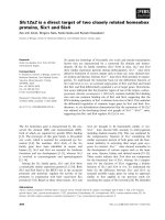
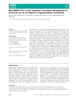
Fig. 1. CTNS encodes a functional homologue of ERS1. Isogenic
wild type (SEY6210) and ers1D cells (XGY51) expressing either the
human CTNS cDNA or the ERS1 gene under the control of the
ERS1 promoter, were streaked on YPAD plates in the presence or
absence of 50 lgÆmL
)1
hygB and grown for 2 days at 30 °C.
ERS1 and CTNS are functional homologues X D. Gao et al.
2498 FEBS Journal 272 (2005) 2497–2511 ª 2005 FEBS
20ERS1 ⁄ ers1D::TRP1 diploids demonstrated complete
cosegregation of hygB sensitivity and tryptophan pro-
totrophy (data not shown). Further, hygB sensitivity
can be completely complemented by expression of the
normal ERS1 gene (Fig. 1), demonstrating this drug
phenotype is a direct consequence of the ERS1 dele-
tion. HygB sensitivity has been described for many
yeast mutants, including those with defects in cell wall
biosynthesis, glycosylation, ion transport, and vacuolar
function (e.g. [12–15]). ers1D strains exhibit no detect-
able cell wall or glycosylation phenotypes (data not
shown). Although the basis for this complex drug sen-
sitivity is not understood, the identification of a pheno-
type associated with loss of ERS1 function provided a
system to analyse the relationship between CTNS and
ERS1.
To ask if CTNS is a functional homologue of ERS1,
we took advantage of the hygB sensitive phenotype to
examine whether or not it can be complemented by
CTNS. The human CTNS gene contains 11 introns.
To analyse complementation of ers1D in yeast, the
human CTNS cDNA was cloned into a yeast expres-
sion vector that places CTNS expression under the
control of the ERS1 promoter (see Experimental pro-
cedures). Under these conditions, the human CTNS
gene completely complemented the hygB sensitive
growth phenotype of an ers1D strain, in a manner
equivalent to its complementation by ERS1, demon-
strating that cystinosin functions in yeast (Fig. 1).
CTNS mutants fail to complement ers1D strains
To further confirm the functional conservation
between ERS1 and CTNS, a series of mutant CTNS
genes was constructed, and their functional activities
were monitored by the complementation of hygB sensi-
tive growth phenotype of ers1D cells. Mutations were
introduced into CTNS that correspond to well-charac-
terized mutations that have been repeatedly isolated
from cystinosin patients. First, two missense mutations
were introduced that had been identified in patients
with infantile nephropathic cystinosis [4]. The glycine
at position 308 was replaced by an arginine (ctns-
G308R) and the leucine at position 338 was replaced
by a proline (ctns-L338P). These amino acids, G308
and L338, lie within regions of cystinosin that are
among the most highly conserved, within the sixth
(G308) or seventh (L338) membrane spanning domains
(see Fig. 2A). These membrane spanning domains have
been postulated to be important for cystinosin function
[4]. Two additional mutations were generated that
encode truncated forms of cystinosin. The first of these
is ctns-ND121, which lacks the first 121 amino acids at
the NH
2
-terminus of cystinosin (Fig. 2A). This domain
is present in cystinosin but completely absent in Ers1.
This region is also the least conserved among CTNS
orthologues from other species, including birds,
worms, flies, mosquitos, and rats (Fig. 2A), so it was
of interest to examine the functional consequence of its
deletion. The second mutant is ctns-CD82, which lacks
the last 82 amino acids at the COOH-terminus of cys-
tinosin. This C-terminal domain of CTNS encompasses
the sixth and seventh predicted transmembrane regions
of cystinosin that have been implicated as functionally
important [4]. This C-terminal deletion is analogous to
several deletion mutations found in severe infantile
nephropathic cystinosis patients [4].
These mutant CTNS alleles were expressed in an
ers1D strain and assayed quantitatively for complemen-
tation of its hygB sensitive growth phenotype. Neither
ctns-G308R nor ctns-L338P alleles can complement the
hygB sensitivity of an ers1D strain at levels observed
by the wild type CTNS (Fig. 2B). Both the G308R
and the L338P mutations were almost 100-fold less
efficient for ers1D complementation than the wild
type CTNS. Thus, these missense mutations in CTNS
mimic their affect in humans when expressed in yeast.
The most severe mutant phenotype was seen in the
ctns-CD82 allele, which completely failed to comple-
ment ers1D. In contrast, the ctns-ND121 allele had no
effect and complemented ers1D in a manner equivalent
to the wild type CTNS gene (Fig. 2B). These results
suggest that the sixth and seventh transmembrane
domains in both CTNS and ERS1 are probably essen-
tial for protein function, while the N-terminal 121
amino acid domain of cystinosin is dispensable.
A trivial explanation for the failure of these mutant
ctns alleles to complement ers1D is that these muta-
tions grossly perturb protein structure and lead to its
instability. To determine if the lack of complementa-
tion by mutant ctns alleles was due to reduced levels of
cystinosin protein, these mutant alleles were tagged
with sequences encoding the haemagglutinin (HA) epi-
tope to compare levels of protein expression. Neither
Ers1 nor cystinosin could be detected by Western blot
analysis of whole cell extracts, suggesting that neither
of these proteins normally accumulate to high steady-
state levels. As described below, these HA-tagged pro-
teins could be readily detected after enrichment from
vacuolar membrane fractions, sedimented by centrifu-
gation at 16 000 g (see Experimental procedures).
Equivalent amounts of protein from these fractions
were separated by SDS ⁄ PAGE and immunoblotted
with antibodies against HA. Ers1 has a predicted
molecular mass of 30 kDa and contains one recogni-
tion site for N-linked glycosylation, in the lumenal
X D. Gao et al. ERS1 and CTNS are functional homologues
FEBS Journal 272 (2005) 2497–2511 ª 2005 FEBS 2499
domain between the sixth and seventh membrane span-
ning regions. Ers1 migrated with molecular weight
markers in this range as a slightly heterogeneous
smear, as expected for a glycosylated protein (Fig. 2C).
Cystinosin is predicted to have a molecular mass of
41.7 kDa and in addition, has seven predicted recog-
nition sites for N-linked glycosylation. Most of the
cystinosin migrated as a large heterogeneous smear,
A
BC
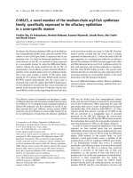
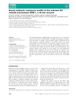
Fig. 2. Mutant forms of CTNS fail to complement ers1D. (A) Schematic diagram of the predicted topology of cystinosin. Magenta and blue
dots represent those amino acids that are invariant in an alignment of representative cystinosin-related proteins from humans (AAH32850.1),
birds (Gallus gallus; XP_415851.1), flies (Drosophila melanogaster; AAM50956.1), mosquitoes (Anopheles gambiae XP_312994.1), worms
(Caenorhabditis elegans NP_495704.1) and yeast (Saccharomyces cerevisiae YCR075C). Mutations that were introduced in this study are
denoted in red. ND120 denotes a deletion of the N-terminal 120 amino acids. CD82 denotes a deletion of the C-terminal 82 amino acids,
which remove the sixth and seventh membrane spanning domains. (B) The parental wild type (SEY6210) and ers1D (XGY51) strains expres-
sing the indicated wild type or mutant alleles of human CTNS under the ERS1 promoter were assayed for complementation of the hygromy-
cin B sensitive phenotype of ers1D. Exponentially growing cells were serially diluted (10-fold), spotted onto YPAD plates with or without
50 lgÆmL
)1
hygB and grown for 2 days at 30 °C. (C) Whole cell lysates from cells expressing HA-tagged ERS1, CTNS, ctns-D121,or
ctnsL338P were subjected to differential centrifugation as described in Experimental procedures and equivalent amounts of protein were
separated by SDS ⁄ PAGE and immunoblotted with anti-HA.
ERS1 and CTNS are functional homologues X D. Gao et al.
2500 FEBS Journal 272 (2005) 2497–2511 ª 2005 FEBS
suggesting that the majority of cystinosin is glycosylated
when expressed in yeast. An additional band corres-
ponding to a molecular mass of 33 kDa was also
seen, though its identity has not been further investi-
gated. Importantly, strains expressing ctns mutant alle-
les expressed altered proteins at levels comparable to
the wild type (Fig. 2C and data not shown). The only
mutation that markedly reduced steady state levels of
cystinosin was the deletion of the N-terminal 121
amino acids. This domain is predicted to face the
lumen and contains all of the sites for N-linked glycan
addition. Consistent with this prediction, cystinosin-
ND120 runs as a single band of about 28 kDa. This
protein accumulated at levels lower than the wild type
cystinosin, but these levels are apparently sufficient as
this mutant allele fully complemented ers1D (Fig. 2B).
Both ctns-L338P (Fig. 2C) and ctns-G308R (not
shown) expressed protein that comigrated with the
wild type although the cystinosin-L338P protein
appeared to have a larger proportion of fully glycosyl-
ated forms than the wild type. As each of these mutant
proteins accumulated to levels that are comparable to
the wild type cystinosin, these results suggest that the
failure to complement ers1D is not due to reduced pro-
tein levels.
Ers1 protein localizes in the endosomes
and vacuole
Cystinosin is a resident lysosomal protein. If cystinosin
and Ers1 perform similar functions, a strong prediction
is that Ers1 resides in the vacuole, the yeast counter-
part of the lysosome. To determine if this is the case,
we analysed the intracellular localization of Ers1 in
living cells by examining a green fluorescent protein
(GFP)–Ers1 fusion protein (see Experimental proce-
dures). While we were unable to detect GFP–Ers1when
expressed from the ERS1 promoter, when driven by
the GAL1 promoter GFP–Ers1 localized in the vacuole
and in a punctate pattern reminiscent of endosomes in
yeast (Fig. 3). To confirm that these puncta represent
components of the endocytic pathway, the localization
of GFP–Ers1 was compared to that of FM4-64, a
fluorescent dye that is a marker for the endocytic com-
partments. At very short times after addition, FM4-64
is first localized on the plasma membrane. With
increasing times of incubation, FM4-64 is found in
endosomes and finally in the vacuole [16]. When cells
expressing GFP–Ers1were stained with Fm4-64 and
viewed after 10 min of incubation, Ers1 largely colo-
calized with FM4-64 fluorescence (Fig. 3), suggesting
that Ers1 is primarily found in endosomes and in the
vacuole.
To rule out the possibility of mislocalization of
GFP–Ers1due to its over expression by the GAL1 pro-
moter, the localization of Ers1 expressed at physiologi-
cal levels was examined by subcellular fractionation.
Yeast strains were constructed that express a low copy
plasmid-borne HA-tagged allele of ERS1 and used to
prepare whole cell lysates. Subcellular organelles were
separated by differences in their densities using differ-
ential centrifugation (see Experimental procedures).
Using this method, we found that the vast majority of
Ers1 sedimented with vacuolar membranes at 16 000 g
(P16), cofractionating with the 100-kDa subunit of the
vacuolar-ATPase, Vph1 (Fig. 3B) and away from other
Golgi markers that sediment in the 100 000 g pellet
(P100) (data not shown). The faint 31 kDa band comi-
grating with Ers1 in Fig. 3B is probably not Ers1 but
rather a nonspecific membrane localized protein that
cross reacts with the anti-HA Ig as it appears in lysates
from strains not expressing Ers1–HA (data not
shown). The simplest interpretation of these results is
that Ers1 does indeed localize in the endosomes and
vacuole, a result that provides additional evidence for
its functional conservation with cystinosin.
Identification of MEH1 as a high copy suppressor
of ers1D
To identify genes involved in regulating ERS1, and
hence CTNS functions, we carried out a screen for
genes that when overexpressed, suppres the hygB sensi-
tivity of an ERS1 deletion mutant. The ers1D strain
was transformed with a yeast genomic DNA library in
a high copy vector. About 10 000 ers1D transformants,
representing at least a fourfold excess of the entire
yeast genome were screened for growth resistance to
hygB (see Experimental procedures). Isolation and
sequence analysis of plasmids conferring this growth
resistance led to the identification of ERS1 itself as
well as nine other genes. At sufficiently high concentra-
tions ( 100–200 lgÆmL
)1
), wild type yeast are sensi-
tive to hygB, and several genes have been identified
that confer hygB resistance at these high concentra-
tions. To rule out the possibility that the genes we
identified are nonspecific high copy suppressers of
hygB sensitivity, each of these genes was further ana-
lysed for the ability to suppress the hygB sensitivity of
a wild type yeast strain on media containing elevated
concentrations of hygB (100 lgÆmL
)1
) at which wild
type cells fail to grow. Indeed we found that over-
expression of five of these genes, including PRP3,
SAT4, HAL5, SKN7 and PDR5, suppress the hygB
sensitivity of wild type cells (data not shown). Thus
four remaining genes were identified as high copy
X D. Gao et al. ERS1 and CTNS are functional homologues
FEBS Journal 272 (2005) 2497–2511 ª 2005 FEBS 2501
suppressors of the ers1D phenotype. Of these four
genes, YKR007W is the strongest suppressor, whose
overexpression reversed the hygB sensitive phenotype
of ers1D as efficiently as ERS1 itself (Fig. 4A). We
henceforth refer to this gene as MEH1 (Multicopy sup-
pressor of ERS1 Hygromycin sensitivity) and describe
its further characterization below.
MEH1 displays genetic interactions with ERS1
and localizes to the vacuole
MEH1 is predicted to encode a 20.2-kDa protein that
is highly conserved among fungi, but its function is
unknown. As an initial investigation of MEH1,we
analysed its null phenotype. A deletion of MEH1
results in a slow growth phenotype, although these
meh1D cells are viable. A deletion of MEH1 also
results in hypersensitivity to hygB (Fig. 4B) and tet-
rad dissection of meh1D::S.p. his3
+
heterozygous
diploids demonstrated complete linkage between the
meh1 deletion and hygB sensitivity (data not shown).
Similar to the ers1D phenotype, meh1D strains do not
display any apparent cell wall or glycosylation defects
(data not shown). To obtain further evidence for the
functional relatedness of ERS1 and MEH1, we ana-
lysed their genetic interactions. We found that meh1D
hygB sensitivity can be suppressed by overexpression
of ERS1 (Fig. 4B). As was seen for the complementa-
tion of ers1D by the wild type ERS1 gene, suppres-
sion of meh1D by ERS1 was most efficient when
ERS1 is expressed from its own promoter (data not
shown).
A
B
GFP-Ers1p FM4-64
DICMerge
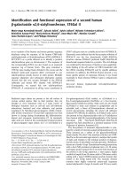
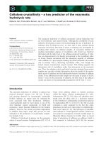
Fig. 3. Ers1p localizes in the vacuole and
endosome-like compartments. (A) Cells
expressing GFP-ERS1 (XGY50) were stained
with FM4-64 and analysed by fluorescence
microscopy as described in Experimental
procedures. GFP–Ers1 is shown in green,
FM4-64 is shown in red and their colocaliza-
tion (merge) is in yellow. Also shown are
cells imaged by Nomarski optics. (B) Whole
cell lysates from cells expressing HA-tagged
Ers1(pRs305ERS1p-ERS1-HA) were subjec-
ted to differential centrifugation and equival-
ent amounts of protein from each fraction
were separated by SDS ⁄ PAGE and immuno-
blotted with anti-HA or anti V-ATPase Igs as
described in Experimental procedures.
ERS1 and CTNS are functional homologues X D. Gao et al.
2502 FEBS Journal 272 (2005) 2497–2511 ª 2005 FEBS
MEH1 is predicted to encode a hydrophilic protein
with no obvious transmembrane spanning domains,
but it contains an N-terminal recognition sequence for
the attachment of a myristate. To determine its subcel-
lular localization, yeast strains were constructed that
expressed an MEH1 allele that was GFP-tagged at the
C terminus. Fluorescence analysis of this Meh1–GFP
fusion suggested that it tightly localized to the vacuo-
lar membrane (Fig. 5B). This result was confirmed by
the determining the localization in these cells of the
vacuole lumen fluorescent marker, CMAC. While
Meh1–GFP and CMAC colocalize to the same com-
partment, Meh1 is found at the membrane, while
CMAC is within the lumen (Fig. 4C). A similar local-
ization pattern was observed by using a Meh1
HA-tagged protein (data not shown). Unlike Ers1,
which localizes to the endosomes as well as the vacu-
ole, Meh1 appears to be largely confined to the vacuo-
lar membrane. Nonetheless, taking together both the
genetic and subcellular localization data, these results
provide good evidence for a functional relationship
between Ers1 and Meh1.
Meh1 is required for vacuolar acidification
To determine if loss of MEH1 plays a role in regula-
ting vacuolar function, we examined the acidity of the
vacuole indirectly using LysoSensor green. LysoSensor
green is a pH-sensitive fluorescent probe that accumu-
lates in the membranes of acidic organelles. In wild
type yeast, LysoSensor green labels the vacuolar mem-
branes and this staining is greatly diminished in
A
B
C
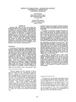
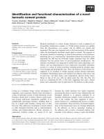
Fig. 4. MEH1, a multisuppressor of ers1D,
encodes a vacuolar protein. (A) ers1D
(XGY51) cells expressing MEH1 (YKR007W)
or ERS1 in YEp213 were streaked on an
YPAD plate containing 50 lgÆmL
)1
hygB.
(B) Isogenic wild type (SEY6210) or meh1D
cells (XGY53) with or without a high copy
plasmid containing the MEH1 or ERS1 gene
were serially diluted (10-fold), spotted onto
YPAD plates with or without 50 lgÆmL
)1
hygB and grown for 2 days at 30 °C.
(C) Yeast cells expressing a GFP-tagged
MEH1 allele (XGY52) were stained with the
vacuolar probe, CMAC, and analysed by
fluorescence microscopy as described in
Experimental procedures. Also shown are
cells imaged by Nomarski optics.
X D. Gao et al. ERS1 and CTNS are functional homologues
FEBS Journal 272 (2005) 2497–2511 ª 2005 FEBS 2503
mutant strains that are defective in the vacuolar (H+)
ATPase (V-ATPase) that pumps protons into the
lumen ([17] and Fig. 5). We qualitatively measured
vacuolar acidification by a visual assay of LysoSensor
green intensity by fluorescence microscopy. For com-
parative purposes, we also assayed LysoSensor green
staining of a vma1D strain, which lacks the 118-kDa
subunit of the V-ATPase (Fig. 5A) and is defective in
vacuolar acidification. Compared to the ers1D strain
(data not shown) or to the isogenic parental wild type
strain, in the meh1D strain LysoSensor staining was
diminished (Fig. 5A) although it was not absent, as it
was in the vma1D strain. No obvious morphological
abnormalities were seen when these different mutant
cells were viewed by bright field microscopy, although
meh1D strains appeared to be slightly swollen
(Fig. 5B).
The yeast V-ATPase is a large membrane associated
complex of proteins containing at least 13 different
subunits. In mutants lacking any of subunits, assembly
of the complex is impaired [18]. As a further test
for an affect of MEH1 on vacuolar acidification, we
compared the steady state levels of the 60-kDa Vma2
protein in meh1D and wild type cells by western immu-
noblotting, using anti-Vma2 antibodies. As expected,
no Vma2 protein was detected in a vma2D mutant
strain, and a 60-kDa protein corresponding to Vma2
was seen in wild type cells. A slightly diminished level
of Vma2 (about twofold) was also observed in ers1D
strains. In contrast to wild type cells, a significant
decrease in Vma2p steady state levels was observed in
meh1D strains (Fig. 6) suggesting that the 60-kDa
V-ATPase subunit is unstable as a consequence of loss
of MEH1 function. While the basis for this instability
is unknown, these results are consistent with the
decreased LysoSensor green staining in meh1D cells
and provide further support for a role of Meh1 in
regulating vacuolar, and hence ERS1 function.
Myristylation of Meh1 is required for its
vacuolar association
The Meh1 protein does not contain any predicted
membrane spanning domains and is quite hydrophilic,
Fig. 5. Loss of MEH1 affects the vacuolar
pH. (A) The isogenic parental strain
(BY4741), meh1D,orvma1D were stained
with the pH-sensitive fluorescent probe,
LysoSensor Green and viewed by fluores-
cence microscopy as described in Experi-
mental procedures. (B) Brightfield view of
the meh1D and vma1D cells imaged in (A).
ERS1 and CTNS are functional homologues X D. Gao et al.
2504 FEBS Journal 272 (2005) 2497–2511 ª 2005 FEBS
raising the question of how it localizes to the vacuolar
membrane. Sequence analysis predicts that Meh1p
contains a conserved N-terminal recognition sequence
for myristyolation (MGAVLSC). Myristate is normally
added to the consensus sequence at glycine-2 (G2)
after removal of the initiator methionine. We wished
to determine if this protein is myristoylated and if so,
whether or not this lipid modification facilitates its
interaction with the vacuolar membrane and is there-
fore important for Meh1 function. To approach these
questions, we created a mutant allele (meh1-ND5) that
replaces the first five N-terminal amino acids with a
methionine residue, and therefore produces an altered
protein that is predicted to lack acylation. To enable
detection of this altered protein, we also tagged the
C terminus with the HA epitope. This plasmid-borne
mutant allele was introduced into an meh1D strain and
tested for complementation of the hygromycin B sensi-
tive phenotype of meh1D. While an identical plasmid
harbouring the wild type MEH1 gene complemented
this phenotype, the mutant meh1ND5 failed to do so,
suggesting these N-terminal five amino acids are essen-
tial for MEH1 function (Fig. 7A). The failure to com-
plement meh1D was not due to the absence of protein,
since this mutant allele produced protein at levels com-
parable to the wild type (e.g. Fig. 7B). To determine if
the absence of this N-terminal region is important for
Meh1 localization to the vacuole, we analysed its local-
ization by subcellular fractionation. Subcellular organ-
elles were separated by differences in their densities
using differential centrifugation (see Experimental pro-
cedures). Using this method, we found that while most
of Meh1 sedimented with vacuolar membranes at
16 000 g (P16), though a proportion was found in the
S14 fraction (Fig. 7B). This result is consistent with
our observation that Meh1–GFP is associated with
vacuolar membranes. It is notable that the majority of
Meh1in the P16 fraction migrated as a smear, while
Meh1 in the S16 fraction migrated as a sharp band,
suggesting the possibility that the soluble portion of
Meh1 lacks a myristate and is therefore not associated
with the membrane. In sharp contrast, Meh1–ND5-
HA, lacking the consensus myristoylation site, largely
fractionated in the S16 fraction and away from the
vacuolar membrane. Unlike most of the wild type
Meh1 protein, this protein migrated as a sharp band.
Taken together, these data demonstrate that the
Fig. 6. Loss of MEH1 results in the instability of the 60-kDa
V-ATPase subunit. Protein extracts ( 50 lg) prepared from equiv-
alent amounts of wild type cells or those containing a deletion of
VMA2, MEH1 or ERS1 were separated by 8% SDS ⁄ PAGE and
immunoblotted with antibodies against Vma2p.
AB
Fig. 7. The N-terminal myristoylation consensus sequence is required for Meh1 function and vacuolar membrane association. (A) meh1ND5
fails to complement an meh1D strain. Wild type (SEY6210) or and meh1D mutant strain (XGY53) harbouring plasmids containing wild type
MEH1 or the mutant meh1ND5 allele, encoding protein lacking the N-terminal myristoylation consensus sequence, were plated on YPAD
media containing 50 lgÆmL
)1
hygromycin B. (B) The N-myristoylation consensus sequence is required for Meh1 membrane association.
Extracts were prepared from the SEY6210 expressing pTiMEH1-HA
3
or pTI-meh1-ND5-HA
3
and subjected to sedimentation centrifugation,
as described in Experimental procedures. Equivalent amounts of each fraction were separated by 10% SDS ⁄ PAGE and analysed by immuno-
blotting with anti-HA Igs.
X D. Gao et al. ERS1 and CTNS are functional homologues
FEBS Journal 272 (2005) 2497–2511 ª 2005 FEBS 2505
N-terminal five amino acids are essential for function
and localization to the vacuole, and provide evidence
to support the idea that Meh1 is myristoylated. Thus,
it is likely that Meh1 localizes to the vacuolar mem-
brane through a myristate tail and probably functions
on the cytosolic face of the vacuole.
Meh1 recruits the small GTPase Gtr1
to the vacuolar membrane
Proteomic analyses ( (-mbi.
ucla.edu/dip/); ( />identify a highly conserved protein, the small Ras-rela-
ted GTP binding protein, Gtr1, as a protein that
Meh1 interacts with. To obtain further information
about the function of Meh1, we examined whether or
not Meh1 and Gtr1 interact with one another, under
physiological conditions, using coimmunoprecipitation
assays. To determine if Meh1 interacts with Gtr11, we
used a coimmunoprecipitation assay. Yeast strains
were constructed that coexpressed HA and myc tagged
GTR1 and MEH1 genes. The chromosomal loci of
GTR1 and MEH1 were replaced with the correspond-
ing HA or myc-tagged alleles (see Experimental proce-
dures). Extracts from each of these strains were
prepared in buffer containing the nonionic detergent,
digitonin, to maintain oligomeric interactions between
membrane proteins, and these extracts were subjected
to coimmunoprecipitation assays. Gtr1–myc protein
was precipitated from these extracts with anti-myc
antibody and the immunoprecipitates were fractionated
by SDS ⁄ PAGE. The relative steady state levels of Gtr–
myc and Meh1–HA in the same extracts used for the
immunoprecipitations were determined by Western
blot analysis of aliquots removed prior to immunopre-
cipitation and were found to be similar (data not
shown; Fig. 8A, lanes 2 and 3). Meh1–HA that copre-
cipitated with Gtr1–myc was detected by immunoblot-
ting with anti-HA Ig (Fig. 8A). The result of this
experiment demonstrates that Meh1 coprecipitated
with Gtr1–myc (Fig. 8A, lanes 6). This interaction is
dependent on the coexpression of Meh1 with Gtr1 as
Meh–HA did not coprecipitate in a control strain that
does not coexpress Gtr1–myc (Fig. 8A, lane 5). We
also find no evidence that Meh1 interacts with another
vacuolar protein, Ers1, further demonstrating the spe-
cificity of this interaction.
If the interaction between Meh1 and Gtr1 is of bio-
logical relevance, we would expect to find Gtr1 locali-
zed in the vacuole, in an Meh1-dependent manner. To
test this idea, we constructed yeast cells whose chro-
mosomal GTR1 locus was replaced with a GFP-tagged
allele, and examined the intracellular localization of
Gtr1–GFP fusion proteins by fluorescence microscopy.
By this analysis, we found that Gtr1–GFP localized in
the vacuole, coincident to the pattern observed by the
vacuolar marker, CMAC. However, unlike CMAC,
which localizes in the lumen of the vacuole, Gtr1–GFP
appears to localize to the vacuolar membrane
(Fig. 8B), in a pattern similar to that of Meh1.
To determine if the vacuolar association of Gtr1 is
dependent on Meh1, we compared the fractionation
behaviour of Gtr1–myc in a wild type or meh1D strain,
by subcellular fraction. Fractions enriched for vacuolar
membranes were prepared and separated using differ-
ential centrifugation (see Experimental procedures).
Consistent with our observation that Gtr1–GFP local-
izes with vacuolar membranes, using this method, we
found that in a wild type MEH1 background, Gtr1
sedimented in the P16 fraction, with very little protein
observed in the S16 fraction (Fig. 8C). In striking con-
trast, in extracts prepared from the meh1D cells that
lack Meh1, the vast majority of Gtr1 appeared soluble,
fractionating in the S16 supernatant. These results dem-
onstrate that the membrane association of Gtr1 is
dependent on Meh1 and suggest that Meh1 is required
to recruit Gtr1 to the vacuolar membrane.
Discussion
In this study we have demonstrated that the S. cerevis-
iae Ers1 and human cystinosin proteins are functional
homologues. Like cystinosin, Ers1 is a vacuolar pro-
tein whose loss of function results in hygB sensitivity.
The human CTNS gene can complement the ers1D
phenotype, reversing its hygB sensitivity. Moreover,
the severity of CTNS mutants, as identified in different
patients afflicted with varying forms of the disease,
mimics the degree with which these CTNS mutant alle-
les can complement ers1D. Importantly, the inability of
these mutant ctns alleles to complement ers1D provides
the proof of principle for the utility of yeast as model
system for functional analyses of cystinosin. We have
used this yeast system to identify novel yeast genes
that regulate Ers1 and other vacuolar functions.
Through a high copy suppressor screen we identify
MEH1, a previously uncharacterized yeast gene that is
required for maintaining vacuolar acidity. We also
identify Gtr1, a small Ras-related GTPase, whose
recruitment to the vacuolar membrane is dependent
upon its interaction with Meh1.
Although cystinosis has been described primarily as
a kidney disease, mutations in CTNS affect a number
of different organs. It is not understood how cystine
accumulation in the lysosome causes cellular damage
or why this accumulation specifically targets the
ERS1 and CTNS are functional homologues X D. Gao et al.
2506 FEBS Journal 272 (2005) 2497–2511 ª 2005 FEBS
different organs affected in patients. Indeed, mice car-
rying loss-of function ctns alleles fail to develop any
renal abnormalities and exhibit only ocular perturba-
tion [19]. Although it is well established that cystinosin
encodes a lysosomal cystine transporter, it has been
suggested that the phenotypic consequences of CTNS
mutations may also be due to secondary affects
[3,19,20]. For instance, CTNS encodes a proton-driven
pump, so defects in cystine transport may affect lyso-
somal pH indirectly via an accumulation of protons or
an affect on the lysosomal ATPase that pumps protons
into the lysosome upon hydrolysis of cytosolic ATP.
These ideas bear on the question of what biological
functions in yeast are regulated by ERS1 and whether
these relate to cystine transport from the vacuole.
Unlike CTNS in mammalian cells, deletion of ERS1
does not dramatically affect cellular growth properties.
Further, ers1D mutants do not display any apparent
abnormal vacuolar morphology and treatment of
ers1D cells with cysteamine does not rescue the ers1D
phenotype (data not shown). These observations sug-
gest that the accumulation of cystine in the vacuole of
yeast does not lead to the types of cellular damage that
is observed in mammalian cells. Two explanations for
these results can be envisaged. First, a formal possibil-
ity that our studies have not ruled out is that, in yeast,
Ers1 and cystinosin are involved in the transport of a
molecule other than cystine, whose accumulation gives
rise to hygB sensitivity. Direct measurements of cystine
levels in the yeast vacuole have been hampered by
technical difficulties (data not shown). A second
explanation is that the mild ers1D phenotype may be
more related to the effect of proton accumulation in
the vacuole than to cystine accumulation. Further
A
B
C
Fig. 8. Meh1 interacts with Gtr1 and is
required for Gtr1 vacuolar localization.
(A) Meh1 and Gtr1 coimmunoprecipitate.
Strains expressing MEH1-HA (TIY3) GTR1–
myc (TIY4), or that coexpress MEH1-HA and
GTR1–myc (TIY5) were constructed as des-
cribed in Experimental procedures. Cell free
extracts were prepared as described [26]
and subjected to immunoprecipitation using
anti-myc Ig to precipitate Gtr1-myc. Immuno-
precipitated proteins were separated by
10% SDS PAGE and any coprecipitated
Meh1-HA was detected by immunoblotting
with anti-HA Ig. In lanes 1–3 aliquots of
extracts were removed prior to immunopre-
cipitation. (B) Gtr1 localizes to the vacuolar
membrane. (C) Yeast cells expressing a
GFP-tagged GTR1 allele (TIY10) were
stained with the vacuolar probe, CMAC, and
analysed by fluorescence microscopy as
described in Experimental procedures. Cells
were also imaged by Nomarski optics (DIC).
(C) Vacuolar localization of Gtr1 is depend-
ent on Meh1. Extracts were prepared from
wild type MEH1 cells (SEY6210) or an iso-
genic meh1D strain (XGY53) expressing
GTR–myc and subjected to sedimentation
centrifugation, as described in Experimental
procedures. Equivalent amounts of each
fraction were separated by 10% SDS ⁄ PAGE
and analysed by immunoblotting with anti-
myc Igs.
X D. Gao et al. ERS1 and CTNS are functional homologues
FEBS Journal 272 (2005) 2497–2511 ª 2005 FEBS 2507
investigation is required to clarify the basis for the
ers1D hygB phenotype in yeast.
We identified MEH1 as a high copy suppressor of
ers1D. Meh1 is localized in the vacuole and this mem-
brane association appears to be dependent on an N-ter-
minal myristate modification. The ability of Meh1 to
associate with the vacuolar membrane is critical for its
function because mutations in this putative myristoyla-
tion site fail to complement an meh1 deletion mutant.
We demonstrated that vacuolar acidification is influ-
enced by MEH1 (Figs 5 and 6) but the precise mechan-
ism by which Meh1 affects vacuolar function remains
unknown. At least one vacuolar subunit, Vma2, is
unstable in an meh1D mutant background (Fig. 6).
While an Meh1 homologue cannot be identified among
mammals, we also identified the highly conserved Ras-
related GTPase Gtr1, as a protein that interacts with
Meh1. A clue that these proteins physically interact
came from the databases and we have demonstrated
this by coimmunoprecipitation assays. Several lines of
evidence suggest that this interaction is of physiological
relevance. First, like Meh1, we find that Gtr1 localizes
to the vacuolar membrane (Fig. 8). While it has been
reported that Gtr1 localizes to the nucleus in yeast
[21], we find no evidence for such a localization. Sec-
ond, the vacuolar association of Gtr1 is completely
dependent upon the presence of Meh1, a result that we
have found by using sedimentation analysis (Fig. 8)
and microscopic analysis of Gtr1–GFP fusions in live
cells (data not shown). In the presence of Meh1, Gtr1
is found at the vacuole, while in the absence of Meh1,
Gtr1 appears to be localized to the cytosol. Given the
degree with which Gtr1, but not Meh1 has been con-
served, it is tempting to speculate that Meh1 has
evolved simply to recruit Gtr1 to the vacuole, where it
may function, like other GTPases, as a molecular
switch to control an as yet vacuolar function. How-
ever, our preliminary analyses of gtr1 demonstrate that
while we observe some vacuolar defects, gtr1 mutants
do not precisely phenocopy meh1 (X. Gao and
N. Dean, unpublished observations). One explanation
for these results is that yeast contain a highly related
GTR1 homologue (GTR2) that may be redundant in
function to GTR1. Further investigation is required to
determine the precise mechanism of suppression of
ers1D by MEH1, and the role of Meh1 and Gtr1 in
regulating vacuolar function. The results we have pre-
sented validate the utility of yeast as a model system
for the functional analysis of cystinosin, both as a sim-
ple plate assay for the detection of functional muta-
tions in CTNS and also as a genetic system to identify
and characterize other genes that may be involved in
the regulation of cystinosin and the lysosome.
Experimental procedures
Yeast strains and media
Standard yeast media, growth conditions and genetic tech-
niques were used [22]. HygB sensitivity was tested on yeast
extract ⁄ peptone ⁄ adenine sulfate ⁄ dextrose plates (YPAD)
supplemented with 50 or 90 lg Æ mL
)1
hygB (Boehringer
Mannheim, Mannheim, Germany) as described previously
[12].
XGY50 (MATa ade2-1 ura3-1 his3-11 trp1-1 leu2-3112
can1-100 GAL1p-GFP-ERS1) was constructed from W303a
(MATa ade2-1 ura3-1 his3-11 trp1-1 leu2-3112 can1-1001)
and contains a replacement of the chromosomal ERS1
locus with a GAL1 promoter-driven ERS1 allele that
encodes Ers1p fused with GFP at its N terminus. XGY51,
XGY52 and XGY53 were constructed from SEY6210
(MATa ura3-52 leu2-3112 his3-D200 trp1-D901 lys2-801
suc2D9). XGY51 (MATa ura3-52 leu2-3112 his3-D200 trp1-
D901 lys2-801 suc2D9 ers1D::TRP1) contains a deletion of
the ERS1 ORF. XGY52 contains a replacement of the
MEH1 ORF (YKR007) with an allele encoding Meh1p
fused to GFP its C terminus. XGY53 contains a deletion of
MEH1 (MATa ura3-52 leu2-3112 his3-D200 trp1-D901 lys2-
801 suc2D9 meh1D::Sp his5
+
). TIY10 (MATa ura3-52 leu2-
3112 his3-D200 trp1-D901 lys2-801 suc2D9 GTR1-GFP-::
Sp his5) produces Gtr1 tagged with GFP at the C terminus.
TIY3 (MATa ura3-52 leu2-3112 his3-D200 trp1-D901 lys2-
801 suc2D9 MEH1-HA-::Sp his5) produces Meh1, tagged
with myc at the C terminus. TIY4 (MATa ura3-52 leu2-
3112 his3-D200 trp1-D901 lys2-801 suc2D9 GTR1-myc-::
TRP1) produces Gtr1, tagged with myc at the C terminus.
TIY5 (MATa ura3-52 leu2-3112 his3- D200 trp1-D901
lys2-801 suc2D9 MEH1-HA-::Sp his5 GTR1-myc-::TRP1)
produces both Gtr1–myc and Meh1-HA. All strains were
constructed using PCR-mediated homologous recombina-
tion [23], using a standard set of plasmid templates and
gene specific primers [24].
Plasmid constructions
For expression of plasmid-borne ERS1 driven by its own
promoter, a DNA fragment containing the ERS1 ORF and
520 base pairs of upstream sequence was amplified by PCR
from yeast genomic DNA and cloned into the EcoR1 ⁄ Xba1
integrative LEU2 plasmid, pRS305 [25] and a 2 l plasmid
YEp213 [26], respectively, to generate pRS305:ERS1 and
Yep213:ERS1. For expression of a plasmid-borne ERS1
under other constitutive promoters, a DNA fragment con-
taining the ERS1 ORF was amplified by PCR. This frag-
ment was cloned into pRS305GAP, which contains the
glyceraldehyde-3-phosphate dehydrogenase (TDH3) promo-
ter, and into pRS305Ti, which contains the triose phosphate
isomerase (TPI1) promoter in pRS305 [27]. This generated
pRS305GAP-ERS1 and pRS305Ti-ERS1. Linearization of
ERS1 and CTNS are functional homologues X D. Gao et al.
2508 FEBS Journal 272 (2005) 2497–2511 ª 2005 FEBS
these plasmids with AflII within the LEU2 gene targets
integration at the leu2-3 locus.
To express the human cystinosin gene ( CTNS)inS. cere-
visiae, a fragment containing the CTNS cDNA was ampli-
fied by PCR from pcDNA-CTNS (kindly provided by
S. Cherqui and C. Antignac, INSERM, Paris, France) [3]
and cloned into pRS305:ERS1pr, which contains a 520-base
pair fragment of 5¢-ERS1 flanking sequences that includes
the ERS1 promoter region. This generated pRS305:ERS1pr-
hCTNS, in which the human CTNS cDNA is expressed
from the ERS1 promoter. All mutant forms of CTNS, gen-
erated by site directed mutagenesis (Quick Change, Invitro-
gen, Carlsbad, CA, USA) of pRS305:ERS1pr-hCTNS (see
below), were cloned downstream of the ERS1 promoter
using a similar strategy. Mutant forms of CTNS that encode
cystinosin lacking its N-terminal 121 amino acids or
C-terminal 82 amino acids (ctns-ND121 and ctns-CD82,
respectively) were amplified by PCR and cloned into
pRS305:ERS1pr to generate pRS305:ERS1pr:hCTNS
ND121 and pRS305:ERS1pr:CTNS-CD82. Single missense
mutations in CTNS corresponding to those identified in
cystinosis patients [4] were generated by PCR mutagenesis
using pRS305:ERS1pr-hCTNS as a template. This created
mutant ctns alleles with amino acids changes G308R and
L338P. The sequence of all PCR-generated CTNS mutants
was verified by DNA sequence analysis. Linearization of
these integrative plasmids with AflII within the LEU2 gene
targets integration at the leu2-3 locus.
To tag the Erd1 protein, a HindIII ⁄ EcoRI fragment con-
taining the ERD1 ORF lacking the stop codon was isolated
by PCR and cloned into pSK
–
P ⁄ XHA
3
[28], a derivative
of Bluescript SK
–
(Stratagene, La Jolla, CA, USA).
pSK:ERD1-HA
3
encodes Erd1p containing an in-frame
fusion to three copies of the HA epitope at the C terminus.
A HindIII ⁄ XbaI fragment from pSK:ERD1-HA
3
, contain-
ing just the ORF was subcloned into pRS305:ERS1pr to
generate into pRS305:ERS1pr-ERS1–HA, which places
ERS1-HA
3
expression under its own promoter in a low
copy, CEN-based plasmid. Linearization of this plasmid
with XhoI within the URA3 gene targets its integration to
the ura3-52 locus.
For expression of plasmid-borne MEH1 under its own
promoter, a DNA fragment containing the MEH1 ORF
and 700 base pairs of 5¢ flanking sequence was amplified
from yeast genomic DNA by PCR and cloned into
YEp213, a 2 l LEU2 plasmid, to generate Yep213:MEH1.
To create Ti-MEH1-HA
3
, a fragment containing the MEH1
ORF but in which the stop codon was removed and
replaced by an Nsi1 site, was amplified by PCR and cloned
into pTiaO [26] to create a plasmid borne, C-terminally
HA-tagged MEH1 gene in a URA3 integration vector. pTi-
meh1-ND5-HA3 was constructed in exactly the same man-
ner except the design of the 5¢ PCR primer was designed to
replace sequences encoding the N-terminal five amino acids
with those encoding a methionine.
Analysis of GFP-fusion proteins and vacuolar
fluorescent staining
To visualize the localization of GFP–Ers1 fusion proteins,
cells that express an GFP-ERS1 allele whose expression is
regulated by the GAL1 promoter (XGY50) were grown to
an D
600
of 1–3 in YPA +2% (w ⁄ v) galactose. After wash-
ing the cells with NaCl ⁄ P
i
, GFP fluorescence was imaged
using a fluorescein isothiocyanate filter set with an excita-
tion wavelength of 470 nm. To visualize the localization of
Meh1–GFP and Gtr1–GFP, cells were grown to a A
600
of
1–3 in YPAD, washed twice in ice cold NaCl ⁄ P
i
and imme-
diately imaged as described above.
The lipophilic dye, FM4-64 (Molecular Probes, Eugene,
OR, USA) was used to visualize the membranes of vacuoles
and endosomes [16]. Cells grown to A
600
¼ 1 were harves-
ted, resuspended at 10 A
600
units per mL in NaCl ⁄ P
i
and
left on ice. FM4-64 was added to a final concentration of
20 lm, and was viewed immediately upon shifting the incu-
bation temperature to 25 °C and after variable lengths of
time by fluorescence microscopy, using a Texas Red filter
set with an excitation wavelength of 560 nm.
To stain the lumen of vacuole, the membrane-permeable
chloromethyl coumarin derivative, CMAC-Ala-Pro (Mole-
cular Probes), was used. Cells were grown to mid-log phase
(A
600
of 1–2), resuspended in 10 mm Hepes buffer [pH 7.4,
containing 5% (w ⁄ v) glucose] and CMAC-Ala-Pro was
added to final concentration of 10 lm. After 15–30 min
incubation at room temperature, the stained cells were visu-
alized by fluorescence microscopy, using a blue filter set,
with an excitation wavelength of 354 nm.
Vacuolar acidity was monitored by the accumulation of
LysoSensor Green (Molecular Probes), followed by fluores-
cence microscopy, as described [17] with the following
minor modifications. After harvesting cells that had grown
to mid-log phase (A
600
¼ 1), cells were resuspended in
YPAD containing 100 mm Hepes (KOH), pH 7.2 and 4 lm
LysoSensor Green and viewed within 10 min of adding the
fluorescent probe by fluorescence microscopy, using a fluo-
rescein isothiocyanate filter. Each sample was individually
resuspended, stained, viewed and imaged because we
observed a general quenching of fluorescence over time.
Subcellular fractionation, western blotting and
immunoprecipitation
Cells were grown to A
600
¼ 1, spheroplasted, lysed by
dounce homogenization and fractionated by differential
centrifugation as described previously [29], except that
vacuole-containing fractions were sedimented at 16 000 g
instead of 14 000 g. Proteins in each fraction, representing
cell equivalents from 10 absorbance units of cells, were sep-
arated by SDS ⁄ PAGE. Proteins were immunoblotted on to
PVDF membranes and detected using anti-HA Ig (12CA5),
X D. Gao et al. ERS1 and CTNS are functional homologues
FEBS Journal 272 (2005) 2497–2511 ª 2005 FEBS 2509
anti-myc Ig (9E10), anti-(V-ATPase
60
) or anti-(V-ATP-
ase
100
) Igs (Molecular Probes) diluted 1 : 2000, to detect
the 60- or100-kDa subunit of the V-ATPase complex. Sec-
ondary antibodies were coupled to horseradish peroxidase
and detected by chemiluminescence (Amersham, ). Immuno-
precipitations of whole cell yeast detergent extracts were
performed exactly as described [26].
Screen for multicopy suppressors of the hygB
sensitivity of ers1D
The high copy plasmid library used for this screen (kindly
provided by B. Futcher, Stony Brook University, NY,
USA) was made from a partial HindIII digest of yeast
genomic DNA cloned into YEp213, and contained inserts
that averaged about 9 kb in length. The ers1D strain
(XGY51) was transformed with this library by the lithium
acetate method [21]. About 10 000 LEU2
+
transformants
were isolated and tested for drug sensitivity by replica pla-
ting onto YPAD plates containing 60 lgÆmL
)1
hygB. After
incubation at 30 °C for 2 days, a total of 300 hygB resistant
colonies grew and were individually streaked out on hygB-
containing plates. To identify transformants whose hygB
resistance resulted from complementation by plasmid-borne
ERS1, candidates were screened by colony PCR using a pair
of primers designed to amplify the ERS1 ORF. Of the trans-
formants 20% were found to contain ERS1 and were elim-
inated from the pool. Plasmids were recovered from the
remaining colonies and digested with several restriction
enzymes to identify those with overlapping inserts. Repre-
sentatives from each group of related plasmids were trans-
formed into the ers1D strain (XGY51) and again tested for
the ability to suppress hygB sensitivity. Nine unique plas-
mids were identified whose inserts were sequenced. For
those containing more than one ORF, each ORF was either
subcloned uniquely or deleted within the plasmid and tested
for ers1D suppression. From this analysis, 10 single genes,
including ERS1, were identified as multicopy suppressors of
ers1D hygB sensitivity. Five of these were subsequently
found to be nonspecific suppressors of hygB sensitivity.
Acknowledgement
We wish to thank Stephanie Cherqui and Corinne
Antignac for providing us with the human CTNS
cDNA. This work was supported by a grant from the
NIH (R01-GM048467) to N.D.
References
1 Gahl WA, Thoene JG & Schneider JA (2002) Cystino-
sis. N Engl J Med 347, 111–121.
2 Gahl WA, Bashan N, Tietze F, Bernardini I & Schul-
man JD (1982) Cystine transport is defective in isolated
leukocyte lysosomes from patients with cystinosis.
Science 217, 1263–1265.
3 Kalatzis V, Cherqui S, Antignac C & Gasnier B (2001)
Cystinosin, the protein defective in cystinosis, is a
H
+
-driven lysosomal cystine transporter. EMBO J 20,
5940–5949.
4 Attard M, Jean G, Forestier L, Cherqui S, van’t Hoff W,
Broyer M, Antignac C & Town M (1999) Severity of
phenotype in cystinosis varies with mutations in the
CTNS gene: predicted effect on the model of cystinosin.
Hum Mol Genet 8, 2507–2514.
5 Pisoni RL, Thoene JG & Christensen HN (1985) Detec-
tion and characterization of carrier-mediated cationic
amino acid transport in lysosomes of normal and cysti-
notic human fibroblasts. Role in therapeutic cystine
removal? J Biol Chem 260, 4791–4798.
6 Pisoni RL, Acker TL, Lisowski KM, Lemons RM &
Thoene JG (1990) A cysteine-specific lysosomal trans-
port system provides a major route for the delivery of
thiol to human fibroblast lysosomes: possible role in
supporting lysosomal proteolysis. J Cell Biol 110,
327–335.
7 Gahl WA, Charnas L, Markello TC, Bernardini I, Ishak
KG & Dalakas MC (1992) Parenchymal organ cystine
depletion with long-term cysteamine therapy. Biochem
Medical Metab Biol 48, 275–285.
8 Gahl WA (2003) Early oral cysteamine therapy for
nephropathic cystinosis. Eur J Pediatr 162 (Suppl. 1),
S38–S41.
9 Hardwick KG & Pelham HR (1990) ERS1 a seven
transmembrane domain protein from Saccharomyces
cerevisiae. Nucleic Acids Res 18, 2177.
10 Hardwick KG, Lewis MJ, Semenza J, Dean N &
Pelham HRB (1990) ERD1, a yeast gene required for
the retention of lumenal endoplasmic reticulum proteins,
affects glycoprotein processing in the Golgi apparatus.
EMBO J 9, 623–630.
11 Cherqui S, Kalatzis V, Trugnan G & Antignac C (2001)
The targeting of cystinosin to the lysosomal membrane
requires a tyrosine-based signal and a novel sorting
motif. J Biol Chem 276, 13314–13321.
12 Dean N (1995) Yeast glycosylation mutants are sensitive
to aminoglycosides. Proc Natl Acad Sci USA 92, 1287–
1291.
13 Gaxiola RA, Rao R, Sherman A, Grisafi P, Alper SL &
Fink GR (1999) The Arabidopsis thaliana proton trans-
porters, AtNhx1 and Avp1, can function in cation
detoxification in yeast. Proc Natl Acad Sci USA 96 ,
1480–1485.
14 Madrid R, Gomez MJ, Ramos J & Rodriguez-Navarro
A (1998) Ectopic potassium uptake in trk1 trk2 mutants
of Saccharomyces cerevisiae correlates with a highly
hyperpolarized membrane potential. J Biol Chem 273,
14838–14844.
ERS1 and CTNS are functional homologues X D. Gao et al.
2510 FEBS Journal 272 (2005) 2497–2511 ª 2005 FEBS
15 Conboy MJ & Cyert MS (2000) Luv1p ⁄ Rki1p ⁄ Tcs3p ⁄
Vps54p, a yeast protein that localizes to the late Golgi
and early endosome, is required for normal vacuolar
morphology. Mol Biol Cell 11, 2429–2443.
16 Vida TA & Emr SD (1995) A new vital stain for visua-
lizing vacuolar membrane dynamics and endocytosis in
yeast. J Cell Biol 128, 779–792.
17 Perzov N, Padler-Karavani V, Nelson H & Nelson N
(2002) Characterization of yeast V-ATPase mutants
lacking Vph1p or Stv1p and the effect on endocytosis.
J Exp Biol 205, 1209–1219.
18 Doherty RD & Kane PM (1993) Partial assembly of the
yeast vacuolar H (+) -ATPase in mutants lacking one
subunit of the enzyme. J Biol Chem 268, 16845–16851.
19 Cherqui S, Sevin C, Hamard G, Kalatzis V, Sich M,
Pequignot MO, Gogat K, Abitbol M, Broyer M, Gubler
MC & Antignac C (2002) Intralysosomal cystine accu-
mulation in mice lacking cystinosin, the protein defec-
tive in cystinosis. Mol Cell Biol 22, 7622–7632.
20 Rosa TG, De Souza Wyse AT, Wajner M & Wannma-
cher CM (2004) Cysteamine prevents and reverses the
inhibition of pyruvate kinase activity caused by cystine
in rat heart. Biochim Biophys Acta 1689, 114–119.
21 Ito HY, Fukuda Y, Murata K & Kimura J (1983)
Transformation of intact yeast with alkalai cations.
J Bacteriol 153, 163–168.
22 Guthrie C & Fink GR (1991) Guide to yeast genetics
and molecular biology. Methods Enzymol 194, 3–20.
23 Baudin A, Ozier-Kalogeropoulos O, Denouel A,
Lacroute F & Cullin C (1993) A simple and efficient
method for direct gene deletion in Saccharomyces cerevi-
siae. Nucleic Acids Res 21, 3329–3330.
24 Longtine, MS, McKenzie A, 3rd Demarini DJ, Shah
NG, Wach A, Brachat A, Philippsen P & Pringle JR
(1998) Additional modules for versatile and economical
PCR-based gene deletion and modification in Saccharo-
myces cerevisiae. Yeast 14, 953–961.
25 Sikorski RS & Hieter P (1989) A system of shuttle vec-
tors and yeast host strains designed for effecient manip-
ulation of DNA in Saccharomyces cerevisiae. Genetics
122, 19–27.
26 Rose AB & Broach JR (1990) Propagation and expres-
sion of cloned genes in yeast: 2-microns circle-based
vectors. Methods Enzymol 185, 234–279.
27 Gao XD & Dean N (2000) Distinct protein domains
of the yeast Golgi GDP-mannose transporter mediate
oligomer assembly and export from the endoplasmic
reticulum. J Biol Chem 275, 17718–17727.
28 Neiman AM, Mhaiskar V, Manus V, Galibert F &
Dean N (1997) Saccharomyces cerevisiae HOC1, a sup-
pressor of pkc1, encodes a putative glycosyltransferase.
Genetics 145, 637–645.
29 Dean N & Poster J (1996) Molecular and phenotypic
analysis of the S. cerevisiae MNN10 gene identifies a
family of related glycosyltransferases. Glycobiology 6,
73–81.
X D. Gao et al. ERS1 and CTNS are functional homologues
FEBS Journal 272 (2005) 2497–2511 ª 2005 FEBS 2511