Trends in Cell Signaling Pathways in Neuronal Fate Decision Edited by Sabine Wislet-Gendebien potx
Bạn đang xem bản rút gọn của tài liệu. Xem và tải ngay bản đầy đủ của tài liệu tại đây (18.5 MB, 366 trang )
TRENDS IN CELL
SIGNALING PATHWAYS
IN NEURONAL FATE
DECISION
Edited by Sabine Wislet-Gendebien
Trends in Cell Signaling Pathways in Neuronal Fate Decision
/>Edited by Sabine Wislet-Gendebien
Contributors
Aviva Symes, Sonia Villapol, Trevor Logan, Eri Hashino, Atsushi Shimomura, Michael Fehlings, Madeleine O'Higgins,
Jenny Wong, Wenhui Hu, Yonggang Zhang, Sabine Wislet-Gendebien, Tanja Vogel, Ann M. Turnley, Harleen Basrai,
Kimberly Christie, Roxana Nat, Galina Apostolova, Georg Dechant, Adam Cole, Liang-Wei Chen, Nibaldo Inestrosa,
Lorena Varela-Nallar, Uwe Ueberham, Thomas Arendt
Published by InTech
Janeza Trdine 9, 51000 Rijeka, Croatia
Copyright © 2013 InTech
All chapters are Open Access distributed under the Creative Commons Attribution 3.0 license, which allows users to
download, copy and build upon published articles even for commercial purposes, as long as the author and publisher
are properly credited, which ensures maximum dissemination and a wider impact of our publications. After this work
has been published by InTech, authors have the right to republish it, in whole or part, in any publication of which they
are the author, and to make other personal use of the work. Any republication, referencing or personal use of the
work must explicitly identify the original source.
Notice
Statements and opinions expressed in the chapters are these of the individual contributors and not necessarily those
of the editors or publisher. No responsibility is accepted for the accuracy of information contained in the published
chapters. The publisher assumes no responsibility for any damage or injury to persons or property arising out of the
use of any materials, instructions, methods or ideas contained in the book.
Publishing Process Manager Iva Simcic
Technical Editor InTech DTP team
Cover InTech Design team
First published March, 2013
Printed in Croatia
A free online edition of this book is available at www.intechopen.com
Additional hard copies can be obtained from
Trends in Cell Signaling Pathways in Neuronal Fate Decision, Edited by Sabine Wislet-Gendebien
p. cm.
ISBN 978-953-51-1059-0
free online editions of InTech
Books and Journals can be found at
www.intechopen.com
Contents
Preface VII
Section 1 TGF-Beta Signaling and Neuronal Fate Decision 1
Chapter 1 Role of TGF-β Signaling in Neurogenic Regions After
Brain Injury 3
Sonia Villapol, Trevor T. Logan and Aviva J. Symes
Chapter 2 Insulin/IGF-Signalling in Embryonic and Adult Neural
Proliferation and Differentiation in the Mammalian Central
Nervous System 37
Tanja Vogel
Chapter 3 The Role of Smad Proteins for Development, Differentiation
and Dedifferentiation of Neurons 75
Uwe Ueberham and Thomas Arendt
Section 2 Wnt Signaling and Neuronal Fate Decision 113
Chapter 4 Wnt Signaling Roles on the Structure and Function of the
Central Synapses: Involvement in Alzheimer’s Disease 115
Nibaldo C. Inestrosa and Lorena Varela-Nallar
Chapter 5 Roles of Wnt/β-Catenin Signaling in Controlling the
Dopaminergic Neuronal Cell Commitment of Midbrain and
Therapeutic Application for Parkinson’s Disease 141
Liang-Wei Chen
Chapter 6 Regulation of Cell Fate in the Brain by GSK3 153
Adam R. Cole
Section 3 Neurotrophin and Neuronal Fate Decision 179
Chapter 7 Neurotrophin Signaling and Alzheimer’s Disease
Neurodegeneration − Focus on BDNF/TrkB Signaling 181
Jenny Wong
Section 4 NF-K-b and Neuronal Fate Decision 195
Chapter 8 NFκB Signaling Directs Neuronal Fate Decision 197
Yonggang Zhang and Wenhui Hu
Section 5 Stem Cells and Signaling Pathways 215
Chapter 9 Telencephalic Neurogenesis Versus Telencephalic
Differentiation of Pluripotent Stem Cells 217
Roxana Nat, Galina Apostolova and Georg Dechant
Chapter 10 Regulation of Basal and Injury-Induced Fate Decisions of Adult
Neural Precursor Cells: Focus on SOCS2 and Related Signalling
Pathways 241
Harleen S. Basrai, Kimberly J. Christie and Ann M. Turnley
Chapter 11 Neural Stem/Progenitor Cells for Spinal Cord
Regeneration 271
Ryan Salewski, Hamideh Emrani and Michael G. Fehlings
Chapter 12 Epigenetic Regulation of Neural Differentiation from
Embryonic Stem Cells 305
Atsushi Shimomura and Eri Hashino
Chapter 13 Neural Fate of Mesenchymal Stem Cells and Neural Crest Stem
Cells: Which Ways to Get Neurons for Cell Therapy
Purpose? 327
Virginie Neirinckx, Cécile Coste, Bernard Rogister and Sabine Wislet-
Gendebien
ContentsVI
Preface
During the last decades, numerous studies about stem cells and regenerative medicine high‐
lighted new therapeutic approaches to treat several neurological disorders. It is noteworthy
that the current optimism over potential stem cell therapies is driven by new understand‐
ings of stem cell biolology leading to specific cell fate decision.
The main objective of this book is to offer a general understanding of signaling pathways
underlying the capacity of differentiation of several types of stem cells into neurons, during
the development. Indeed, in this book, we deeply described TGF-beta signaling, Wnt Signal‐
ing, neurotrophin and NF-κ-B signaling and their implication in neuronal fate decision.
The second objective of this book is to understand how those pathways are altered in pathologi‐
cal conditions. We consequently analyzed those pathways in several pathological conditions.
Finally the third objective of this book is to describe advances in cellular therapy that could
be use to restore central nervous system dysfunction in pathological conditions, based on
new molecular biology findings. Several sources of stem cells and their potential benefits
were described in the last part of this book.
Finally, I would like to conclude this preface by expressing my deepest gratitude to all au‐
thors who contributed to the elaboration of this book.
Sabine Wislet-Gendebien, PhD
GIGA Neurosciences
University of Liège, Belgium
Section 1
TGF-Beta Signaling and Neuronal Fate Decision
Chapter 1
Role of TGF-β Signaling in
Neurogenic Regions After Brain Injury
Sonia Villapol, Trevor T. Logan and Aviva J. Symes
Additional information is available at the end of the chapter
/>1. Introduction
In 1928 Santiago Ramón y Cajal penned what became the accepted view about neurons in the
central nervous system; “everything may die, nothing can be regenerated”. He later exhibited his
wisdom by adding; “It’s the job of science to rewrite, if possible, this cruel phrase” [1]. Up until 20
years ago, the scientific literature had emphasized that neurogenesis only occurs during
development with no new neurons generated in the adult mammalian brain. However, since
the discovery of adult neurogenesis, an extensive literature has emerged supporting the
constant generation of new neurons in two neurogenic regions of the adult brain: the subven‐
tricular zone around the lateral ventricles (SVZ) and the subgranular zone (SGZ) of the
hippocampal dentate gyrus (DG) [2].
The existence of adult neurogenesis gave hope for recovery and regeneration from the many
different insults that can damage the brain. After stroke or traumatic brain injury (TBI),
immediate massive necrosis occurs followed by a subsequent prolonged period of inflamma‐
tion and further neuronal death [3]. Although brain injury induces massive cell loss, it also
induces an increase in proliferation of NSCs residing in the neurogenic niches [4]. The
environment of the neurogenic niche in adult animals is exquisitely regulated, with a finely-
tuned balance of soluble and cell-intrinsic factors that regulate the many different processes
that are critical to neurogenesis: cell survival, proliferation, differentiation, and migration [5].
Dramatic changes occur in this environment as a consequence of the injury. The careful
regulation of neurogenesis is disrupted by the many different cellular, soluble and vascular
signals detected by the different cell types in the SVZ and DG. This major environmental
alteration leads to increased proliferation of progenitor cells for long periods after the acute
injury, yet the ability of the neural progenitor cells to fully differentiate, migrate and integrate
into the lesioned area is limited [6]. Understanding the signals that regulate adult neurogenesis
© 2013 Villapol et al.; licensee InTech. This is an open access article distributed under the terms of the
Creative Commons Attribution License ( which permits
unrestricted use, distribution, and reproduction in any medium, provided the original work is properly cited.
in the naïve and injured animals is key to ultimately being able to harness the potential of
neuronal replacement and improve stem cell therapy.
There are many different factors important to regulation of neurogenesis, many of which are
discussed in other chapters in this book. Here we will focus on the role of the transforming
growth factor-β (TGF-β) superfamily and its associated signaling pathways in regulating
neurogenesis after brain injury. Members of this family, including the bone morphogenetic
proteins (BMPs), Activin, and TGF-β1, -β2 and -β3 have a profound influence on the neuro‐
genic process in naïve animals [7]. Many of these cytokines are induced by injury and play
critical roles in many kinds of brain damage related processes around the lesion [3]. We and
others recently started to accumulate data on their induction in the neurogenic niches after
different types of injury. Here we will focus on the relevance of their induction in these specific
brain regions, and the mechanisms through which they may influence the neurogenic response
to injury. As there are significant differences between the behavior of cells contributing to
neurogenesis during development and in the adult, we will restrict our analysis to that
observed in adult animals after injury. Delineation of the specific role of members of the TGF-
β superfamily in injury-induced neurogenesis may provide specific therapeutic targets for
enhancing neurogenesis after trauma.
2. The TGF-β superfamily; cytokines, receptors and signaling
The TGF-β cytokine superfamily is a large group of proteins comprising 33 different members
that include: bone morphogenetic proteins (BMPs), growth differentiation factors (GDFs),
activins, inhibins, nodal, lefty, mülllerian inhibiting substance (MIS) together with the TGF-
β proteins [8, 9]. All members of this cytokine family mediate their effects in a broadly
analogous manner, binding specific type I and II transmembrane serine threonine kinase
receptors and transducing their signal through similar intracellular Smad proteins [10]. These
cytokines are divided into two distinct groups: those of the TGF-β/Activin group which mainly
signal through the type I receptors ALK4, -5 and -7 activating Smad2 and -3, and those of the
BMP/GDF group [11, 12] which employ ALK1, -2, -3 and -6 to activate Smad1, -5 and -8 [13,
14]. The specificity of Smad activation is therefore mainly determined by the identity of the
type I receptor used to transduce the cytokine signal [15] (Figure 1).
TGF-β1, -β2 and -β3 together with some GDFs are unique in that they are synthesized as a
large precursor molecule that is cleaved but remains non-covalently linked to its latency
associated peptides, in either a small or large complex [18]. The bioavailability of TGF-βs is
tightly regulated by the release of active TGF-β from these complexes in the extracellular
matrix, so synthesis of TGF-β does not necessarily provide a reliable indication of available
cytokine to initiate signaling. Similarly, the bioavailability of BMPs is regulated by binding to
secreted extracellular antagonists that prevent BMP (and sometimes Activin) from binding to
their receptor [19]. Expression levels of endogenous antagonists, including noggin, chordin,
follistatin, gremlin and cerberus, thereby regulate the availability, and therefore, active
signaling by their associated ligands [20]. TGF-β signaling is the archetype for signaling by
Trends in Cell Signaling Pathways in Neuronal Fate Decision4
this cytokine family. TGF-β binds to the constitutively active TGF-β receptor II (TβRII) which
can then recruit the type I receptor TGF-β receptor I (TβRI/ALK5). Activation of TβRI by
transphosphorylation activates it, initiating downstream signaling [21]. Canonical signaling
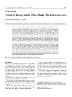
Figure 1. TGF-β superfamily signal transduction. TGF-β, nodal or activin ligands bind to Type II receptors, which
then recruit Type I receptors leading to transphosphorylation of type 1 receptors. Activated type I receptors phosphor‐
ylate Smad 2/3 (i.e. R-Smads) which then complex with the co-Smad, Smad4 and translocate to the nucleus to bind
DNA at specific DNA motifs. Smad proteins activate or repress transcription through association with various co-activa‐
tor (Co-Act) or co-repressor proteins. This pathway is inhibited by Smad7. BMP signaling operates by a similar para‐
digm. BMP6 and BMP7 bind to their Type II receptor before the complex recruits the Type I receptors, Alk-3 or Alk-6.
BMP2 and BMP4, however bind first to their type I receptor before recruiting the type II receptor BMPRII. BMP binding
to either receptor can be inhibited by first binding to various extracellular inhibitor proteins, such as noggin. Activa‐
tion of the receptor complex leads to phosphorylation of the receptors and subsequent phosphorylation of Smad1,
Smad5, or Smad8, allowing them to form a complex with Smad4. This heteromeric complex translocates to the nu‐
cleus, to target BMP-regulated genes through interaction with co-activators or repressors. Smad 6 and Smad7 may act
similarly to inhibit the BMP pathway through interactions with the receptor complex and thus inhibiting R-Smad acti‐
vation. TGF-β and BMP pathways induce the expression of proteins involved in proliferation, differentiation, survival
and apoptosis. The diagram is adapted from [16] and [17].
Role of TGF-β Signaling in Neurogenic Regions After Brain Injury
/>5
by these cytokines is through the receptor regulated Smads (R-smads). As previously men‐
tioned, TGF-β and activin signal through activation of Smad2 and Smad3, which are phos‐
phorylated by the Type I receptor, and form a heteromeric complex with the common or co-
Smad, Smad4 [22]. This Smad complex translocates to the nucleus where it regulates the
transcription of numerous genes in cooperation with other transcription factors, coactivators
and corepressors. Inhibitory Smads, or I-smads, are Smad-activated proteins that provide
negative feedback to the Smad pathway through a variety of mechanisms [16, 23]. BMP
signaling is similar in form to TGF-β signaling, although the specifics of individual receptors
and R-Smads (1, 5, 8/9) involved vary according to the specific cytokine. For a full review of
signaling and receptor nomenclature by this cytokine family please refer to some excellent
reviews [14, 24]. The Smad pathway is by no means the only mechanism through which TGF-
β cytokine signals are transduced from the receptor to the nucleus. Smad-independent
pathways include activation of MAPKs, Ras/ERK, JNK, p38, PI3K-Akt, NF-kappaB, JAK/STAT,
PP2A/S6 phosphatases and small Rho-related GTPases (16, 25). Some of the non-Smad kinases
can influence Smad directed signaling by complexing with, or modifying the Smad proteins
directly [16, 25]. Another level of control was found when it was shown that TGF-β/BMP
signaling is both regulated by, and can regulate transcription of miRNAs [26]. Smads can also
influence miRNA biogenesis by binding directly to the pri-miRNA to enhance Drosha
processing of these molecules to pre-miRNA [27]. An intricate balance between Smad and non-
Smad signaling superimposed on cell intrinsic and environmental conditions determines the
specificity and the ultimate response of each cell to TGF-β signaling. Thus, there is a complexity
to TGF-β superfamily signaling that befits cytokines that signal to multiple different cell types,
in context dependent manners to influence many different physiologic processes [28].
Genetic evidence indicates that TGF-β family members regulate embryonic, perinatal or
neonatal development of the mouse embryo. Most mice null for one TGF-β superfamily ligand,
receptor, protein or signaling protein fail in either gastrulation or mesoderm differentiation.
Table 1 lists known phenotypes of mice that are null for specific proteins in the TGF-β
superfamily signaling pathways.
Conventional
knockout mouse
model of TGF-β
proteins
Phenotype References
TβRI Failed angiogenesis, Embryonic lethality (E8) [29]
TβRII Embryonic lethality (E10.5) [30]
TβRIII Failed coronary vessel development accompanied by reduced
epicardial cell invasion. Embryonic lethality (E14.5)
[31]
TGFβ-1 Loss of a critical regulator of immune function [32, 33]
TGFβ-2 Perinatal lethal, craniofacial defects [34]
TGFβ-3 Perinatal lethal, delayed lung development [33]
Trends in Cell Signaling Pathways in Neuronal Fate Decision6
Conventional
knockout mouse
model of TGF-β
proteins
Phenotype References
Smad1 Embryonic lethality (E10) [35, 36]
Smad2 Embryonic lethality (E7.5–E12.5) [37]
Smad3 Viable and fertile. Impaired immune function, including defective
neutrophil chemotaxis, and impaired mucosal immunity
[38, 39]
Smad4 Increased number of Olig2-expressing progeny [40]
Smad5 Embryonic lethality: defective vascular development [41, 42]
Smad7 Significantly smaller than wild-type mice, died within a few
days of birth
[43]
Smad8 Viable and fertile [41, 44]
BMPRIA Embryonic lethality (E9.5) [45]
BMPRIB Viable and exhibit defects in the appendicular skeleton [46]
BMPRII Embryonic lethality (E9.5), arrest at gastrulation [47]
BMP2 Embryonic lethality (E7.5-10.5), defective cardiac development
and have defects in cardiac development
[48]
BMP3 Increased bone density in adult [49]
BMP4 Embryonic lethality (E6.5-E9.5), no mesoderm differentiation
and show little or no mesodermal differentiation
[50]
BMP5 Viable, skeletal and cartilage abnormalities [51]
BMP6 Viable and fertile; slight delay in ossification. [52]
BMP7 Perinatal lethal because of poor kidney development, eye defects
that appear to originate during lens induction.
[53-56]
BMP8A Viable: male infertility due to germ cell degeneration [57]
BMP8B Viable: male infertility due to germ cell depletion [58]
BMP15 Viable: female subfertility [59]
Endoglin Embryonic lethality (E11.5) [60, 61]
Activin receptor IA
(ALK2)
Embryonic lethality (E9.5) [62]
Activin receptor IIB
(ActR2B)
Perinatal lethal [63]
Activin-βA Neonatal lethal, craniofacial defects (cleft palate and loss of
whiskers, upper incisors, lower incisors and molars)
[64]
Role of TGF-β Signaling in Neurogenic Regions After Brain Injury
/>7
Conventional
knockout mouse
model of TGF-β
proteins
Phenotype References
Activin-βB Large litters but delayed parturition; nursing defects;
Eye lid closure defects at birth
[65]
Noggin Perinatal lethal, cartilage hyperplasia [66]
Follistatin Neonatal lethal, craniofacial defects, growth retardation and skin
defects retardation and skin defects
[67]
Table 1. Phenotype of mice that do not express specific TGF-β ligands, receptors or signaling molecules.
3. TGF-β superfamily expression and function in normal adult brain: Role
in neurogenesis
Adult neurogenesis involves proliferation of neural stem cells (NSCs), cell cycle exit, differ‐
entiation, maturation, and integration into the neural circuits, in a process that is involved in
learning and memory in the normal adult brain [68]. The neurogenic niche of the adult
forebrain subventricular zone (SVZ) is comprised of three major proliferative cell types; A, B
and C. Multipotent, self-renewing type B cells occur earliest in the neurogenic lineage of the
SVZ and give rise to the rapidly dividing type C cells, or transit amplifying progenitors. Type
A cells or neuroblasts differentiate from Type C cells and are migratory neuronal progenitors
with proliferative capacity, which migrate to the olfactory bulb where they differentiate into
interneurons (reviewed in [69-71]. In the subgranular zone (SGZ) of the hippocampal dentate
gyrus (DG), type 1 and type 2 slowly-dividing progenitors give rise to more rapidly dividing
intermediate progenitor cells, and these in turn differentiate into immature neuroblasts, which
migrate into the granule cell layer, then differentiate into mature neurons and integrate with
the existing hippocampal circuitry [71].
Within the CNS, all three isoforms of TGF-β are produced by both glial and neuronal cells [72].
Immunohistochemical studies show widespread expression of TGF-β2 and -β3 in the devel‐
oping CNS, and these proteins play a role in regulation of neuronal migration, glial prolifer‐
ation and differentiation [73-76]. In adult brain, TGF-β receptors are found in all areas of the
CNS including the cortex, hippocampus, striatum, brainstem and cerebellum [77, 78]. Immu‐
noreactivity for TβRI and TβRII is detected on neurons, astrocytes and microglia and endo‐
thelial cells located in the cortical gray matter, suggesting that almost every cell type in the
CNS is a potential target for TGF-β signaling [79].
The TGF-β superfamily and its downstream targets are capable of controlling proliferation,
differentiation, maturation and survival of stem cells and precursors in the neurogenic niches
of adult brain [18]. TβRI and TβRII are expressed by Nestin-positive type B and C cells in the
SVZ [80, 81]. Our data show mRNA expression of TGF-β1, -β2, and -β3 in both the adult SVZ
Trends in Cell Signaling Pathways in Neuronal Fate Decision8
and DG [82]. In the adult human brain, TGF-β1 protein expression has been reported in the
hippocampus, and the protein levels significantly increased with the age of the individual [83].
As neurogenesis declines with age [84], it has been suggested that TGF-β is a possible regulator
of this age-related decline [83]. Signaling by the Smad2/3 pathway is high in the hippocampus
and specifically the dentate gyrus, indicating a role for TGF-β and/or activin in regulation of
neurogenesis [85, 86]. When TGF-β protein is overexpressed or infused directly into the lateral
ventricles of uninjured animals, hippocampal neurogenesis is dramatically inhibited [81, 87].
This may be due to a direct anti-proliferative effect of TGF-β on type 1 and 2 primary NSCs
[17]. A direct effect of TGF-β on NSCs is supported by in vitro studies showing that TGF-β1
treatment of cultured adult NSCs induces the cyclin-dependent kinase inhibitor (p21) and
leads to cell cycle termination, without altering the differentiation choices of the NSCs [81].
Additionally, overexpression studies lead to increased TGF-β signaling in many different cell
types within the neurogenic niche, making the exact contribution of more restricted, endoge‐
nous TGF-β difficult to determine. Recent data have suggested that TGF-β signaling at later
stages of neurogenesis is critical for newborn neuron survival and maturation in the DG.
Conditional deletion of the TβRI (ALK5) gene specifically in immature and mature neurons,
leads to decreased neurogenesis and reduced survival of newborn neurons [85]. Thus, TGF-
β potentially has opposing roles at different stages of neurogenesis, providing an additional
example of the contextual nature of TGF-β action.
Activin receptors are expressed throughout the brain, with strong expression in the neuronal
layers of the hippocampus [88-90]. We have found that mRNA for activin-A and for activin’s
endogenous high affinity inhibitor, follistatin, are expressed in both the SVZ and DG of the
adult mouse [82] and several recent reports have demonstrated that activin-A modulates
adult neurogenesis [88, 91, 92]. Chronic overexpression of follistatin by neurons of the
hippocampus almost entirely ablates adult DG neurogenesis, due to drastically lowered
survival of adult-generated neurons [91], although short-term infusion of follistatin does
not affect neurogenesis in uninjured animals [88]. Infusion of activin to the lateral ventri‐
cle of uninjured mice mildly increases the rate of NSC proliferation and neuron genera‐
tion in the DG, indicating that activin might stimulate division of NSCs. This effect may be
indirect as activin has a potent anti-inflammatory effect in the CNS, and may modulate
local microglia to stimulate neurogenesis [88]. Smad3 knockout mice have decreased levels
of cell proliferation in the SVZ and along the rostral migratory stream, and decreased levels
of olfactory bulb neurogenesis [93]. As these mice have defective signaling by both TGF-β
and activin, these data suggest that activin signaling in the SVZ may be the predominant
Smad3-utilizing cytokine in defining basal levels of neurogenesis. In the DG pSmad2 is
normally absent from Sox2-positive type 1 and 2 primary NSCs in the DG of adult mice
[17]. However, Smad3 knockout mice also have reduced proliferation in the DG potential‐
ly pointing to a different role for Smad2 and Smad3 in the DG [93].
The BMP family of proteins regulates cell proliferation and fate commitment throughout
development and within the adult neurogenic niches [19]. Expression of BMP2, -4 and -7
mRNAs have been reported in neurogenic regions of adult rodent brain [94], and the BMP
receptors BMPRIA, -IB and -II are expressed abundantly in neurons, as well as in astrocytes
Role of TGF-β Signaling in Neurogenic Regions After Brain Injury
/>9
and ependymal cells [95]. All three of these receptors are expressed in type A cells of the SVZ,
while type B and C cells express BMPRIA and BMPRII [96]. In the DG, radial stem cells of the
SGZ marked with glial fibrillary acidic protein (GFAP) and Nestin or Sox2 primarily express
BMPRIA but not BMPRIB, while mature neurons express only BMPRIB [97]. BMP ligands are
also expressed in the adult rat brain [98, 99]. BMP2, -4, -6, and -7 are expressed by cells of the
SVZ and DG [96, 97]. In the DG, the BMP signal transducer pSmad1 is strongly expressed in
non-dividing primary NSCs and neuroblasts, but is absent in dividing primary NSCs [97],
while in the SVZ, pSmad1/5/8 has been reported in primary NSCs and transit amplifying
progenitors, but not in DCX-positive neuroblasts [40]. The soluble BMP inhibitor noggin is
also expressed by ependymal cells of the SVZ [96] and by cells of the DG [100].
Changing the ratio of BMP to noggin alters the rates of NSC proliferation and neurogenesis in
adult animals, indicating that these proteins are primary regulators of basal adult neurogene‐
sis [96, 97, 100]. Administration of exogenous BMP4 or BMP7 potently inhibits the division of
NSCs and generation of new neurons in vivo and in vitro [96, 97], as does inhibition of noggin
expression [101]. Conversely, infusion of noggin or genetic deletion of the BMPRIA receptor
causes an increase in NSC proliferation and generation of NeuN-expressing neurons in the DG
[96, 97]. However this increase is transient, there is an eventual depletion of the primary NSC
pool and a drastically reduced level of neurogenesis [97]. Decreased BMP signaling in the DG is
thought to be responsible for increased neurogenesis driven by exercise [102]. It has been
proposed that secretion of noggin from ependymal cells inhibits BMP signaling allowing a low
level of basal neurogenesis to occur, while BMP signaling maintains the overall quiescence of
the primary NSC pool [96, 97, 100]. Exogenous noggin infusion potentially has a different effect
on SVZ NSCs, leaving their proliferation rate unaffected, but causing an increase in the generation
of oligodendrocyte precursor cells from primary NSCs at the expense of immature neuro‐
blasts [40]. This noggin infusion phenocopies the effect of conditionally deleting Smad4 in NSCs
using GLAST-cre [40] and is in contrast to the pro-neurogenic effects of noggin described by Lim
et al [96]. Thus, although there is still some controversy in the field it its clear that the balance
between BMP and noggin is critical to proper maintenance of the adult NSC population.
4. Expression of TGF-β related cytokines in the adult rodent brain after
injury
TGF-β family proteins are present in the brain immediately after injury as they are carried into
the wound by the blood [103]. Additionally, extracellular TGF-β proteins are activated and
released from their latent protein complexes in the brain parenchyma [104]. Local CNS expres‐
sion of TGF-β, activin, and BMP proteins is increased after many different injuries [72, 105, 106].
Following acute brain injury, TGF-β1 levels are elevated in astrocytes, microglia, macrophag‐
es, neurons, ependymal cells and choroid plexus cells with peak expression around 3 days
[107-110]. TGF-β2 and -β3 expression has also been found in astrocytes, microglia, endothelial
cells and neurons after both ischemic and TBI [111, 112]. We have recently found TGF-β2
expression in oligodendrocytes in the lesioned cortex and corpus callosum [113]. Ischemic lesions
as well as TBI show elevated activin-A mRNA as well as mRNA for the BMPRII receptor [90, 94,
Trends in Cell Signaling Pathways in Neuronal Fate Decision10
114]. Smad proteins are also upregulated after injury and were mainly located in the cerebral
cortex, typically in the nucleus and/or in the cytoplasm of astrocytes, oligodendrocytes or neurons
[86, 108, 115, 116]. We have summarized many studies that have examined changes in the TGF-
β superfamily of cytokines after central nervous system injury in Table 2.
TGF-β
protein
Acute brain
Insult
(Animal model)
Expression in
Brain
Expression in
neurogenic
niche
Cell types in which
protein is expressed
mRNA
and/or
protein
References
TGF-β1 Ischemia Cerebral cortex _ _ _ _ _ Microglia, neurons,
oligodendrocytes,
endothelial cells,
astrocytes,
macrophages, and
ependymal cells
mRNA,
protein
[107-110]
Transient
ischemia
Cerebellum,
Cerebral cortex
Hippocampus,
Subventricular
zone
Microglia, T cells,
neuroblasts and
neurons
mRNA,
protein
[117-120]
Permanent
ischemia
Cerebral cortex,
Striatum
_ _ _ _ _ Neurons, neuroblasts mRNA,
protein
[121-123]
Bilateral cerebral
ischemia
Cerebellum,
Cerebral cortex
Dentate gyrus Neurons, vessels Protein [124, 125]
Hypoxic-ischemic Cerebral cortex,
Corpus callosum
_ _ _ _ _ Astrocytes, Microglia
and blood vessels
Protein [126]
Stab wound Cerebral cortex _ _ _ _ _ Neurons Protein [116]
Traumatic brain
injury
Cerebral cortex Hippocampus,
Subventricular
zone
Microglia, astrocytes
and neurons
mRNA,
protein
[82, 112, 127,
128]
Excitotoxic lesion
(NMDA)
Gray matter
surrounding the
lesion
_ _ _ _ _ Astrocytes, neurons Protein [129]
Triethyltin
exposure
Cerebral cortex Hippocampus Neurons mRNA,
protein
[130, 131]
Penetrating
brain Injury
Cerebral cortex _ _ _ _ _ Activated glia,
meningeal
cells, choroid plexus
mRNA,
protein
[132]
Excitotoxic Injury _ _ _ _ _ Hippocampus Neurons Protein [133]
Irradiation Cerebral cortex _ _ _ _ _ Macrophages and
astrocytes
Protein [134]
Role of TGF-β Signaling in Neurogenic Regions After Brain Injury
/>11
TGF-β
protein
Acute brain
Insult
(Animal model)
Expression in
Brain
Expression in
neurogenic
niche
Cell types in which
protein is expressed
mRNA
and/or
protein
References
Excitotoxicity
with kainic acid
Cerebral cortex Hippocampus Microglia/macrophages,
neurons and astrocytes
mRNA,
protein
[86, 135-137]
Stab wound Cerebral cortex _ _ _ _ _ Astrocytes Protein [138]
TGF-β2 Ischemia Cerebral cortex,
cerebellum,
striatum
Hippocampus Neurons and
endothelial cells,
microglia and astrocytes
mRNA,
protein
[108, 109,
111]
TGF-β3 Ischemia Cerebral cortex Dentate gyrus Neurons mRNA,
protein
[111]
Traumatic brain
injury
Cerebral cortex Hippocampus Astrocytes Protein [112]
TβRI Permanent
ischemia
Cerebral cortex _ _ _ _ _ Astrocytes and neurons mRNA,
protein
[122]
TβRII Ischemia Cerebral cortex,
midbrain,
cerebellum, and
brainstem
_ _ _ _ _ Neurons, astrocytes,
microglia, endothelial
cells, and other non-
neuronal cells found in
the choroid plexus
mRNA,
protein
[122, 139,
140]
Traumatic brain
injury
Cerebral Cortex _ _ _ _ _ Endothelial cells Protein [141]
Smad2 Excitotoxicity Cerebral Cortex Hippocampus Neurons, astrocytes and
microglia
Protein [86]
pSmad2 Stroke Cerebral Cortex _ _ _ _ _ Astrocytes, activated
microglia
Protein [108]
pSmad
1,5,8
Cuprizone-
induced
demyelination
_ _ _ _ _ Subventricular
zone
Oligodendrocytes mRNA,
protein
[115]
BMPRII Traumatic brain
injury
_ _ _ _ _ Dentate
gyrus
Neurons mRNA,
protein
[90]
BMPs and
receptors
Ischemia Cerebral Cortex,
cerebellum
Hippocampus Neurons mRNA,
protein
[124, 142,
143]
Bilateral cerebral
ischemia
Cerebral cortex,
cerebellum
Subventricular
zone, dentate
gyrus
Neurons mRNA,
protein
[94]
Traumatic brain
injury
Cerebral cortex Subventricular
zone
Astrocytes mRNA,
protein
[144]
Trends in Cell Signaling Pathways in Neuronal Fate Decision12
TGF-β
protein
Acute brain
Insult
(Animal model)
Expression in
Brain
Expression in
neurogenic
niche
Cell types in which
protein is expressed
mRNA
and/or
protein
References
BMP4 Cuprizone-
induced
demyelination
_ _ _ _ _ Subventricular
zone
Astrocytes and
oligodendrocytes
mRNA,
protein
[115]
BMP7 Traumatic brain
injury
Cerebral cortex _ _ _ _ _ Astrocytes Protein [144]
Stroke Cerebral cortex,
corpus callosum
Subventricular
zone
Progenitors cells and
neurons
Protein [145]
Noggin Traumatic brain
injury
Cerebral cortex Subventricular
zone
Astrocytes and
progenitors cells
Protein [144]
ActR-1A Traumatic brain
injury
_ _ _ _ _ Dentate
gyrus
Neurons mRNA,
protein
[90]
Activin Ischemia Cerebral Cortex,
striatum
Hippocampus Neurons mRNA,
protein
[89, 146]
Hypoxia-ischemia Cerebral Cortex Dentate
gyrus
Microglia and blood
vessels
mRNA,
protein
[114]
Excitotoxicity Amygdala,
Piriform cortex,
and thalamus
Dentate
gyrus
Neurons, blood vessels mRNA,
protein
[105, 146-148]
Table 2. TGF-β superfamily cytokine and signaling intermediate expression after different forms of injury.
Relatively few studies have examined changes in expression of the TGF-β superfamily of
cytokines specifically within the neurogenic regions after brain injury. TGF-β1 expression
increases in the SVZ [119] and DG [117, 118, 124] after ischemic injury. Its expression is also
induced in neurons of the DG after a demyelinating lesion [131] or after local kainic acid
injection [133]. Our group recently found that controlled cortical impact injury increased
mRNA expression of many TGF-β cytokines, including TGF-β1 and -β2, activin-A, and BMPs
-4, -5, -6, and -7 in the DG and SVZ, demonstrating that a distal injury can alter TGF-β signaling
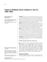
pathways in the neurogenic regions [82]. We have observed upregulation of TGF-β1 and -β3
in GFAP and Nestin positive progenitors in the SVZ and DG after TBI (Figure 2 and unpub‐
lished data). TβRII is expressed in these Nestin positive progenitors in the lateral SVZ (Figure
2d). Phospho-Smad3 (pSmad3) shows strong nuclear localization in these cells as well (Figure
2i and unpublished data) suggesting a role for TGF-β/activin signaling in the regulation of
post-injury neurogenesis. In the DG, TβRII is expressed in GFAP-positive precursors with
strong pSmad3 nuclear staining (Figure 2m, 2r) suggesting a similar role for TGF-β cytokines
in this neurogenic niche.
Role of TGF-β Signaling in Neurogenic Regions After Brain Injury
/>13
Figure 2. Confocal images of the TGF-β ligands, receptors and signaling proteins in the SVZ and DG in the in‐
jured adult mice brain. Double and triple labelled inmmunofluorescence staining for TGF-β proteins and receptors,
with the following cell-type specific markers: Nestin (for undifferentiated neuronal precursors), NeuN (for mature neu‐
rons), GFAP (for progenitor and astroglial cells), DCX (for neuroblasts). The left column shows coronal sections within
Trends in Cell Signaling Pathways in Neuronal Fate Decision14
the subventricular zone (SVZ) at 3 (a-g) and 7 (h and i) days after traumatic brain injury (TBI). TβRII (a, red) is expressed
in Nestin positive (b, green) neural stem cells (NSCs) in the SVZ, and also in ependymal cells (d), lining the walls of the
lateral ventricle (LV). Light TGFβ−1 (green) and predominant TGFβ−3 (red) expression is also found in the walls of the
LV where the adult NSCs reside (e). (f) Neurons (NeuN, green) are co-localized with TGFβ−2 (red) in the damaged stria‐
tum. (h) The majority of Smad 1,5,8 proteins (red) are co-expressed with Nestin (green). (i) pSmad3 (red) colocalizes
with GFAP (green) in the dorsolateral corner of the SVZ. The right column shows coronal sections within the dentate
gyrus (DG) of the hippocampus at 3 (j-q) and 7 (r) days after TBI. (j-m) TGFβ−1 (red, j) and TβRII (green) are colocalized
in astrocytes (GFAP, blue) in the hilus and GCL (granule cell layer) of the hippocampus (n) TGFβ−1 (red) is co-localized
with astrocytes (GFAP positive cells) located in the subgranular zone (SGZ) of the hippocampus. In (o) TGFβ−2 (red) is
co-localized with NeuN (green) positive neurons in the hilus of the dentate gyrus. (p) TGFβ−3 (red) is co-localized with
GFAP positive (blue) immature progenitors in the SGZ but not with DCX (green) positive neuroblasts. (q) Immunos‐
taining with TGFβ−1 (green) and TGFβ−3 (red) show they are almost entirely colocalized in the SGZ. (r) pSmad3 stain‐
ing in the nuclei of GFAP positive progenitor cells in the SGZ and hilus of the hippocampus. Scale bars: (c, d, f, (inset in
i), m, (inset in n), o, (inset in o), p, (inset in r)) 20 µm; (e, g, h, i, q, r) 50 µm.
Local injury to the hippocampus via saline injection produces a strong induction of activin-βA
mRNA in the DG, which can be blocked by inhibiting NMDA receptors [114]. Activin expres‐
sion in the DG is potently induced by seizures, local excitotoxic lesions, hypoxia/ischemia, TBI
or permanent MCAO [89, 114, 146, 148, 149]. Cortical weight drop injury also elevates the
expression of the activin receptor ActR-I and the BMP receptor BMPRII in the DG [90]. BMPRII
expression is also elevated in the DG after global cerebral ischemia [94], and BMP4 levels
increase in the SVZ after a demyelinating lesion [115].
The limited studies available indicate that TGF-β, BMP, and activin signaling may all be active
in the neurogenic regions after injury. However, it is currently unclear the manner in which
they affect the behavior of neural stem cells. Given that these cytokines clearly regulate adult
neurogenesis in the uninjured adult, more research in this area is necessary to fully elucidate
the effect of brain injury on these signaling pathways, and the mechanisms through which
these changes alter post-injury neurogenesis.
5. Injury-induced neurogenesis and its regulation by TGF-β family
proteins
We have described the role of TGF-β proteins in the regulation of neurogenesis under basal
conditions. In response to various injuries, the rate of neurogenesis is increased and the fate
and migration of the neural progenitors is changed. Cerebral ischemia, excitotoxicity and TBI
can all promote neurogenesis in the adult DG and SVZ [88, 150-153]. After injury, the altered
environment changes the basic processes of proliferation, differentiation, migration and
integration. TGF-β related cytokines have the potential to regulate many of these processes.
Alteration in the destination of progenitor cells means that many of the neuroblasts change
their usual trajectory and migrate towards and into the lesion [154]. The cell fate of progenitor
cells can be altered by the changed environment of the injured brain, in both the neurogenic
niche and at the lesion site to which the progenitor cells migrate. The environment around the
lesion is now very different than the normal location of these progenitors and thus further
differentiation and integration occurs in an entirely unique environment [155]. Additionally,
the actions of TGF-β cytokines are highly context dependent, and they can have very different
effects in the injured as compared to the uninjured brain.
Role of TGF-β Signaling in Neurogenic Regions After Brain Injury
/>15
A major component of the brain post-injury in comparison to the uninjured brain is the
inflammatory response, both of local CNS cells and invading macrophages. While the
majority of studies have indicated that inflammation is detrimental to neurogenesis, it is
now appreciated that the effect of inflammation on neurogenesis is multifaceted [156]. Of
particular importance is the response of local microglia and astrocytes in the neurogenic
regions. Microglia are potent regulators of neurogenesis, and in certain contexts can
powerfully inhibit the process [157]. However microglia have also been shown to pro‐
mote neurogenesis [158, 159], and studies have described differential action of acute vs.
chronically activated microglia on NSC division and neurogenesis, as well as for micro‐
glia activated by different mechanisms or by different cytokines [160, 161]. As TGF-β
proteins are prominent anti-inflammatory molecules [162], their actions after brain injury
can regulate neurogenesis by acting directly on NSCs as well as indirectly through their
effects on the glial inflammatory response [163].
Due to their pleiotropic actions, TGF-β superfamily proteins have been investigated as
potential treatments for a variety of CNS injuries, and several studies have demonstrated
potential uses for these cytokines as therapeutic molecules (see Table 3). They have also
provided insights into the action of these molecules as regulators of neural stem/progenitor
cell (NSPC) proliferation and differentiation, with respect to both endogenous and transplant‐
ed stem cell populations.
TGF-β
related
protein
Animal Model
Mode of
administration
Effect on cell proliferation
and neurogenesis
Behavioral
Outcome
Reference
TGFβ-1
Transient ischemia
Intranasal aerosol
spray
Decreased NSC proliferation
and induce the number of
DCX expressing neuronal
precursors
Reduced Neurological
Severity Score deficits
[164]
Adrenalectomy
Intraventricular
infusion
Decreased the percentage of
dividing cells which co-express
PSA-NCAM in the DG
None measured [163]
Adrenalectomy
Adenoviral
overexpression
Increased NSC proliferation
and neurogenesis in the SVZ
None measured [165]
Prenatal LPS
inflammation
Adult adenoviral
overexpression
Inhibited chronic microglial
activation and restored
neurogenesis
None measured [166]
Naïve animals
Injected into the
cerebrospinal
fluid
Number of proliferating cells
in the hippocampus and in the
lateral ventricle wall is
substantially reduced, fewer
neuronal precursor cells
None measured [81]
Trends in Cell Signaling Pathways in Neuronal Fate Decision16
TGF-β
related
protein
Animal Model
Mode of
administration
Effect on cell proliferation
and neurogenesis
Behavioral
Outcome
Reference
Naïve animals
Transgenic
astrocytic
overexpression
Decreased DG cell
proliferation and generation
of neuroblasts and neurons
None measured [87]
Noggin
Permanent MCAO
Transgenic
neuronal
overexpression
Increased immature
oligodendrocyte generation
Reduced motor
deficits
[167]
Naïve animals
Intraventricular
infusion
Promoted neuronal
differentiation of SVZ
precursor cells transplanted to
the striatum
None measured [96]
Cuprizone-induced
corpus callosum
demyelination
Intraventricular
infusion
Decreased astrocyte and
increased oligodendrocyte
generation from the SVZ
None measured [115]
Spinal cord injury
Overexpression
by transplanted
NPCs.
Increased neuronal and
oligodendroglial
differentiation of transplanted
NPCs
Improved motor
recovery
[168]
BMP7
Transient ischemia
Intraventricular
infusion
Increased SVZ proliferation
and neurogenesis
Reduced motor
deficits
[145]
Naïve animals
Intraventricular
infusion
Inhibited SVZ proliferation None measured [96]
Chordin
Lysolectithin-
induced corpus
callosum
demyelination
Intraventricular
infusion
Increased NPCs migrating to
lesion, and increased
oligodendrocyte
differentiation
None measured [169]
Activin-A
Excitotoxic
hippocampal
lesion
Continuous
intraventricular
infusion
Decreased astrocyte and
microglial inflammation, and
increases neurogenesis
None measured [88]
Naïve mice
Transgenic
overexpression
Increases new neuron survival
Reduced anxiety-like
behavior
[91]
Activin-A or
Activin-B
Naïve mice ICV injection Not examined
Reduced depression-
like behavior
[170]
Follistatin
Excitotoxic
hippocampal
lesion
Continuous
intraventricular
infusion
Increased NSC proliferation
and neurogenesis.
None measured [88]
Naïve mice
Transgenic
overexpression
Potently inhibited
neurogenesis
Increased anxiety-like
behavior
[91]
Table 3. Therapeutic application of TGF-β proteins in the normal and injured brain that affect neurogenesis.
Role of TGF-β Signaling in Neurogenic Regions After Brain Injury
/>17