Báo cáo khoa học: The study of G-protein coupled receptor oligomerization with computational modeling and bioinformatics doc
Bạn đang xem bản rút gọn của tài liệu. Xem và tải ngay bản đầy đủ của tài liệu tại đây (440.19 KB, 13 trang )
MINIREVIEW
The study of G-protein coupled receptor oligomerization
with computational modeling and bioinformatics
Marta Filizola
1
and Harel Weinstein
1,2
1 Department of Physiology and Biophysics, Weill Medical College of Cornell University, NY, USA
2 Institute for Computational Biomedicine (ICB), Weill Medical College of Cornell University, NY, USA
Introduction
The growing experimental evidence showing that
GPCR oligomerization has pharmacological and func-
tional implications [1–4] (recent reviews), has prompted
the search for detailed structural information of the
receptor–receptor interface(s) in order to enable a
mechanistic understanding of these complex biological
systems. Despite the results from experimental studies
suggesting the participation of C- [5] and N-terminal
[6] regions in the self association of rhodopsin-like
GPCRs, more recent evidence points to the transmem-
brane helices (TMs) as the most probable structural
elements involved in oligomerization [7–12]. In partic-
ular, the recent atomic-force microscopy map of rho-
dopsin molecules in native mouse disk membranes
[10–12] provides direct evidence for the organization
of rhodopsin protomers into two-dimensional arrays of
Keywords
bioinformatics; GPCRs; interface; molecular
modeling; oligomerization
Correspondence
M. Filizola, Department of Physiology and
Biophysics, Box 75, Weill Medical College
of Cornell University, 1300 York Avenue
New York, NY 10021, USA
Fax: +01 212 746 8690
Tel: +01 212 746 6348
E-mail:
(Received 16 February 2005, accepted
8 April 2005)
doi:10.1111/j.1742-4658.2005.04730.x
To achieve a structural context for the analysis of G-protein coupled recep-
tor (GPCR) oligomers, molecular modeling must be used to predict the
corresponding interaction interfaces. The task is complicated by the paucity
of detailed structural data at atomic resolution, and the large number of
possible modes in which the bundles of seven transmembrane (TM) seg-
ments of the interacting GPCR monomers can be packed together into
dimers and ⁄ or higher-order oligomers. Approaches and tools offered by
bioinformatics can be used to reduce the complexity of this task and, com-
bined with computational modeling, can serve to yield testable predictions
for the structural properties of oligomers. Most of the bioinformatics meth-
ods take advantage of the evolutionary relation that exists among GPCRs,
as expressed in their sequences and measurable in the common elements
of their structural and functional features. These common elements are
responsible for the presence of detectable patterns of motifs and correlated
mutations evident from the alignment of the sequences of these complex
biological systems. The decoding of these patterns in terms of structural
and functional determinants can provide indications about the most likely
interfaces of dimerization ⁄ oligomerization of GPCRs. We review here
the main approaches from bioinformatics, enhanced by computational
molecular modeling, that have been used to predict likely interfaces of
dimerization ⁄ oligomerization of GPCRs, and compare results from their
application to rhodopsin-like GPCRs. A compilation of the most fre-
quently predicted GPCR oligomerization interfaces points to specific
regions of TMs 4–6.
Abbreviations
CMA, correlated mutation analysis; GPCR(s), G-protein coupled receptor(s); SCM, subtractive correlated mutation; TM, transmembrane.
2926 FEBS Journal 272 (2005) 2926–2938 ª 2005 FEBS
dimers. Inference from this map led to the construction
of an oligomeric molecular model of rhodopsin with
TM4 and TM5 involved in intradimeric contact, while
TM1, TM2, and the cytoplasmic loop connecting TM5
and TM6, facilitate the formation of rhodopsin dimer
rows.
Two modes of association of the TM helices of
GPCR monomers into dimers have been proposed.
One of them, which is termed contact dimerization,
corresponds to the packing of two different TM bun-
dles with separate binding sites through interactions at
interfaces that otherwise would face the lipid environ-
ment [13]. The other one, termed ‘domain-swapped
dimerization’, involves interpenetration of transmem-
brane bundles, where the interacting TMs from two
different polypeptides appear as interlaced units
[14–17]. Experimental data can be found in the litera-
ture to support either contact dimers [10–13,18–21] or
domain-swapped dimers [22–29] of GPCRs.
Regardless of the type of geometry assumed for the
association of the rhodopsin-like GPCRs, the specific
interacting residues that form the dimerization inter-
face remain unknown for most receptor subtypes. To
guide specific experiments aimed at producing such
information, it is necessary to organize the available
data in a structural context. At this stage, only
molecular modeling offers such a structural context,
and even the most direct experimental evidence for
dimer geometry in rhodopsin makes use of such mod-
els [11]. The construction of the molecular models of
dimers ⁄ oligomers for other GPCRs, in the absence of
the type of direct data available for rhodopsin, is quite
complex. Thus, even for contact dimers in which the
seven TM bundles of two GPCR monomers are
packed together, there is a large number of alternative
possibilities {at least 49 (¼ 7 · 7) for hetero-dimers
and 28 [¼ 7(7 + 1) ⁄ 2] for homo-dimers}. This number
of different alternatives for the possible interfaces can
be reduced significantly with the use of a variety of
computational methods, including genome level bio-
informatics tools and homology models [30,31].
Computational methods designed to help identify
protein–protein interaction interfaces (e.g. [32,33]) are
especially appropriate for this task. In general, the com-
putational methods that can serve in the modeling of
oligomerization interfaces fall into two categories. If 3D
structural information is available, approaches generally
known as ‘docking methods’ [33] (review) can be used
to identify protein–protein interfaces, based on a variety
of selection criteria and exhaustive searches of the
‘interaction space’. Still, the accuracy of the predictions
from such computational techniques remains quite
limited [34]. In contrast, even if structural information
about the interacting G-proteins in a complex is not
available, bioinformatics methods based on sequence
and genomic information can be used to predict pro-
bable regions involved in protein–protein interactions.
Many of the limitations and considerations of accuracy
and reliability of these methods have been reviewed
recently [32].
Some of the computational techniques developed to
predict protein–protein interactions have been applied
to modeling of GPCR interactions. Most of these
approaches utilize sequence and genomic information
to predict putative functionally important residues, such
as those involved in the interaction of GPCRs with
cognate G-proteins [35–41], as well as in signal trans-
duction [35,42–44]. Similarly, bioinformatic methods
were tapped for approaches used to predict possible
interfaces of GPCR dimerization ⁄ oligomerization
[16,17,40,45–50]. We review here the underlying princi-
ples of these methods, as well as results of their
application to rhodopsin-like GPCRs. To enable com-
parisons, we use the generic numbering system of
GPCR sequences (N1.N2) originally described in Bal-
lesteros & Weinstein [51]. Specifically, this numbering
scheme consists of a number (N1) corresponding to
the TM number, and another (N2) counting the posi-
tion relative to the most conserved residue in the
particular TM. The conserved locus is assigned the
number 50, and the N2 values of the other loci
decrease towards the N-terminus and increase toward
the C-terminus.
The compilation of the predicted dimerization ⁄ oligo-
merization interfaces obtained from the comparison
shows that regions in TMs 4–6 have the highest num-
ber of occurrences. From specific examples, it becomes
evident that this type of analysis for GPCR interaction
interfaces can provide valuable structure-based hypo-
theses for further probing of functional mechanisms,
because: (a) they can be tested specifically (e.g. with
mutagenesis) for sequence determinants of dimeriza-
tion ⁄ oligomerization, as well as for disruption of the
dimers and, (b) they provide criteria for the design of
experiments to probe the functional effects of dimeriza-
tion ⁄ oligomerization on mechanisms of ligand binding
and extent of activation (i.e. pharmacological efficacy),
as well as on the involvement of protein–protein inter-
actions in GPCR function (e.g. transactivation).
Methods employed
The evolutionary trace method
The evolutionary trace method is an adaptation of an
earlier strategy for the hierarchical analysis of residue
M. Filizola and H. Weinstein Computational study of GPCR oligomerization
FEBS Journal 272 (2005) 2926–2938 ª 2005 FEBS 2927
conservation in protein sequence alignments [52]. It
was first described by Lichtarge et al. [38,53,54] as a
technique that predicts functionally important residues
(e.g. active sites and functional interfaces) in proteins
of known structure. The set of assumptions used to
extract such evolutionary trace residues from sequence
conservation patters in homologous proteins includes
the following: (a) protein structures descendant from a
common ancestor retain their fold (even if sequence
identities are as low as 25% [55]), as well as the loca-
tion of their functional sites and (b) functionally
important residues undergo fewer mutations than other
residues [56], and their lower mutation rate is inter-
rupted mainly by mutations that cause divergence.
Using a dendrogram (or phylogenetic tree) to represent
graphically a multiple sequence alignment of homo-
logous proteins, an evolutionary trace residue can be
identified as a residue which, upon partitioning of the
dendrogram at a certain level of sequence divergence,
is conserved within each group into which the dendro-
gram is divided, but may vary from one group to
another.
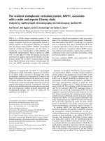
An example of the steps involved in identifying evo-
lutionary trace residues from a hypothetical multiple
sequence alignment is shown in Fig. 1. Based on a
dendrogram, sequences in a family can be divided into
subfamilies at selected sequence identity cutoffs. At
high sequence identity cutoffs, a subfamily is com-
posed of a smaller number of groups of sequences,
which are expected to show functional specificity. In
contrast, at low sequence identity cutoffs, the sub-
families contain more groups of sequences with less
specificity, but perhaps reflecting functional common
elements. In general, the exact cutoff value that must
be used to obtain reliable predictions is depending on
the protein of interest.
In the example shown in Fig. 1, the dendrogram of
a hypothetical multiple sequence alignment was parti-
tioned into four subfamilies using the sequence identity
cutoff shown as a vertical dotted line. For each sub-
family, a consensus sequence can be compiled by
transcribing conserved residues, and leaving variable
positions blank, as shown on the right of Fig. 1. Com-
parison of these consensus sequences allows for the
identification of evolutionary trace residues, which
are supposed to be part of a protein active site, or a
functional interface. More specifically, such residues
include: (a) class-specific residues, i.e. residues con-
served within a subfamily that are different between
subfamilies, but never a gap (e.g. X in Fig. 1) and
(b) conserved residues, i.e. residues conserved across
the entire sequence family (e.g. E and R in Fig. 1).
Residues that cannot be classified as conserved or
class-specific residues are called neutral (shown as
underscore characters in Fig. 1). Once the evolutionary
trace residues have been identified, they can be
mapped (e.g. by color-coding) to the structure of one
of the proteins in the sequence family, and clustered in
3D space, as shown schematically in Fig. 1.
In the application to over 700 aligned GPCR
sequences from classes A (rhodopsin-like), B (secretin-
like), and C (metabotropic glutamate-like), an enhan-
ced evolutionary trace method using Monte–Carlo
techniques [57] suggested a potential functional site on
the lipid-exposed faces of TM5 and TM6 in common
to each family or subfamily of receptors [40]. Although
this analysis did not result in the identification of
exactly the same residues for all the GPCR families
and subfamilies studied, the presence of a functional
site in the same lipid-exposed region of TM5 and TM6
suggested these helices as candidates for the dimeriza-
tion interface of GPCRs. As these studies only used
the TM regions of GPCRs, the authors could not dis-
tinguish between contact and domain-swapped dimers,
and suggested that, for the purpose of signaling, the
two alternative models are equivalent [16,17,40]. Using
the enhanced evolutionary trace method [40], a second
functional site on the lipid-exposed faces of TM2 and
TM3 was also predicted. Specifically, this functional
site was suggested to be implicated either in hetero-
dimerization, or in the formation of higher-order oligo-
mers [17,40]. On the other hand, considerably less
functionality was observed on TM1, TM4, and TM7
when using this enhanced evolutionary trace method
[40].
Fig. 1. Steps involved in identifying evolutionary trace residues
from a hypothetical multiple sequence alignment. The vertical dot-
ted line identifies the sequence identity cutoff used to partition the
dendrogram into four subfamilies. Class-specific and conserved resi-
dues, shown in blue and red, respectively, are mapped to the
structure of one of the proteins in the sequence family, and clus-
tered in 3D space.
Computational study of GPCR oligomerization M. Filizola and H. Weinstein
2928 FEBS Journal 272 (2005) 2926–2938 ª 2005 FEBS
In order to identify clusters of residues that might
be responsible for global and class-specific functions,
the evolutionary trace method was applied recently to
a multiple sequence alignment of visual opsin, bio-
amine, olfactory, and chemokine class A GPCRs [43].
Among the trace residues suggested to mediate a gen-
eric signal transduction mechanism, only one (position
4.47, according to the generic numbering scheme) was
predicted to be lipid-exposed based on solvent accessi-
bility values (> 45%) calculated with the getarea
software version 1.1 [58] using the rhodopsin crystal
structure [59]. Interestingly, a mutation at this position
in chemokine receptors was recently suggested to affect
receptor homodimerization [49].
Correlated mutation analysis
Correlated mutations are typically identified in multiple
sequence alignments as loci that mutate simultaneously.
A correlated mutation algorithm was described by
Gobel et al. [60] as a powerful tool to correctly predict
physical contacts in homologous proteins. Oliveira
et al. [61] used a similar approach as Gobel et al. [60]
to first determine the correlation between residue posi-
tions in GPCRs. Such inferences based on correlated
mutation analysis (CMA) have been verified experi-
mentally to indicate spatial adjacencies between GPCR
TMs in the intramolecular portion of the proteins (e.g.
[62,63]). In addition, the observation that the type of
compensatory changes identified by CMA tend to accu-
mulate at protein interfaces [61,64] led to the extension
of the concept of correlated mutations to predict pro-
tein–protein contacts. This approach is based on the
reasoning that sequence changes that occur during evo-
lution at one of the interaction interfaces must be com-
pensated by changes in the other interacting protein
in order to preserve the protein–protein interface. This
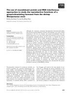
implementation is illustrated schematically in Fig. 2.
Specifically, sequence changes that occur during evolu-
tion at the interaction interface of a given protein
(Fig. 2B) must be compensated by changes in the inter-
acting protein (Fig. 2C) to preserve the protein–protein
interface. In the hypothetical multiple sequence align-
ment shown in Fig. 2, these compensatory changes
occur at positions 36 and 140. This basic principle was
used not only to identify likely interfaces of GPCR–
G-protein interactions [35–37,41,43,45], but also to
predict interfaces of homo- and heterodimerization in
GPCRs [16,17,30,31,45,47–49].
Correlated mutation analysis performed by Gould-
son et al. [45] on multiple sequence alignments of
bioaminergic, somatostatin, neurokinin, opioid, thyro-
trophin, and chemokine receptors, using the whatif
molecular graphics software [65], showed an accumula-
tion of correlated mutations on the lipid-exposed sur-
face of these receptors. Specifically, the occurrence of
correlated mutations on the external faces of TM1,
TM5, TM6, and TM7 was interpreted as an indication
that these helices may be involved in the formation of
domain-swapped dimers. In contrast, conformational
changes and ⁄ or the formation of higher order struc-
tures were invoked to explain the simultaneous appear-
ance of correlated mutations on the external faces of
TM2, TM3, and particularly TM4.
This type of approach was enhanced with filtering
algorithms to enable identification of the likely hetero-
and homo-oligomerization interfaces of family A
GPCRs [47,48]. These methods take advantage of an
improved CMA-based algorithm [66] that combines
correlated mutations with other types of sequence
properties (e.g. sequence conservation and contact den-
sity), and utilize the structural information from the
rhodopsin crystal structure [59] as the basis to predict
functionally important residues at the dimerization
Fig. 2. Schematic representation of the concept of correlated mutations applied to protein–protein interactions. In the specific example,
compensatory changes occur at positions 36 and 140 of the hypothetical multiple sequence alignment.
M. Filizola and H. Weinstein Computational study of GPCR oligomerization
FEBS Journal 272 (2005) 2926–2938 ª 2005 FEBS 2929
interfaces of GPCRs. Specifically, the approach we
developed to identify probable interfaces of GPCR
heterodimerization, termed Subtractive Correlated
Mutation (SCM) method [47], consists of a modified
version of the original algorithm [66] that provides a
means to filter out the intramolecular pairs of correla-
ted residues within each interacting monomer from the
complete list of intra- and intermolecular pairs of cor-
related residues. These correlated mutations are identi-
fied in a multiple sequence alignment of concatenated
monomeric sequences of the two different GPCRs,
obtained from the same organisms. The concatenation
step is essential for the identification of the hetero-
dimerization interface, as outlined in detail [47]. A sim-
ilar approach was developed independently by Pazos &
Valencia [67].
Although a powerful bioinformatics tool that was
demonstrated from specific tests to identify residues
that are functionally essential, the CMA in itself does
not usually achieve specific identification of the residue
composition of the dimerization interfaces of GPCRs
(e.g. results of Gouldson et al. 2001, which essentially
show correlated mutations in all seven TM helices).
Consequently, additional stringency criteria must be
added to the CMA approaches, especially in the appli-
cation to GPCR homo-dimerization, in order to
achieve reliable predictions of the dimerization ⁄ oligo-
merization interface of GPCRs. To this end, we intro-
duced the filtering criteria described below, which are
applied to the list of calculated correlated mutations
to reduce the number of false positives [31], and are
combined with the geometric filtering derived from the
construction of 3D molecular models of putative
configurations of GPCR dimers ⁄ oligomers [48]. Experi-
ence shows that application of these criteria may cause
the procedure to overlook some correct predictions of
residues involved in the actual interface, i.e. false negat-
ives. Nevertheless, this multistep filtering remains
advantageous because it ensures elimination of isolated
residues (as opposed to complete interfaces), which
have high correlation index values. Briefly, we devel-
oped an approach in which the correlated pairs of resi-
dues are first sorted by decreasing correlation values
(from 1 to 0). To increase the chance of obtaining cor-
rectly predicted contacts, only highly correlated pairs
are taken into account for each case. This is achieved
by limiting the predictions to a maximum of L ⁄ 2,
where L is the length of the sequence of each receptor
subtype (i.e. only the most significant L ⁄ 2 predictions
are retained, and the others discarded). This specific fil-
tering is carried out because a list of L ⁄ 2 was demon-
strated to be enriched in correctly predicted contacts
[66]. However, even the L ⁄ 2 list is purged of correlated
pairs with a correlation index £ 0.7, in order to reduce
the number of false positives. Conversely, however, if
the number of correlated pairs with a correlation index
equal to 1 exceeds the number of L ⁄ 2 correlated pairs,
they are all taken into account. Finally, the location of
the identified residues on the ‘outward’ facing portions
of the monomers is established using the information
contained in the crystal structure of the cognate rho-
dopsin receptor [59]. Any pair of correlated residues
with either one, or both inaccessible to solvent (thus
not considered to be outward-facing) is eliminated from
the list of predictions. Specifically, only pairs of correla-
ted residues where both positions have a surface expo-
sure of more than 45 A
˚
2
are considered as candidates
for intermolecular contacts (the implicit assumption
is that association of GPCRs occurs only via contact
dimers ⁄ oligomers). A final filtering criterion is based on
the requirement for interaction neighborhoods on the
proposed helix interface. Specifically, one definition
of the neighborhood that we have used [47,48] is that
residues in a TM helix are considered to define an inter-
face only if at least three appear close to each other,
within i + 7. Application of this final filter further redu-
ces the number of false positives obtained from the bio-
informatics approach of CMA.
In a recent application to rhodopsin-like GPCR sub-
types for which homo-dimerization has been demon-
strated experimentally [31], our enhanced CMA-based
approach identified TM1 and TM4 most often as puta-
tive interfaces among the studied GPCRs. The fre-
quency with which these two TMs appear in the
predictions suggested them as the most probable seg-
ments of rhodopsin-like GPCRs to be involved in
dimerization ⁄ oligomerization interfaces. This finding is
intriguing given the recent experimental data suggest-
ing a role for precisely TM1 and TM4 in the dimeri-
zation ⁄ oligomerization of rhodopsin-like GPCRs
(including rhodopsin [11], dopamine D2 [9], a1 adren-
ergic [7], and C5a [68] receptors).
Methods that detect tree-determinant positions
Two of the fully automatic methods recently imple-
mented in Valencia’s lab to detect tree-determinant
positions in multiple sequence alignments [69] (specific-
ally, the Level Entropy and the SequenceSpace Auto-
matization methods) were combined recently with
CMA [60,67] to identify probable interfaces of dimeriza-
tion in chemokine receptors [49]. Tree-determinant posi-
tions correspond to residues that are conserved within
a subfamily of proteins, but differ between subfamilies.
Several algorithms have been developed to search for
the best tree-determinants involved in the function of a
Computational study of GPCR oligomerization M. Filizola and H. Weinstein
2930 FEBS Journal 272 (2005) 2926–2938 ª 2005 FEBS
protein family [52,53,70–79]. It is clear that each of
the different implementations of this bioinformatics
approach presents special advantages as well as specific
drawbacks. However, comparing the results of all these
methods is beyond the scope of this review.
The main features of the approach are illustrated
here for the combination of the Level Entropy method
and the SequenceSpace Automatization method with
CMA. The Level Entropy method searches automatic-
ally for different partitions of the phylogenetic tree of
a protein family in order to identify an optimal parti-
tion according to the number of tree-determinants
involved in the function of the protein family, normal-
ized by the number of conserved positions in each
subfamily. The distance between the distribution of
tree-determinants and the product of the distributions
of conserved positions in each subfamily is calculated
using the concept of Relative Entropy from Informa-
tion Theory [80]. In some protein families there could
be more than one optimal dendrogram partition that
features tree-determinants involved in different func-
tions, but this method has been shown to identify the
most informative partition according to the number of
tree-determinants involved in biological activity [69].
The SequenceSpace Automatization Method consists
of an automated version of the earlier SequenceSpace
analysis method [70], which had been shown to work
more effectively than other approaches in the predic-
tion of functionally important residues [81]. This new
automatic implementation of the method attempts to
reduce human intervention in the recognition of resi-
dues with similar tendencies by using a geometric
criterion that identifies clusters of residues in the multi-
dimensional space. Both the Level Entropy method
and the SequenceSpace Automatization Method have
been tested on nonredundant lists of protein families,
and demonstrated to predict residues that have a clear
tendency to be close to functionally important resi-
dues.
A combination of these two methods together with
CMA [60,67] was applied to the chemokine receptor
family to predict a specific interface of dimerization in
the chemokine receptor corresponding to the SWISS-
PROT sequence identity code CKR5_HUMAN [49].
Specifically, TM1, TM2, and TM4 were proposed as
candidates of the homodimerization interface of chemo-
kine receptors. The predicted helices were then used as
a guide to build 3D models of the CKR5_HUMAN
homodimer using the automated docking procedure
embedded into the global range molecular match-
ing docking program [82]. The potential homodimer
model was selected from among 100 initial solutions
proposed by this docking program. Using the correct
membrane orientation and the proximity of the calcu-
lated lipid-exposed tree-determinants and ⁄ or correlated
residues as criteria for selection, this proposed homo-
dimer model produced an asymmetric interface invol-
ving TM1 and TM4 helices. Interestingly, experimental
evaluation of several mutants with alterations in TM1
and TM4 identified a two-point mutation I52V ⁄ V150A
at positions 1.54 and 4.47, respectively, as responsible
for the disruption of the homodimer of CKR5_
HUMAN [49].
Hidden-site class model of evolution
As shown for the methods described above, most of
the bioinformatics techniques that detect putative func-
tional sites in proteins from multiple sequence align-
ments, share the assumption that proteins that are
evolutionarily related might exhibit common structural
and functional features corresponding to detectable
patterns in their sequences. As a result, a suitable rep-
resentation of the evolutionary relationships between
proteins under study is an essential requirement for the
prediction of sequence locations bearing structural or
functional significance. In general, evolutionary rela-
tionships between proteins can be represented by a
matrix indicating the rate at which every amino acid
substitution occurs during evolution. Current models
of evolution use a single substitution matrix for all
locations in all protein sequences. This is, however, a
limitation of these models because the probability that
an amino acid substitution at a particular location in
the sequence of a protein would produce any func-
tional effect is not the same at all locations. A novel
model of evolution, termed hidden-site class model
[83–85], was proposed to overcome this limitation by
using different substitution matrices to represent amino
acid substitutions at different locations in a protein
sequence. Specifically, each location in a multiple
sequence alignment can be described by one of the dif-
ferent types of sites, thus creating site classes that are
each associated with a specific substitution model.
While the assignment of locations to different site clas-
ses is unknown a priori, it can be calculated iteratively
after optimization of the corresponding substitution
models using a maximum likelihood formulation. This
hidden-site class method was demonstrated to attain
better phylogenetic inferences by identifying locations
in the protein sequences that are considered to be
under similar selective pressure, and by characterizing
changes in this selective pressure. Specifically, locations
that are assigned to site classes with the slowest rate of
substitution are expected to correspond to structural
or functional important positions.
M. Filizola and H. Weinstein Computational study of GPCR oligomerization
FEBS Journal 272 (2005) 2926–2938 ª 2005 FEBS 2931
In an application to 199 aminergic receptors from
the class A family of GPCRs [50], this hidden-site class
model of evolution identified 56 locations that
belonged to the slowest evolving site classes in one of
the site class models expected to provide the most rea-
sonable insights into the structural and functional fea-
tures of the aminergic receptors. Among them, 16 of
33 locations that are known to be involved in ligand
binding in aminergic receptors were identified. The
method also detected lipid-exposed evolutionarily con-
served locations on TM4, TM5 and TM6 in different
subfamilies. Specifically, a general abundance of lipid-
exposed locations on TM5 and TM6 of most
aminergic receptors was interpreted to suggest the
involvement of these helices in the dimerization of the
aminergic receptors, whereas TM4 and TM5 were sug-
gested to be involved in the dimerization of muscari-
nic, opsin, and serotonin receptors.
Common elements in the prediction
of GPCR–GPCR interaction interfaces
The various computational studies that looked for
possible dimerization ⁄ oligomerization interfaces of
GPCRs, using the type of bioinformatics approaches
described above, did not predict exactly the same inter-
faces for all the GPCR subfamilies studied. This is not
entirely surprising given the differences not only in
methodology, but also in the selected data sets and the
corresponding multiple sequence alignments. Both the
assumptions underlying the computational algorithms
and the selection of the sequences in the alignment
determine the nature of the answers returned by the
application of the computational tools. The statistical
nature of these tools makes their success in predict-
ing likely dimerization ⁄ oligomerization interfaces of
GPCRs strongly dependent on the number of sequences
available for each family or subfamily of these
proteins. As more sequences become available with the
completion of sequencing of more genomes, the power
of these approaches is expected to increase signifi-
cantly.
In spite of the specific differences in the results from
different methods, it is notable that some TM seg-
ments appear more often than others in the prediction
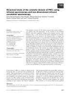
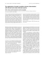
of GPCR interfaces. To zoom into the level of specific
lipid-exposed residues, we mined the prediction data
(Fig. 3) – to see whether some loci were more fre-
quently predicted to be at interfaces than others – by
comparing the results of the different studies
Fig. 3. Occurrence of the lipid-exposed residues in the predictions from bioinformatics methods applied to search for dimerization ⁄ oligo-
merization interfaces of GPCRs. Residues within a TM region that are predicted more than a baseline of four times are indicated for each
TM (purple, blue, red, green and magenta for residues in TM1, TM2, TM4, TM5 and TM6, respectively).
Computational study of GPCR oligomerization M. Filizola and H. Weinstein
2932 FEBS Journal 272 (2005) 2926–2938 ª 2005 FEBS
[16,17,30,31,40,45,47,48,50] that have looked so far for
possible dimerization ⁄ oligomerization interfaces of
rhodopsin-like GPCRs. Among all predicted residues
in each study, we only compared the lipid-exposed res-
idues within TM regions, and subject to the solvent
accessibility criterion of values > 45%. These values
were calculated with the getarea software version 1.1
[58] using the atomic coordinates of the rhodopsin
crystal structure [59]. It is important to keep in mind,
however, that rhodopsin and rhodopsin-like GPCRs
may exhibit structural differences that affect local
exposure of residues to the environment [86]. This can
create differences in the details of GPCR interfaces,
and therefore inaccurate estimation of solvent accessi-
bility values assigned to equivalent positions in differ-
ent receptors.
Figure 3 presents a histogram plot of the number
of times that each lipid-exposed residue has been pre-
dicted with the methods discussed here to belong to
dimerization ⁄ oligomerization interfaces of GPCRs.
Residues within a TM region that were predicted more
often than a base line of four times, are indicated for
each TM (purple, blue, red, green, and magenta for
residues in TM1, TM2, TM4, TM5 and TM6, respect-
ively). Most of these residues are within TM4, TM5,
and TM6, indicating that the prediction of dimeriza-
tion ⁄ oligomerization interfaces of GPCRs with various
computational methods has thus far pointed to a speci-
fic role for the lipid-exposed regions of these three heli-
ces. Among the loci identified within each of these
three helices, 4.58, 5.48 and 6.42 have the greatest
number of occurrences. In particular, the latter has
almost the same frequency of occurrence as 6.30.
(Note that the large frequency of occurrence for 6.30,
at the boundary between TM6 and the cytoplasmic
loop connecting TM5 and TM6, could be explained by
an involvement of this locus in a broader oligomeriza-
tion scheme of GPCRs. This is suggested by the
atomic force microscopy map of rhodopsin in native
membranes [11], which indicates that the cytoplasmic
loop connecting TM5 and TM6 facilitates the forma-
tion of rows of rhodopsin-dimers.)
Interestingly, position 4.58, which corresponds to a
cysteine in dopamine D
2
receptor, was shown recently
to form a copper phenanthroline-induced disulfide
cross-link resulting in the appearance of a dimeric
band [9]. This finding is consistent with the hypothesis
that TM4 is involved in a symmetrical interface in
dopamine D
2
receptor dimers, and that 4.58 is part of
this interface.
To the best of our knowledge, no information about
the involvement of position 5.48 in the dimeriza-
tion ⁄ oligomerization of rhodopsin-like GPCRs exists
in the literature. In contrast, a phenylanine at this
position in the 5HT2 subfamily of serotonin receptors
has been suggested to be involved in ligand binding
[87]. On the other hand, early experimental studies
pointed to the involvement of 6.42 based on inhibition
assays with synthetic peptides comprising the amino
acid sequence of TM6 of b2-adrenergic receptor that
contains the glycophorin-like dimerization motif
GXXXG [88]. Thus, the involvement of position 6.42
(G280 in human b2-adrenergic receptor) in the dimeri-
zation ⁄ oligomerization of GPCRs was inferred. How-
ever, these data do not necessarily establish TM6 as
the dimer interface in b2-adrenergic receptor, because
a specific peptide–receptor interaction at one site may
modulate the ability of the receptor to form dimers at
a different interface.
Last but not least, it is important to emphasize here
that the specific lipid-exposed positions shown in
Fig. 3 may also have been picked out by the bioinfor-
matics tools due to functional roles different from
dimerization ⁄ oligomerization. Such functional roles
may include correct protein folding, required interac-
tions with the lipid bilayer, or interactions with other
proteins. The high number of occurrences of position
2.59 seems to relate to this hypothesis because the pro-
line residue found at this position in almost all aminer-
gic receptors, on which most of the studies focused,
may induce a functional kink in this helix [86] that
could affect the structural integrity of the receptors.
The specific reason for the structure ⁄ function role of
the predictions shown in Fig. 3 notwithstanding, they
probably represent valuable hypotheses for further
experimental exploration of functionally important resi-
dues located on the surface of rhodopsin-like GPCRs.
Interpreted in the structural context offered by models
of the GPCRs [30,86,89] and their oligomers [11,48],
the putative interfaces are ripe for specific probing
with dimerization-disrupting mutations, cross-linking,
and derivatization. Such experiments should yield valu-
able insight not only about the structural details of the
GPCR–GPCR interaction in both homo- and hetero-
meric complexes, but will offer specific tools for the
elucidation of the functional roles of GPCR inter-
actions and the molecular details of signaling.
Structural details from computational
modeling and bioinformatics
As anticipated above, structural details from computa-
tional modeling and bioinformatics may provide valu-
able hypotheses for the experimental exploration of
interfaces of GPCR oligomerization. The goal is to
identify key residues responsible for disrupting the
M. Filizola and H. Weinstein Computational study of GPCR oligomerization
FEBS Journal 272 (2005) 2926–2938 ª 2005 FEBS 2933
GPCR oligomeric structures in order to probe effects
on receptor function, and achieve a better understand-
ing of the mechanisms that regulate these complex
systems.
Such a process of inquiry is illustrated by an
approach developed to identify the molecular determi-
nants for the oligomerization of rhodopsin-like GPCRs
as an iterative protocol of computational prediction
and experimental validation [31]. In this protocol,
results from CMA-based methods are used to guide
the construction of 3D molecular models of GPCR
homo- and heteromers [47,48]. Inferences from these
models (and not from correlated mutations alone) are
then implemented in the design of key collaborative
experiments that serve to probe, validate, and refine
the hypotheses of interaction between monomers (e.g.
an important validation probe is the design of dimeri-
zation-disrupting mutants). Specifically, the experimen-
tal designs can use a cysteine cross-linking method that
was recently implemented to identify residues at the
homo–dimerization interfaces of dopamine D2 recep-
tors. The success of the application of this experimen-
tal procedure is documented in the literature [9].
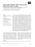
An illustration of the structural details that were
identified for dopamine D
2
receptor dimers using this
combined computational and experimental strategy
is given in Fig. 4. Our CMA-based approach for
the identification of homodimerization interfaces of
GPCRs [48] predicted that 4.58, 4.48, 4.55, and 4.60
are part of the interface of dopamine D
2
receptor
dimers. These predictions were then used to build geo-
metrically feasible configurations of dopamine D
2
receptor homodimers using the criteria reported in
[48], as reviewed above. Notably, the initial (unrefined)
models suggested that position 4.60, which is located
at the extracellular boundary of TM4, should not be
considered in the construction of the dimer models,
because of its possible inaccurate solvent accessibility
value (loop regions are missing in these models). Fig-
ure 4 shows the three residues that were used to guide
the construction of the receptor homodimeric interface
in CPK representation. Among them, C4.58 at the
extracellular end of TM4 had already been proposed
from cysteine cross-linking experiments to be part of a
symmetrical interface in dopamine D
2
receptor dimers
[9]. Additional cysteine cross-linking experiments (with
copper-phenanthroline) were then carried out at the
predicted positions 4.48 and 4.55, and these positions
were also suggested to be at the interface between
interacting monomers of the dopamine D
2
receptor
(W. Guo, L. Shi, M. Filizola, H. Weinstein, J. A.
Javich, unpublished results). The refined 3D models
that incorporate the new information are currently
being used to refine the scheme of dimerization disrup-
tion by single mutations (or a stretch identifiable as a
motif) of dopamine D
2
receptor homodimers. The pre-
dictive ability of this combined computational and
experimental strategy in the application to dopamine
Fig. 4. Proposed interface for dopamine D
2
receptor homodimers. Specific residues predicted with our CMA-based method to be at the
interface between monomers are shown in CPK representations.
Computational study of GPCR oligomerization M. Filizola and H. Weinstein
2934 FEBS Journal 272 (2005) 2926–2938 ª 2005 FEBS
receptors creates exciting expectations for the study of
the structural and functional properties of oligomeriza-
tion in other GPCRs as well, and possibly also in any
other membrane protein. Moreover, it points to the
exiting possibilities for exploration of functional
mechanisms of GPCRs in a structural context offered
by models of the protein–protein interaction. The
types of structure-based hypotheses that can be derived
from such models are essential for the development of
mechanistic understanding of the multibody, time-
dependent elements of GPCR function in cellular-
signaling pathways.
Experimentally tested predictions
Most experimental methods that have been used so far
to study GPCR oligomerization, such as inhibition
assays using synthetic peptides, coexpression, biolu-
minescence resonance energy transfer (BRET), and
fluorescence resonance energy transfer (FRET), do not
reveal the details of the GPCR oligomerization inter-
face. In particular, they do not reveal the specific resi-
dues that are in actual contact. As a result, only a few
positions have been reported in the literature to parti-
cipate directly to the oligomerization interface(s) of
GPCRs based entirely on experimental data. Specific-
ally, these positions are: 1.54 and 4.47 in the chemo-
kine receptors [49]; 4.58 in the dopamine D
2
receptor
[9] and 6.42 in the b
2
-adrenergic receptor [88]. Nota-
bly, three of these four positions had been predicted
with bioinformatics approaches to lie at the dimeriza-
tion interface more often than the four times consid-
ered as a base line (see Fig. 3).
As detailed information about the oligomerization
interface of GPCRs is essential for designing dimeriza-
tion-disrupting mutants, and thus for probing structural
and mechanistic hypotheses by interfering with GPCR
function, more incisive experimental approaches are
needed that are better suited for the identification of
specific residues at the dimeric ⁄ oligomeric interface of
GPCRs. The close collaboration established between
the computational and experimental approaches to
investigation of GPCR oligomerization, as illustrated
in this review, should then enable rapid success in
unraveling the mechanisms and functional implications
of GPCR oligomerization in GPCR signaling processes.
Acknowledgements
We are grateful to our colleagues Drs Wen Guo,
Jonathan Javitch, and Lei Shi for illuminating collabor-
ative studies, and to Dr Masha Niv for comments on
the manuscript. Computational support was provided
by the National Science Foundation Terascale Compu-
ting System at the Pittsburgh Supercomputing Center.
The authors also acknowledge access to the computer
and bioinformatics facilities at the Institute of Compu-
tational Biomedicine (ICB) of Weill Medical College.
The work was supported in part by NIH grants P01
DA12923, and K05 DA-00060.
References
1 Breitwieser GE (2004) G protein-coupled receptor oligo-
merization: implications for G protein activation and
cell signaling. Circ Res 94, 17–27.
2 Bai M (2004) Dimerization of G-protein-coupled recep-
tors: roles in signal transduction. Cell Signal 16, 175–
186.
3 Terrillon S & Bouvier M (2004) Roles of G-protein-
coupled receptor dimerization. EMBO Report 5, 30–34.
4 Milligan G (2004) G protein-coupled receptor dimeriza-
tion: function and ligand pharmacology. Mol Pharmacol
66, 1–7.
5 Cvejic S & Devi LA (1997) Dimerization of the delta
opioid receptor: implication for a role in receptor inter-
nalization. J Biol Chem 272, 26959–26964.
6 AbdAlla S, Zaki E, Lother H & Quitterer U (1999)
Involvement of the amino terminus of the B (2) receptor
in agonist-induced receptor dimerization. J Biol Chem
274, 26079–26084.
7 Carrillo JJ, Pediani J & Milligan G (2003) Dimers of
class A G protein-coupled receptors function via ago-
nist-mediated trans-activation of associated G proteins.
J Biol Chem 278, 42578–42587.
8 Carrillo JJ, Lopez-Gimenez JF & Milligan G (2004)
Multiple interactions between transmembrane helices
generate the oligomeric {alpha}1b-adrenoceptor. Mol
Pharmacol 66, 1123–1137.
9 Guo W, Shi L & Javitch JA (2003) The fourth trans-
membrane segment forms the interface of the dopamine
D2 receptor homodimer. J Biol Chem 278, 4385–4388.
10 Fotiadis D, Liang Y, Filipek S, Saperstein DA, Engel A
& Palczewski K (2003) Atomic-force microscopy: Rho-
dopsin dimers in native disc membranes. Nature 421,
127–128.
11 Liang Y, Fotiadis D, Filipek S, Saperstein DA,
Palczewski K & Engel A (2003) Organization of the G
protein-coupled receptors rhodopsin and opsin in native
membranes. J Biol Chem 278, 21655–21662.
12 Fotiadis D, Liang Y, Filipek S, Saperstein DA, Engel A
& Palczewski K (2004) The G protein-coupled receptor
rhodopsin in the native membrane. FEBS Lett 564,
281–288.
13 Schulz A, Grosse R, Schultz G, Gudermann T & Scho-
neberg T (2000) Structural implication for receptor oligo-
merization from functional reconstitution studies of
M. Filizola and H. Weinstein Computational study of GPCR oligomerization
FEBS Journal 272 (2005) 2926–2938 ª 2005 FEBS 2935
mutant V2 vasopressin receptors. J Biol Chem 275,
2381–2389.
14 Gouldson PR & Reynolds CA (1997) Simulations on
dimeric peptides: evidence for domain swapping in
G-protein-coupled receptors? Biochem Soc Trans 25,
1066–1071.
15 Gouldson PR, Snell CR & Reynolds CA (1997) A new
approach to docking in the beta 2-adrenergic receptor
that exploits the domain structure of G-protein-coupled
receptors. J Med Chem 40, 3871–3886.
16 Gouldson PR, Snell CR, Bywater RP, Higgs C &
Reynolds CA (1998) Domain swapping in G-protein
coupled receptor dimers. Prot Eng 11, 1181–1193.
17 Gouldson PR, Higgs C, Smith RE, Dean MK,
Gkoutos GV & Reynolds CA (2000) Dimerization and
domain swapping in G-protein-coupled receptors: a
computational study. Neuropsychopharmacology 23,
S60–S77.
18 Lee SP, O’Dowd BF, Ng GY, Varghese G, Akil H,
Mansour A, Nguyen T & George SR (2000) Inhibition
of cell surface expression by mutant receptors demon-
strates that D2 dopamine receptors exist as oligomers in
the cell. Mol Pharmacol 58, 120–128.
19 Hamdan FF, Ward SD, Siddiqui NA, Bloodworth LM
& Wess J (2002) Use of an in situ disulfide cross-linking
strategy to map proximities between amino acid residues
in transmembrane domains I and VII of the M3 mus-
carinic acetylcholine receptor. Biochemistry 41, 7647–
7658.
20 Hadac EM, Ji Z, Pinon DI, Henne RM, Lybrand TP &
Miller LJ (1999) A peptide agonist acts by occupation
of a monomeric G protein-coupled receptor: dual
sites of covalent attachment to domains near TM1 and
TM7 of the same molecule make biologically significant
domain-swapped dimerization unlikely. J Med Chem 42,
2105–2111.
21 Overton MC & Blumer KJ (2002) The extracellular
N-terminal domain and transmembrane domains 1 and
2 mediate oligomerization of a yeast G protein-coupled
receptor. J Biol Chem 277, 41463–41472.
22 Maggio R, Vogel Z & Wess J (1993) Coexpression stu-
dies with mutant muscarinic ⁄ adrenergic receptors pro-
vide evidence for intermolecular ‘cross-talk’ between
G-protein-linked receptors. Proc Natl Acad Sci USA 90,
3103–3107.
23 Ridge KD, Lee SS et al. (1996) Examining rhodopsin
folding and assembly through expression of polypeptide
fragments. J Biol Chem 271, 7860–7867.
24 Kobilka BK, Kobilka TS, Daniel K, Regan JW, Caron
MG & Lefwoeits RJ (1988) Chimeric a2-, b2-adrenergic
receptors: delineation of domains involved in effector
coupling and ligand binding specificity, Science 240,
1310–1316.
25 Schoneberg T, Liu J & Wess J (1995) Plasma membrane
localization and functional rescue of truncated forms of
a G protein-coupled receptor. J Biol Chem 270, 18000–
18006.
26 Schoneberg T, Yun J, Wenkert D & Wess J (1996)
Functional rescue of mutant V2 vasopressin receptors
causing nephrogenic diabetes insipidus by a co-expressed
receptor polypeptide. EMBO J 15, 1283–1291.
27 Gudermann T, Schoneberg T & Schultz G (1997) Func-
tional and structural complexity of signal transduction
via G-protein-coupled receptors. Annu Rev Neurosci 20,
399–427.
28 Nielsen SM, Elling CE & Schwartz TW (1998) Split-
receptors in the tachykinin neurokinin-1 system – muta-
tional analysis of intracellular loop 3. Eur J Biochem
251, 217–226.
29 Bakker RA, Dees G, Carrillo JJ, Booth RG, Lopez-
Gimenez JF, Milligan G, Strange PG & Leurs R (2004)
Domain swapping in the human histamine h1 receptor.
J Pharmacol Exp Ther 311, 131–138.
30 Filizola M, Visiers I, Skrabanek L, Campagne F &
Weinstein H (2003) Functional mechanisms of GPCRs
in a structural context. In Strategies in molecular neuro-
pharmacology (Schousboe A & Brauner-Osborne H,
eds), pp. 235–266. Humana Press, Totowa, NJ.
31 Filizola M, Guo W, Javitch JA & Weinstein H (2005)
Oligomerization domains of G-protein coupled recep-
tors: insights into the structural basis of GPCR associa-
tion. In Contemporary Clinical Neuroscience: the
G-protein Coupled Receptor Handbook (Devi LA, ed),
pp. 243–265. Humana Press, Totowa, NJ.
32 Valencia A & Pazos F (2002) Computational methods
for the prediction of protein interactions. Curr Opin
Struct Biol 12, 368–373.
33 Smith GR & Sternberg MJ (2002) Prediction of pro-
tein–protein interactions by docking methods. Curr Opin
Struct Biol 12, 28–35.
34 Dixon JS (1997) Evaluation of the CASP2 docking sec-
tion. Proteins Supplement 1, 198–204.
35 Oliveira L, Paiva AC & Vriend G (2002) Correlated
mutation analyses on very large sequence families.
Chembiochem 3, 1010–1017.
36 Oliveira L, Paiva AC & Vriend G (1999) A low resolu-
tion model for the interaction of G proteins with G pro-
tein- coupled receptors. Protein Eng 12 , 1087–1095.
37 Horn F, van der Wenden EM, Oliveira L, AP IJ &
Vriend G (2000) Receptors coupling to G proteins: is
there a signal behind the sequence? Proteins 41, 448–459.
38 Lichtarge O, Bourne HR & Cohen FE (1996) Evolutio-
narily conserved Galphabetagamma binding surfaces
support a model of the G protein-receptor complex.
Proc Natl Acad Sci USA 93, 7507–7511.
39 Lichtarge O, Sowa ME & Philippi A (2002) Evolution-
ary traces of functional surfaces along G protein signal-
ing pathway. Methods Enzymol 344, 536–556.
40 Dean MK, Higgs C, Smith RE, Bywater RP, Snell CR,
Scott PD, Upton GJ, Howe TJ & Reynolds CA (2001)
Computational study of GPCR oligomerization M. Filizola and H. Weinstein
2936 FEBS Journal 272 (2005) 2926–2938 ª 2005 FEBS
Dimerization of G-protein-coupled receptors. J Med
Chem 44, 4595–4614.
41 Moller S, Vilo J & Croning MD (2001) Prediction of
the coupling specificity of G protein coupled receptors
to their G proteins. Bioinformatics 17, S174–S181.
42 Oliveira L, Paiva PB, Paiva AC & Vriend G (2003)
Sequence analysis reveals how G protein-coupled recep-
tors transduce the signal to the G protein. Proteins 52,
553–560.
43 Madabushi S, Gross AK, Philippi A, Meng EC, Wensel
TG & Lichtarge O (2004) Evolutionary trace of G pro-
tein-coupled receptors reveals clusters of residues that
determine global and class-specific functions. J Biol
Chem 279, 8126–8132.
44 Suel GM, Lockless SW, Wall MA & Ranganathan R
(2003) Evolutionarily conserved networks of residues
mediate allosteric communication in proteins. Nat Struct
Biol 10, 59–69.
45 Gouldson PR, Dean MK, Snell CR, Bywater RP,
Gkoutos G & Reynolds CA (2001) Lipid-facing corre-
lated mutations and dimerization in G-protein coupled
receptors. Prot Eng 14, 759–767.
46 Gkoutos GV, Higgs C, Bywater RP, Gouldson PR &
Reynolds CA (1999) Evidence for dimerization in the
b2-adrenergic receptor from the evolutionary trace
method. Intl J Quantum Chem Biophys Q74, 371–379.
47 Filizola M, Olmea O & Weinstein H (2002) Prediction
of heterodimerization interfaces of G-protein coupled
receptors with a new subtractive correlated mutation
method. Prot Eng 15, 881–885.
48 Filizola M & Weinstein H (2002) Structural models for
dimerization of G-Protein coupled receptors: the opioid
receptor homodimers. Biopolymers (Peptide Sci) 66,
317–325.
49 Hernanz-Falcon P, Rodriguez-Frade JM, Serrano A,
Juan D, del Sol A, Soriano SF, Roncal F, Gomez L,
Valencia A, Martinez AC & Mellado M (2004) Identifi-
cation of amino acid residues crucial for chemokine
receptor dimerization. Nat Immunol 5, 216–223.
50 Soyer OS, Dimmic MW, Neubig RR & Goldstein RA
(2003) Dimerization in aminergic G-protein-coupled
receptors: application of a hidden-site class model of
evolution. Biochemistry 42, 14522–14531.
51 Ballesteros JA & Weinstein H (1995) Integrated meth-
ods for the construction of three-dimensional models
and computational probing of structure-function rela-
tions in G protein-coupled receptors. Methods Neurosci
25, 366–428.
52 Livingstone CD & Barton GJ (1993) Protein sequence
alignments: a strategy for the hierarchical analysis
of residue conservation. Comput Appl Biosci 9, 745–
756.
53 Lichtarge O, Bourne HR & Cohen FE (1996) An evolu-
tionary trace method defines binding surfaces common
to protein families. J Mol Biol 257, 342–358.
54 Lichtarge O, Yamamoto KR & Cohen FE (1997) Iden-
tification of functional surfaces of the zinc binding
domains of intracellular receptors. J Mol Biol 274, 325–
337.
55 Chothia C & Lesk AM (1986) The relation between the
divergence of sequence and structure in proteins. Embo
J 5, 823–826.
56 Zvelebil MJ, Barton GJ, Taylor WR & Sternberg MJ
(1987) Prediction of protein secondary structure and
active sites using the alignment of homologous
sequences. J Mol Biol 195, 957–961.
57 Upton G & Fingleton B (1985) Spatial Data Analysis by
Example. Wiley, Chichester.
58 Fraczkiewicz R & Braun W (1998) Exact and efficient
analytical calculation of the accessible surface areas and
their gradients for macromolecules. J Computational
Chem 19, 319–333.
59 Palczewski K, Kumasaka T, Hori T, Behnke CA,
Motoshima H, Fox BA, LeTrong I, Teller DC, Okada T,
Stenkamp RE et al. (2000) Crystal structure of rhodo-
psin: a G protein-coupled receptor. Science 289,
739–745.
60 Gobel U, Sander C, Schneider R & Valencia A (1994)
Correlated mutations and residue contacts in proteins.
Proteins 18, 309–317.
61 Oliveira L, Paiva ACM & Vriend G (1993) A common
motif in G-protein coupled seven transmembrane helix
receptors. J Comp Aid Mol Des 7, 649–658.
62 Zhou W, Flanagan C, Ballesteros JA, Konvicka K,
Davidson JS, Weinstein H, Millar RP & Sealfon SC
(1994) A reciprocal mutation supports helix 2 and helix
7 proximity in the gonadotropin-releasing hormone
receptor. Mol Pharmacol 45, 165–170.
63 Sealfon SC, Chi L, Ebersole BJ, Rodic V, Zhang D,
Ballesteros JA & Weinstein H (1995) Related contribu-
tion of specific helix 2 and 7 residues to conformational
activation of the serotonin 5-HT2A receptor. J Biol
Chem 270, 16683–16688.
64 Pazos F, Helmer-Citterich M, Ausiello G & Valencia
A (1997) Correlated mutations contain information
about protein–protein interaction. J Mol Biol 271,
511–523.
65 Vriend G (1990) WHAT IF: a molecular modeling and
drug design program. J Mol Graph 8 (52–6), 29.
66 Olmea O & Valencia A (1997) Improving contact pre-
dictions by the combination of correlated mutations and
other sources of sequence information. Fold Des 2, S25–
S32.
67 Pazos F & Valencia A (2002) In silico two-hybrid sys-
tem for the selection of physically interacting protein
pairs. Proteins 47, 219–227.
68 Klco JM, Lassere TB & Baranski TJ (2003) C5a recep-
tor oligomerization. I. Disulfide trapping reveals oligo-
mers and potential contact surfaces in a G protein-
coupled receptor. J Biol Chem 278, 35345–35353.
M. Filizola and H. Weinstein Computational study of GPCR oligomerization
FEBS Journal 272 (2005) 2926–2938 ª 2005 FEBS 2937
69 del Sol Mesa A, Pazos F & Valencia A (2003) Auto-
matic methods for predicting functionally important
residues. J Mol Biol 326, 1289–1302.
70 Casari G, Sander C & Valencia A (1995) A method to
predict functional residues in proteins. Nat Struct Biol
2, 171–178.
71 Dorit RL & Ayala FJ (1995) ADH evolution and the
phylogenetic footprint. J Mol Evol 40, 658–662.
72 Andrade MA, Casari G, Sander C & Valencia A (1997)
Classification of protein families and detection of the
determinant residues with an improved self-organizing
map. Biol Cybern 76, 441–450.
73 Zhang B, Rychlewski L, Pawlowski K, Fetrow JS, Skol-
nick J & Godzik A (1999) From fold predictions to
function predictions: automation of functional site con-
servation analysis for functional genome predictions.
Protein Sci 8, 1104–1115.
74 Goh CS, Bogan AA, Joachimiak M, Walther D &
Cohen FE (2000) Co-evolution of proteins with their
interaction partners. J Mol Biol 299, 283–293.
75 Landgraf R, Xenarios I & Eisenberg D (2001) Three-
dimensional cluster analysis identifies interfaces and
functional residue clusters in proteins. J Mol Biol 307,
1487–1502.
76 Armon A, Graur D & Ben-Tal N (2001) ConSurf: an
algorithmic tool for the identification of functional
regions in proteins by surface mapping of phylogenetic
information. J Mol Biol 307, 447–463.
77 Madabushi S, Yao H, Marsh M, Kristensen DM,
Philippi A, Sowa ME & Lichtarge O (2002) Structural
clusters of evolutionary trace residues are statistically
significant and common in proteins. J Mol Biol 316,
139–154.
78 Lichtarge O & Sowa ME (2002) Evolutionary predic-
tions of binding surfaces and interactions. Curr Opin
Struct Biol 12, 21–27.
79 Pupko T, Bell RE, Mayrose I, Glaser F & Ben-Tal N
(2002) Rate4Site: an algorithmic tool for the identifica-
tion of functional regions in proteins by surface
mapping of evolutionary determinants within
their homologues. Bioinformatics 18 (Suppl. 1), S71–
S77.
80 Shannon C & Weaver W (1963) Mathematical Theory of
Communication. University of Illinois press, Champaign,
IL.
81 Pazos F, Sanchez-Pulido L, Garcia-Ranea JA, Andrade
MA, Atrian S & Valencia A (1997) Comparative analy-
sis of different methods for the detection of specific
regions in protein families. In Biocomputing and Emer-
gent Computation (Lundh, D, Olsson, B & Narayanan,
A, eds), pp. 132–145. World Scientific, Singapore.
82 Vakser IA & Jiang S (2002) Strategies for modeling the
interactions of transmembrane helices of G protein-
coupled receptors by geometric complementarity using
the GRAMM computer algorithm. Methods Enzymol
343, 313–328.
83 Koshi JM & Goldstein RA (1995) Context-dependent
optimal substitution matrices. Protein Eng 8, 641–645.
84 Koshi JM & Goldstein RA (1998) Models of natural
mutations including site heterogeneity. Proteins 32, 289–
295.
85 Koshi JM, Mindell DP & Goldstein RA (1999) Using
physical-chemistry-based substitution models in phylo-
genetic analyses of HIV-1 subtypes. Mol Biol Evol 16,
173–179.
86 Ballesteros JA, Shi L & Javitch JA (2001) Structural
mimicry in G Protein-coupled receptors. Implications of
the high-resolution structure of rhodopsin for structure-
function analysis of rhodopsin-like receptors. Mol
Pharmacol 60, 1–19.
87 Shapiro DA, Kristiansen K, Kroeze WK & Roth BL
(2000) Differential modes of agonist binding to 5-hydro-
xytryptamine (2A) serotonin receptors revealed by muta-
tion and molecular modeling of conserved residues in
transmembrane region 5. Mol Pharmacol 58, 877–886.
88 Hebert TE, Moffett S, Morello JP, Loisel TP, Bichet
DG, Barret C & Bouvier M (1996) A peptide derived
from a beta2-adrenergic receptor transmembrane
domain inhibits both receptor dimerization and activa-
tion. J Biol Chem 271, 16384–16392.
89 Visiers I, Ballesteros JA & Weinstein H (2002) Three-
dimensional representations of G protein-coupled recep-
tor structures and mechanisms. Methods Enzymol 343,
329–371.
Computational study of GPCR oligomerization M. Filizola and H. Weinstein
2938 FEBS Journal 272 (2005) 2926–2938 ª 2005 FEBS