Báo cáo khoa học: Molecular cloning and functional expression of the human sodium channel b1B subunit, a novel splicing variant of the b1 subunit potx
Bạn đang xem bản rút gọn của tài liệu. Xem và tải ngay bản đầy đủ của tài liệu tại đây (397.84 KB, 9 trang )
Molecular cloning and functional expression of the human sodium
channel b
1B
subunit, a novel splicing variant of the b
1
subunit
Ning Qin
1
, Michael R. D’Andrea
1
, Mary-Lou Lubin
1
, Navid Shafaee
2,
*, Ellen E. Codd
1
and Ana M. Correa
2
1
Department of Drug Discovery, Johnson & Johnson Pharmaceutical Research & Development, Spring House, PA, USA;
2
Department of Anesthesiology, University of California, Los Angeles, CA, USA
The voltage gated sodium channel comprises a pore-forming
a subunit and regulatory b subunits. We report here the
identification and characterization of a novel splicing variant
of the human b
1
subunit, termed b
1B
. The 807 bp open
reading frame of the human b
1B
subunit encodes a 268
residue protein with a calculated molecular mass of
30.4 kDa. The novel human b
1B
subunit shares an identical
N-terminal half (residues 1–149) with the human b
1
subunit,
but contains a novel C-terminal half (residues 150–268) of
less than 17% sequence identity with the human b
1
subunit.
The C-terminal region of the human b
1B
is also significantly
different from that of the rat b
1A
subunit, sharing less than
33% sequence identity. Tissue distribution studies reveal
that the human b
1B
subunit is expressed predominantly in
human brain, spinal cord, dorsal root ganglion and skeletal
muscle. Functional studies in oocytes demonstrate that the
human b
1B
subunit increases the ionic current when coex-
pressed with the tetrodotoxin sensitive channel, Na
V
1.2,
without significantly changing voltage dependent kinetics
and steady-state properties, thus distinguishing it from the
human b
1
and rat b
1A
subunits.
Keywords: sodium channel; b
1B
subunit; splicing variant.
By mediating the rapid entry of sodium ions into excitable
cells in response to voltage changes across the plasma
membrane, voltage gated sodium channels (VGSCs) play a
fundamental role in the control of neuronal excitability in
the central and peripheral nervous systems. The VGSC is a
heteromeric protein complex that comprises at least a large
(200–300 kDa) pore-forming a subunit and several smaller
(30–40 kDa) regulatory b subunits [1–4]. It is well known
that sodium channel a subunits determine the basic
properties of the channel, while b subunits modulate the
channel properties. Functional studies in a heterologous
system have demonstrated that, depending on the type of
coexpressed a subunit, b subunits are able to modulate
almost all aspects of the channel properties, including
voltage dependent gating, activation and inactivation, as
well as greatly increasing the number of functional channels
present on the plasma membrane [5,6]. Currently, at least
nine different a subunits, three b subunits, and a splicing
variant of the b
1
subunit, rat b
1A
[7], have been cloned and
characterized.
The rat b
1A
subunit is a splicing variant of the b
1
subunit via intron retention. The N-terminal half of the
b
1A
subunit is identical to that of the rat b
1
subunit,
whereas its C-terminal half, encoded by a retained intron
with an in-frame stop codon, is completely different from
that of the rat b
1
subunit (to which it shows less than
17% identity). Coexpression of the rat b
1A
subunit with
the pore forming alpha subunit, Na
V
1.2, in Chinese
hamster lung 1610 cells, increased the sodium current
density and produced subtle changes in voltage depend-
ent activation and inactivation [7]. To further explore the
function and physiological relevance of the sodium
channel b
1
splicing variant, we first tried to clone the
same splicing variant from human tissue. Here, we report
the cloning and characterization of a novel, splicing
variant of the human b
1
subunit by rapid amplification
of cDNA end polymerase chain reaction (RACE-PCR)
based on the human b
1
sequence. The novel b
1
subunit
splicing variant, named b
1B
, is produced via extension of
exon 3 with an in-frame stop codon. The human b
1B
subunit is significantly different from the rat b
1A
subunit
in sequence, expression pattern and regulatory properties,
although they share a similar splicing pattern. Functional
studies indicate that the human b
1B
subunit performs a
physiological function distinct from that of the human b
1
subunit when it is coexpressed with Na
V
1.2 in Xenopus
oocytes.
Experimental procedures
Molecular cloning of the human sodium channel b
1B
subunit
Full-length human b
1B
cDNA was cloned using a strat-
egy that combined reverse transcription polymerase chain
Correspondence to N. Qin, Drug Discovery, Johnson & Johnson
Pharmaceutical Research and Development, PO Box 776, Welsh and
McKean Roads, Spring House, PA 19477-0776, USA.
Fax: + 1 215 628 3297, Tel.: + 1 215 540 4886,
E-mail:
Abbreviations: DRG, dorsal root ganglia; RACE-PCR, rapid ampli-
fication of cDNA end-polymerase chain reaction; RT-PCR, reverse
transcription–polymerase chain reaction; VGSC, voltage gated
sodium channel.
*Present address: Royal College of Surgeons, Dublin, Ireland.
(Received 28 July 2003, revised 6 October 2003,
accepted 15 October 2003)
Eur. J. Biochem. 270, 4762–4770 (2003) Ó FEBS 2003 doi:10.1046/j.1432-1033.2003.03878.x
reaction (RT-PCR) and RACE-PCR. Marathon-Ready
TM
human adrenal gland and fetal brain cDNA libraries were
purchased from Clontech (Palo Alto, CA, USA). The
RACE-PCR was performed according to the supplier’s
instructions. The reaction mixture (50 lL final volume)
contained 5 lL of Marathon-Ready
TM
human adrenal
gland cDNA, 200 l
M
dNTP, 200 n
M
AP1 primer (Clon-
tech), 200 n
M
human b
1
subunit specific primer (SB1-10:
5¢-TGGACCTTCCGCCAGAAGGGCACTG-3¢), and
1 lLof50· Advatage2 DNA polymerase mixture (Clon-
tech). The thermal cycling parameters for RACE–PCR were
as follows: an initial denaturation at 94 °C for 30 s; five cycles
of 94 °Cfor5sand72°C for 4 min; five cycles of 94 °Cfor
5sand70°C for 4 min; and 20 cycles of 94 °Cfor5sand
68 °C for 4 min. The RACE–PCR product was then cloned
into the PCR-Script
TM
Amp Cloning vector (Statagene,
La Jolla, CA, USA), according to the protocol provided
by the supplier.
The full-length b
1B
subunit was cloned from the Mara-
thon-Ready
TM
human fetal brain cDNA library based on
the C-terminal sequence of human b
1B
subunit obtained
using the RACE-PCR. The PCR was performed in a final
volume of 50 lL, containing 5 lL of Marathon-Ready
TM
humanfetalbraincDNA,5lLof10· reaction buffer,
200 l
M
dNTP, 200 n
M
SB1-6 primer (5¢-GCCATGGG
GAGGCTGCTGGCCTTAGTGGTC-3¢) and SB1-19 pri-
mer (5¢-GTGTGCCTGCAGCTGCTCAA-3¢), and 1 lL
of 50· HF2 DNA polymerase mixture (Clontech). Four
independent clones were selected and subjected to double
stranded DNA sequencing analysis. All four independent
clones from the human fetal brain were found to contain
sequences identical to that of the RACE-PCR cloned b
1B
subunit from human adrenal gland.
Generation of polyclonal antibody
A peptide (RWRDRWQAVDRTGC), derived from the
C terminus of the human b
1B
subunit, was synthesized
and used for raising polyclonal antibodies in rabbits.
(This peptide was chosen because the seqeunce shows
the highest homology between human and rat b
1A
subunits.) The antibody was raised and affinity purified
by BioSource International, Inc. The resulting affinity
purified antibody was used for immunohistochemical
analysis.
Northern blot analysis
Human Multiple Tissue Northern blot (MTN
TM
)and
human Brain II MTN
TM
blot were purchased from
Clontech. The cDNA fragment encoding residues 217–
268 of the human b
1B
subunit was used as a probe. The
antisense single stranded DNA probe was synthesized
using the Strip-EZ
TM
PCR kit (Ambion, Austin, TX,
USA), in the presence of antisense primer SB1-20 (5¢-TC
AAACCACACCCCGAGAAA-3¢)and[
32
P]dATP[aP]
(3000 CiÆmmol
)1
; Amersham Pharmacia Biotech.), follow-
ing the manufacturer’s instructions. The labeled probe was
then separated from free [
32
P]dATP[aP] using a Micro-
Spin
TM
G-50 column (Amersham Pharmacia Biotech.). The
cDNA fragment encoding the human b
1
subunit from amino
acids 150 to 218 was used as a human b
1
subunit specific
probe. The single stranded antisense b
1
specific probe was
labeled and purified as described above. A 2 kb human
b-actin cDNA fragment was used as the control probe
and labeled with Ready-To-Go
TM
DNA Labelling Bead
(–dCTP) (Amersham Pharmacia Biotech.), followed by
purification as described above.
The blots were prehybridized with 5 mL of UltraHyb
Solution (Ambion), at 42 °C for 2 h, and then hybridized
in the presence of 1 · 10
6
c.p.m.ÆmL
)1
probe (b
1B
, b
1
and
b-actin separately) at 42 °C overnight. The blots were
washed with 2 · 200 mL of 0.2· NaCl/Cit/0.1% SDS, at
65 °C for 2 h. Finally, the blots were exposed to X-ray
film in a )80 °C freezer for 2–18 h. The same blots were
used for all three probes (b
1B
, b
1
and b-actin) after
stripping at 68 °C for 15 min and reconstitution at room
temperature for 15 min using the Strip-EZ
TM
removal kit
(Ambion).
Immunohistochemistry
Protocols for immunohistochemistry have been described
previously [8]. All incubations were performed at room
temperature. After microwaving the slides in Target
(Dako, Carpenturia, CA, USA), the slides were placed
in NaCl/P
i
andthenin3%H
2
O
2
,rinsedinNaCl/P
i
and
then the appropriate blocking serum was added for
10 min. Subsequently, primary antibody, rabbit polyclonal
anti-(human b
1B
), at a titer of 1 : 200, was applied to the
slides for 30 min. After several washes in NaCl/P
i
,a
biotinylated secondary antibody (Vector Laboratories)
was placed on the slides for 30 min. Subsequently, the
slides were washed in NaCl/P
i
and the avidin–biotin
complex (ABC; Vector Laboratories) was applied to the
cells for 30 min. The presence of the primary antibody was
detected after two 5 min incubations in 3¢-diaminobenzi-
dine-HCl (Biomeda, Foster City, CA, USA). Slides were
briefly exposed to Mayer’s hematoxylin for 1 min, dehy-
drated and coverslipped. Antibody specificity controls
included (a) replacement of the primary antibody with
nonimmune serum, (b) omission of the primary antibody
with the antibody dilution buffer (Zymed Laboratories
Inc., San Francisco, CA, USA), and (c) preincubation
with specific antigen (preabsorption). Preabsorption was
carried out using a 10-fold titer excess of the antigen
peptide preincubated with antibody overnight at 4 °C.
This mixture was then used as the Ôprimary antibodyÕ. b
1B
subunit immunolabeling was not detected in the preab-
sorption controls or in other negative controls. Specimens
were examined and photographed using an Olympus
BX-50 microscope.
In vitro
synthesis of cRNA
The expression constructs of b
1B
and Na
V
1.2 were linearized
following digestion with restriction enzymes. The cRNAs
were synthesized in vitro with T7 RNA polymerase
using reagents and protocols from the mMESSAGE
mMACHINE
TM
transcription kit (Ambion), except that
the LiCl precipitation was repeated twice. To ensure full-
length clones of the Na
V
1.2 a subunit, the reaction mixture
was supplemented, halfway through transcription, with
additional enzyme and nucleotides. The cRNAs were
Ó FEBS 2003 Cloning and characterization of Na
+
channel b
1B
(Eur. J. Biochem. 270) 4763
suspended in diethylpyrocarbonate-treated H
2
O at a final
concentration of 1–2 mgÆmL
)1
.
Oocyte preparation and RNA injection
Conventional methods were followed for oocyte isolation
and removal of the follicular membrane [9]. Adult female
Xenopus laevis (Xenopus One, Ltd, Dexter, MI, USA) were
anesthetized by immersion in 0.1% tricaine. Ovaries
were removed through an abdominal incision. Ovarian sacs
were rinsed in Ca
2+
-free medium and teased apart to expose
the oocytes. The follicular layer was removed by treat-
ment with collagenase (200 UÆmL
)1
;Gibco)inCa
2+
-free
medium, followed by rinsing and storage in saline medium
containing Ca
2+
and 50 lgÆmL
)1
gentamicin. Stage V–VI
oocytes were separated for injection the following day.
Normally, 20–25 oocytes per RNA sample were micro-
injected, each with 50 nL of 1 mgÆmL
)1
cRNA. Combina-
tion of subunits was obtained by injecting the premixed
cRNAs. Microinjection was performed under sterile,
RNase-free conditions. After injection, oocytes were
maintained at 18 °C.
Recording solutions
External and internal recording solutions contained mostly
impermeant anions and were made iso-osmolar to the
oocyte media (120 m
M
; 220–240 mOsmÆkg
)1
). Sodium
currents were recorded in external 120 m
M
sodium methane
sulfonate, 1.8 m
M
CaCl
2
,10m
M
Hepes-sodium, pH 7.2;
and internal 120 m
M
cesium methane sulfonate, 10 m
M
sodium methane sulfonate, 10 m
M
Hepes-sodium, 1 m
M
EGTA-sodium, pH 7.2. Voltage electrodes were filled with
2.7
M
TMA, which comprised 2.7
M
tetra methyl-ammo-
nium, 10 m
M
NaCl, and 10 m
M
Hepes-sodium, pH 7.0.
Recording and analysis of macroscopic ionic
and gating currents
The cut open oocyte Vaseline gap technique [10] was used
to record macroscopic ionic currents. This technique,
described previously [11], greatly improves the temporal
resolution over that of the conventional two-electrode
voltage clamp. The currents were recorded from an area of
the oocyte equivalent to 20–25% of the total surface.
Voltage electrodes had resistances of 0.2–0.5 MW.Custom-
made software and hardware were used for acquisition and
analysis of data. Leakage and linear capacity currents were
subtracted by using P/4 protocols. Data were sampled once
every 5 ls and were filtered at 1/5 of the sampling frequency.
Conventional pulse protocols were used to record mac-
roscopic sodium currents in response to changes in mem-
brane voltage. Test pulses of 15 ms were applied from
holding potentials of )80 or )100 mV; the range of test
potentials used to cover the whole activation curve was
typically )60 mV to 100 mV, at 5 mV intervals. For steady-
state inactivation curves, a 15 ms test pulse to 0 mV was
preceded by a preconditioning 100 ms pulse spanning
)140 mV to 20 mV, at intervals of 10 mV. Conductance
vs. voltage (G-V) curves were obtained from the I-V plots
fitting the data to: I ¼ G(V)Æ(V
m
–Vrev), where I is the
current amplitude, G(V) is the voltage-dependent conduct-
ance, V
m
is the membrane voltage, and V
rev
is the voltage
for current reversal. Once V
rev
was determined from the I-V
fits, the individual I-V curves were divided by V
m
–V
rev
to
obtain the G(V). The G-V plots were fitted to: G ¼ G
max
/
(1 + exp[–zÆ(V-V
½
)/25]), where G
max
is the maximum
conductance, z is the valence of the process and V
½
is the
midpoint voltage of activation.
Ionic current expression levels were determined from
batches of oocytes injected with Na
V
1.2 alone or with
Na
V
1.2 combined with b
1B
subunit at an a: b ratio of 1 : 5
or 1 : 20. Only data from batches expressing all three a : b
combinations were included in the analysis. Unpaired t-test
statistics were used to compare the different current
amplitude data sets.
Results
Cloning and analysis of the human VGSC b
1B
subunit
In order to clone the human b
1
splicing variant, we first
used RT-PCR with a forward primer based on the
human b
1
subunit and degenerated reverse primers based
on the rat b
1A
C-terminal sequence. However, these
attempts failed to clone the human b
1A
subunit from
cDNA libraries of adrenal gland, brain and fetal brain.
BLAST
searches of the human genomic sequence with the
cDNA encoding the rat b
1A
subunit C terminus also
failed to identify any homologous region in the human
b
1
gene. These results suggested that, if the human b
1
gene also undergoes alternative splicing, it might have a
very different sequence from that of the rat b
1A
subunit.
Therefore, a RACE-PCR technique was applied to clone
a novel, splicing variant of the human b
1
subunit. To
perform RACE-PCR of a novel C terminus of the human
b
1
subunit, we designed a forward primer (SB1-10) based
on the N-terminal cDNA sequence. The resulting
RACE-PCR product was directly cloned into the PCR
cloning vector. As the human b
1
subunit specific
primer was used for 3¢ extension, both the b
1
subunit
and its splicing variant would be amplified and cloned.
To exclude the b
1
subunit clones, each individual clone
was characterized by PCR using a pair of primers for
specific amplification of the b
1
subunit C terminus. All
PCR negative, non-b
1
subunit clones were subjected to
further sequencing analysis, which revealed that one of
the clones had a continuous reading frame from the
N-terminal sequence of the human b
1
. However, the
C-terminal sequence was significantly different from that
of the human b
1
, suggesting that it might encode a novel
splice variant of the human b
1
subunit.
BLAST
searches of
the National Institutes of Health (NIH) database with
this sequence also identified a shorter, but identical,
unannotated EST clone (accession number: AI742310) in
the human EST database, which was cloned from a pool
of five normalized cDNA libraries.
Based on the novel C-terminal sequence of the human b
1
subunit obtained by RACE-PCR, a full-length splice
variant was cloned from the human fetal brain cDNA
library. The full-length cDNA contained a 979 bp sequence
encoding 268 amino acids and a 172 bp 3¢ untranslated
region (GenBank accession number: AY391842). The
amino acid sequence deduced from the cDNA sequence is
4764 N. Qin et al. (Eur. J. Biochem. 270) Ó FEBS 2003
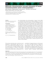
Fig. 1. Sequence analysis and genomic struc-
ture of the human sodium channel b
1B
subunit.
(A) Amino acid sequence comparisons
between human b
1B
and b
1
subunits. The
signal peptide sequence and transmembrane
domain (TM) are indicated in (A), and an
IgG-like motif is located between residues 22
and 150. (B) C-terminal amino acid sequence
comparisons of human b
1B,
rat b
1A
and
putative mouse b
1A
, which is predicated based
on the mouse genomic sequence. Conserved
residues are underlined. (C) Genomic struc-
ture of the human b
1
gene, SCN1B.The
SCN1B gene spans 9kbonchromosome19
across six exons. Exon 3A is an extended exon
3(retentionofpartofintron3)viaalternative
splicing. Exons 1, 2, 3, 4 and 5 (solid boxes)
encode the b
1
subunit, while exons 1, 2 and 3A
(solid and diagonally shaded boxes) encode
the b
1B
subunit. The 5¢ and 3¢ untranslated
regions are indicated by solid thin lines (b
1
)
andshadedthinlines(b
1B
), and the unidenti-
fied 3¢ untranslated region of b
1B
is indicated
using the thin interrupted broken line. The
stop codon is indicated by an asterisk. The
RACE-PCR primer, SB1-10 (indicated by an
arrow), is located at the end of exon 2.
Table 1. Intron–exon boundary sequence of the sodium channel b
1
/b
1B
gene (SCN1B). UT, untranslated.
Exon (bp)
cDNA
location Codon Acceptor Donor Intron (kb)
Exon 1 (> 136) ) 89 to +22 1–14 GCA CTG G– – gtgagt Intron 1 (1.67)
A L (V)
Exon 2 (166) 23–207 14–69 ccacag –TG TCC TCA TTT GTC AAG gtgtgc Intron 2 (1.80)
(V) S S F V K
Exon 3 (240) 208–458 70–149 ccctag ATC CTG CGC GAC AAA G– – gtgagt Intron 3 (5.38)
I L R D K (A)
Exon 3A (> 770) 208–978 70–268 ccctag ATC CTG CGC GTG GTT TGA xxxxxx Intron 3A (?)
I LRVV*
Exon 4 (141) 459–580 150–197 ctgcag –CC AAC AGA GAG AAT GC– gtgagt Intron 4 (0.38)
(A) N R E N (A)
Exon 5 (71) 581–662 198–218 ccacag – –C TCG GAA TAG CCC TG– gtaagg Intron 5 (0.09)
(A) S E *
Exon 6 (> 641) 663–1307 3¢ UT (b1) cttcag GCC CTG GGC
Ó FEBS 2003 Cloning and characterization of Na
+
channel b
1B
(Eur. J. Biochem. 270) 4765
shown in Fig. 1A. The open reading frame, designated b
1B
,
is related to the sodium channel b
1
subunit. Conserved
motifs of the sodium channel b subunit family were also
presented in the human sodium channel b
1B
subunit,
including a signal peptide sequence, the extracellular
immunoglobulin fold domain and the C-terminal trans-
membrane domain. The predicted peptide contained a
hydrophobic N-terminal residue (1–16 residues) with
sequences highly predictive of signal cleavage sites that
would result in mature proteins initiating at amino acid 17
(alanine). The hydrophobic C-terminal region (residues
243–262) may serve as a transmembrane domain. The
estimated protein molecular mass was 30.4 and 28.9 kDa
before and after removing the signal peptide from the N
terminus, respectively. The in vitro translated human b
1B
subunit migrated with an apparent molecular mass of
30 kDa (with signal peptide) when analyzed by 8–20%
SDS/PAGE (data not shown). Peptide sequence compar-
ison revealed that the predicted peptide was 72% identical
to both that of human (Fig. 1A) and rat sodium channel b
1
subunits and rat b
1A
subunit (Fig. 1B). Like the rat b
1A
subunit, the human b
1B
subunit contained an N-terminal
region (residues 1–149) of 100% identity to the b
1
subunit
and a novel C-terminal region (residues 150–268) with an
identity to the b
1
subunit of less than 17% (Fig. 1A). The
C-terminal region of the human b
1B
subunit was also
significantly different from the rat b
1A
and putative mouse
b
1A
subunits (The amino acid sequence of mouse b
1A
subunit is deduced from mouse genomic sequence. The
presence of such a splicing variant has not been confirmed
by any experiment.) The C-terminal portion of human b
1B
shares less than 33% and 36% peptide sequence identity
with rat and mouse b
1A
subunits, respectively, while the
same region of rat and mouse b
1A
shares at least 77%
identity (Fig. 1B).
A genomic organization study of the human sodium
channel b
1
subunit gene, SCN1B [12], revealed that the gene
spans 9 kb over six exons and five introns on chromo-
some 19 (19q13.1-q13.2).
BLAST
searches of the human
genomic database, using the cDNA sequence of human b
1B
,
revealed that the N-terminal region of the human b
1B
subunit (residues 1–149) was encoded by exons 1–3, whereas
the novel C-terminal region was encoded by the part of
intron 3 adjacent to exon 3 (Fig. 1C and Table 1). As the
site of divergence between the b
1
and b
1B
subunit cDNAs
was located precisely at the exon 3/intron 3 boundary of the
SCN1B gene, the human sodium channel b
1B
subunit
should be considered as a splicing variant of the b
1
subunit
via the extension of exon 3 to intron 3 (or partial intron 3
retention) with an in-frame stop codon.
Tissue distribution of the human b
1B
subunit
Northern blot analysis, using a human b
1B
specific probe,
showed that the b
1B
transcript is abundant in human brain
and skeletal muscle (Fig. 2A), and present at a very low level
Fig. 2. Northern blot analysis of the gene
expression of human b
1B
(A,B) and b
1
(C,D)
subunits, using human b-actin mRNA level as
the control (E,F). (A), (C) and (E) are human
multiple tissue blots; (B), (D) and (F) are
human brain II blots. The cDNA fragment
encoding residues 217–268 of the human b
1B
subunit, and the cDNA fragment encoding the
human b
1
subunit from amino acids 150 to
218, were used as probes for detecting the
messages of human b
1B
and b
1
subunits,
respectively. A 2 kb human b-actin cDNA
fragment was used as the control probe. The
blots were incubated at 42 °Covernightand
washed with 0.2· NaCl/Cit/0.1% SDS at
65 °C for 2 h. Finally, the blots were exposed
to X-ray film in a )80 °C freezer for
2–18 h.The same blots were used for all three
probes in the order b
1B
, b
1
and b-actin, after
they were stripped at 68 °C for 15 min and
reconstituted at room temperature for 15 min
using the Strip-EZ
TM
removal kit provided by
Ambion.
4766 N. Qin et al. (Eur. J. Biochem. 270) Ó FEBS 2003
in heart, placenta, lung, liver, kidney and pancreas. In
human brain, the b
1B
transcript was most abundant in the
cerebellum, followed by the cerebral cortex and occipital
lobe (Fig. 2B). The overall expression pattern of human b
1B
was very similar to that of human b
1
(Fig. 2C,D), except
that human b
1
was more abundant in cerebral cortex than in
cerebellum. If the transcript of the human b
1B
subunit is
spliced only from exon 1 (111 bp), exon 2 (185 bp), exon 3
(250 bp) and either partial or entire intron 3 (5.3 kb), the
calculated size of the transcript should be less than 6 kb.
However, the major transcript of b
1B
, as determined by
Northern blot, is 7.5 kb. This suggests that an additional
unidentified splicing event must be present to generate a
longer 3¢ untranslated region, which needs to be identified
by further experiments. In addition, a second transcript of
the human b
1B
,of 1.5 kb, was observed in skeletal
muscle.
Expression of the novel b
1B
subunit was further investi-
gated by immunohistochemistry with affinity purified anti-
b
1B
(see Experimental procedures). The anti-b
1B
was
generated against a peptide derived from the retained intron
3 in the human cDNA clone. As shown in Fig. 3,
immunohistochemical analyses revealed that the b
1B
sub-
unit was expressed in many different regions in the human
brain, including cerebellar Purkinje cells (Fig. 3A), cortex
pyramidal neurons, and many of the neuronal fibers
throughout the brain (data not shown), consistent with
the results of Northern blot analysis. Strong immunolabe-
ling was also observed in human dorsal root ganglion
(DRG) (Fig. 3C), in fibers (arrowheads) of the spinal nerve
(Fig. 3D) and in cortical neurons (large arrowheads) and
their processes (small arrowheads) (Fig. 3E). The specific
b
1B
labeling in the Purkinje cells (arrowhead) was abolished
when the primary antibody was preabsorbed with the
specific peptide.
Functional expression of the human b
1B
subunit
with Na
V
1.2 in
Xenopus
oocytes
To explore the regulatory function of the human b
1B
subunit, we injected cRNA of human b
1B
,aswellascRNA
of the sodium channel pore forming subunit Na
V
1.2, into
Xenopus oocytes. As shown in Fig. 4A–D, the rates of
activation and inactivation of the sodium current via
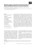
Fig. 3. Immunohistochemical analysis of b
1B
subunit expression in human tissues. (A) The presence of b
1B
in Purkinje cells (large arrowheads) and in
their processes (small arrowheads) of the cerebellum. (B) Specific b
1B
labeling was abolished in the Purkinje cells (arrowhead) when the primary
antibody was preabsorbed with the specific peptide. (C) Human b
1B
was also detected in dorsal root ganglia (large arrowheads) as well as in the
surrounding capsule cells (small arrowheads). (D) b
1B
was present in fibers (arrowheads) of the spinal nerve. (E) b
1B
was present in cortical neurons
(large arrowheads) and their processes (small arrowheads). Bar ¼ 25 lm (A, B, D, E); 50 lm(C).
Ó FEBS 2003 Cloning and characterization of Na
+
channel b
1B
(Eur. J. Biochem. 270) 4767
Fig. 4. The effect of b
1B
and b
1
subunits on the function and expression levels of the Na
V
1.2 channel expressed in Xenopus oocytes. Representative
sodium current traces from oocytes expressing the sodium channel a subunit, Na
V
1.2, in the absence (A) and presence of b
1
(B) and b
1B
(C)
subunits. Sodium currents were evoked by 15 ms long depolarizing pulses (as indicated) from a holding potential of )80 mV. (D) The effect of b
1B
and b
1
subunits on current time courses. Representative currents at )10mVinthepresenceorabsenceofb
1B
or b
1
subunits are shown normalized
to their individual peak value. (E) Inset: current-voltage relationship (I-V curve) for Na
V
1.2 alone (s)andNa
V
1.2 : hb
1B
(1 : 5) (d). Sodium
currents were evoked by 15 ms long depolarization steps, ranging from )60 to 80 mV, at 10 mV increments, from a holding potential of )100 mV.
The peak magnitude of the currents, elicited by test depolarizations to the various potentials, were measured and used to construct I-V curves. Data
represent average currents from a single batch of oocytes. Main panel: voltage dependence of the conductance (G-V curve). Data from the average
G-V curves for Na
V
1.2 alone (s), Na
V
1.2 : hb
1B
(d), and Na
V
1.2 : hb
1
(d)werefittedtoG¼ G
max
/(1 + exp[–zÆ(V-V
½
)/25]) with parameters
G
max
,z,andV,asdescribedinthetext.CurveswerenormalizedbydividingbyG
max
. (F) Voltage dependence of inactivation (steady-state
inactivation curves). Channels were inactivated by 100 ms conditioning pulses ranging from )140 to 20 mV, at 10 mV increments, then activated by
a 15 ms test pulse to 0 mV (symbols as in part E: main panel). The relative fraction of channels available for activation was measured as the peak
current during the test pulse to 0 mV. Data from individual oocytes were fitted by I ¼ I
max
/G ¼ G
max
/(1 + exp[–zÆ(V-V
½
)/25]) + I
min
and
normalized by I
m
/I
max
obtained from the fit. All data points (E, F) correspond to the mean ± SEM of the averaged normalized currents for the
number of oocytes indicated. (G) Effect of b
1B
on the current amplitude of Na
V
1.2. Each individual data point in the histogram represents the peak
inward current for a single oocyte. Also shown are the mean (j) and standard deviation (bars) for each cRNA a:b ratio, and the sample size per
cRNA combination is shown in parenthesis. Data are from five different batches of oocytes, each batch injected with cRNA for the a subunit alone
or with the human b
1B
subunitatratiosof1 :5and1:20.Unpairedt-test statistical analysis resulted in P-values of 0.056 (a vs. a:b
1B
; 1 : 5), 1.3e-5
(a vs. a:b
1B
;1:20)and0.035(a:b
1B
1:5vs.a:b
1B
;1:20).
4768 N. Qin et al. (Eur. J. Biochem. 270) Ó FEBS 2003
Na
V
1.2 did not change significantly in the presence or
absence of the b
1B
subunit, whereas, under the same
conditions, the rate of inactivation was increased in the
presence of the b
1
subunit. The effects of b
1B
were further
assessed by studying the current-voltage (I-V) relationships,
the voltage-dependence of the conductance (G-V curve),
and the voltage dependence of steady-state inactivation.
Except for a minor negative shift (3–4 mV) in the voltage-
dependence of activation (Fig. 4E), no significant effect of
the human b
1B
subunit on the regulation of Na
V
1.2 sodium
channel properties was observed (Fig. 4E,F, and Table 2).
Under the same conditions, the human b
1
subunit also
shifted the G-V relationship left, to a similar extent (Fig. 4E
and Table 2), but caused a significant shift of the steady
state inactivation curve by 10 mV, towards more negative
potentials (Fig. 4F and Table 2). Although no significant
modulatory effect of the b
1B
subunit on channel kinetics and
steady-state properties was observed, we found that the b
1B
subunit increased the ionic current conducted by Na
V
1.2
sodium channels (e.g. Fig. 4E, inset). At cRNA ratios of
1:5 and 1:20 (Na
V
1.2 : b
1B
), the average (n ¼ 16–22)
peak ionic current densities were increased by two- and
threefold, respectively (Fig. 4G). Despite significant vari-
ability in current densities within and between batches of
oocytes, statistical analysis indicated that the difference
between Na
V
1.2 expressing oocytes and those expressing
Na
V
1.2 : b
1B
at a ratio of 1 : 20 was significant
(P < 0.0001).
Discussion
We report here the cloning and characterization of the
human VGSC b
1B
subunit. The human b
1B
subunit is a
novel splicing variant of the b
1
subunit via alternative
intron 3 retention. The retained intron encodes a novel
extracellular, a transmembrane, and an intracellular region,
sharing little homology with the human and rat b
1
(17%
identity) and the rat b
1A
(33% identity) subunits. Although
the novel b
1B
subunit has a structure similar to other sodium
channel b subunits, it exhibits regulatory properties in
Xenopus oocytes that distinguish it from the b
1
and b
1A
subunits.
It is interesting that the only regulatory function of the
b
1B
subunit, observed in this study, was its ability to increase
the sodium current density when coexpressed with the
tetrodotoxin sensitive channel, Na
V
1.2, in oocytes without
affecting any of its voltage dependent properties. Several
previous studies have shown that the b
1
subunit not only
increases the levels of functional sodium channel on the cell
surface, but that it also changes voltage dependent activa-
tion and inactivation [3,4,13]. In the present study, we also
observed that the simultaneous injection of b
1
with Na
V
1.2
into oocytes resulted in an increase of the inactivation rate
and a shift of the steady state inactivation curve to a more
negative potential ( 10 mV), as well as an increase in ionic
current amplitude (not shown), consistent with other studies
in oocytes [3]. However, under the same conditions, the b
1B
subunit had little effect on the properties of Na
V
1.2. The
increase in ionic currents induced by coexpression of the
human b
1B
could result from an increase in the number of
channels present in the membrane, N
o
,anincreaseinthe
probability of opening of the channels, P
o
, and/or an
increase in the single channel conductance, c
o
. Discrimin-
ation among these options, however, requires evaluation of
single channel parameters by other means (single channel
recording or mean-variance analysis).
The modulatory property of the b
1B
subunit is also
different from that of the b
1A
subunit reported by Isom’s
group. In their studies [7], the coexpression of b
1A
with
Na
V
1.2 in Chinese hamster lung cells resulted in a 2.5-fold
increase in the sodium current density, slightly shifted the
steady state inactivation curve to a more positive potential
(which also distinguishes it from the b
1
subunit) and had no
effect on channel activation. However, we are unable to rule
out the possibility that the different regulatory properties
observed between rat b
1A
and human b
1B
results from the
use of different expression systems in the two studies.
Although b
1
, b
1A
and b
1B
have different effects on Na
V
1.2
channel properties (b
1
affects both activation and inactiva-
tion, b
1A
affects inactivation only, and b
1B
has no effect on
either), the subunits all share a common regulatory prop-
erty, i.e. they increase the sodium current density regardless
of expression system. These results suggest that alteration of
channel kinetics and steady state properties may be a
function distinct from the increase in current density on the
cell surface induced by b
1
subunits. The functional differ-
ences of the b
1
, b
1A
and b
1B
subunits suggest that the
C-terminal half of b
1
and its splicing variants play an
important role in the modulation of sodium channel
properties. Based on the sequence differences between b
1
and its splicing variants, there are three regions on the
human b
1B
subunit that may alter its regulatory properties
(a) the additional extracellular region ( 90 residues in the
human b
1B
vs. 55 residues in rat b
1A
), (b) the transmem-
brane domain, and (c) the intracellular region. The trans-
membrane region of the human b
1B
is located at the C
terminus (241–262 residues) with five intracellular residues.
This unique structure of the human b
1B
subunit is very
similar to that of the calcium channel a
2
d subunit, which
also has six residues downstream from the transmembrane
Table 2. Steady-state properties of the sodium channel Na
V
1.2 in the presence or absence of the b
1B
or b
1
subunit.
Activation:
G=G
max
/(1 + [exp()z
*
(V)V
1/2
)/25])
Inactivation:
I=I
max
/(1 + [exp()z
*
(V)V
1/2
)/25]) + I
min
G
max
zV
1/2
(mV) I
max
zV
1/2
(mV) I
min
NaV
1/2
1.00 4.18 )9.94 1.00 )2.63 )36.11 )0.03
NaV
1/2
/b
1
1.00 4.19 )12.14 0.98 3.37 )48.46 )0.01
NaV
1/2
/b
1B
1.00 4.20 )10.92 1.00 )2.70 )37.75 )0.02
Ó FEBS 2003 Cloning and characterization of Na
+
channel b
1B
(Eur. J. Biochem. 270) 4769
domain serving as an intracellular segment [14]. To date, no
regulatory function related to the single transmembrane
domain and the short, five-residue intracellular segment of
the calcium channel a
2
d subunit has been reported, except
that the transmembrane domain is essential for anchoring
the protein into the membrane [14]. Therefore, the addi-
tional extracellular region is probably responsible for the
differences of regulatory functions between the b
1B
and b
1
subunit or rat b
1A
subunit. Recently, Meadows et al.[15]
reported that the intracellular segment of the b
1
subunit is
required for the interaction with the a subunit, probably a
crucial step for the regulation of channel properties.
The tissue distribution of the human b
1B
subunit is similar
to that of the human b
1
subunit. Its message was detected in
skeletal muscle and in a variety of subregions in the brain
(Figs 2 and 3). More interestingly, the human b
1B
subunit is
also expressed in DRG neuron and fibers (Fig. 3). It is well
known that the number of functional sodium channels and
magnitude of sodium currents are differentially changed
following peripheral nerve injury [16–19]. Our observations
of the existence of the b
1B
subunit in human DRG, and its
ability to increase sodium current density when coexpressed
with Na
V
1.2a in Xenopus oocytes, suggest that the human
b
1B
subunit may be another candidate useful for studying
the mechanism of upregulation of functional sodium
channels on the cell surface and increasing the rate of
spontaneous firing in peripheral neurons after nerve injury.
It will be interesting to determine whether the human b
1B
subunit is up-regulated, and whether its up-regulation is
correlated with the increase of sodium channel activity, in
injured human DRG neuron.
Acknowledgements
We thank Drs Mike X. Zhu, Rich R. Ryan and Yi Liu for their critical
discussion of the manuscript, Ms S. Yagel for her help in subcloning,
and Ms Patti A. Reiser, Norah A. Gumula, Brenda M. Hertzog and
Debbie Polkovitch for their histological and immunohistochemical
expertise.
References
1. Catterall, W.A. (1993) Structure and modulation of Na
+
and
Ca
2+
channels. Ann. NY Acad. Sci. 707, 1–19.
2. Isom, L.L., De Jongh, K.S., Patton, D.E., Reber, B.F., Offord, J.,
Charbonneau, H., Walsh, K., Goldin, A.L. & Catterall, W.A.
(1992) Primary structure and functional expression of the beta 1
subunit of the rat brain sodium channel. Science 256, 839–842.
3. Isom, L.L., Ragsdale, D.S., De Jongh, K.S., Westenbroek, R.E.,
Reber, B.F., Scheuer, T. & Catterall, W.A. (1995) Structure and
function of the beta 2 subunit of brain sodium channels, a trans-
membrane glycoprotein with a CAM motif. Cell 83, 433–442.
4. Morgan, K., Stevens, E.B., Shah, B., Cox, P.J., Dixon, A.K., Lee,
K., Pinnock, R.D., Hughes, J., Richardson, P.J., Mizuguchi, K. &
Jackson, A.P. (2000) Beta 3: an additional auxiliary subunit of the
voltage-sensitive sodium channel that modulates channel gating
with distinct kinetics. Proc.NatlAcad.Sci.USA97, 2308–2313.
5. Catterall, W.A. (1992) Cellular and molecular biology of voltage-
gated sodium channels. Physiol. Rev. 72, S15–S48.
6. Isom, L.L., De Jongh, K.S. & Catterall, W.A. (1994) Auxiliary
subunits of voltage-gated ion channels. Neuron 12, 1183–1194.
7. Kazen-Gillespie, K.A., Ragsdale, D.S., D’Andrea, M.R., Mattei,
L.N., Rogers, K.E. & Isom, L.L. (2000) Cloning, localization, and
functional expression of sodium channel beta1A subunits. J. Biol.
Chem. 275, 1079–1088.
8. D’Andrea, M.R., Derian, C.K., Leturcq, D., Baker, S.M., Brun-
mark, A., Ling, P., Darrow, A.L., Santulli, R.J., Brass, L.F. &
Andrade-Gordon, P. (1998) Characterization of protease-acti-
vated receptor-2 immunoreactivity in normal human tissues.
J. Histochem. Cytochem. 46, 157–164.
9. Goldin, A.L. (1992) Maintenance of Xenopus laevis and oocyte
injection. Methods Enzymol. 207, 266–279.
10. Taglialatela, M., Toro, L. & Stefani, E. (1992) Novel voltage
clamp to record small, fast currents from ion channels expressed in
Xenopus oocytes. Biophys. J. 61, 78–82.
11. Stefani, E. & Bezanilla, F. (1998) Cut-open oocyte voltage-clamp
technique. Methods Enzymol. 293, 300–318.
12. Makita, N., Sloan-Brown, K., Weghuis, D.O., Ropers, H.H. &
George, A.L. Jr (1994) Genomic organization and chromosomal
assignment of the human voltage-gated Na
+
channel beta 1
subunit gene (SCN1B). Genomics 23, 628–634.
13. Isom, L.L., Scheuer, T., Brownstein, A.B., Ragsdale, D.S., Mur-
phy, B.J. & Catterall, W.A. (1995) Function co-expression of the
b1 and type IIA a subunits of sodium channels in a mammalian
cell line. J. Biol. Chem. 270, 3306–3312.
14. Gurnett, C.A., De Waard, M. & Campbell, K.P. (1996) Dual
function of the voltage-dependent Ca
2+
channel alpha 2 delta
subunit in current stimulation and subunit interaction. Neuron 16,
431–440.
15. Meadows, L., Malhotra, J.D., Stetzer, A., Isom, L.L. & Ragsdale,
D.S. (2001) The intracellular segment of the sodium channel beta 1
subunit is required for its efficient association with the channel
alpha subunit. J. Neurochem. 76, 1871–1878.
16. Tanaka, M., Cummins, T.R., Ishikawa, K., Dib-Hajj, S.D., Black,
J.A. & Waxman, S.G. (1998) SNS Na
+
channel expression
increases in dorsal root ganglion neurons in the carrageenan
inflammatory pain model. Neuroreport 9, 967–972.
17. Okuse, K., Chaplan, S.R., McMahon, S.B., Luo, Z.D., Calcutt,
N.A., Scott, B.P., Akopian, A.N. & Wood, J.N. (1997) Regulation
of expression of the sensory neuron-specific sodium channel SNS
in inflammatory and neuropathic pain. Mol. Cell. Neurosci. 10,
196–207.
18. Novakovic, S.D., Eglen, R.M. & Hunter, J.C. (2001) Regulation
of Na
+
channel distribution in the nervous system. Trends Neu-
rosci. 24, 473–478.
19. Cummins, T.R. & Waxman, S.G. (1997) Downregulation of
tetrodotoxin-resistant sodium currents and upregulation of a
rapidly repriming tetrodotoxin-sensitive sodium current in small
spinal sensory neurons after nerve injury. J. Neurosci. 17, 3503–
3514.
4770 N. Qin et al. (Eur. J. Biochem. 270) Ó FEBS 2003