Báo cáo khoa học: In vitro gamma-secretase cleavage of the Alzheimer’s amyloid precursor protein correlates to a subset of presenilin complexes and is inhibited by zinc potx
Bạn đang xem bản rút gọn của tài liệu. Xem và tải ngay bản đầy đủ của tài liệu tại đây (478.92 KB, 14 trang )
In vitro gamma-secretase cleavage of the Alzheimer’s
amyloid precursor protein correlates to a subset
of presenilin complexes and is inhibited by zinc
David E. Hoke, Jiang-Li Tan, Nancy T. Ilaya, Janetta G. Culvenor, Stephanie J. Smith,
Anthony R. White, Colin L. Masters and Genevie
`
ve M. Evin
Department of Pathology, The University of Melbourne and the Mental Health Research Institute, Parkville, Victoria, Australia
Gamma-secretase is an aspartyl protease that cleaves
type I integral membrane proteins intramembranously.
The amyloid precursor protein (APP) undergoes
sequential cleavages by beta-site APP cleaving enzyme
and c-secretase to form amyloid-b (Ab). The beta-site
APP cleaving enzyme cleavage releases an APP ecto-
domain leaving a 99-amino acid membrane spanning
C-terminal fragment (CTF), C99. C99 then undergoes
intramembranous cleavage to form Ab peptides of
different lengths. c-secretase also releases an APP
intracellular domain (AICD or e-CTF) by cleaving 9–7
amino acids from c 40 and 42 sites at the e site [1–3].
Several lines of evidence support the pathogenic role
of c-cleavage of APP in Alzheimer’s disease (AD). The
genes encoding presenilin 1 and 2 (PS) are essential for
c-secretase activity and 150 mutations in the PS genes
have been found associated with autosomal dominant
early onset familial AD [4]. Although only 16 muta-
tions have been found in the APP gene, the majority
linked to early onset familial AD occur in the
Keywords
Alzheimer’s disease; amyloid precursor
protein; gamma-secretase; amyloid beta
Correspondence
D. E. Hoke, Department of Microbiology,
Monash University, Clayton, Vic 3800,
Australia
Fax: +61 39905 4811
Tel: +61 39905 4807
E-mail:
G. M. Evin, Department of Pathology, The
University of Melbourne, Parkville, Vic 3010,
Australia
Fax: +61 38344 4004
Tel: +61 38344 4205
E-mail:
(Received 20 May 2005, revised 4 August
2005, accepted 30 August 2005)
doi:10.1111/j.1742-4658.2005.04950.x
The c-secretase complex mediates the final proteolytic event in Alzheimer’s
disease amyloid-b biogenesis. This membrane complex of presenilin, ante-
rior pharynx defective, nicastrin, and presenilin enhancer-2 cleaves the
C-terminal 99-amino acid fragment of the amyloid precursor protein intra-
membranously at c-sites to form C-terminally heterogeneous amyloid-b
and cleaves at an e-site to release the intracellular domain or e-C-terminal
fragment. In this work, two novel in vitro c-secretase assays are developed
to further explore the biochemical characteristics of c-secretase activity.
During development of a bacterial expression system for a substrate based
on the amyloid precursor protein C-terminal 99-amino acid sequence, frag-
ments similar to amyloid-b and an e-C-terminal fragment were observed.
Upon purification this substrate was used in parallel with a transfected
source of substrate to measure c-secretase activity from detergent extracted
membranes. With these systems, it was determined that recovery of size-
fractionated cellular and tissue-derived c-secretase activity is dependent
upon detergent concentration and that activity correlates to a subset of
high molecular mass presenilin complexes. We also show that by changing
the solvent environment with dimethyl sulfoxide, detection of e-C-terminal
fragments can be elevated. Lastly, we show that zinc causes an increase in
the apparent molecular mass of an amyloid precursor protein c-secretase
substrate and inhibits its cleavage. These studies further refine our know-
ledge of the complexes and biochemical factors needed for c-secretase
activity and suggest a mechanism by which zinc dysregulation may contrib-
ute to Alzheimer’s disease pathogenesis.
Abbreviations
AD, Alzheimer’s disease; APP, amyloid precursor protein; CTF, C-terminal fragment; NTF, N-terminal fragment; PS, presenilin.
5544 FEBS Journal 272 (2005) 5544–5557 ª 2005 FEBS
transmembrane region near c and e cleavage sites (as
reviewed in [5]). Gamma-secretase activity is attributed
to an integral membrane complex of the four trans-
membrane proteins: PS, nicastrin (Nct), anterior pha-
rynx-defective, and presenilin enhancer 2 (as reviewed
in [6]).
In vitro c-secretase assays have been essential in elu-
cidating the mechanism of inhibitors [7–9], the struc-
ture of active c-secretase complexes [10,11], and have
aided in the finding of activity-modulating factors
[12,13]. These assays have shown that peripheral mem-
brane proteins are not necessary for activity as car-
bonate washing retains activity [14]. Additionally,
detergent solubilization has allowed solution-based
biochemical manipulation to show that all four of the
genetically determined c-secretase components interact
to form high molecular mass, enzymatically active
c-secretase complexes [10,11,15]. In this paper we des-
cribe two novel in vitro c-secretase assays that differ in
substrate and enzyme source to monitor c-secretase
activity without the need to overexpress the c-secretase
complex components. These assays are used to test the
effects of detergent concentration, solvents and metals
on the c-secretase cleavage of APP substrates.
These studies show that extracts from Escherichia
coli transformed with a c-secretase substrate contain
products similar to those expected from c-secretase
cleavage. Furthering the characterization of c-secretase
activity, we show that the detergent concentration used
during gel filtration affects the recovery of activity.
These studies also show that dimethylsulfoxide is a
solvent that allows greater detection of c-secretase
activity. Lastly, zinc causes structural changes in a
c-secretase substrate and acts as an inhibitor of c-
secretase cleavage of APP.
Results
Design of a novel APP c-secretase substrate and
standards
Sensitive western blot assays for c-secretase were
based on the production of a 3FLAG-tagged e-CTF
from an APP substrate. An E. coli expression vector
was made to encode a starting methionine, the C-ter-
minal 99 amino acids of APP and a C-terminal triple
FLAG tag. The resulting protein was named MC99-
3FLAG (Fig. 1A). Escherichia coli expression vectors
encoding 3FLAG-tagged APP-CTFs mimicking prod-
ucts from cleavage at position 40 (gamma-3FLAG
standard) and 49 (epsilon-3FLAG standard) were also
made to aid in the identification of 3FLAG-tagged
CTFs (Fig. 1A).
Ab-like and e-CTF-like products are present in
extracts from E. coli expressing MC99-3FLAG
Upon expression of MC99-3FLAG in E. coli,we
observed three predominant anti-FLAG immunoreact-
ive peptides. The major product migrated at 18 kDa
and was also detected by anti-Ab antibody, WO2.
From its apparent molecular mass and its immunore-
activity, it can be concluded that it corresponds to
MC99-3FLAG (Fig. 1B and C). There were also sev-
eral higher molecular mass and degraded species iden-
tified by both antibodies. The higher molecular mass
forms may correspond to aggregated MC99-3FLAG.
One anti-FLAG immunoreactive peptide migrated
similarly to the gamma and epsilon standards at
9 kDa (Fig. 1B) and the corresponding N-terminal
fragment resembling Ab was identified by WO2 west-
ern blot analysis (Fig. 1C). Further identification of
the anti-FLAG-immunoreactive CTFs was made by
coelectrophoresis with gamma and epsilon standards.
Co-electrophoresis obviates subtle lane-to-lane varia-
tions that may occur for these low molecular mass
proteins. The 9-kDa peptide comigrated with the
e-3FLAG standard (Fig. 1D) but faster than the
c-3FLAG standard (Fig. 1E). No anti-FLAG immuno-
reactivity was detected in lysates of mock-transformed
cells (Fig. 1E). Collectively, these data indicate that
upon expression or during purification, a small frac-
tion of MC99-3FLAG is degraded into multiple spe-
cies including peptides resembling products expected
from c-secretase cleavage.
Development of in vitro c-secretase assays using
purified MC99-3FLAG as a substrate
Peptides similar to an e-CTF present in substrate prep-
arations would interfere with the detection of e-CTF
production from mammalian tissue extracts. Therefore
a purification strategy was devised to minimize this
contamination. An initial nondenaturing size-exclusion
chromatography step was performed to separate
MC99-3FLAG from lower molecular mass fragments.
Unexpectedly, MC99-3FLAG eluted at the void vol-
ume of the column while fragments eluted according
to their apparent molecular mass determined by
SDS ⁄ PAGE and western blot analysis (data not
shown). These MC99-3FLAG enriched void volume
fractions were purified in a second-step by anti-FLAG
chromatography. This two-step purified material was
used in c-secretase assays. MC99-3FLAG was tes-
ted for cleavage by c-secretase from PS1A246E trans-
genic mouse brain [16] (Fig. 2A) prepared by
solubilization of carbonate-washed membranes with
D. E. Hoke et al. Characterization of in vitro c-secretase activity
FEBS Journal 272 (2005) 5544–5557 ª 2005 FEBS 5545
1% [3-[(3-cholamidopropyl)dimethylammonio-]-2-hyd-
roxy-1-propanesulfonate] (CHAPSO). Upon incuba-
tion at 37 °C, generation of an 9 kDa CTF was
detected by western blotting with anti-FLAG anti-
body. The c-secretase inhibitor L-685,458 was used to
confirm that the fragment detected was produced by
c-secretase activity. L-685,458 inhibited formation of
this e-CTF-3FLAG in a dose-dependent manner, with
an effect still observed at concentrations as low as
3.3 nm, consistent with previous reports [17]. The
preparation of MC99-3FLAG contained additional
anti-FLAG immunoreactive peptides but these did not
interfere with the assay (Fig. 2A, long exposure).
Assay sensitivity was tested by varying the enzyme
amount, enzyme dilution, and CHAPSO concentra-
tion. For this experiment, c-secretase activity was pre-
pared by extracting carbonate-washed guinea pig brain
membranes with 1% CHAPSO. A 2 mgÆmL
)1
extract
was diluted to obtain a final concentration of 0.5%
CHAPSO, and dilutions were made in 0.5% CHAPSO
to 20 lgÆmL
)1
. Incubation of these dilutions with sub-
strate showed c-secretase activity to be enzyme dose-
dependent, and that signal was detectable using the
40 lgÆmL
)1
dilution of extract. Therefore, as little as
1 lg of membrane extract was sufficient to obtain a
signal (Fig. 2B). Secondly, the 2 mgÆ mL
)1
extract was
A
BC
DE
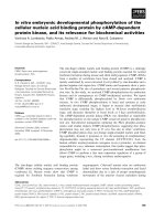
Fig. 1. Escherichia coli produces peptides
similar to Ab and an e-CTF when trans-
formed with MC99-3FLAG. (A) Schematic of
proteins. (B) Lysate from MC99-3FLAG-
expressing E. coli, c and e standards were
separated by SDS ⁄ PAGE and western blot-
ted for FLAG immunoreactivity. MC99-
3FLAG migrates between the 20 and
14-kDa molecular mass markers. One lower
molecular mass FLAG immunoreactive
protein has a mobility similar to the c and e
standards. (C) Lysate from E. coli trans-
formed with the MC99-3FLAG expression
vector was probed with monoclonal anti-
body WO2, directed to the N-terminal region
of Ab. Besides MC99-3FLAG, this antibody
detected several peptides of higher and
lower molecular masss, one of them with a
similar mobility as synthetic Ab40. (D) Anti-
FLAG western blot analysis of lysate from
MC99-3FLAG-transformed E. coli alone or
spiked with different amounts of the
e-3FLAG standard. An uncharacterized anti-
FLAG immunoreactive protein migrating just
below the e-CTF-like peptide is indicated
by *. Note that ‘e std.’ migrated identically
to the E. coli product marked ‘e’. (E) A sim-
ilar experiment to that shown in (D) was
performed by spiking MC99-3FLAG-trans-
formed E. coli lysate with c-3FLAG stand-
ard. This standard migrated slower than the
E. coli product. Mock-transformed E. coli
had no background anti-FLAG immuno-
reactivity.
Characterization of in vitro c-secretase activity D. E. Hoke et al.
5546 FEBS Journal 272 (2005) 5544–5557 ª 2005 FEBS
diluted to 0.25% CHAPSO and subsequently diluted
in 0.25% CHAPSO to 40 lgÆmL
)1
. In contrast to the
0.5% CHAPSO dilution, 0.25% dilution did not show
a dose-dependent relation of enzyme amount to prod-
uct formed (Fig. 2C). Rather the highest concentration
and amount showed little activity while the least con-
centrated sample (1.2 lgof40lgÆmL
)1
) showed the
highest activity and greater than the corresponding
dilution in 0.5% CHAPSO. Perhaps this 0.25% con-
centration was not enough to keep high concentrations
of extract solubilized leading to an apparent loss of
activity. Collectively, these data show that two-step
purified MC99-3FLAG is an appropriate substrate to
study tissue-derived c-secretase activity that is inhibited
by a specific c-secretase inhibitor and is detergent con-
centration-sensitive.
Mammalian expression and proteolytic
processing of SPC99-3FLAG
An alternative approach to monitoring c-secretase
activity was developed using a novel mammalian
expression vector. The SPA4CT sequence, which cor-
responds to the C-terminal 99 amino acids of human
APP fused to the APP signal peptide [18], was ligated
into a C-terminal 3-FLAG repeat expression vector
and the resulting construct, SPC99-3FLAG (Fig. 3A),
was used for expression of c-secretase substrate in
mammalian cells. Anti-FLAG western blot analysis of
COS-7 cells transfected with SPC99-3FLAG (COS-7-
SPC99-3FLAG cells) shows the expected cleavage
product by signal peptidase (Fig. 3B and C). Previous
data with the SPA4CT construct showed that signal
peptidase cleavage resulted in a 101 amino acid protein
with the amino acids LE fused to the N terminus of
Ab [19], thus the protein was named C101-3FLAG.
C-terminal fragments produced from COS7-SPC99-
3FLAG cells were analysed by co-electrophoresis of
cell lysates with c-3FLAG or e-3FLAG standards.
Anti-FLAG western blot analysis shows that the CTF
from COS7-SPC99-3FLAG cells has an electrophoretic
mobility indistinguishable from that of e-3FLAG
standard (Fig. 3B, lane 1) but a slightly faster mobility
than the c-3FLAG standard (Fig. 3C, lane 1). Using
C101-3FLAG, c-3FLAG and e-3FLAG standards as
molecular mass markers, the FLAG-reactive band
migrating below C101-3FLAG is calculated to be a
protein resulting from the expected a-secretase cleav-
age [20] (see Experimental procedures). Lastly, longer
exposures allowed the detection of a protein with a
calculated molecular mass of 11.1 kDa migrating
between a- and c-3FLAG standard proteins that may
correspond to a minor a-secretase cleavage product [1]
(Fig. 3C). Therefore, SPC99-3FLAG is expressed in
mammalian cells as a C101-3FLAG protein that
undergoes the expected processing by a- and c-cleav-
ages to produce a CTF corresponding to cleavage at
the e-site.
AB
C
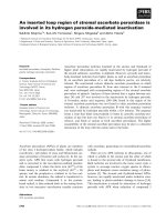
Fig. 2. Detection of c-secretase activity in tissue extracts using exogenous MC99-3FLAG substrate. (A) Purified MC99-3FLAG substrate was
added to 0.5% CHAPSO soluble c-secretase from PS1 A246E transgenic mouse brain and incubated at 37 or 4 °C for 15 h with or without
L-685,458 inhibitor. These reactions were analysed by anti-FLAG western blot analysis. Gamma-secretase activity was defined as the gen-
eration of e-CTF-3FLAG (e) signal upon incubation at 37 °C over background 4 °C levels; this was inhibited by L685,458 in a dose-dependent
manner. Longer exposures showed a contaminating CTF (indicated by *) that migrated slightly faster than the e-CTF. (B) Sensitivity of exo-
genous substrate c-secretase assay in the presence of 0.5% CHAPSO. Guinea pig brain soluble c-secretase was diluted to 0.5% CHAPSO
and further dilutions in 0.5% CHAPSO were incubated with MC99-3FLAG at 37 or 4 °C for 15 h. The reactions were analysed by anti-FLAG
Western blot. Gamma-secretase activity was detected using as little as 1 lg of membrane extract. (C) A similar experiment to that in (B)
except that guinea pig brain soluble c-secretase was diluted to 0.25% CHAPSO with further dilutions in 0.25% CHAPSO incubated with
MC99-3FLAG. The most highly concentrated reaction shows little activity while the lowest concentration shows the greatest activity.
D. E. Hoke et al. Characterization of in vitro c-secretase activity
FEBS Journal 272 (2005) 5544–5557 ª 2005 FEBS 5547
In vitro c-secretase assay with COS7-SPC99-
3FLAG solubilized membranes
To complement our MC99-3FLAG based in vitro
c-secretase assays, an in vitro assay using COS7-
SPC99-3FLAG CHAPSO extracts was developed. The
substrate in this assay is synthesized, processed, and
trafficked in the cell and would theoretically be presen-
ted to the c-secretase complex in a more native state
than E. coli-derived substrate. Anti-FLAG western
blot analysis was used to monitor the generation of
e-CTF. Upon 16 h incubation of a 0.5% CHAPSO-
solubilized membrane preparation at 37 °C, a robust
e-CTF signal was detected while a similar signal was
not observed upon incubation at 4 °C (Fig. 3D). This
activity was inhibited in a dose-dependent manner by
the c-secretase inhibitor L-685,458 with a similar
potency as seen for the MC99-3FLAG-based assay
(Fig. 2A) and previous reports [17]. The sensitivity of
the COS-7 SPC99-3FLAG c-secretase assay was
explored in relation to extract amount and CHAPSO
content. e -C-terminal fragment production could be
detected in a dose-dependent fashion with as little
as 2 lg of cell membrane extract diluted in 0.5%
CHAPSO (Fig. 3E). Using COS7-SPC99-3FLAG
extracts diluted in 0.25% CHAPSO, dose-dependent
c-secretase activity was detected but the sensitivity was
increased, allowing activity to be detected from 1 lgof
extract (Fig. 3F). Collectively these data show that
COS7-SPC99-3FLAG extracts can be used to mon-
itor c-secretase activity and that this activity is sensi-
tive to CHAPSO concentration.
PS molecular mass and c-secretase activity from
COS7-SPC99-3FLAG cells is altered by size
exclusion chromatography in a CHAPSO
concentration-dependent fashion
Much controversy exists within the literature concern-
ing the molecular mass of c-secretase complexes and
activity. Since our assays measure activity without the
need of overexpressing the c-secretase complex compo-
nents and are highly sensitive under diluting conditions,
we set out to determine the molecular mass of activity
by size exclusion chromatography. Unlike blue native
PAGE, this method allows the simultaneous determin-
ation of c-secretase complexes size and activity. A 1%
CHAPSO extract from COS-7-SPC99-3FLAG cells
A
DEF
BC
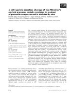
Fig. 3. Expression of SPC99-3FLAG in COS-7 cells and detection of c-secretase activity in whole-cell and cell-free assays. (A) Schematic of
the SPC99-3FLAG protein. (B) Anti-FLAG Western blot analysis of extracts from COS7-SPC99-3FLAG cells. Lane 1, spiked with e-3FLAG
standard; Lane 2, lysate sample alone. Note that the intensity of the e-CTF-3FLAG band was greater in lane 1 spiked with e-3FLAG standard.
(C) A similar experiment to that in (B) was performed except that lane 1 is a sample spiked with c-3FLAG standard indicated by the arrow
marked ‘c std’. Lane 2, lysate sample alone. Note that a separation between c-3FLAG standard and e-CTF-3FLAG was achieved in lane 1.
An uncharacterized anti-FLAG immunoreactive protein migrating between the a and c peptides was detected on long exposures (indicated
by the arrow on the right of panel C). (D) In vitro assay with CHAPSO-solubilized c-secretase from COS7-SPC99-3FLAG cells. 1% CHAPSO
extracts from COS7-SPC99-3FLAG cells were diluted to 0.5% and incubated at 37 or 4 °C for 16 h with dimethylsulfoxide or the c-secretase
inhibitor L-685,458 at the concentrations indicated. The reactions were analysed by anti-FLAG Western blot. Note that activity was abolished
in a dose-dependent manner upon addition of L-685,458. (E) Sensitivity of COS-7-SPC99-3FLAG soluble c-secretase assay in 0.5% CHAPSO:
4, 2, or 1 lg soluble c-secretase was diluted to 0.5% CHAPSO, incubated at 37 or 4 °C and analysed by anti-FLAG Western blot. Note that
e-CTF production was detected from 2 lg of membrane extract. (F) COS7-SPC99-3FLAG soluble c-secretase was diluted to 0.25% CHAPSO
and 2, 1, and 0.5 lg of extract tested for activity. Note that faint activity was seen with 1 lg extract.
Characterization of in vitro c-secretase activity D. E. Hoke et al.
5548 FEBS Journal 272 (2005) 5544–5557 ª 2005 FEBS
was diluted to 0.5% CHAPSO and chromatographed
on a Superose 6 column equilibrated with 0.5%
CHAPSO. This CHAPSO concentration was chosen as
it is compatible with c-secretase activity (as shown in
Fig. 3E) and it results in a lesser dilution of sample
than the previously published 0.25% CHAPSO con-
centration [21]. Because the c-secretase complex com-
ponents were endogenous, only low amounts were
present such that detection by western blot analysis
was limited to our most sensitive assay for PS1
N-terminal fragment (NTF). Fractions were analysed
for the presence of C101-3FLAG and PS1 NTF and
the signals quantified by image densitometry (Fig. 4A).
A broad peak of C101-3FLAG immunoreactivity was
found in fractions corresponding to 440–25 kDa while
PS1 NTF was detected in a 669-kDa peak. These data
indicate that very little of the substrate co-fractionates
with c-secretase complexes.
Gamma-secretase activity from 0.5% CHAPSO
columns was tested by pooling fractions, adding
phospholipids, and incubating at 37 or 4 °C, followed
by immunoprecipitation with anti-FLAG agarose. No
generation of e-CTF was observed in any of the
pooled fractions. We hypothesized that not enough
substrate cofractionated with PS complexes to allow
the production of a detectable signal. However, when
exogenous MC99-3FLAG substrate and phospholipid
was added to fractions, activity was not detected.
Thus, substrate limitation is not the reason that PS1
complexes of this size range were unable to sustain
robust c-secretase activity.
Original reports on the size of PS1 and c-secretase
activity by size exclusion chromatography showed
that both eluted at the void volume [21]. However,
these authors used 0.25% CHAPSO during column
chromatography. As we observed that the CHAPSO
concentration had an effect on c-secretase activity in
unseparated materials, we repeated the size exclusion
experiment in the presence of 0.25% CHAPSO. Immu-
noblots for PS1 NTF showed elution at the void vol-
ume (Fig. 4B), a result in contrast to the 669-kDa
peak obtained with chromatography in presence of
0.5% CHAPSO. Because most of C101-3FLAG
immunoreactivity was again found in fractions
between 440 and 25 kDa, separate from the fractions
containing PS1, c-secretase activity acting upon trans-
fected C101-3FLAG was not tested. Rather, fractions
were tested by adding exogenous MC99-3FLAG and
phospholipids (Fig. 4C). Under these conditions, the
fractions eluting at the void volume were able to pro-
duce a strong e-CTF signal upon incubation at
37 °C. It was noted that activity did not directly cor-
relate to the amount of PS1-NTF present in these
pooled fractions. These results indicate that endo-
genous c-secretase activity from COS-7 cells is associ-
ated with a CHAPSO concentration-sensitve complex
A
B
C
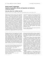
Fig. 4. Superose 6 size fractionation of COS7-SPC99-3FLAG soluble
c-secretase. (A) Sixty-seven micrograms of 1% CHAPSO cell mem-
brane extract was diluted to 0.5% CHAPSO and loaded onto a
Superose 6 column equilibrated in 0.5% CHAPSO. Arrows at the
top indicate elution of molecular mass standards. PS1 NTF fractio-
nates in 669-kDa fractions while C101-3FLAG fractionates between
440 and 25 kDa. (B) Twenty micrograms of CHAPSO extract from
COS-7 SPC99-3FLAG cells was diluted to 0.25% CHAPSO and
applied to a Superose 6 column equilibrated in 0.25% CHAPSO.
Presenilin-1 NTF immunoreactivity was detected from column frac-
tions with a peak near the void volume. C101-3FLAG immuno-
reactivity was detected as a peak between 440 and 25 kDa. (C)
Fractions from (B) were assayed for c-secretase activity using exo-
genous MC99-3FLAG substrate and phospholipids as described.
These reactions were incubated at 37 or 4 °C for 17 h and analysed
by anti-FLAG Western blot. Lanes containing the e-3FLAG standard
are indicated by ‘e’. Gamma-secretase activity was detected in void
volume fractions only. Note that e-3FLAG production per fraction
pool did not correlate directly to the amount of presenilin-1 NTF
present in those fractions.
D. E. Hoke et al. Characterization of in vitro c-secretase activity
FEBS Journal 272 (2005) 5544–5557 ª 2005 FEBS 5549
in the megaDalton range and suggest that only a sub-
set of PS-containing c-secretase complexes are enzy-
matically active.
Gamma-secretase activity from guinea pig brain
membrane is altered by size exclusion
chromatography in a CHAPSO concentration-
dependent fashion
To extend these results, 1% CHAPSO membrane
extracts of guinea pig brain were subjected to Superose
6 chromatography in the presence of 0.25% or 0.5%
CHAPSO and assayed for c-secretase activity on exo-
genous MC99-3FLAG substrate. A 2 mgÆmL
)1
1%
CHAPSO extract was diluted to 500 lgÆmL
)1
in 0.25%
CHAPSO and 400 lL (200 lg) loaded onto the col-
umn. Gamma-secretase activity was detected in high
molecular mass fractions in duplicate column runs
(Fig. 5A). The c-secretase complex components Nct,
PS1, and PS2 were likewise found primarily in high
molecular mass fractions but not exactly overlapping
with c-secretase activity (Fig. 5B). Aph1a, Aph1b, and
Pen2 could not be detected in any fraction due to
sample dilution during chromatography. Similarly, a
2mgÆmL
)1
1% CHAPSO extract was diluted to
1mgÆmL
)1
in 0.5% CHAPSO and 400 lL (400 lg)
loaded onto a Superose 6 column. Gamma-secretase
activity was not detected in any fraction (Fig. 5C),
confirming that column chromatography in the pres-
ence of 0.5% CHAPSO resulted in a loss of c-secretase
activity. Interestingly, when Nct, PS1, and PS2 immu-
noreactivity was tested in these 0.5% CHAPSO frac-
tions it was found that a significant amount of these
proteins were present in high molecular mass fractions
A
BD
C
Fig. 5. Size fractionation of guinea pig brain soluble c-secretase by Superose 6 column chromatography. (A) Guinea pig brain soluble
c-secretase (200 lg) was diluted to 0.25% CHAPSO, and chromatographed on a Superose 6 column equilibrated in 0.25% CHAPSO.
One-ml fractions were collected and aliquots assayed for c-secretase activity using exogenous MC99-3FLAG substrate and phospho-
lipids. Gamma-secretase activity was measured by densitometry as described. Gamma-secretase activity was detected mainly in frac-
tions 10 and 11. (B) Fractions from the 0.25% CHAPSO column were analysed for the presence of mature (mat) and immature (imm)
nicastrin (Nct), PS1 NTF, PS1 CTF, and PS2 by western blot. These proteins were present mainly in fractions 11 and 12. Note that
c-secretase complex component levels did not directly correlate to the amount c-secretase activity. (C) Guinea pig brain soluble c-secret-
ase (400 lg) was diluted to 0.5% CHAPSO and separated in 0.5% CHAPSO. No activity was detected in any fraction tested. (D) Frac-
tions from the 0.5% CHAPSO column were analysed for c-secretase complex components by western blot. Note increases in Nct and
PS2 immunoreactivity migrating between 440 and 25 kDa in 0.5% CHAPSO fractions compared to 0.25% CHAPSO fractions. These
results (A–D) are typical of duplicate column runs.
Characterization of in vitro c-secretase activity D. E. Hoke et al.
5550 FEBS Journal 272 (2005) 5544–5557 ª 2005 FEBS
(Fig. 5D). However, in contrast to 0.25% CHAPSO
chromatography, equivalent amounts of PS2 and
mature and immature Nct could be found in low
molecular mass fractions. Thus 0.5% CHAPSO during
chromatography abolishes c-secretase activity and cau-
ses a subset of both Nct isoforms and PS2 to migrate
in lower molecular mass fractions.
Dimethylsulfoxide can modulate detection of
COS7-SPC99-3FLAG in vitro c-secretase activity
While performing control reactions for inhibitor
experiments, a two- to fivefold increase in the detection
of products arising from in vitro c-secretase activity
was observed when adding 2.5% v ⁄ v dimethylsulfoxide
(the inhibitor solvent) in the assay. This observation
was complemented by performing a dimethylsulfoxide
dose–response in the COS7-SPC99-3FLAG c-secretase
assay in 0.5% CHAPSO. Epsilon-CTF detection
was enhanced fivefold by 2.5%, enhanced slightly by
5%, and decreased by 10% dimethylsulfoxide when
compared to non-dimethylsulfoxide control reactions
(Fig. 6). These data show that dimethylsulfoxide can
enhance or decrease detection of c-secretase activity
depending on the concentration used.
Zinc treatment of COS7-SPC99-3FLAG CHAPSO
extracts causes C101-3FLAG to elute at a high
molecular mass
Zinc binding to Ab has been shown to promote Ab
oligomerization [22–25]. Since a functioning zinc-bind-
ing domain may be present in the Ab sequence of C99,
we hypothesized that zinc may affect the oligomeriza-
tion state of C101-3FLAG. Therefore, the molecular
mass of C101-3FLAG before and after zinc treatment
was determined by size exclusion chromatography
(Fig. 7). Without the addition of zinc, C101-3FLAG
eluted as a peak in the 67–43-kDa molecular mass
range. After treatment with ZnCl
2
, C101-3FLAG eluted
as a high molecular mass peak corresponding to the
void volume of this column. These data show that zinc
can alter the apparent molecular mass of an APP-
derived c-secretase substrate.
Zinc inhibits c-secretase activity in COS7-SPC99-
3FLAG and MC99-3FLAG based assays
We hypothesized that zinc-induced substrate oligomeri-
zation may affect its ability to be cleaved. Therefore,
the effect of zinc on the two in vitro c-secretase assays
was determined. Firstly CHAPSO extracts from COS7-
SPC99-3FLAG membranes were incubated with ZnCl
2
(Fig. 8A). This inhibited c-secretase substrate cleavage
with a 50% inhibitory concentration (IC
50
)of7lm
Zn. To verify these findings, they were repeated in a
second assay system using MC99-3FLAG as a sub-
strate for guinea pig brain membrane-derived c-secret-
ase activity. The counterion dependence for zinc was
tested by using ZnCl
2
(Fig. 8B) and ZnSO
4
(Fig. 8C).
Regardless of the counterion, zinc inhibited cleavage
of MC99-3FLAG with comparable IC
50
values of 22
and 9 lm Zn. Using two assay systems, these results
show that zinc can inhibit in vitro c -secretase cleavage
of an APP substrate.
Discussion
The amyloid hypothesis of AD states that low molecu-
lar mass oligomers of Ab initiate cellular toxicity lead-
ing to memory loss and dementia [26,27]. Thus
blocking Ab formation by inhibiting c-secretase is a
Fig. 7. C101-3FLAG size fractionated by Superose 12 chromato-
graphy in the presence of zinc shows an increased molecular mass.
CHAPSO extracts of COS7-SPC99-3FLAG cells were incubated in
buffer with or without 234 l
M Zn before loading onto a column
equilibrated in the same buffer with or without zinc. The fractions
were analysed by anti-FLAG western blot for C101-3FLAG immuno-
reactivity with the resulting C101-3FLAG signal quantified by image
densitometry. This data (y-axis) was plotted according to fraction
number (x-axis). The elution points for blue dextran (void), BSA
(67 kDa), ovalbumin (43 kDa), and chymotrypsinogen (25 kDa) are
indicated by arrows.
Fig. 6. Effects of dimethylsulfoxide on the detection of in vitro
c-secretase cleavage of C101-3FLAG. CHAPSO-solubilized (0.5%)
c-secretase from COS7-SPC99-3FLAG cells was incubated in the
absence or presence of dimethylsulfoxide at the concentrations
indicated. Adding 2.5% dimethylsulfoxide significantly increased
e-CTF signal compared to 0% and 10% dimethylsulfoxide reactions.
D. E. Hoke et al. Characterization of in vitro c-secretase activity
FEBS Journal 272 (2005) 5544–5557 ª 2005 FEBS 5551
strategy for the prevention of AD. An initial step in
discovering c-secretase inhibitors is the development of
assays that monitor c -secretase activity. This paper
describes two novel in vitro c-secretase assays. During
the development of these assays we identified an
Ab-like NTF and e-like CTF from extracts of MC99-
3FLAG-transformed E. coli. Secondly, we show that
detergent concentration can affect the apparent size of
the c-secretase complex components and affect c-secre-
tase activity which correlates to a subset of PS com-
plexes. Thirdly, dimethylsulfoxide can modulate the
detection of in vitro c-secretase activity. Lastly we
show that zinc causes a change in the apparent
molecular mass of a c-secretase substrate and inhibits
c-secretase cleavage.
Using a purification protocol that minimized the
E. coli-derived e-CTF-like contamination, purified
MC99-3FLAG was used to detect c-secretase activity
from rodent brains. An alternative in vitro assay was
developed by solubilizing membranes from COS-7 cells
transfected with the SPC99-3FLAG construct. Previ-
ous studies have shown that MC99 tagged with a sin-
gle FLAG motif forms SDS-insoluble aggregates [28].
We also found higher molecular mass species of
MC99-3FLAG and C101-3FLAG after SDS ⁄ PAGE.
Therefore, like Ab it would appear that C99 is inher-
ently aggregating. When comparing the molecular
mass of E. coli-to COS-7-derived substrates, significant
differences are seen. While nondenaturing size exclu-
sion chromatography of MC99-transformed E. coli
extracts yields a void volume molecular mass determin-
ation, C101-3FLAG is found mainly in 67–43-kDa
fractions. This shows that E. coli and mammalian cells
have different mechanisms to control the aggregation
states of these substrates and supports our original
hypothesis that mammalian cellular factors enable
endogenous proteins to be presented to the c-secretase
complex in a different state than exogenous substrate.
When Superose 6 size exclusion chromatography
was used to separate COS7-SPC99-3FLAG and guinea
pig brain membrane extracts in the presence of 0.5%
CHAPSO, c-secretase activity was not detected despite
numerous attempts and the addition of exogenous
phospholipids. Calculations allowing for a 50% theor-
etical loss during chromatography, and the fact that
only an aliquot of each fraction was assayed still
placed the theoretical yield well within the detection
limits of our assay which showed that activity could be
detected with 2 lg of extract regardless of enzyme
source, dilution, or CHAPSO concentration. There-
fore, the reason for a lack of c-secretase activity can-
not be attributed to low assay sensitivity.
Size-separation of c-secretase using 0.25% CHAPSO
as the column buffer allowed detection of c-secretase
activity despite using less starting material than for
0.5% CHAPSO columns. Analysis of fractions from
COS7-SPC99-3FLAG separations showed a shift for
PS1 NTF to low molecular mass fractions after chroma-
tography in the presence of 0.5% CHAPSO as com-
pared to elution at the void volume of the column in the
presence of 0.25% CHAPSO. Fractionation of guinea
pig brain membrane extracts did not show as dramatic a
decrease in the c-secretase complex molecular mass
upon 0.5% CHAPSO chromatography as all of the
AB C
Fig. 8. Zinc inhibits in vitro c-secretase activity. (A) 0.5% CHAPSO-solubilized c-secretase from COS7-SPC99-3FLAG cells was incubated
with ZnCl
2
. This resulted in a dose-dependent inhibition of activity. (B, C) CHAPSO-solubilized (0.5%) c-secretase from guinea pig brain
acting upon the MC99-3FLAG substrate was incubated with ZnCl
2
(B), and ZnSO
4
(C) to show a dose-dependent decrease in c-secretase
activity with increasing zinc content. The quantitated data is shown in graphical form under each panel.
Characterization of in vitro c-secretase activity D. E. Hoke et al.
5552 FEBS Journal 272 (2005) 5544–5557 ª 2005 FEBS
components examined were present in a high molecular
mass complex. However, a partial decomposition of the
complex had occurred since equivalent amounts of
mature and immature Nct and PS2 were detected in high
and low molecular mass fractions of the 0.5% CHAPSO
separations when compared to the recovery of these pro-
teins predominantly in high molecular mass fractions
during 0.25% CHAPSO separation. Collectively these
data show that increasing detergent upon column chro-
matography can partially dissociate PS1 and PS2 c-secr-
etase complexes. While preparing this manuscript, a
report by Wrigley et al. [29] showed that overexpressed
c-secretase complex components yielded c-secretase
activity that was abolished during chromatography on a
Superose 6HR column in the presence of 0.5% CHA-
PSO. However they found that activity could be
restored by adding exogenous phospholipids. While we
were not able to restore activity with the addition of
phospholipids, these results show that by keeping the
CHAPSO concentration at 0.25%, significant activity
can be recovered from size exclusion chromatography
separations.
When comparing c-secretase activity from 0.25%
CHAPSO-separated fractions to the presence of PS1
NTF in those fractions, we noted that activity and PS
levels did not directly correlate. The greatest amount of
activity was always present in the highest molecular
mass fractions before PS levels had peaked. These data
suggest that a subset of PS involved in the highest com-
plexed state yields significant activity as has been sugges-
ted by other methods previously [30] and by inhibitor
binding assays [31,32]. A restrospective analysis of the
work by Li et al. [21] also indicates an imperfect rela-
tionship between activity and PS NTF ⁄ CTF levels. The
successful recovery of native activity after size-exclusion
chromatography, described in this work, is an important
step in identifying the factors that enable c-secretase
cleavage in these highest molecular mass fractions.
Our data show that detection of e-CTFs from
in vitro c-secretase activity can be increased two- to
fivefold by the addition of 2.5% dimethylsulfoxide.
Three hypotheses for this effect can be made. Firstly,
dimethylsulfoxide can alter c-secretase enzyme kinet-
ics through its ability to interact with the phospho-
lipid bilayer [33–36]. Secondly, dimethylsulfoxide
could stabilize the c-secretase complex making it act
longer without altering the rate of proteolysis. As di-
methylsulfoxide affects the phase behaviour of bilay-
ers it probably affects the c-secretase complex which
is composed of at least 18 transmembrane domains
and its interaction with transmembrane substrates.
This is supported by our work and by other studies
showing its activity is highly sensitive to factors that
modulate membrane structure and stability, inclu-
ding detergent type [21], detergent concentration
[21,28,37], and phospholipid content [12,28]. How-
ever, until a detailed kinetic analysis is made we can-
not exclude a third hypothesis that the endproduct
of proteolysis is stabilized by dimethylsulfoxide in a
concentration-dependent fashion.
Our results indicate that in vitro c-secretase cleavage
of APP substrates is inhibited by zinc and that zinc
increases the apparent molecular mass of C101-
3FLAG as determined by size exclusion chromato-
graphy. Residues 6–28 within Ab constitute a domain
that binds metal ions such as zinc, copper, and iron
and mediates Ab aggregation ([23] reviewed [38]). This
is the first report suggesting that this metal binding
domain is functional within APP C99 causing oligo-
merization with the biochemical consequence of inhib-
iting c-secretase cleavage. This mechanism is supported
by correlations between the metal-dependent IC
50
for
c-secretase inhibition and the affinity constants for Ab
interaction with metals. Firstly, the 7–22 lm IC
50
for
zinc inhibition of c-secretase activity correlates with
the reported 5.2-lm dissociation constant for a low
affinity Ab interaction with zinc [23]. Secondly, just as
zinc is the most potent metal mediating Ab aggrega-
tion, we found that c-secretase inhibition by zinc was
approximately 10 times more potent than copper
(D.E.H., unpublished data). These results suggest that
the Ab metal-binding site within APP C99 causes
oligomerization to a noncleavable state. An alternate
explanation for the effect of zinc and copper inhibition
is an interaction between metals and phospholipid
bilayers. Zinc has been shown to be the most potent
metal in dehydrating lipid bilayers with copper being
the second most potent [39,40]. As water molecules are
necessary for most proteoytic processes, zinc and cop-
per modulation of the hydration state of lipid bilayers
may control c-secretase activity regardless of substrate.
Future experiments with c-secretase substrates that do
not bind metals will clarify the mechanism by which
zinc and copper inhibit in vitro c-secretase activity.
A universal characteristic of AD pathology is the
post-mortem detection of Ab plaques, thus confirming
the pathological relevance of c-secretase cleavage of
APP. Since only a small subset of AD cases are linked
to mutant PS or APP proteins, it has been hypothes-
ized that disease modifying genes and environmental
factors account for the common pathology of Ab pla-
que formation in sporadic cases. Here we have shown
that dimethylsulfoxide, and detergent concentrations
alter in vitro c-secretase activity. While these experi-
mental manipulations could not be compared to envir-
onmental factors they do show that agents known to
D. E. Hoke et al. Characterization of in vitro c-secretase activity
FEBS Journal 272 (2005) 5544–5557 ª 2005 FEBS 5553
modify phospholipid bilayers can modulate in vitro
c-secretase activity positively or negatively. Likewise,
high cholesterol levels have been shown to increase the
risk of AD [41] and some reports have suggested that
this occurs through modulation of c-secretase activity
by changes in membrane structure [29]. A large body
of literature has suggested that zinc and copper levels
in the brain could be environmental-derived factors in
AD pathogenesis [22,42]. Recently, several mouse
models have confirmed a key role of zinc [43–46] and
copper [47,48] in Ab plaque formation. The finding
that in vitro c -secretase cleavage of APP is inhibited by
physiologically relevant concentrations of zinc places
metal-mediated modulation of this activity as a poten-
tial mechanism for the metal-mediated modification of
Ab plaque formation and Ab biogenesis.
Experimental procedures
Construction of expression vectors for
SPC99-3FLAG, MC99-3FLAG, c-3FLAG standard
and e-3FLAG standard
The following primer pairs were used to PCR amplify the
SPA4CT sequence [18]: forward, 5¢-CCCAAGCTTGGGT
GCCCCGCGCAGGGTCGCG-3¢; reverse, 5¢-GGGGGG
GATCCGTTCTGCATCTGCTC-3¢. This product was then
ligated into the HindIII ⁄ BamHI site of p3XFLAG-CMV-14
and the resulting vector named SPC99-3FLAG. The follow-
ing primer pairs were used to amplify the C99-3FLAG
sequence from SPC99-3FLAG: forward, 5¢-GGGGGGCC
ATGGATGCAGAATTCCGAC-3¢; reverse, 5¢-GGGGGG
AAGCTTTTACTTGTCATCGTCATCC-3¢ (reverse 3FLAG
HindIII). This product was ligated into the NcoI ⁄ HindIII
site of pTrcHisA (Invitrogen, Carlsbad, CA, USA) resulting
in a plasmid named MC99-3FLAG. The following primers
were used to amplify the 40-3FLAG sequence from the
SPC99-3FLAG vector: forward, 5¢-GGGGGGCCAT
GGCGACAGTGATCGTC-3¢; reverse, 3FLAG HindIII
creating the plasmid c-3FLAG standard. Finally, the pri-
mer pairs forward, 5¢-GGGGGGCCATGGTGATGCTGA
AGAAGAACAG-3¢ and reverse 3FLAG HindIII were
used to generate the plasmid e-3FLAG standard.
Preparation of MC99-3FLAG
Escherichia coli was grown, induced, and harvested as in
[49]. Eshcherichia coli pellets were then sonicated in Hepes
buffer (50 mm Hepes, 5 mm MgCl
2
,5mm CaCl
2
, 0.15 m
KCl) +1% (w ⁄ v) protease inhibitor cocktail and centri-
fuged at 100 000 g to create a soluble and membrane frac-
tion. The 100 000 g pellet was homogenized in Hepes
buffer + 1% (v ⁄ v) CHAPSO by repeated passage through
a 25-G needle and incubated with end-over-end rocking for
1 h. This mixture was then centrifuged at 18 000 g and the
supernatant transferred to a separate tube. This supernatant
was brought up to 10% glycerol (v ⁄ v) and loaded onto a
Superdex-75 (Pharmacia, Fairfield, CT, USA) column
equilibrated with 0.5% (v ⁄ v) Triton X-100 in NaCl ⁄ P
i
.
Fractions were analysed by anti-FLAG western blot analy-
sis. Fractions rich in MC99-3FLAG but depleted in lower
molecular mass cleavage products were pooled. These
pooled fractions were then applied to an anti-FLAG, M2
agarose column (Sigma, St Louis, MO, USA), washed with
Hepes buffer + 0.5% (v ⁄ v) Triton X-100 and eluted with
2 mL 0.1 m glycine, 0.15 m NaCl, 20% (v ⁄ v) glycerol,
pH 4.0 into 80 lL1m Tris ⁄ HCl pH 9.0. The 2-mL eluate
was used in exogenous substrate c-secretase assays.
Preparation of c- and e-3FLAG proteins
Escherichia coli transformed with the c- and e-3FLAG vec-
tors were prepared as above. Pellets were sonicated in lysis
buffer [1% (v ⁄ v) Triton X-100, 1% (v ⁄ v) NP40, 5 mm
MgCl
2
,1mm EDTA, 50 mm Tris ⁄ HCl pH 7.5] with 1 : 100
protease inhibitor cocktail solution and centrifuged at
3000 g. The supernatant was purified by anti-FLAG affinity
chromatography as above.
Preparation of soluble c-secretase from
PS1 A246E transgenic mouse brain, guinea pig
brain, and COS7-SPC99-3FLAG cells
Whole brains minus the cerebellum were minced with a
razor blade in Hepes buffer plus 1% protease inhibitor
cocktail (Sigma). COS7-SPC99-3FLAG cell pellets stored at
)80 °C were thawed and suspended in Hepes buffer. This
suspension was then subjected to repeated passages through
successively smaller needles down to 25 G. The homogenate
was centrifuged at 3000 g for 20 min 4 °C and the super-
natant subjected to a 100 000 g centrifugation for 1 h at
4 °C. The pellet was then homogenized in carbonate buffer
(0.1 m Na
2
CO
3
pH 11.2) and centrifuged at 100 000 g for
1h 4°C. The final carbonate-washed pellet was washed
twice with Hepes buffer before resuspension in Hepes buf-
fer + 1% (v ⁄ v) CHAPSO and mixing end-over-end at 4 °C
for 1 h. The suspension was centrifuged at 18 000 g for
5 min at room temperature and the supernatant, named
‘soluble c-secretase’, was aliquotted and stored at )80 °C.
MC99-3FLAG c-secretase assay with soluble
c-secretase from mouse brain extracts, guinea
pig brain membrane extracts and column
fractions
MC99-3FLAG was added to soluble c-secretase or size-
fractionated soluble c-secretase at a 1 : 60 dilution. Experi-
ments in which the CHAPSO content was not indicated
Characterization of in vitro c-secretase activity D. E. Hoke et al.
5554 FEBS Journal 272 (2005) 5544–5557 ª 2005 FEBS
were performed in 0.5% CHAPSO. Dimethylsulfoxide or
L685,458 in dimethylsulfoxide, were added in equivalent
volumes to make control and inhibitor reactions. Metal
inhibition assays were performed by incubating soluble
c-secretase activity from guinea pig brain membrane
extracts with equal volumes of glycine buffer (0.1 m glycine
pH 7.0), or ZnCl
2
⁄ ZnSO
4
dissolved in glycine buffer to
make control and experimental reactions that were incuba-
ted at 37 °C for 9 h. 3-sn-Phosphatidylethanolamine from
bovine brain and l-a-phosphatidylcholine from egg yolk
were added to fractions from sizing columns at a final con-
centration of 2.5 lgÆmL
)1
each as described previously [28].
Reactions were then subjected to 37 or 4 °C incubation for
12–18 h. Reactions were stopped by adding SDS sample
buffer and then analysed by anti-FLAG western blot with
M2 monoclonal antibody (Sigma).
Cell lines and transfections
COS-7 cells were tranfected by lipofectamine 2000 accord-
ing to the manufacturer’s protocol (Invitrogen). Stable
COS7-SPC99-3FLAG cell lines were established by selec-
tion with 300 lgÆmL
)1
geneticin G418.
Determining the molecular mass of FLAG
immunoreactivities from COS7-SPC99-3FLAG
cells
COS7-SPC99-3FLAG cell extracts were separated by elec-
trophoresis using tricine gels [50] and Anti-FLAG western
blot analysis was performed. An electrophoretic mobility
vs. molecular mass graph was prepared using C101-
3FLAG, c-3FLAG and e-3FLAG standards with the
resulting line having a correlation coefficient of 0.998. This
line was used to predict the molecular mass of the CTF
migrating faster than C101-3FLAG as an a-CTF within
27 Da of the calculated molecular mass. Finally the
same line was used to predict the molecular mass of a
third FLAG-reactive protein between alpha and c-3FLAG
proteins as 11.1 kDa.
In vitro c-secretase assays with COS7-SPC99-
3FLAG cells
Assays were performed by thawing soluble c-secretase, dilu-
ting to 0.5% or 0.25% (v ⁄ v) CHAPSO in Hepes buffer and
incubating at 4 °Cor37°C for 2–16 h. Experiments in
which the CHAPSO content was not indicated were per-
formed in 0.5% CHAPSO. Inhibitor assays were incubated
with equivalent volumes of dimethylsulfoxide or L-685,458
diluted in dimethylsulfoxide. Metal inhibition assays were
performed by incubation with equal volumes of Hepes
buffer or ZnCl
2
dissolved in Hepes buffer to make con-
trol ⁄ experimental reactions that were incubated at 37 °C
for 9 h. The assays were stopped by adding SDS sample
buffer and the reactions were separated on tricine gels [50].
Size exclusion chromatography
A1· 30 cm column was packed with Superose 6 resin and
calibrated with blue dextran (void volume), ferritin (880-
kDa dimer eluted at the void volume and 440-kDa mono-
mer), thyroglobulin (669 kDa), and chymotrypsinogen A
(25 kDa). A 1 · 30-cm column was packed with Superose
12 resin and calibrated with blue dextran (void volume),
BSA (67 kDa), ovalbumin (43 kDa), and chymotrypsinogen
(25 kDa). All solutions were filtered through a 0.2-lm filter
prior to the addition of CHAPSO. CHAPSO solutions were
then filtered through Whatman paper. Glycerol was added
to soluble c-secretase (10% glycerol, v ⁄ v) plus Hepes buffer
making the final CHAPSO concentration 0.5% or 0.25%
(v ⁄ v). The column was equilibrated with at least five col-
umn volumes of buffer with the same CHAPSO ⁄ Hepes
composition as the sample. Zinc or control columns were
equilibrated with 0.25% (v ⁄ v) CHAPSO in Hepes buffer
with or without 487 lm ZnCl
2
. Finally, the sample
was applied to the column, separated at a flow rate of
0.1 mLÆmin
)1
, and fractions collected.
Quantitation of c-secretase activity and inhibition
by zinc
nih image software (version 1.63) was used to quantify the
density of e-CTF immunoreactivity in 37 °C, 4 °C, and
zinc-treated reactions.
Acknowledgements
We thank L. D. Canterford and K. Uaesoontrachoon
for technical assistance and Dr M. Shearman for pro-
viding L-685,458 inhibitor. We would also like to
thank Drs D. A. Caruso, K. J. Barnham, A. I. Bush,
and R. A. Cherny for helpful discussion. The graphics
expertise of J. C. Hoke is also appreciated. This work
was supported by a Ruth L. Kirschstein NRSA indi-
vidual fellowship from the United States NIH-NIA to
D.E.H. (AG05887) and by the Australian NHMRC
(program grant 208978).
References
1 Weidemann A, Eggert S, Reinhard FB, Vogel M, Paliga
K, Baier G, Masters CL, Beyreuther K & Evin G
(2002) A novel epsilon-cleavage within the trans-
membrane domain of the Alzheimer amyloid precursor
protein demonstrates homology with Notch processing.
Biochemistry 41, 2825–2835.
D. E. Hoke et al. Characterization of in vitro c-secretase activity
FEBS Journal 272 (2005) 5544–5557 ª 2005 FEBS 5555
2 Sastre M, Steiner H, Fuchs K, Capell A, Multhaup G,
Condron MM, Teplow DB & Haass C (2001) Preseni-
lin-dependent gamma-secretase processing of beta-amy-
loid precursor protein at a site corresponding to the S3
cleavage of Notch. EMBO Rep 2, 835–841.
3 Sato T, Dohmae N, Qi Y, Kakuda N, Misonou H,
Mitsumori R, Maruyama H, Koo EH, Haass C, Takio
K, Morishima-Kawashima M, Ishiura S & Ihara Y
(2003) Potential link between amyloid beta-protein 42
and C-terminal fragment gamma 49–99 of beta-amyloid
precursor protein. J Biol Chem 278, 24294–24301.
4 Tanzi RE & Bertram L (2005) Twenty years of the
Alzheimer’s disease amyloid hypothesis: a genetic
perspective. Cell 120, 545–555.
5 Evin G & Weidemann A (2002) Biogenesis and meta-
bolism of Alzheimer’s disease Abeta amyloid peptides.
Peptides 23, 1285–1297.
6 De Strooper B (2003) Aph1, Pen2, and nicastrin with
presenilin generate an active gamma-secretase complex.
Neuron 38, 9–12.
7 Kornilova AY, Das C & Wolfe MS (2003) Differential
effects of inhibitors on the gamma-secretase complex.
Mechanistic implications. J Biol Chem 278, 16470–
16473.
8 Beher D, Clarke EE, Wrigley JD, Martin AC, Nadin A,
Churcher I & Shearman MS (2004) Selected non-steroi-
dal anti-inflammatory drugs and their derivatives target
gamma-secretase at a novel site. Evidence for an allo-
steric mechanism. J Biol Chem 279, 43419–43426.
9 Tian G, Ghanekar SV, Aharoney D, Shenvi AB, Jacobs
RT, Liu X & Greenberg RD (2003) The mechanism of
gamma-secretase: multiple inhibitor binding sites for
transition state analogs and small molecule inhibitors.
J Biol Chem 278, 28968–28975.
10 Takasugi N, Tomita T, Hayashi I, Tsuruoka M, Niim-
ura M, Takahashi Y, Thinakaran G & Iwatsubo T
(2003) The role of presenilin cofactors in the gamma-
secretase complex. Nature 422, 438–441.
11 Kimberly WT, LaVoie MJ, Ostaszewski BL, Ye W,
Wolfe MS & Selkoe DJ (2003) Gamma-secretase is a
membrane protein complex comprised of presenilin,
nicastrin, Aph1, and Pen2. Proc Natl Acad Sci USA
100, 6382–6387.
12 Fraering PC, Ye W, Strub JM, Dolios G, LaVoie MJ,
Ostaszewski BL, van Dorsselaer A, Wang R, Selkoe DJ
& Wolfe MS (2004) Purification and characterization of
the human gamma-secretase complex. Biochemistry 43,
9774–9789.
13 Netzer WJ, Dou F, Cai D, Veach D, Jean S, Li Y,
Bornmann WG, Clarkson B, Xu H & Greengard P
(2003) Gleevec inhibits beta-amyloid production but not
Notch cleavage. Proc Natl Acad Sci USA 100, 12444–
12449.
14 McLendon C, Xin T, Ziani-Cherif C, Murphy MP,
Findlay KA, Lewis PA, Pinnix I, Sambamurti K, Wang
R, Fauq A & Golde TE (2000) Cell-free assays for
gamma-secretase activity. FASEB J 14, 2383–2386.
15 Culvenor JG, Ilaya NT, Ryan MT, Canterford L, Hoke
DE, Williamson NA, McLean CA, Masters CL & Evin
G (2004) Characterization of presenilin complexes from
mouse and human brain using Blue Native gel electro-
phoresis reveals high expression in embryonic brain and
minimal change in complex mobility with pathogenic
presenilin mutations. Eur J Biochem 271 , 375–385.
16 Dewachter I, Van Dorpe J, Smeijers L, Gilis M, Kuiperi
C, Laenen I, Caluwaerts N, Moechars D, Checler F,
Vanderstichele H & Van Leuven F (2000) Aging
increased amyloid peptide and caused amyloid plaques
in brain of old APP ⁄ V717I transgenic mice by a differ-
ent mechanism than mutant presenilin1. J Neurosci 20,
6452–6458.
17 Shearman MS, Beher D, Clarke EE, Lewis HD, Harri-
son T, Hunt P, Nadin A, Smith AL, Stevenson G &
Castro JL (2000) L-685,458, an aspartyl protease transi-
tion state mimic, is a potent inhibitor of amyloid beta-
protein precursor gamma-secretase activity. Biochemistry
39, 8698–8704.
18 Dyrks T, Dyrks E, Monning U, Urmoneit B, Turner J
& Beyreuther K (1993) Generation of beta A4 from the
amyloid protein precursor and fragments thereof. FEBS
Lett 335, 89–93.
19 Lichtenthaler SF, Multhaup G, Masters CL & Beyreuther
K (1999) A novel substrate for analyzing Alzheimer’s
disease gamma-secretase. FEBS Lett 453, 288–292.
20 Lichtenthaler SF, Ida N, Multhaup G, Masters CL &
Beyreuther K (1997) Mutations n the transmembrane
domain of APP altering gamma-secretase specificity.
Biochemistry 36, 15396–15403.
21 Li YM, Lai MT, Xu M, Huang Q, DiMuzio-Mower J,
Sardana MK, Shi XP, Yin KC, Shafer JA & Gardell SJ
(2000) Presenilin 1 is linked with gamma-secretase activ-
ity in the detergent solubilized state. Proc Natl Acad Sci
USA 97, 6138–6143.
22 Bush AI (2003) The metallobiology of Alzheimer’s
disease. Trends Neurosci 26, 207–214.
23 Bush AI, Pettingell WH Jr, Paradis MD & Tanzi RE
(1994) Modulation of A beta adhesiveness and secretase
site cleavage by zinc. J Biol Chem 269, 12152–12158.
24 Bush AI, Pettingell WH, Multhaup G, d Paradis M,
Vonsattel JP, Gusella JF, Beyreuther K, Masters CL &
Tanzi RE (1994) Rapid induction of Alzheimer A beta
amyloid formation by zinc. Science 265, 1464–1467.
25 Esler WP, Stimson ER, Jennings JM, Ghilardi JR,
Mantyh PW & Maggio JE (1996) Zinc-induced aggrega-
tion of human and rat beta-amyloid peptides in vitro.
J Neurochem 66, 723–732.
26 Walsh DM, Klyubin I, Fadeeva JV, Rowan MJ &
Selkoe DJ (2002) Amyloid-b oligomers: their produc-
tion, toxicity and therapeutic inhibition. Biochem
Soc Trans 30, 552–557.
Characterization of in vitro c-secretase activity D. E. Hoke et al.
5556 FEBS Journal 272 (2005) 5544–5557 ª 2005 FEBS
27 Lashuel HA, Hartley D, Petre BM, Walz T & Lansbury
PT (2002) Neurodegenerative disease: amyloid pores
from pathogenic mutations. Nature 18, 291.
28 Kimberly WT, Esler WP, Ye W, Ostaszewski BL, Gao
J, Diehl T, Selkoe DJ & Wolfe MS (2003) Notch and
the amyloid precursor protein are cleaved by similar
gamma-secretase (s). Biochemistry 42, 137–144.
29 Wrigley JD, Schurov I, Nunn EJ, Martin AC, Clarke
EE, Ellis S, Bonnert TP, Shearman MS & Beher D
(2005) Functional overexpression of gamma -secretase
reveals protease independent trafficking functions and a
critical role of lipids for protease activity. J Biol Chem
280, 12523–12535.
30 Gu Y, Sanjo N, Chen F, Hasegawa H, Petit A, Ruan
X, Li W, Shier C, Kawarai T, Schmitt-Ulms G, Westa-
way D, St George-Hyslop P & Fraser PE (2004) The
presenilin proteins are components of multiple mem-
brane-bound complexes that have different biological
activities. J Biol Chem 279, 31329–31336.
31 Beher D, Fricker M, Nadin A, Clarke EE, Wrigley JD,
Li YM, Culvenor JG, Masters CL, Harrison T & Shear-
man MS (2003) In vitro characterization of the preseni-
lin-dependent gamma-secretase complex using a novel
affinity ligand. Biochemistry 42, 8133–8142.
32 Evin G, Canterford LD, Hoke DE, Sharples RA,
Culvenor JG & Masters CL (2005) Transition-state analo-
gue gamma-secretase inhibitors stabilize a 900 kDa
presenilin ⁄ nicastrin complex. Biochemistry 44, 4332–4341.
33 Smondyrev AM & Berkowitz ML (1999) Molecular
dynamics simulation of DPPC bilayer in DMSO.
Biophys J 76, 2472–2478.
34 Sum AK & de Pablo JJ (2003) Molecular simulation
study on the influence of dimethylsulfoxide on the struc-
ture of phospholipid bilayers. Biophys J 85, 3636–3645.
35 Yamashita Y, Kinoshita K & Yamazaki M (2000) Low
concentration of DMSO stabilizes the bilayer gel phase
rather than the interdigitated gel phase in dihexadecyl-
phosphatidylcholine membrane. Biochim Biophys Acta
1467, 395–405.
36 Yu ZW & Quinn PJ (1995) Phase stability of phospha-
tidylcholines in dimethylsulfoxide solutions. Biophys J
69, 1456–1463.
37 Fraering PC, LaVoie MJ, Ye W, Ostaszewski BL, Kim-
berly WT, Selkoe DJ & Wolfe MS (2004) Detergent-
dependent dissociation of active gamma–secretase
reveals an interaction between Pen-2 and PS1-NTF
and offers a model for subunit organization within the
complex. Biochemistry 43, 323–333.
38 Cuajungco MP, Frederickson CJ & Bush AI (2005)
Amyloid–beta metal interaction and metal chelation.
Subcell Biochem 38, 235–254.
39 Binder H & Zschornig O (2002) The effect of metal
cations on the phase behavior and hydration character-
istics of phospholipid membranes. Chem Phys Lipids
115, 39–61.
40 Binder H, Arnold K, Ulrich AS & Zschornig O (2001)
Interaction of Zn2+ with phospholipid membranes.
Biophys Chem 90, 57–74.
41 Wolozin B (2002) Cholesterol and Alzheimer’s disease.
Biochem Soc Trans 30, 525–529.
42 Cuajungco MP & Lees GJ (1997) Zinc and Alzheimer’s
disease: is there a direct link? Brain Res Rev 23, 219–
236.
43 Lee JY, Cole TB, Palmiter RD, Suh SW & Koh JY
(2002) Contribution by synaptic zinc to the gender-dis-
parate plaque formation in human Swedish mutant APP
transgenic mice. Proc Natl Acad Sci USA 99, 7705–
7710.
44 Lee JY, Kim JH, Hong SH, Lee JY, Cherny RA, Bush
AI, Palmiter RD & Koh JY (2004) Estrogen decreases
zinc transporter 3 expression and synaptic vesicle zinc
levels in mouse brain. J Biol Chem 279, 8602–8607.
45 Friedlich AL, Lee JY, van Groen T, Cherny RA, Voli-
takis I, Cole TB, Palmiter RD, Koh JY & Bush AI
(2004) Neuronal zinc exchange with the blood vessel
wall promotes cerebral amyloid angiopathy in an
animal model of Alzheimer’s disease. J Neurosci 24,
3453–3459.
46 Maynard CJ, Cappai R, Volitakis I, Cherny RA, White
AR, Beyreuther K, Masters CL, Bush AI & Li QX
(2002) Overexpression of Alzheimer’s disease amyloid-
beta opposes the age-dependent elevations of brain
copper and iron. J Biol Chem 277, 44670–44676.
47 Phinney AL, Drisaldi B, Schmidt SD, Lugowski S,
Coronado V, Liang Y, Horne P, Yang J, Sekoulidis J,
Coomaraswamy J et al. (2003) In vivo reduction of
amyloid-beta by a mutant copper transporter. Proc Natl
Acad Sci USA 100, 14193–14198.
48 Bayer TA, Schafer S, Simons A, Kemmling A, Kamer
T, Tepest R, Eckert A, Schussel K, Eikenberg O, Stur-
chler-Pierrat C et al. (2003) Dietary Cu stabilizes brain
superoxide dismutase 1 activity and reduces amyloid
Abeta production in APP23 transgenic mice. Proc Natl
Acad Sci USA 100, 14187–14192.
49 Hoke DE, LaBrenz SR, Hook M & Carson DD (2000)
Multiple domains contribute to heparin ⁄ heparan sulfate
binding by human HIP ⁄ L29. Biochemistry 39, 15686–
15694.
50 Schagger H & Von Jagow G (1987) Tricine-sodium
dodecyl sulfate-polyacrylamide gel electrophoresis
for the separation of proteins in the range from 1 to
100 kDa. Anal Biochem 166, 368–379.
D. E. Hoke et al. Characterization of in vitro c-secretase activity
FEBS Journal 272 (2005) 5544–5557 ª 2005 FEBS 5557