Báo cáo khoa học: In vitro expansion of DNA triplet repeats with bulge binders and different DNA polymerases pdf
Bạn đang xem bản rút gọn của tài liệu. Xem và tải ngay bản đầy đủ của tài liệu tại đây (523.01 KB, 12 trang )
In vitro expansion of DNA triplet repeats with bulge
binders and different DNA polymerases
Di Ouyang, Long Yi, Liangliang Liu, Hong-Tao Mu and Zhen Xi
State Key Laboratory of Elemento-Organic Chemistry and Department of Chemical Biology, Nankai University, Tianjin, China
Triplet repeats are the most abundant simple sequence
repeats in the coding and non-coding sequences of all
known eukaryotic genomes [1]. The frequency of spe-
cific types of triplet repeats and their localization in
genes vary significantly between genomes, reflecting
their important role in genome evolution [1,2]. Expan-
sions of DNA triplet repeat sequences are associated
with 16 inherited neurological disorders known as
triplet repeat expansion diseases [3–5], which can lead
to total disability and death. The severity of a triplet
repeat expansion disease is increased anticipatively
and the age of onset is reduced with each successive
generation [6,7]. The high mutation rate of triplet
repeats makes them a rich source of quantitative
genetic variation [8–11]. The tendency for repeating
DNA strands to form hairpin loops or slipped confor-
mations, and their inherent conformational properties,
for example their high degree of flexibility, writhing
and the stability of the hairpin formation, are impor-
tant in the investigation of DNA slippage phenomena
[3,11,12].
Among the non-B-form DNA conformations formed
by triplet repeats, simple bulged structures (one or
more unpaired bases) have been postulated as inter-
mediates in the synthesis of slipped DNA and are
associated with the unstable expansion of triplet
repeats on the basis of their entropy [13]. Several
groups have shown an interest in developing small
molecules that possess specific effects for DNA triplet
repeat strand slippage [14–23]. The most promising
and successful bulge-specific agent discovered to date
originated from studies on the enediyne natural prod-
uct neocarzinostatin chromophore (NCS-chrom) [24].
Its isostructural mimic, NCSi-gb (Scheme 1A) binds
bulge DNA at sub-micromolar concentrations [25],
and is also able to induce formation of the bulge-bind-
ing pocket by stacking between the base pairs that
flank the bulge site in the oligonucleotide [26,27].
Keywords
bulge binder; DNA polymerase; DNA
slippage; drug–DNA interaction; repeat
sequences
Correspondence
Z. Xi, State Key Laboratory of Elemento-
Organic Chemistry and Department of
Chemical Biology, Nankai University, Tianjin,
300071, China
Fax: +86 022 2350 4782
Tel: +86 022 2350 4782
E-mail:
(Received 11 May 2008, revised 8 July
2008, accepted 10 July 2008)
doi:10.1111/j.1742-4658.2008.06593.x
The expansion of DNA repeat sequences is associated with many genetic
diseases in humans. Simple bulge DNA structures have been implicated as
intermediates in DNA slippage within the DNA repeat regions. To probe
the possible role of bulged structures in DNA slippage, we designed and
synthesized a pair of simple chiral spirocyclic compounds [Xi Z, Ouyang D
& Mu HT (2006) Bioorg Med Chem Lett 16, 1180–1184], DDI-1A and
DDI-1B, which mimic the molecular architecture of the enediyne antitumor
antibiotic neocarzinostatin chromophore. Both compounds strongly stimu-
lated slippage in various DNA repeats in vitro. Enhanced slippage synthesis
was found to be synchronous for primer and template. CD spectra and UV
thermal stability studies supported the idea that DDI-1A and DDI-1B
exhibited selective binding to the DNA bulge and induced a significant
conformational change in bulge DNA. The proposed mechanism for the
observed in vitro expansion of long DNA is discussed.
Abbreviations
DDI, Double Deck Intercalater; NCS-chrom, neocarzinostatin chromophore.
4510 FEBS Journal 275 (2008) 4510–4521 ª 2008 The Authors Journal compilation ª 2008 FEBS
Molecular studies have shown that the affinity of
NCSi-gb for DNA bulges is mostly dependent on the
spirocyclic ring junction being at an appropriate angle,
the pendant aminosugar group that enhances binding
at the bulged site, and the two discrete aromatic moie-
ties for p-stacking that mimic a base pair. Molecules
that mimic the wedge-shaped natural product have
been designed and synthesized, with the expectation
that they may be used to study the role of bulged
structures in nucleic acid function [16]. For example,
the compound double deck intercalater (DDI), which
has an spirocyclic backbone almost identical to that of
NCSi-gb (Scheme 1B), was able to enhance slippage
synthesis of various repeat DNA strands [16,20,28].
Analogs of NCSi-gb, the aminoglucose in a-glycosidic
linkage or the natural sugar N-methylfucosamine in
b-glycosidic linkage to the backbone, were found to
interfere with bulge-specific cleavage by NCS-chrom
and to inhibit DNA synthesis involving DNA poly-
merase-dependent primer extension on two-base bulge
templates [14]. Of these, enantiomers possessing the
natural sugar in a b-glycosidic linkage have been
shown to be the most potent inhibitors. An NMR
study [17] found that another designed stable analog
of NCSi-gb, SCA-R2, binds specifically and tightly at
a two-base bulge in DNA via stacking of its helically
oriented aromatic ring systems on the bulge-flanking
base pairs that define the long sides of the triangular
prism binding pocket, with its amino sugar anchored
in the major groove of the DNA pointing toward the
3¢-bulge-flanking base pair.
We were interested in small molecules that can selec-
tively bind bulge DNA and control DNA repeat
expansion. In our previous studies, some molecules
targeted at the bulge site were found to enhance repeat
nucleotide slippage during in vitro DNA replication
[20,29]. DDI-1A and DDI-1B (Scheme 1C,D) with one
benzene ring fewer than the spirocyclic backbone of
DDI, showed comparative activities in simulating
ATTÆAAT triplet expansion [20]. To gain insight into
the stimulation effect of DDI-1A and DDI-1B on
DNA strand slippage synthesis, we studied the effect
of drug-stimulated DNA slippage synthesis using vari-
ous repeat sequences (DNA doublet or triplet with 3–7
repeats) and different prokaryotic DNA polymerases
(sequenase, Taq, pfu, T4, T7, etc.) on the DNA exten-
sion reaction by using
32
P-labeling primer or template
in the presence or absence of DNA-binding agents
(DDIs and doxorubicin). The DNA bulge binding
of both compounds was detected by CD and UV
melting experiments. Possible slippage mechanisms are
discussed.
Results and Discussion
Effect of DDI-1A and DDI-1B on repeats
expansion
DDI-1A and DDI-1B were tested for their effect on
the expansion of several doublet and triplet repeats in
the presence of the Klenow fragment of DNA poly-
merase I. The reaction contained 5¢-
32
P-end-labeled
9-mer primer, unlabeled template, dNTPs and the
Klenow fragment. Figure 1 shows the extension prod-
ucts on a denaturing polyacrylamide gel. Band intensi-
ties in each lane were measured using a Phosphor
Imager. In the control reaction (Fig. 1, lane 2), the
9-mer primer with different sequences was extended to
different lengths. Sequences with relatively unstable
secondary structures, such as the triplet repeats
(AAT)
3
⁄ (ATT)
5
and (ATT)
3
⁄ (AAT)
5
and doublet
repeats (CA)
4
C ⁄ (GT)
7
G and (GT)
4
G ⁄ (CA)
7
C, slipped
in such a way that they were unable to form stable
secondary structures, such as (CAG)
3
⁄ (CTG)
5
and
(CTG)
3
⁄ (CAG)
5
tracts.
In the presence of DDI-1A and DDI-1B, slippage
synthesis was greatly enhanced, as indicated by the
presence of much longer DNA products (Fig. 1A–F,
lanes 3 and 4). Slippage enhancement for sequences
with less stable secondary structures was much stronger
(Fig. 1A–D) than for sequences with relatively stable
secondary structures (Fig. 1E,F). The stimulation effect
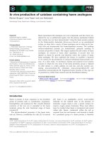
Scheme 1. (A) DNA bulge-specific compound derived from NCS-
chrom upon base catalysis. (B–D) Synthetic compounds mimicking
NCS-chrom, which showed selectivity for binding to DNA bulge site
[16], and strongly enhanced the repeat nucleotide slippage during
in vitro DNA synthesis [20].
D. Ouyang et al. Expansion of DNA repeat sequences
FEBS Journal 275 (2008) 4510–4521 ª 2008 The Authors Journal compilation ª 2008 FEBS 4511
of DDI-1A was better than that of DDI-1B, presum-
ably because of the different conformation of the agly-
con moiety. DDI-1A has a right-handed aglycon helix
with geometry mimicking the DNA helix, and is there-
fore more effective at intercalating into DNA base
pairs [20]. Although DDI-1B also mimics the structure
of NCSi-gb, it has a left-handed aglycon helix and
may be less effective at base pair intercalation. It
should be noted that 2-deoxy-2-aminoglucose (with
concentrations of 10–1000 lm) or the aglycon back-
bone (concentrations of 10–100 lm) of DDI-1A and
DDI-1B did not affect DNA slippage (data not
shown). Interestingly, there was a similar hierarchy of
intensities for the three bands in the (AAT)
3
⁄ (ATT)
5
and (ATT)
3
⁄ (AAT)
5
systems (Fig. 1A,B), each appar-
ently separated by two nucleotides, and this was
repeated every three nucleotides. This band spacing
appeared to reflect the triplet repeat unit, implying
that the in vitro DNA strand slippage syntheses of
(AAT)
3
⁄ (ATT)
5
and (ATT)
3
⁄ (AAT)
5
tracts mainly
occurred by triplet step expansion. Addition of both
DDI-1A and DDI-1B did not influence this pattern
(Fig. 1, inset). Similarly, the doublet repeat (GT)
4
G ⁄
(CA)
7
C and (CA)
4
C ⁄ (GT)
7
G also produced a regular
two-band repeat (Fig. 1C,D), suggesting that slippage
of these repeats occurred by two nucleotides each time.
Extension products from other two-triplet repeat
sequences, (CAG)
3
⁄ (CTG)
5
and (CTG)
3
⁄ (CAG)
5
, were
too short to generate a similar pattern on the gel.
Doxorubicin, an anthracycline glycoside that inter-
calates between DNA base pairs [30], inhibited the
expansion of all the repeat sequences used (Fig. 1,
lane 5). When both DDI-1A or DDI-1B and doxo-
rubicin were present, similar inhibition was found at
experimental concentrations (data not shown).
In vitro studies show that single-stranded tracts con-
taining (CTG)
n
repeats have a higher propensity to
form hairpin structures than similar tracts containing
the complementary (CAG)
n
repeats [31]; possibly
accounting for the orientation-dependent behavior of
these repeats in replication. Hairpin stability is attrib-
uted to the TÆT mismatch which stacked more effi-
ciently on the CTG strand than the AÆA mispair on
the complementary CAG strand, resulting in expanded
CTG fragments that are shorter than those of the
CAG strand (Fig. 1E,F). This rule is also the same for
other repeat sequences. As a result, the slippage effects
of AAT and CA repeats (Fig. 1A,D) are better than
those of their complementary strands, ATT and GT
(Fig. 1B,C). As such, the enhancement effects of
AB CDE F
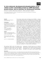
Fig. 1. Expansion of the various repeats and the effect of diastereomers DDI-1A and DDI-1B with
32
P-primer strands. A standard reaction
(23 °C, 24 h) containing 5¢-
32
P-end-labeled primer and unlabeled template was catalyzed by the Klenow fragment at 0.0177 unitÆlL
)1
. (A–F)
Lane 1, control to which no DNA polymerase was added; lane 2, control reaction system lacking compound, but receiving an equal volume
of dimethyl sulfoxide; lanes 3 and 4, reaction system to which DDI-1A and DDI-1B at a concentration of 60 l
M were added, respectively;
lane 5, reaction system to which 40 l
M doxorubicin was added. The products were resolved on a 15% sequencing gel. The numbers indi-
cate size markers of 26 and 41 nucleotides (random sequence) in length. *The 5¢ -
32
P-end-labeled strand. (Inset) Special attention of triple
band pattern in the gel.
Expansion of DNA repeat sequences D. Ouyang et al.
4512 FEBS Journal 275 (2008) 4510–4521 ª 2008 The Authors Journal compilation ª 2008 FEBS
DDI-1A and DDI-1B on AAT, CAG and CA repeats
are relatively better than for ATT, CTG and GT.
State of the template during slippage extension
Several repeated templates with 5¢-
32
P-end labeling
were used to investigate template extension. Figure 2
shows the extension result of six different sequences
under similar reaction conditions to those in Fig. 1. In
the control reaction, the 15-mer template of various
sequences (Fig. 2, lane 2) was extended to different
lengths depending on the stability of the secondary
structure formed between the primer and template
(Fig. 1). Enhancement of sequences with less stable
secondary structures was stronger than that with rela-
tively stable secondary structures. After addition of
DDI-1A and DDI-1B, slippage synthesis was greatly
enhanced for all sequences, as reflected by the presence
of much longer products (Fig. 2, lanes 3 and 4) in
comparison with the control. The stimulation effect of
DDI-1A in the template extension was obviously better
than that of DDI-1B, which was similar in the primer
extension reaction. As expected, doxorubicin inhibited
template expansion for all the repeated sequences
chosen (Fig. 2, lane 5). Again, the gel band pattern of
the synthesized DNA products reflected the particular
nucleotide repeat unit. A similar band pattern in both
the labeled primer and the template expansion system
implied that template and primer extension took place
synchronously.
Time course of DNA expansion
A time course for the extension of the repeat sequences
was performed (Table 1) in the assays shown in Figs 1
and 2. In the control, longer DNA fragments were
generated with the increase in reaction time, indicating
that primer and template slippage occurred during
DNA synthesis. In the presence of DDI-1A and DDI-
1B, radioactivity bands (both primer and template)
with long fragments increased steadily over time for all
the sequences tested. The slippage of less stable repeat
sequences almost reached saturation after being incu-
bated for > 48 h, and the differences in length
between the drug-containing samples and the control
was remarkable.
Effect of different polymerases on drug-
stimulated replication of the ATTÆAAT triplet
As shown in Fig. 3, we also investigated the effect of a
series of different prokaryotic polymerases proficient
or deficient in 3¢ to 5¢ exonuclease activity on
ATTÆAAT triplet slippage synthesis in vitro. The exten-
ABC DEF
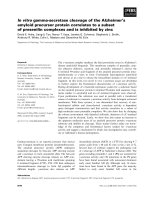
Fig. 2. Expansion of various templates with
32
P-template strands. (A–F) Lane 1, control to which no DNA polymerase was added; lane 2,
control reaction system lacking the compound, but receiving an equal volume of dimethyl sulfoxide; lanes 3 and 4, reaction system to which
DDI-1A and DDI-1B (60 l
M) was added, respectively; lane 5, reaction system to which 40 lM doxorubicin was added. Products were
resolved on a 15% sequencing gel. The numbers indicate size markers of 26 and 41 nucleotides (random sequence) in length. *The 5¢-
32
P-
end-labeled strand.
D. Ouyang et al. Expansion of DNA repeat sequences
FEBS Journal 275 (2008) 4510–4521 ª 2008 The Authors Journal compilation ª 2008 FEBS 4513
sion behavior of different polymerases was completely
different. The primer itself slipped in the control
reaction when using polymerases deficient in 3¢ to 5¢
exonuclease activity, such as the Klenow fragment
(Fig. 3B, lane 2), Sequenase Version 2.0 DNA poly-
merase (Fig. 3C, lane 2), Taq DNA polymerase
(Fig. 3E, lane 2) and pfu DNA polymerase (Fig. 3F,
lane 2). The addition of DDI-1A and DDI-1B strongly
increased the slippage effect in these polymerase
systems. Among these, Sequenase showed the weakest
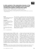
ABCDEF
Fig. 3. Effect of different polymerases on the stimulation of a triplet repeat expansion. A standard reaction (23 °C, 24 h) containing 5¢-
32
P-
end-labeled (AAT)
3
and unlabeled template (ATT)
5
was catalyzed by different prokaryotic polymerases (indicated). The concentration of pri-
mer–template and deoxynucleoside triphosphates in the reaction system is 4 l
M and 1 mM, respectively. The amount of polymerase used
was almost equal, i.e. 0.0177 unitÆlL
)1
of each enzyme. (A–F) Lane 1, control to which no DNA polymerase was added; lane 2: control reac-
tion system lacking drug, but with an equal volume of dimethyl sulfoxide; lanes 3 and 4, reaction system to which DDI-1A or DDI-1B (60 l
M)
was added; lane 5, reaction system to which 40 l
M doxorubicin was added. Products were resolved on a 15% sequencing gel. The numbers
indicate size markers of 26 and 41 nucleotides (random sequence) in length.
Table 1. Time course of primer ⁄ template expansion in the presence or absence of DDI-1A or DDI-1B. The concentration of DDI-1A or DDI-
1B is 60 l
M. Data are from experiments similar to those described in Figs 1 and 2 using
32
P-labeled primer ⁄ templates. After gel analysis of
the products, the band intensities were quantitated by Phosphor Imager (Molecular Dynamics). *5¢-
32
P-end-labeled primer or template.
Primer ⁄ template
Fragments > 15-mer (%) at 23 °C
12 h 24 h 48 h
Control DDI-1A DDI-1B Control DDI-1A DDI-1B Control DDI-1A DDI-1B
(AAT)
3
* ⁄ (ATT)
5
23.3 56.4 32.2 29.8 86.4 45.5 33.4 92.1 63.5
(AAT)
3
⁄ (ATT)
5
* 16.8 76.7 44.6 22.3 95.9 62.8 26.7 97.6 85.4
(ATT)
3
* ⁄ (AAT)
5
17.6 52.2 30.5 18.9 72.2 35.5 22.6 88.4 56.3
(ATT)
3
⁄ (AAT)
5
* 9.7 50.2 25.9 15.6 71.1 42.6 19.2 89.4 67.7
(CAG)
3
* ⁄ (CTG)
5
10.3 12.9 8.2 15.0 25.9 21.4 18.5 33.6 28.7
(CAG)
3
⁄ (CTG)
5
* 2.2 25.1 12.3 3.5 38.7 19.4 5.2 49.6 26.8
(CTG)
3
* ⁄ (CAG)
5
0 7.8 1.0 0 15.8 2.0 0 23.2 4.5
(CTG)
3
⁄ (CAG)
5
* 6.9 37.5 18.1 9.4 52.0 28.2 10.9 67.2 44.3
(GT)
4
G* ⁄ (CA)
7
C 0 32.7 9.4 1.5 67.7 12.0 12.0 89.7 36.8
(GT)
4
G ⁄ (CA)
7
C* 32.9 70.4 55.7 46.3 93.1 83.7 52.5 95.2 90.7
(CA)
4
C* ⁄ (GT)
7
G 15.7 50.2 19.5 18.6 85.2 28.5 23.7 92.2 48.7
(CA)
4
C ⁄ (GT)
7
G* 25.6 69.3 51.8 35.8 89.7 80.7 37.3 96.4 90.5
Expansion of DNA repeat sequences D. Ouyang et al.
4514 FEBS Journal 275 (2008) 4510–4521 ª 2008 The Authors Journal compilation ª 2008 FEBS
slippage effect under uniform conditions, whereas, the
difference between DDI-1A and DDI-1B is undistin-
guishable for Taq DNA polymerase. An inhibition
effect of doxorubicin was also observed (Fig. 3,
lane 5). Under the extension conditions used, pfu
DNA polymerase did not excise the extruded nucleo-
tides in the oligomer or eliminate the secondary struc-
ture formed by the repeated sequences, but it did
amplify the ATT repeats faithfully.
By contrast, for T7 DNA polymerase (Fig. 3D) and
T4 DNA polymerase (data not shown), although their
extension activities are very similar to that of the Kle-
now fragment [32], the 3¢ to 5¢ exonuclease activity
was so strong that the overhanging nucleotides were
excised completely from the 3¢-terminus in the anneal-
ing oligomer, and consequently, no expanded band
was observed during incubation. Doxorubicin did not
inhibit the exonuclease activity of T7 DNA polymer-
ase, whereas for Escherichia coli DNA polymerase I,
both exonuclease and polymerase activities were seen.
In the control (Fig. 3A, lane 2) and drug-addition
(Fig. 3A, lanes 3 and 4) reactions, the enzyme both
extended the primer to some extent, and excised the 3¢
overhung nucleotides from the duplex to give smaller
fragments. Because of the presence of excised short
oligomers, the extension bands in these lanes were
much lighter than the others, and various types of
duplex were formed by the primer and template. We
did not observe any strong stimulation to DNA slip-
page synthesis in the gel pattern by the addition of
DDI-1A and DDI-1B in these cases. These results may
be due to DNA polymerases with strong 3¢ to 5¢
exonuclease activity (including T7 DNA polymerase
and DNA polymerase I) degrading the product. To
our surprise, the addition of doxorubincin did not
obviously inhibit expansion, but did inhibit the exonu-
clease activity of DNA polymerase I to some extent;
the excised short oligomers were obviously less
(Fig. 3A, lane 5) than in the control and drug-addition
reactions.
Again, a triple band pattern was apparent through-
out the gel. Although the pattern in the Taq and
pfu DNA polymerase system differed from that in the
Escherichia coli DNA polymerase I-based system, the
expanded primary bands were almost all seen in
the three-nucleotide unit, which indicated that the
in vitro DNA strand slippage synthesis of (ATT)
3
⁄
(AAT)
5
tract was mainly a triplet expansion pattern. It
is suggested that the complete complementary structure
formed by the two triplet complementary strands
might be more stable than the others during slippage
synthesis, and be similar whatever DNA polymerases
are used.
Selective binding of DDI-1A and DDI-1B to
bulge DNA
Because formation of the bulge structure might be
important in DNA slippage [16], we speculate that the
enhancement of repeat slippage by DDI-1A and DDI-
1B might be caused by their specific recognition of
bulge DNA. Accordingly, CD spectropolarimetry can
be used to monitor conformational transitions as the
ligand–nucleic acid complex is formed. To gain insight
into the binding of drugs to bulge DNA, several bulge-
containing oligonucleotides were selected as binding
hosts for DDI-1A and DDI-1B.
CD spectroscopy of DDI-1A (Fig. 4) showed a
positive Cotton effect at 246 and 310 nm, and a neg-
ative Cotton effect at 220 and 290 nm, whereas the
CD of DDI-1B was almost complementary to that of
DDI-1A. These peaks are Cotton effect-associated
with corresponding p to p* transitions in the UV
spectra. The positive CD spectra for DDI-1A suggests
that the helix was right-handed, hence in the P con-
formation [14].
In order to observe the conformational transitions
of DNA directly and to eliminate drug interference,
the CD spectra of native DNA and altered DNA, after
subtracting the spectrum for the drug alone from that
of the complex, are also presented, assuming that the
conformation of the drug was not significantly altered
because the molecular models of DDI-1A and DDI-1B
are fairly rigid. The differential CD spectra of the
complex formed between DNA and the drugs are
shown in Fig. 4. The observed CD spectrum of the
native DNA (solid line) consists of a distinct positive
band at 280 nm caused by base stacking and a negative
band at 250 nm caused by helicity [33], which is char-
acteristic of DNA in the right-handed B-form. CD
spectra of DNA with DDI-1A (dashed line) and DDI-
1B (dotted line) consistently revealed an isoelliptic
point at 260 nm, except for the oligomer without a
bulge structure (Fig. 4A), suggesting formation of a
drug–DNA complex. For oligomer with a hairpin
structure (HT3AT), the band at 252 nm shifted to
241 nm (Fig. 4A), whereas for DNA with simple bulge
structures (one to three unpaired bases), the band at
252 nm shifted to 244 nm for DDI-1A and to 248 nm
for DDI-1B. There was no overall change in ellipticity
detected from the differential spectra of DNA
(Fig. 4B) for the oligomer HT3AT. In this case, the
binding of DDI-1A or DDI-1B to DNA might be via
simple groove binding and ⁄ or electrostatic interaction
that showed fewer or no perturbations on the base
stacking and helicity bands [34], ruling out the possi-
bility of conformational change.
D. Ouyang et al. Expansion of DNA repeat sequences
FEBS Journal 275 (2008) 4510–4521 ª 2008 The Authors Journal compilation ª 2008 FEBS 4515
AB
CD
EF
GH
IJ
Fig. 4. CD spectra and differential spectra
of DDI-1A and DDI-1B and their complexes
with selected DNA sequences. Solid line,
20 l
M free DNA. Dashed line: (A,C,E,G,I)
complex of DNA with DDI-1A (50 l
M),
(B,D,F,H,J) drug-alone has been subtracted.
Dotted line: (A,C,E,G,I) complex of DNA
with DDI-1B (50 l
M), (B,D,F,H,J) drug-alone
has been subtracted. The numbers indicate
size markers of 26 and 41 nucleotides (ran-
dom sequence) in length.
Expansion of DNA repeat sequences D. Ouyang et al.
4516 FEBS Journal 275 (2008) 4510–4521 ª 2008 The Authors Journal compilation ª 2008 FEBS
For oligomers containing single (one to three base)
bulges, the differential spectrum (dashed line for DDI-
1A and dotted line for DDI-1B) was changed signifi-
cantly, compared with that for the native DNA (solid
line). As shown in Fig. 4D, the new band at 305 nm
proved the formation of a DNA–drug complex [33].
The significant change in the band at 250 and 280 nm
implied an alteration in the DNA conformation
because of an overall bending of the DNA backbone
[33,35]. Both DDI-1A and DDI-1B exhibited binding
behaviors obviously different to that of the bulge
DNA host. The addition of DDI-1A and DDI-1B to
the two-base bulge (HT3AGTT) and three-base bulge
(HT3AATTT and HT3AAATT) caused the DNA
spectrum to be altered significantly. The trend and
characteristics of the conformational transformation
were similar to that of the one-base bulge oligomer.
However, the aglycon unit of DDI-1A and DDI-1B
(10–500 lm), lacking any CD signal itself, did not
affect the conformation of DNA (data not shown).
From the CD results, both compounds can interact
with oligomers containing a simple bulge and induce
significant conformational change. Therefore, the addi-
tion of DDI-1A and DDI-1B may induce formation of
the bulge or stabilize the bulge structure. The UV
melting temperature (T
m
) of oligonucleotides with a
three-base bulge increased upon intercalating with
DDI-1A and DDI-1B (Table 2), implying that DNA
secondary structures were stabilized by interaction with
the drug. For example, the change in T
m
(DT
m
) for the
ATT bulge increased by 3.4 and 1.7 °C in the presence
of DDI-1A and DDI-1B, respectively. The increase in
DT
m
for DDI-1A was higher than that for DDI-1B,
implying that intercalation to the bulge site was better
for DDI-1A than for DDI-1B due to its right-handed
aglycon helix, which might be suitable for stacking
into natural helical bases. The CD and melting temper-
ature data were consistent with the greater stimulation
effect of DDI-1A than of DDI-1B in the repeat slip-
page.
Conclusion
It has previously been shown that long DNA prod-
ucts can be generated in polymerase extension reac-
tions containing short complementary oligomers (e.g.
9-mer ⁄ 15-mer combinations) of di- or trinucleotide
repeats [36]. The efficiency of reiterative synthesis
depended on several factors including the length of
the repetitive unit, its sequence and the characteristics
of polymerase. In vitro studies on the expansion of
triplet repeats such as CAG, CGG and GAA, which
are associated with human hereditary disease genes,
helped in understanding the possible mechanism of
slippage and the molecular basis of the diseases
[37,38].
Given the size of DNA products made by the DNA
polymerase-based system using short repeat primers
and templates, slippage must be involved during repli-
cation. Furthermore, slippage occurs synchronously on
both strands. Slippage synthesis was enhanced mark-
edly by our synthetic diastereomers DDI-1A and DDI-
1B, which bind preferentially to simple bulges of one
to three unpaired bases in DNA. These results suggest
a process of stimulated slippage synthesis (Scheme 2).
After denaturing and annealing, the primers and tem-
plates form various types of duplex DNA. The small
DNA primer–template may have gone through multi-
ple rounds of slippage to reach the large expanded
products observed. Each cycle is initiated by the disso-
ciation of polymerase to re-associate at a new inter-
mediate. The intermediate is a combination of various
DNA strands with an unorthodox structure, such as
hairpin, bulged and slipped DNA, and may be the
main contributor to expansion. Under the experimen-
tal conditions used, various combinations of these
unstable intermediates are in homeostasis. When one
round of extension finishes, the extended primer and
template separate and realign to form new intermedi-
ates for the next round of replication, and longer
extended products are obtained through multiple
rounds of replication. For example, following
bulge ⁄ hairpin formation on the AAT strand of an
AAT ⁄ ATT repeat tract, replication extends the fore-
shortened AAT strand. The AAT bulge ⁄ hairpin may
then come apart to allow the complementary ATT
strand to be extended by DNA polymerase along the
previously extended AAT strand, and vice versa. In
fact, template extension is the same as primer exten-
sion. We call it template extension to distinguish the
Table 2. T
m
values of oligomers (P3 and P4) and DT
m
values by addition of DDI-1A and DDI-1B.
DNA sequence
P3 5¢-GTCCGATGCGTG-3¢ 3¢-CAGGCTACGCAC-5¢ ATT P4 5¢-GTCCGATGCGTG-3¢ 3¢-CAGGCTACGCAC-5¢ TAA
Native 20 l
M DDI-1A 20 lM DDI-1B Native 20 lM DDI-1A 20 lM DDI-1B
T
m
(°C) 27.5 30.9 29.2 29.2 30.7 29.8
DT
m
(°C) 3.4 1.7 1.5 0.6
D. Ouyang et al. Expansion of DNA repeat sequences
FEBS Journal 275 (2008) 4510–4521 ª 2008 The Authors Journal compilation ª 2008 FEBS 4517
expanded product of labeled primer from that of
labeled template. If doxorubicin intercalates between
DNA base pairs, a bulge structure cannot form. As a
result, DNA expansion of the repeated sequences is
inhibited (Scheme 2).
The propensity for the unwinding of DNA unwind-
ing elements, for example AATÆATT triplet repeats
[39,40], enables accessibility to chemical probes within
the region, as well as oligonucleotide hybridization,
which lead to aberrant DNA replication. At the reac-
tion temperature, the bulge ⁄ hairpin structures of these
types of sequences form and come apart easily, as does
realignment of the expanded primer and template,
allowing the complementary strand to be extended fur-
ther (Scheme 2). By contrast, the repeat sequence
CTGÆCAG associated with myotonic dystrophy type 1
has been observed to form slipped structures and hair-
pins in a length- and orientation-dependent manner
under physiological conditions [41–43]. Once the non-
B structure has formed, it is difficult for the CTG or
CAG strand to re-anneal to its complementary strand,
nor would realignment of primer and template and
further extension be easy. Thus, the expanded frag-
ments are relatively short.
Scheme 2. Mode for primer and template extensions stimulated by drug. The crooked region of two swallow-tailed shapes represent the
unstable intermediates that are composed of bulge, hairpin and slipped DNA etc. The compound formula represents DDI-1A or DDI-1B. One
cycle of simple extension and drug stimulation is shown for each pathway. It is assumed that multiple cycles through these pathways are
required to reach the dramatic expansion.
Expansion of DNA repeat sequences D. Ouyang et al.
4518 FEBS Journal 275 (2008) 4510–4521 ª 2008 The Authors Journal compilation ª 2008 FEBS
Once simple extension of the primer and template is
accomplished, slippage synthesis in the presence of
DDI-1A and DDI-1B becomes more pronounced as
incubation proceeds. In our experiment, the more
DDI-1A and DDI-1B were added and the longer incu-
bation time, the longer the expanded products
obtained; this may be due to two or more bulged inter-
mediates formed or induced by the additional drug
(Scheme 2). Compared with stimulation slippage and
bulge binding specificity, it is proposed that the associ-
ation and disassociation of the compound with the
bulged structure is also in a dynamic equilibrium,
whereas a molecule with moderate binding affinity and
binding dynamics to the bulged structure would facili-
tate further slippage, and yield a good stimulation
result [29].
In summary, DDI-1A and DDI-1B were designed
according to a DNA bulge binder, the enediyne natu-
ral product NCS-chrom [28], which exhibited selective
bulge-binding properties. To date, both compounds
are the smallest bulge-binding molecules shown to suc-
cessfully stimulate DNA strand slippage [20]. Detailed
investigation into the effects on in vitro DNA replica-
tion leads to several conclusions: (a) DNA sequences
with relatively unstable secondary structures slipped
more than DNA sequences with stable secondary
structures; (b) slippage of these repeats occurred by
two or three nucleotides each time, depending on the
DNA sequences; (c) template and primer extension
were synchronous; (d) the stimulation effect of DDI-
1A containing a right-handed aglycon helix was
greater than that of DDI-1B; (e) the enhancement
effects of DDI-1A and DDI-1B on AAT, CAG and
CA repeats are stronger than that of the ATT, CTG
and GT strand, which may be attributed to the TÆT
mismatch as opposed to the AÆA mismatch; (f) doxo-
rubincin inhibited the exonuclease activity of DNA
polymerase I to some extent. Considering these results
and previous publications [16,17,20,28,29], we propose
that the bulge selectivity of drugs is due to the wedge-
shaped spirocyclic part which fits into the DNA bulge
pocket, and aromatic aminosugar compounds with
bulge-binding selectivity may be anticipated to stimu-
late DNA slippage synthesis. This study provides
insight into the development of agents that interfere
with nucleotide expansion, as found in various disease
states. Given the relationship between repeat length
and both disease severity and age of onset, treatment
that interferes with triplet expansion or the generation
of ineffectual DNA triplet templates, might make sense
for RNA regulation and prevent the formation of toxic
proteins, such as polyglutamine [44] and polyalanine
tract [45].
Experimental procedures
Materials
Oligodeoxyribonucleotides were synthesized on a EXPE-
DITEÔ 8909 nucleic acids synthesis system (Applied
Biosystems, Foster City, CA, USA), and purified by elec-
trophoresis on a denaturing polyacrylamide gel using a
standard procedure [46]. The product was recovered from
the gel by phenol ⁄ chloroform extraction and ethanol pre-
cipitation. T4 polynucleotide kinase, E. coli DNA polymer-
ase I, the Klenow fragment of E. coli DNA polymerase I
lacking 3¢ to 5¢ exonuclease activity, Taq DNA polymerase
and pfu DNA polymerase were from Takara Biotechnology
(Dalian City, China). T7 DNA polymerase was from MBI
Company (Tangshan City, China). Sequenase Version 2.0
DNA polymerase was from U.S. Biochemical Corporation
(London, UK). Radioactive materials were from Beijing
Furui Biological Engineering Company (Beijing, China).
Other chemicals were from Sigma (St Louis, MO, USA).
The oligonucleotides were 5¢-
32
P-end labeled using
[
32
P]ATP[cP] and polynucleotide kinase.
DNA polymerase assays
A standard reaction (15 lL) contained 4 lm each of the
primer and template and 1 mm each of deoxynucleoside tri-
phosphate, DNA polymerase and the corresponding reac-
tion buffer. The DNA was in a several fold molar excess of
the enzyme. Unless otherwise indicated, the enzyme was at
a level of 0.0177 unitÆ lL
)1
of the reaction. A mixture of
5-
32
P-end-labeled primer and unlabeled template, generally
in equimolar concentrations, was annealed by heating in
Tris ⁄ HCl and MgCl
2
to 95 °C for 5 min followed by slow
cooling to room temperature. Following the addition of
dithiothreitol and deoxynucleoside triphosphates to the
annealed mixture, it was distributed for assays. The com-
pounds to be tested were added as a solution in dimethyl
sulfoxide. Controls lacking the compound received an equal
volume of dimethyl sulfoxide, the final concentration of
which was 2%. The reaction was started by addition of the
enzyme. Incubation was at 23 or 37 °C for the times indi-
cated. To terminate the reaction, 98% formamide contain-
ing 100 mm EDTA and marker dyes was added to the
reaction mixtures at a 1 : 1 vol. The reaction mixtures with
formamide, EDTA and marker dyes were loaded onto a
15% polyacrylamide sequencing gel for analysis. Gels were
exposed to a storage phorsphor screen, and the band inten-
sities were quantitated on a Phosphor Imager (Molecular
Dynamics, Sunnyvale, CA, USA).
UV melting experiments
Ultraviolet absorptions of 2 lm oligonucleotides were mea-
sured using a Cary-Bio100 UV-Visible spectrophotometer
D. Ouyang et al. Expansion of DNA repeat sequences
FEBS Journal 275 (2008) 4510–4521 ª 2008 The Authors Journal compilation ª 2008 FEBS 4519
with heating at 0.5 °CÆmin
)1
in phosphate buffer containing
10 mm phosphate, 10 mm NaCl, pH 7.0. The T
m
for DNA
in the presence of DDI-1A or DDI-1B was determined
when the concentration of drug was 10-fold that of DNA,
and calculated using the derivative method supplied in the
cary winuv software package for T
m
calculation.
CD spectropolarimetry
CD spectra were performed on a Jasco-715 spectropolarim-
eter, using a cylindrical quartz cell of 1 mm path length.
The cell compartment was purged continuously with dry
N
2
. Data were recorded at a bandwidth of 1.0 nm and mea-
sured every 0.2 nm over 210–325 nm at 20 ± 1 °CinTE
buffer (10 mm Tris, 1 mm EDTA, pH 8.0) containing
10 mm NaCl. All oligonucleotides were heated to 95 °Cin
the same buffer for 5 min and then cooled slowly to room
temperature before use. Conformations of bulge DNA-
bound drug were obtained by subtracting the drug-only
CD signal from that of the complex made by 20 lm of
DNA mixed with 50 lm drug.
Acknowledgements
We thank the two referees for the useful discussion.
This study was supported by the National Key Project
for Basic Research of China (2003CB114403), National
Natural Science Foundation of China (20272029,
20572053, 20421202, 20432010), Ministry of Education
of China (104189, B06005) and Nankai University ISC.
References
1 Toth G, Gaspari Z & Jurka J (2000) Microsatellites
in different eukaryotic genomes: survey and analysis.
Genome Res 10, 967–981.
2 Nag DK, Suri M & Stenson EK (2004) Both CAG
repeats and inverted DNA repeats stimulate
spontaneous unequal sister-chromatid exchange in
Saccharomyces cerevisiae. Nucleic Acids Res 32,
5677–5684.
3 Wells RD, Dere R, Hebert ML, Napierala M & Son LS
(2005) Advances in mechanisms of genetic instability
related to hereditary neurological diseases. Nucleic Acids
Res 33, 3785–3798.
4 Wells RD & Warren ST (eds) (1998) Genetic Instabilities
and Hereditary Neurological Diseases. Academic Press,
San Diego, CA.
5 Cummings CJ & Zoghbi HY (2000) Trinucleotide
repeats: mechanisms and pathophysiology. Annu Rev
Genomics Hum Genet 1, 281–328.
6 Ashizawa T & Zoghbi HY (1997) Diseases with trinu-
cleotide repeat expansion. In Current Neurology, Vol.
17 (Appel SH, ed.), pp. 79–135. IOS Press, Amsterdam.
7 Kunkel TA (1993) Slippery DNA and diseases. Nature
365, 207–208.
8 Kashi Y, King D & Soller M (1997) Simple sequence
repeats as a source of quantitative genetic variation.
Trends Genet 13, 74–78.
9 Napierala M, Bacolla A & Wells RD (2005) Increased
negative superhelical density in vivo enhances the genetic
instability of triplet repeat sequences. J Biol Chem 280,
37366–37376.
10 Parniewski P, Jaworski A, Wells RD & Bowater RP
(2000) Length of CTGÆCAG repeats determines the
influence of mismatch repair on genetic instability.
J Mol Biol 299, 865–874.
11 Wells RD (1996) Molecular basis of genetic instability
of triplet repeats. J Biol Chem 271, 2875–2878.
12 Perutz MF (1996) Glutamine repeats and inherited
neurodegenerative disease: molecular aspects. Curr Opin
Struct Biol 6, 848–858.
13 Harvey SC (1997) Slipped structures in DNA triplet
repeat sequences: entropic contributions to genetic
instabilities. Biochemistry 36, 3047–3049.
14 Kappen LS, Lin Y, Jones GB & Goldberg IH (2007)
Probing DNA bulges with designed helical spirocyclic
molecules. Biochemistry 46, 561–567.
15 Xi Z, Jones GB, Qabaja G, Wright J, Johnson FS &
Goldberg IH (1999) Synthesis and DNA binding of
spirocyclic model compounds related to the neocarzino-
statin chromophore. Org Lett 1, 1375–1377.
16 Xi Z, Hwang GS, Goldberg IH, Harris JL, Pennington
WT, Fouad FS, Qabaja G, Wright JM & Jones GB
(2002) Targeting DNA bulged microenvironments with
synthetic agents: lessons from a natural product. Chem
Biol 9, 925–931.
17 Zhang N, Lin Y, Xiao Z, Jones GB & Goldberg IH
(2007) Solution structure of a designed spirocyclic heli-
cal ligand binding at a two-base bulge site in DNA.
Biochemistry 46, 4793–4803.
18 Lin Y, Jones GB, Hwang GS, Kappen L & Goldberg
IH (2005) Convenient synthesis of ncs-chromophore
metabolite isosteres: binding agents for bulged DNA
microenvironments. Org Lett 7, 71–74.
19 Trotta E, Grosso ND, Erba M, Melino S, Cicero D &
Paci M (2003) Interaction of DAPI with individual
strands of trinucleotide repeats. Effects on replication
in vitro of the AATATT triplet. Eur J Biochem 270,
4755–4761.
20 Xi Z, Ouyang D & Mu HT (2006) Stimulation on
DNA triplet repeat strand slippage synthesis by the
designed spirocycles. Bioorg Med Chem Lett 16, 1180–
1184.
21 Jones GB, Lin YQ, Xiao ZW, Kappen L & Goldberg
IH (2007) Molecular probes of DNA bulges: functional
assay and spectroscopic analysis. Bioorg Med Chem 15,
784–790.
Expansion of DNA repeat sequences D. Ouyang et al.
4520 FEBS Journal 275 (2008) 4510–4521 ª 2008 The Authors Journal compilation ª 2008 FEBS
22 Xiao Z, Kappen LS & Goldberg IH (2006) Develop-
ment of new simple molecular probes of DNA
bulged structures. Bioorg Med Chem Lett 16, 2895–
2899.
23 Hwang GS, Jones GB & Goldberg IH (2004) Stereo-
chemical control of small molecule binding to bulged
DNA: comparison of structures of spirocyclic
enantiomer-bulged DNA complexes. Biochemistry 43,
641–650.
24 Kappen LS & Goldberg IH (1993) DNA conformation-
induced activation of an enediyne for site-specific cleav-
age. Science 261, 1319–1321.
25 Yang CF, Stassinopoulos A & Goldberg IH (1995) Spe-
cific binding of the biradical analog of neocarzinostatin
chromophore to bulged DNA: implications for thiol-
independent cleavage. Biochemistry 34, 2267–2275.
26 Gao X, Stassinopolous A, Ji J, Kwon Y, Bare S &
Goldberg IH (2002) Induced formation of a DNA bulge
structure by a molecular wedge ligand-postactivated
neocarzinostatin chromophore. Biochemistry 41, 5131–
5143.
27 Xi Z & Goldberg IH (1999) Comprehensive Natural
Products Chemistry, Vol. 7 (Barton DHR & Nakanishi
K, eds), pp. 553–592. Elsevier Science, Oxford.
28 Kappen L, Xi Z, Jones GB & Goldberg IH (2003) Stim-
ulation of DNA strand slippage synthesis by a bulge
binding synthetic agent. Biochemistry 42, 2166–2173.
29 Liu L, Yi L, Yang X, Yu Z, Xi Z & Wen X (2008) Syn-
thesis and spectroscopic characterization of binaphthol
aminosugars for stimulation of DNA strand slippage
synthesis. Tetrahedron, 64, 5885–5890.
30 Kornberg A (1980) DNA Replication (Bartlett AC, ed.),
pp. 415–440. W. H. Freeman, San Francisco, CA.
31 Kang S, Ohshima K, Shimizu M, Amirhaeri S & Wells
RD (1995) Pausing of DNA synthesis in vitro at specific
loci in CTG and CGG triplet repeats from human
hereditary disease genes. J Biol Chem 270, 27014–27021.
32 Tuntiwechapikul W & Salazar M (2002) Mechanism of
in vitro expansion of long DNA repeats: effect of tem-
perature, repeat length, repeat sequence, and DNA
polymerase. Biochemistry 41, 854–860.
33 Fasman GD (1996) Circular Dichroism and the Confor-
mational Analysis of Biomolecules . Plenum Press, New
York, NY.
34 Johnson WC (2000) Circular Dichroism: Principles and
Applications (Berova N, Nakanishi K & Woody RW,
eds), p. 738. Wiley, New York, NY.
35 S
ˇ
tefl R, Trantı
´
rek L, Vorlı
´
c
ˇ
kova
´
M, Koc
ˇ
a J, Sklena
´
r
ˇ
V
& Kypr J (2001) A-like guanine-guanine stacking in the
aqueous DNA duplex of d(GGGGCCCC). J Mol Biol
307, 513–524.
36 Schlotterer C & Tautz D (1992) Slippage synthesis
of simple sequence DNA. Nucleic Acids Res 20
, 211–
215.
37 Lyons-Darden T & Topal MD (1999) Effects of temper-
ature, Mg
2+
concentration and mismatches on triplet-
repeat expansion during DNA replication in vitro.
Nucleic Acids Res 27, 2235–2240.
38 Pearson CE & Sinden RR (1998) Slipped strand DNA,
dynamic mutations, and human disease. In Genetic
Instabilities and Hereditary Neurological Diseases (Wells
RD & Warren ST, eds), pp. 585–626. Academic Press,
New York, NY.
39 Ohshima K, Kang S, Larson JE & Wells RD
(1996) TTAÆTAA triplet repeats in plasmids form a
non-H bonded structure. J Biol Chem 271, 16784–
16791.
40 Trotta E, Grosso ND, Erba M & Paci M (2000) The
ATT stand of AATÆATT trinucleotide repeats adopts
stable hairpin structures induced by minor groove
binding ligands. Biochemistry 39, 6799–6808.
41 Tam M, Montgomery ES, Kekis M, Stollar DB,
Price GB & Pearson CE (2003) Slipped (CTG)Æ(CAG)
repeats of the myotonic dystrophy locus: surface
probing with anti-DNA antibodies. J Mol Biol 332,
585–600.
42 Pearson CE, Tam M, Wang YH, Montgomery SE,
Dar AC, Cleary JD & Nichol K (2002) Slipped-strand
DNAs formed by long (CAG)Æ(CTG) repeats: slipped-
out repeats and slip-out junctions. Nucleic Acids Res 30,
4534–4547.
43 Kang S, Jaworski A, Ohshima K & Wells RD (1995)
Expansion and deletion of CTG repeats from human
disease genes are determined by the direction of replica-
tion in E. coli. Nat Genet 10, 213–218.
44 Taylor JP, Hardy J & Fischback KH (2002) Toxic
proteins in neurodegenerative disease. Science 296,
1991–1995.
45 Brown LY & Brown SA (2004) Alanine tracts: the
expanding story of human illness and trinucleotide
repeats. Trends Genet 20, 51–58.
46 Maniatis T, Fritsch EF & Sambrook J (1982) Molecular
Cloning: A Laboratory Manual. Cold Spring Harbor
Laboratory Press, Cold Spring Harbor, NY.
D. Ouyang et al. Expansion of DNA repeat sequences
FEBS Journal 275 (2008) 4510–4521 ª 2008 The Authors Journal compilation ª 2008 FEBS 4521