Báo cáo khoa học: Regulated interaction of endothelin B receptor with caveolin-1 potx
Bạn đang xem bản rút gọn của tài liệu. Xem và tải ngay bản đầy đủ của tài liệu tại đây (348.08 KB, 12 trang )
Regulated interaction of endothelin B receptor with caveolin-1
Tomohiro Yamaguchi
1
, Yasunobu Murata
1
, Yoshinori Fujiyoshi
1,2
and Tomoko Doi
1
1
Department of Biophysics, Graduate School of Science, Kyoto University, Oiwake, Japan;
2
Japan Biological Information Research
Centre, Tokyo, Japan
The peptide hormone endothelin transmits various signals
through G protein-coupled receptors, the endothelin type A
(ET
A
R) and B (ET
B
R) receptors. Caveolae are specialized
lipid rafts containing polymerized caveolins. We examined
the interaction of ET
B
R with caveolin-1, expressed in Sf9,
COS-1, and HEK293 cells, and its effects on the subcellular
distribution and the signal transduction of ET
B
R. ET
B
R
formed a complex with caveolin-1 in cells in which these
two proteins were coexpressed and in the mixture after
purification and reconstitution (as examined by immuno-
precipitation) suggesting the direct binding of ET
B
Rwith
caveolin-1. The complex formed efficiently only when the
ET
B
R was ligand-free or bound to an antagonist, RES-
701-1, whereas the addition of ET-1 or another antagonist,
BQ788, dissociated the complex, suggesting the structural
recognition of ET
B
R by caveolin-1. In contrast, the
ET
A
R bound to caveolin-1 regardless of ligand binding.
Caveolin-1 utilized its scaffolding domain (residues 82–101)
and the C-terminal domain (residues 136–178) to bind to
ET
B
R, as for other signalling molecules. Furthermore, the
amount of ET
B
R localized in caveolae increased significantly
with the expression of caveolin-1 and decreased with the
addition of ET-1. The disruption of caveolae by filipin
reduced the ET-1-derived phosphorylation of ERK1/2.
These results suggest the possibility that the binding to
caveolin-1 retains the ligand-free ET
B
R in caveolae and
regulates the ET signal.
Keywords: caveolae; caveolin; endothelin; endothelin type B
receptor; lipid raft.
Endothelins (ETs) are 21-amino acid peptides that mediate
diverse physiological effects on vasoconstriction, cellular
development, differentiation, mitogenesis and other func-
tions, in various tissues via their G protein-coupled recep-
tors (GPCRs) ) endothelin receptor type A (ET
A
R) and B
(ET
B
R) [1,2]. For example, an ET
A
R mediates vasocon-
striction in vascular smooth muscle cells, whereas an ET
B
R
mediates the release of nitric oxide to stimulate vasodilata-
tion in vascular endothelial cells. The ET ligand–receptor
complex exhibits a slow dissociation rate and almost
irreversible binding, which could explain the prolonged
vasoconstriction by ET [3]. This property highlights the
importance of understanding ET signal regulation. Both
ET
A
RandET
B
R undergo rapid desensitization [4–6] and
internalize differently in transfected cells, as shown for many
GPCRs [7]. Concerning the intracellular trafficking path-
ways, ET
A
R is internalized rapidly, either via caveolae or
clathrin-coated pits, upon ligand binding [3,8,9] and follows
a recycling pathway, whereas ET
B
R follows a degradative
pathway after internalization via coated-pits, implicated for
clearance of plasma ET-1 [10]. Moreover, the ET
B
Rinthe
plasma membrane is constitutively transported to this
pathway independently of ligand stimulation, indicating
that ET
B
R is hardly retained in the plasma membrane at
steady state [9]. Differences in the final fates and subcellular
localization of the ETRs are determined by the C-terminal
sequences of the two receptors [9,11]. However, the effects of
the cell surface localization of the ETRs on the ET signals
have not been studied carefully.
Lipid rafts are microdomains of cell membranes that are
rich in cholesterol and sphingolipid and serve as sites for
gathering signalling molecules [12–14]. Caveolae are vesi-
cular invaginations of the plasma membrane, formed from
lipid rafts with polymerized caveolins [15]. The three
caveolin genes have been cloned (caveolin-1, caveolin-2,
and caveolin-3). The caveolin-1 and caveolin-2 show the
same distribution pattern, whereas caveolin-3 is specific to
muscle cells. Caveolins act as scaffolding proteins to cluster
and regulate signalling molecules targeted to the caveolae,
such as Src-family tyrosine kinases, Ha-Ras, G protein
a subunits, endothelial nitric oxide synthase, protein kinase
C, and epidermal growth factor (EGF) receptor, among
others [16]. Caveolins are cholesterol-binding integral
membrane proteins that are thought to form an unusual
hairpin-like structure, with the central hydrophobic domain
(residues 102–134 of caveolin-1) in the membrane and the
N- (residues 1–101) and C-terminal (residues 135–178)
domains inside the cell. In this structure, the scaffolding
domain of caveolin (residues 82–101) has been shown to
Correspondence to T. Doi, Department of Biophysics,
Graduate School of Science, Kyoto University, Oiwake,
Kitashirakawa, Sakyo-ku, Kyoto 606-8502 Japan.
Fax: + 81 75 753 4218, Tel.: + 81 75 753 4216,
E-mail:
Abbreviations: ET, endothelin; ETR, endothelin receptor; ET
A
Rand
ET
B
R, endothelin receptor type A and type B; GPCR, G protein-
coupled receptor; HEK293, human embryo kidney 293; CHO,
Chinese hamster ovary; EGF, epidermal growth factor; ERK1/2,
extracellular signal-regulated kinase 1 and 2; GST, glutathione
S-transferase; Cav.1-H6, His
6
-tagged caveolin-1; OG, n-octyl-b-D-
glucopyranoside; DRM, detergent-resistant membrane;
FL,fulllength.
(Received 27 November 2002, revised 11 February 2003,
accepted 27 February 2003)
Eur. J. Biochem. 270, 1816–1827 (2003) Ó FEBS 2003 doi:10.1046/j.1432-1033.2003.03544.x
mediate the interactions with the signalling molecules
described above [17]. On the other hand, the caveolin-
binding sequence motif (FXFXXXXF or FXXXXFXXF,
where F is an aromatic amino acid, Trp, Phe, or Tyr) has
been suggested to exist in the signalling molecules, by which
they bind to the caveolin scaffolding domain [18].
Several GPCRs, including the B2 bradykinin, b-adrener-
gic, cholecystokinin, ET
A
, angiotensin II type 1, and
adenosine A
1
receptors, are localized within caveolae
[19–25]. Particularly, the B2 bradykinin, angiotensin II type
1, and m2 muscarinic acetylcholine receptors are translo-
cated to caveolae upon agonist binding, whereas adenosine
A
1
and b
2
-adrenergic receptors are translocated out of
caveolae upon activation, and the binding of ET
A
Rwith
caveolins is not affected by agonist binding. Furthermore,
the m2 muscarinic acetylcholine and ET
A
receptors could be
internalized via caveolae [8,26]. The mitogenic signal
through ET
B
R in primary astrocytes, where the ET
B
Ris
relatively well expressed, originates from caveolae micro-
domains [27]. In addition, a number of proteins mediating
intracellular Ca
2+
signalling are concentrated in caveolae
[15,16]. Thus, the localization to caveolae and the inter-
action with caveolins appear to play fundamental roles in
the signal transduction by GPCRs. However, the inter-
actions of GPCRs with caveolins are not fully understood.
To examine the molecular mechanisms of the cell surface
localization, and its effects on signal transduction, we
studied the interactions of ET
B
R with caveolins in vivo and
in vitro, in terms of agonist-regulated localization on the cell
surface and signal transduction downstream. The results
revealed that only the ET
B
R in the ground-state structure
bound to caveolin-1, through the caveolin scaffolding and
C-terminal domains, and that a fraction of ET
B
Rwas
targeted to caveolae by the expression of caveolin-1 and was
gradually released from caveolae by ET-1 stimulation. In
addition, the disruption of caveolae impaired the down-
stream signals from ET
B
R. These results suggest feasible
regulations for ET
B
R signal transduction by the interaction
with caveolin-1. The residence of ET
B
R in caveolae might
be a way of ensuring the cell surface localization of ET
B
R
against rapid internalization.
Experimental procedures
Materials
The cyclic-peptide antagonist for ET
B
R, RES701-1, was
generously provided by M. Yoshida (Kyowa Hakko Kogyo
Co., Ltd, Tokyo, Japan). The cDNAs encoding human
ET
A
RandET
B
R were kindly provided by T. Masaki
(National Cardiovascular Research Institute, Osaka, Japan)
and Y. Furuichi (AGENE Research Institute, Kanazawa,
Japan), respectively. The hybridoma producing the anti-
(bovine rhodopsin) mAb, 1D4, was generously provided by
R. Molday (University of British Columbia, Vancouver,
Canada). The antagonist for ET
B
R, BQ788, was from
Phoenix Pharmaceuticals, Inc. The anti-ET
B
R mAb, 2A5,
was generated against Sf9-expressed human ET
B
R(T.
Yamaguchi, I. Arimoto, Y. Fujiyoshi & T. Doi, unpublished
data). Goat anti-mouse and anti-(rabbit IgG)–alkaline
phosphatase and –horseradish peroxidase conjugates were
from Promega Biotech.
DNA construction
The caveolin-1 cDNA was produced from human lung
mRNA by RT-PCR using appropriate oligonucleotide
primers, and was subsequently subcloned into the BamHI–
NotI sites of the mammalian expression vector pcDNA3.1
(Invitrogen). The ET
B
R cDNA was subcloned into the
BamHI–NotI sites of pcDNA3.1. Mutagenesis of the ET
B
R
cDNA was carried out with appropriate oligonucleo-
tide primers by a PCR-based site-directed mutagenesis
approach. In each ET
B
R mutant, the aromatic amino acid
residue (Tyr127, Tyr200, Trp206, Trp217, Phe326, Tyr387,
Phe393, Phe397, Trp404, or Phe408) was replaced with
alanine. These cDNAs were subcloned into the BamHI–
NotI sites of pcDNA3.1.
IntheCav.1-H6cDNA,aHis
6
-tag was attached to the
C terminus of caveolin-1 for affinity purification using
Ni
2+
–NTA agarose (Qiagen). The 1D4 epitope sequence
(KTETSQVAPA, an epitope for the anti-rhodopsin mAB)
was fused to the C terminus of ET
A
R. For expression in
insect cells, the cDNAs encoding caveolin-1, Cav.1-H6,
ET
B
R, and ET
A
R-1D4 were subcloned into a transfer
vector, pVL1393 (BD Biosciences), and the isolation of
recombinant viruses was carried out with linearized Bacu-
logold DNA according to the manufacture’s instructions
(BD Biosciences).
To generate glutathione S-transferase (GST)-fused
caveolin-1 proteins, selected regions of caveolin-1 [amino
acids 1–101, 1–81, 61–101, 136–178, 101–136, and 1–136
(full length; FL)] were amplified by PCR. GST-Cav.1
(1–101), GST-Cav.1(1–81), GST-Cav.1(61–101), and
GST-Cav.1(101–136) were subcloned into the BamHI–
XhoI sites of pGEX-4T-3 (Amersham Biosciences Inc.),
whereas GST-Cav.1 (136–178) and GST-Cav.1-FL were
subcloned into the BamHI–NotI sites. After expression
in Escherichia coli (BL21 strain), the GST fusion pro-
teins were affinity-purified on glutathione–agarose beads
(Amersham Biosciences Inc.) according to the manufac-
turer’s instructions.
Cell culture
COS-1, HEK293, and CHO-K1 cells were cultured in
Dulbecco’s modified Eagle’s medium (Sigma) containing
10% foetal bovine serum (Invitrogen) and penicillin/strep-
tomycin at 37 °C in a 5% CO
2
atmosphere. For transient
expression, LipofectAmine Plus reagent (Invitrogen) was
used to transfect COS, HEK293, and CHO cells in a
100-mm diameter plate or in a 6-well plate with 1–8 lg
pcDNA3.1 encoding the wild type or mutant ET
B
Rs,
caveolin-1, or vector only. These cells were subjected to each
study 36 h after transfection.
For the isolation of stably transfected HEK293 cell lines
expressing ET
B
R, the cells were transfected with the plasmid
pcDNA3.1 containing the ET
B
R cDNA, using the calcium
phosphate precipitation technique (Invitrogen). Two days
after transfection, the cells were plated in selective medium
containing 500 lgÆmL
)1
G418 (Invitrogen). The G418-
resistant colonies were selected, and the single colonies were
purified further. The ET
B
R expression level by the HEK293
cell lines isolated for further studies was 1–2 pmol per mg
membrane protein.
Ó FEBS 2003 Endothelin-1 dissociates the ETBR–caveolin-1 complex (Eur. J. Biochem. 270) 1817
The culture of Sf9 insect cells and the expression of ET
B
R
in Sf9 cells were performed as described previously [28]. The
expression of caveolin-1 and His
6
-tagged caveolin-1 (Cav.1-
H6) was also performed similarly. Co-expression of ET
B
R
and caveolin-1 in Sf9 cells was carried out by a double
infection with ET
B
R and caveolin-1 recombinant viruses.
Purification of ET
B
R and caveolin-1 from Sf9 cells
All operations were carried out at 4 °C. Purification of
ET
B
R by ligand-affinity chromatography using biotinylated
ET-1, and reconstitution of the purified ligand-free ET
B
R
into phospholipid vesicles were performed as described
previously [28,29].
Cav.1-H6 was purified from infected Sf9 cells using
Ni
2+
–NTA-agarose beads. The membranes were prepared
as described ( 100 mg protein from a 500-mL suspension
culture) and were solubilized in 20 mL NaCl/Tris (20 m
M
Tris/HCl pH 7.5, 150 m
M
NaCl) with protease inhibitors
(1 m
M
phenylmethylsulfonyl fluoride, 10 lgÆmL
)1
aproti-
nin, 10 lgÆmL
)1
leupeptin, 10 lgÆmL
)1
pepstatin), 60 m
M
n-octyl-b-
D
-glucopyranoside (OG; Dojindo), and 1% Tri-
ton X-100 (Wako). After 30 min solubilization, the lysates
were ultracentrifuged for 1 h at 100 000 g. The super-
natants were adjusted to 10 m
M
imidazole and were
incubated with 200 lLNi
2+
-nitrilotriacetic acid-agarose
beads overnight. After the incubation, the beads were
washed extensively with washing buffer (NaCl/Tris con-
taining 20 m
M
imidazole, 30 m
M
OG, and 0.03% Triton X-
100) four times by rotating for 5 min. After washing, the
bound proteins were eluted with 400 lL elution buffer
(NaCl/Tris containing 100 m
M
imidazole, 30 m
M
OG, and
0.03% Triton X-100) by rotating for 1 h.
Immunoprecipitation
All preparations were carried out at 4 °C unless stated
otherwise.
ETRs expressed with caveolin-1 in Sf9 membranes. Sf9
membranes (10 mg membrane proteins) were prepared as
described above [28,29], resuspended in NaCl/Tris/EDTA
(20 m
M
Tris/HCl pH 7.5, 150 m
M
NaCl, 1 m
M
EDTA)
containing protease inhibitors (1 m
M
phenylmethanesulfo-
nyl fluoride, 10 lgÆmL
)1
aprotinin, 10 lgÆmL
)1
leupeptin,
10 lgÆmL
)1
pepstatin), and incubated with or without 2 l
M
ET-1 for 1 h at room temperature. After the incubation, the
membranes were solubilized with 60 m
M
OG and 1%
Triton X-100 for 30 min, and were centrifuged at 100 000 g
for 30 min. The supernatants were incubated with 30 lLof
either 1D4 or 2A5 monoclonal antibody-immobilized resin
(1 mg antibody per mL resin) overnight. After this
incubation, the resins were washed extensively at least four
times with NaCl/Tris/EDTA containing the aforemen-
tioned detergents, and then the bound proteins were eluted
with 100 l
M
of either the 1D4 or 2A5 epitope peptides for
1 h at room temperature.
Purified ET
B
R and Cav.1-H6. Reconstituted phospho-
lipid vesicles with or without ET
B
R(1–2pmol)were
incubated with 1 l
M
ET-1 for 1 h at room temperature,
and then were incubated with 10–20 lg purified Cav.1-H6
overnight. In these mixtures, the concentrations of OG and
Triton X-100 derived from the Cav.1-H6 solution were far
below the critical micelle concentrations (OG, 2–3 m
M
;
Triton X-100, 0.002–0.003%) so that the membranes were
not solubilized. After incubation, the membranes were
solubilized with 60 m
M
OG and 1% Triton X-100,
incubated with 2A5-immobilized resin, washed, and eluted
with 2A5-epitope peptides.
GST–Cav.1 fusion proteins. Sf9 membranes containing
ET
B
R (2.5 mg protein per sample) were incubated with the
respective GST–Cav.1 fusion proteins (50 lg) overnight
and then centrifuged. The concentrations of detergents in
these mixtures derived from the GST–Cav.1 fusion proteins
were far below the critical micelle concentration. The
membranes were solubilized in 1 mL NaCl/Tris containing
1% n-decyl-b-
D
-maltopyranoside (Dojindo), and were
centrifuged again. After ultracentrifugation, the superna-
tants were incubated with 2A5-immobilized resin, washed,
and eluted with 2A5-epitope peptides.
Mutant ET
B
Rs expressed in COS cells. Thirty six hours
after transfection with the wild type ET
B
R cDNA, each
mutant ET
B
R cDNA or the vector only, COS cells (one
100-mm diameter plate per sample) were washed twice with
NaCl/P
i
, lysed with 1 mL NaCl/Tris/EDTA containing
60 m
M
OG and 1% Triton X-100, and centrifuged at
100 000 g (Fig. 3B). In addition, the washed cells were
collected in 1 mL hypotonic buffer (20 m
M
Tris/HCl pH 7.5,
1m
M
EDTA, and protease inhibitors) to burst the cells,
incubated with 1 l
M
ET-1 for 1 h at room temperature,
solubilized with 60 m
M
OG and 1% Triton X-100, and
centrifuged at 100 000 g (Fig. 3C). The supernatants were
incubated with 2A5-immobilized resin, washed, and eluted
with 2A5-epitope peptides.
Effects of antagonists on the interaction. The membranes
prepared from HEK293 cells, which expressed ET
B
Rstably
and caveolin-1 transiently (1–2 mgÆprotein per sample),
were resuspended in NaCl/Tris/EDTA containing protease
inhibitors, and incubated with 1 l
M
ET-1, 10 l
M
RES-
701-1, or 10 l
M
BQ788 for 1 h at room temperature. After
this incubation, the samples were solubilized with 60 m
M
OG and 1% Triton X-100, incubated with 2A5-immobilized
resin, washed, and eluted with 2A5-epitope peptides.
Electrophoresis and immunoblotting
Samples prepared in Laemmli sample buffer were subjected
to SDS/PAGE and transferred to nitrocellulose membranes
(Schleicher and Schuell). The nitrocellulose membranes
were blocked with 2.5% BSA and 0.5% gelatin for 30 min,
and were then incubated with the 2A5 mAb (1 lgÆmL
)1
),
anti-GST (1 lgÆmL
)1
, Amersham Biosciences Inc.), anti-
caveolin-1 mAb (2297) (1 : 1000, Transduction Laborator-
ies), anti-caveolin polyclonal Ig (1 : 10 000, Transduction
Laboratories), anti-(transferrin receptor) mAb (1 lgÆmL
)1
,
Zymed Laboratories Inc.), anti-flotillin mAb (1 : 250,
Transduction Laboratories), anti-(extracellular signal-regu-
lated kinase) (ERK)1 mAb (1 : 2000, Transduction Labor-
atories), or anti-phosphoERK1/2 mAb (E10) (1 : 2000; Cell
Signaling Technology) overnight at 4 °C. The membranes
1818 T. Yamaguchi et al. (Eur. J. Biochem. 270) Ó FEBS 2003
were washed with buffer (5 m
M
Tris/HCl pH 7.5, 150 m
M
NaCl, 0.05% Tween 20), incubated with secondary anti-
bodies for 1 h, washed, and then visualized by the BCIP/
NBT colour development substrates or the ECL+ plus
Western blotting detection system (Promega Biotech and
Amersham Biosciences Inc., respectively).
Preparation of caveolin-enriched, low buoyant
membrane fractions
Thirty-six hours after the transfection with the Cav.1 gene,
the HEK293 cells stably expressing ET
B
Rwereculturedin
serum-free medium for 6 h, and were then incubated with
or without 100 n
M
ET-1 for 30 min at 37 °C.The
following operations, except for the elution, were carried
out at 4 °C. After the incubation, the confluent HEK293
cells of two 100-mm diameter plates ( 8 mg total protein)
per sample were washed twice with NaCl/P
i
, scraped into
2 mL buffer (25 m
M
Mes pH 6.5, 150 m
M
NaCl and
protease inhibitors) containing 1% Triton X-100, and
homogenized using a loose-fitting Dounce homogenizer
(10 strokes). The homogenates were then adjusted to 40%
sucrose by the addition of an equal volume of an 80%
sucrose solution prepared in the above buffer, but lacking
Triton X-100, placed in the bottom of ultracentrifuge
tubes, and then overlaid with a discontinuous sucrose
gradient of 5 mL 30% (w/v) sucrose and 2 mL 5% (w/v)
sucrose, both prepared in buffer lacking Triton X-100. The
samples were centrifuged at 39 000 r.p.m. (200 000 g)in
an SW41 rotor (Beckman Instruments) for 16–20 h,
fractionated into 11 1 mL fractions sequentially from the
top of the gradient, and concentrated by precipitation with
trichloroacetic acid or by ultracentrifugation following
dilution.
Assay for phosphorylation of ERK1/2
Forty-eight hours after transfection in a 6-well plate, the
CHO cells were cultured in serum-free medium for 24 h,
preincubated with 1 or 2 lgÆmL
)1
filipin III, or vehicle for
10 min at 37 °C, and then incubated with 100 n
M
ET-1 or
vehicle for 5 min at 37 °C. After stimulation, the cells were
washed twice with ice-cold NaCl/P
i
, and were lysed with
100 lL RIPA buffer (50 m
M
Tris/HCl pH 7.5, 150 m
M
NaCl, 1% Triton X-100, 0.1% SDS, 0.5% sodium
deoxycholate, 0.5 m
M
Na
3
VO
4
,50m
M
NaF, 5 m
M
EDTA
and protease inhibitors) at 4 °C. The lysates were trans-
ferred into centrifuge tubes, sonicated for 15 s to shear the
DNA and reduce the sample viscosity, and then boiled for
5 min at 95 °C. The cooled samples were subjected to SDS/
PAGE and immunoblotted with anti-phosphoERK1/2 or
anti-ERK1 Igs.
Other methods
Protein concentrations were measured by the modified
Lowry method (DC-protein assay, Bio-Rad) with BSA as
standard. The amounts of ET
B
R contained in the samples
were measured by the
125
I-labelled ET-1 (NEN Life Science
Products) binding assay as described previously [29]. The
relative intensities of indicated proteins were measured by
comparative densities of reactive bands on immunoblots
with
IMAGEMASTER VDS
-
CL
and
TOTALLAB
version 1.10
software (Amersham Biosciences Inc.).
Results
ET
B
R directly binds to caveolin-1 in an ET-1-dependent
manner
To examine the interaction of ET
B
R with caveolin-1, we
took advantage of an insect cell expression system, in which
ET
A
RandET
B
R are expressed well, with K
D
values for
ET-1 of 55 ± 8 and 83 ± 11 p
M
,theB
max
values of
21 ± 1.7 and 60 ± 7.8 pmol per mg membrane protein,
respectively [28]. In addition, it has also been shown that the
caveolin-1 expressed in insect cells forms oligomers, and is
incorporated into caveolae-sized vesicles [30]. The human
ET
A
R-1D4 (ET
A
R fused with the 1D4 epitope at the
C terminus) and ET
B
R were each coexpressed with
caveolin-1 in insect Sf9 cells, incubated with or without
ET-1, and immunoprecipitated with the 1D4 and 2A5 anti-
ET
B
R mAbs, respectively, as shown in Fig. 1A. We
extensively used 60 m
M
OG/1% Triton X-100 mixed-
micelle conditions for the membrane solubilization before
immunoprecipitation, which allowed the recovery of ligand-
free ET
B
R, as described later, and the solubilization of
many Triton-insoluble proteins [31]. The eluates from the
antibody-immobilized resin were analysed for caveolin-1,
ET
A
R-1D4, and ET
B
R by immunoblotting. The caveolin-1
coimmunoprecipitated with the ET
A
R-1D4, regardless of
the absence or presence of ET-1 (lanes 2 and 3), but not
without the expression of the receptor (lane 1). This
observation is consistent with those of the previous report
[21]. In contrast, ET
B
R coimmunoprecipitated caveolin-1
only when the receptor was ligand-free (lane 5). The
addition of ET-1 significantly reduced the amount of
coimmunoprecipitated caveolin-1 (lane 6). This coprecipi-
tation of caveolin-1 was not detected in the absence of
ET
B
R expression (lane 4). These results suggest that ET
A
R
binds to caveolin-1 regardless of ET-1 binding, whereas
ET
B
R binds to it in an ET-1-sensitive manner.
Although several GPCRs have been suggested to interact
with caveolin, it is not clear if the interactions occur directly
or indirectly. To clarify the interaction of ET
B
Rwith
caveolin-1, we purified ET
B
R and caveolin-1 in detergent-
micelles. ET
B
R, expressed in Sf9 cells, was purified on a
ligand-affinity column eluted with 2
M
NaSCN to yield
ligand-free ET
B
R [29] (Fig. 1B, lane 1). Caveolin-1, with a
His
6
-tag fusion at the C terminus (Cav.1-H6), was also
expressed in Sf9 cells and purified by nickel-affinity
chromatography, by which Cav.1-H6 became the predom-
inant protein, as described in Experimental procedures
(Fig. 1B, lane 2). The purified Cav.1-H6 could interact with
a purified G protein a
i
subunit in detergent micelles, as
detected by immunoprecipitation, and as described previ-
ously (data not shown) [32]. The purified ET
B
Rwas
reconstituted into phospholipid vesicles, into which the
purified Cav.1-H6 was added, and the interaction between
them was assessed by immunoprecipitation (Fig. 1C). While
no Cav.1-H6 was immunoprecipitated from the phospho-
lipid vesicles without ET
B
R (lane 1), it coimmunoprecipi-
tated from the ET
B
R-containing vesicles (lane 2). Moreover,
the addition of ET-1 significantly reduced the amount of the
Ó FEBS 2003 Endothelin-1 dissociates the ETBR–caveolin-1 complex (Eur. J. Biochem. 270) 1819
precipitated Cav.1-H6 (lane 3). These results using the
heterologously expressed and purified proteins completely
reproduced the observations in the Sf9 membranes
(Fig. 1A), suggesting the direct interaction of ET
B
Rwith
caveolin-1. Interestingly, ET
B
R and caveolin-1 did not form
the complex in a detergent/micelle solution, but they bound
to each other only when the receptor was reconstituted into
vesicles. This could be due to that either the interaction of
ET
B
R and caveolin-1 occurs by two-dimensional molecular
movements along the membranes, or that the integration of
ET
B
R and caveolin-1 into the lipid bilayer stabilizes
the structures required for the interaction, or that the
detergent molecules binding to the ET
B
R interfere with
caveolin-1 binding.
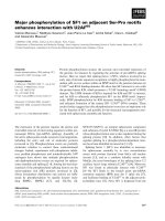
Fig. 1. ET
B
R directly interacts with caveolin-1 and dissociates from it upon ET-1 binding. (A) Sf9 membranes containing ET
A
R-1D4 (lanes 1, 2 and
3) or ET
B
R (lanes 4, 5 and 6) expressed with caveolin-1 were incubated with (lanes 3 and 6) or without 2 l
M
ET-1 (lanes 1, 2, 4 and 5) for 1 h at
room temperature, solubilized, and immunoprecipitated with the 1D4 or 2A5 mAbs. The eluates from the resin were analysed by SDS/PAGE,
followed by immunoblotting with the anti-(caveolin-1) mAb (upper panel), the 1D4 mAb (lower panel, lanes 1–3), or the 2A5 mAb (lower panel,
lanes 4–6). ET
A
R-1D4 coimmunoprecipitated with caveolin-1, regardless of the absence or presence of ET-1 (lanes 2 and 3), whereas ET
B
R
coimmunoprecipitated with caveolin-1 only when the receptor was ligand-free (lanes 5 and 6). Immunoprecipitation of caveolin-1 was not detected
in the absence of the receptor (lanes 1 and 4). (B) ET
B
R and Cav 1-H6 were individually expressed and purified from infected Sf9 cells, as described
in the Experimental procedures. The purified ET
B
R (lane 1) and Cav.1-H6 (lane 2) were visualized by silver staining, following SDS/PAGE. (C)
Phospholipid vesicles reconstituted with (lanes 2 and 3) or without (lane 1) the purified ET
B
R (1–2 pmol) were incubated with vehicle (lanes 1 and 2)
or 1 l
M
ET-1 for 1 h at room temperature, and were then incubated with the purified Cav.1-H6 overnight at 4 °C. The binding was analysed by
immunoprecipitation with the 2A5 mAb after solubilization. The eluates from the resin were analysed by immunoblotting with the anti-(caveolin-1)
mAb (upper panel) or the 2A5 mAb (lower panel). While the phospholipid vesicles without ET
B
R did not show any coimmunoprecipitated Cav.1-H6
(lane 1), the Cav.1-H6 coimmunoprecipitated from the ET
B
R-containing vesicles (lane 2). The addition of ET-1 significantly reduced the amount of
coprecipitated Cav.1-H6 (lane 3).
1820 T. Yamaguchi et al. (Eur. J. Biochem. 270) Ó FEBS 2003
The scaffolding domain and the C-terminal domain
of caveolin-1 both interact with ET
B
R
The caveolin scaffolding domain (residues 82–101 of
caveolin-1) is responsible for the binding with the
aforementioned signalling molecules, protein kinase A
catalytic subunit, connexin 43, and others [16,33,34].
Conversely, these proteins contain the caveolin-binding
motifs (UXUXXXXU and UXXXXUXXU,whereU is an
aromatic amino acid residue), which have been suggested
to be responsible for the binding to caveolin [18]. To
investigate the interaction between ET
B
R and caveolin-1,
we constructed a set of GST fusion proteins with various
caveolin-1 domains, as shown in Fig. 2A. These fusion
proteins were expressed in E. coli and purified by GST-
affinity chromatography, as shown in Fig. 2B. Sf9 cell
membranes containing ET
B
R were incubated with these
purified fusion proteins, and immunoprecipitataed with
the 2A5 mAb. While the ET
B
Rs in each eluate were
recovered to similar extents (data not shown), certain
GST-fusions, including GST–Cav.1-FL, GST–Cav.1
(1–101), GST–Cav.1(61–101), and GST–Cav.1(136–178)
coimmunoprecipitated, whereas GST–Cav.1(1–81) and
GST itself did not (Fig. 2C). GST–Cav.1(101–136) also
did not coimmunoprecipitate (data not shown). These
results suggest that caveolin-1 also utilizes the scaffolding
and C-terminal domains to interact with ET
B
R, as with
other signalling molecules. The binding of the C-terminal
domain fusion, GST–Cav.1(136–178), appeared to be
weaker, as compared with that of the scaffolding domain,
in repeated experiments.
Structure of ET
B
R recognized by caveolin
Since caveolin interacts with ET
B
R via the scaffolding and
C-terminal domains, the caveolin-binding motifs could be
the sites of these interactions in ET
B
R. However, these
motifs are not present in the cytoplasmic and transmem-
brane domains of ET
B
R, at least in the primary structure,
which would contain the caveolin-binding region, because
caveolin resides inside the cell. In fact, we mutated the
aromatic residues in the cytoplasmic and transmembrane
regions close to the cytoplasmic side, one by one
(Fig. 3A). The ET
B
RsexpressedinCOScells,shownin
Fig. 3B, were observed as two bands in the immunoblot-
ting because of N-terminal proteolysis, which was also
found with HEK293 and CHO cells, as described later
[35–37]. These mutated ET
B
Rs expressed in COS cells
bound caveolin-1 in the ligand-free form (Fig. 3B), and
dissociated from caveolin-1 following ET-1 binding,
similar to the wild type (Fig. 3C). The measurement of
band intensities showed no obvious differences between
the wild type and mutant ET
B
Rs in the ligand-sensitive
caveolin-1 binding. The C-terminal truncated ET
B
R
(residues 408–442 deleted) also showed these properties
in the COS cell system (data not shown). Therefore, single
mutations of these residues in the ET
B
R do not substan-
tially affect the caveolin-1 binding.
The fact that the addition of ET-1 reduced the amount of
caveolin-1 bound to ET
B
R (Fig. 1) indicates that caveolin-1
could distinguish the structure of the ligand-free ET
B
Rfrom
that of the ligand-bound form. To examine the contribu-
tions of the ET
B
R tertiary structure to the recognition by
caveolin-1, we further analysed the interactions of caveo-
lin-1 with ET
B
R in the presence of two types of antagonists,
RES-701-1 [38] or BQ788 [39] (Fig. 4). Previously, we
showed that RES-701-1 displayed an inverse-agonist acti-
vity that stabilizes ET
B
R structure in the ground-state, but
BQ788 did not [28]. The HEK293 cells stably expressing
ET
B
R were transfected with the caveolin-1 gene. The
membranes prepared from these cells were incubated with
ET-1, RES-701-1 or BQ788 and were immunoprecipitated
with the 2A5 mAb. The eluates were analysed for caveolin-1
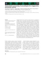
Fig. 2. The scaffold and C-terminal domains of caveolin-1 recognize
ET
B
R. (A) Schematic diagram summarizing the construction of a set
of GST–Cav.1 fusion proteins: GST–Cav.1-FL, GST–Cav.1(1–101),
GST–Cav.1(1–81), GST–Cav.1(61–101), GST–Cav.1(136–178) and
GST–Cav.1(101–136). (B) GST–Cav.1 fusion proteins, purified
by GST-affinity chromatography, were resolved by SDS/PAGE and
visualized by Coomassie blue staining. (C) Sf9 membranes containing
ET
B
R were incubated with each GST–Cav 1 fusion protein (50 lg)
overnight at 4 °C and then were subjected to immunoprecipitation
with the 2A5 mAb after solubilization. The eluates from the resin were
analysed by immunoblotting with an anti-GST mAb. GST–Cav.1-FL
retained binding activity to ET
B
R.WhileGSTaloneandGST–
Cav.1(1–81) were not coimmunoprecipitated with ET
B
R, GST–
Cav.1(1–101), GST–Cav.1(61–101) and GST–Cav.1(136–178) were
coimmunoprecipitated (bands marked by asterisks). GST–Cav.1(101–
136) did not coimmunoprecipitate (data not shown). Among these
constructs, the amount of coimmunoprecipitated GST–Cav.1(136–
178) appeared to be less than the others.
Ó FEBS 2003 Endothelin-1 dissociates the ETBR–caveolin-1 complex (Eur. J. Biochem. 270) 1821
and ET
B
R by immunoblotting (Fig. 4A). The ET
B
R
expressed in HEK293 cells was also observed as two bands
in immunoblotting. Fig. 4B shows the extents of caveolin-1
bound to ET
B
R, as shown in Fig. 4A, relative to the binding
to the ligand-free ET
B
R (lane 1) as 1.0. As observed with the
Sf9 membranes (Fig. 1), the extent of caveolin-1 binding to
ET
B
R was reduced to 0.35 ± 0.03 by the addition of ET-1
(lane 2). However, the inverse-agonist, RES-701-1-bound
ET
B
R retained caveolin-1-binding activity (0.98 ± 0.13,
lane 3) similar to that of the ligand-free form, whereas the
BQ788-bound ET
B
R reduced the activity (0.42 ± 0.13,
lane 4). The results suggest that the ET
B
R, in the ligand-free
or ground-state structure, exhibits higher affinity to cave-
olin-1 than that in the BQ788-bound or an activated
structure, and that the recognition by caveolin-1 is influ-
enced highly by structure.
Caveolin-1 targets ET
B
R to the caveolae membrane
and ET-1 releases ET
B
R from caveolae
To examine the effects of caveolin-1 on the localization of
ET
B
R,acelllinestablyexpressingET
B
R was isolated using
HEK 293 cells, which do not express endogenous caveolin.
The expression level of ET
B
R in this cell line is approxi-
mately 1–2 pmolÆmg
)1
membrane protein. The ET
B
R
distribution in these cell membranes was analysed before
or after transfection with the caveolin-1 gene. Fig. 5A shows
the results of sucrose-density gradient centrifugation of the
Triton-insoluble fractions of the caveolin-1-transfected cells.
The low buoyant density and bottom fractions have been
shown to contain the caveolae membranes and the Triton-
soluble membrane proteins, respectively [31]. Indeed, cave-
olin-1 and another caveolae marker, flotillin, were present
within the low-density fractions around fraction 3, whereas
a noncaveolae marker, the transferrin receptor, was fract-
ionated to the bottom, high-density fractions. When cave-
olin-1 was transfected, the ET
B
R was fractionated to the
low-density and the bottom fractions, suggesting that a
fraction of ET
B
R was targeted to the caveolae membranes.
Since the treatment with Triton X-100 denatured the ET
B
R
that was solubilized from the nonlipid raft membranes, the
approximate distribution of ET
B
R, as assessed by ligand
binding, was examined using fractions prepared by cell
disruption with sodium carbonate and sucrose density-
gradient centrifugation. Approximately 7% of the cell
surface ET
B
R was fractionated to the low-density fraction,
based on the ligand binding activities of ET
B
Rinthelow-
density fraction and in the plasma membrane fraction (data
not shown).
The ET
B
R found in the detergent-resistant, low-density
fraction (DRM, combined fractions 2 and 3 in Fig. 5A) is
shown in Fig. 5B, while Fig. 5C represents the averaged
band intensities of ET
B
RobservedinFig.5Bfromfive
repeated experiments, relative to the band intensity of
total ET
B
R [band I (full-length isoform) plus band II
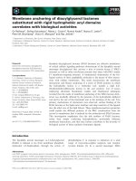
Fig. 3. Single mutations of aromatic amino acids in or close to the
cytoplasmic domain of ET
B
R did not affect the interaction with
caveolin-1. (A) Secondary structure model of human ET
B
R, showing
the 10 aromatic residues (marked with circles) that were each mutated
to Ala (Tyr127, Tyr200, Trp206, Trp217, Phe326, Tyr387, Phe393,
Phe397, Trp404, and Phe408). The putative seven helices are boxed.
(B) COS cells transiently expressing the wild type or each mutant ET
B
R
were subjected to immunoprecipitation with the 2A5 mAb. The eluates
from the resin were analysed by immunoblotting with the anti-caveolin
polyclonal Ig (upper panel) or the 2A5 mAb (lower panel). The band
intensities of caveolin-1 per ET
B
R were compared to that of the wild
type ET
B
R as 1.0. The three independent experiments were averaged.
All of the mutant ET
B
Rs interacted with caveolin-1, in a similar
manner to that of the wild type. (C) COS cells transiently expressing the
wild type or each mutant ET
B
R were lysed with a hypotonic buffer,
incubated with 1 l
M
ET-1 for 1 h at room temperature, and then
subjected to immunoprecipitation with the 2A5 mAb. The eluates from
the resin were analysed by immunoblotting with the anti-caveolin
polyclonal Ig (upper panel) or the 2A5 mAb (lower panel). The band
intensities of caveolin-1 were compared as in (B). The two independent
experiments were averaged. All ligand-bound mutant ET
B
Rs disso-
ciated from caveolin-1 significantly, similar to the wild type.
1822 T. Yamaguchi et al. (Eur. J. Biochem. 270) Ó FEBS 2003
(N-terminally cleaved isoform)] without the expression of
caveolin-1 (lane 1). The intensities of the total and the
band II ET
B
R in each lane are shown separately. Similar
amounts of proteins were recovered in the low-density
fractions in each experiment. When the caveolin-1 gene was
not transfected, the ET
B
R was scarcely recovered in the
low-density fraction (lane 1). On the other hand, when
caveolin-1 was expressed, the total amount of ET
B
R found
in the low-density fraction (lane 2) increased about three-
fold. Furthermore, when the caveolin-1-expressing cells
were treated with ET-1 for 30 min at 37 °C, the total
amount of ET
B
R was slightly reduced. In addition, the
amount of N-terminally cleaved ET
B
R (band II) in the
low-density fraction was reduced to about 64% (lane 3), as
compared to that of the ET-1-untreated cells (lane 2). Two
isoforms of ET
B
R corresponding to bands I and II,
observed in mammalian tissue culture cells and in native
tissues [35,36], have been shown to be caused by proteases
activated or released from cells during membrane prepar-
ation. The increased band II ET
B
R intensities in Fig. 5B
compared to those in Fig. 5A could be due to the
proteolysis during further ultracentrifugation to concentrate
the fraction 2 and 3 membranes. It was also shown that the
stably expressed ET
B
R in HEK293 cells is not cleaved at the
cell surface without agonist stimulation, and that metallo-
proteases cleave the N terminus of agonist-bound ET
B
Rat
the cell surface [37]. We assume that the band II isoform in
ET-1-untreated cells was derived from proteolysis during
the membrane preparation, whereas the band II in ET-1-
treated cells was derived from metalloprotease cleavage, in
addition to proteolysis during the membrane preparation.
Furthermore, the band II might predominantly contain the
ET-1-bound form as compared to the band I isoform,
because ET-1 binding to full-length ET
B
R might not be
quantitative at the cell surface in a 30-min assay at 37 °C.
Therefore, the decrease in the band II intensity of ET-1-
treated cells suggests that a fraction of the ET-1-bound
ET
B
R is gradually exiting out from caveolae. The reason
why the band I isoform did not decrease is not clear at
present. Thus, some of the ET
B
R is targeted to the caveolae
membranes by the expression of caveolin-1, and is gradually
excluded from the caveolae by agonist stimulation in
HEK293 cells.
Disruption of caveolae impairs ET-1-induced ERK
activation
Cholesterol binding agents such as filipin have been shown
to disrupt lipid rafts, probably by altering biophysical
characteristics [40,41]. The ET
B
R activates mitogen-activa-
ted protein kinases, such as ERK, c-Jun N-terminal kinase
and p38 kinase, to mediate mitogenic and cell-proliferation
signals [42–44]. To study the significance of the compart-
mentalization to caveolae membranes, the ET
B
Rwas
expressed transiently in CHO cells, which expressed cave-
olin-1 abundantly, and the ET-1-induced phosphorylation
of ERK was examined. Fig. 6 shows that the addition of
ET-1 greatly increased the amount of phosphorylated
ERK1/2 in CHO cells, while the amounts of the recovered
ERK1/2 remained unchanged, as shown by the immuno-
blotting. However, pretreatment with increasing amounts of
filipin III before the addition of ET-1 significantly reduced
the amount of accumulated phosphorylated ERK1/2. In
untransfected CHO cells, no ET-1-induced phosphorylation
of ERK1/2 was observed (data not shown). These results
suggest that the caveolae microdomain plays fundamental
roles in efficient signal propagation in the ERK pathway by
the ET
B
R, although the effects of filipin III on caveolae and
the ET
B
R are not exactly clear. In addition, these results are
consistent with the report showing impaired ERK and focal
adhesion kinase signal transduction via the ET
B
R in filipin
III-treated primary astrocytes [27].
Fig. 4. Effects of antagonists on the interaction of ET
B
Rwith
caveolin-1. The membranes prepared from HEK293 cells expressing
ET
B
R and caveolin-1 (1–2 mg of proteins per sample) were incubated
with either vehicle, 1 l
M
ET-1, 10 l
M
RES-701-1, or 10 l
M
BQ788 for
1 h at room temperature, and subsequently were subjected to immu-
noprecipitation with the 2A5 mAb. (A) The eluates from the resin were
analysed by immunoblotting with the anti-caveolin-1 mAb (upper
panel) or the 2A5 mAb (lower panel). (B) The extents of caveolin-1
bound to ET
B
R observed in (A) are represented by normalizing
the binding to the ligand-free ET
B
R (lane 1) as 1. The data are
means ± SE of three independent experiments. The extents of
caveolin binding was decreased significantly by the addition of either
ET-1 (lane 2) or BQ788 (lane 4), whereas the RES-701-1-bound ET
B
R
retained an activity similar to that of the ligand-free form.
Ó FEBS 2003 Endothelin-1 dissociates the ETBR–caveolin-1 complex (Eur. J. Biochem. 270) 1823
Discussion
We studied the interaction of ET
B
R and caveolin-1 in vitro
and in vivo, using the expressed proteins in insect and
mammalian cells. The ligand-free ET
B
R in the reconstituted
phospholipid vesicles formed a complex with caveolin-1,
which dissociated upon agonist binding. The significance of
this interaction could be perceived in a model cultured cell
system. The expression of caveolin-1 targeted some of the
ET
B
R to the membrane microdomain, caveolae, and ET-1
stimulation translocated the ET
B
R out of caveolae, which
when disrupted, diminished the ET
B
R-derived signal pro-
pagation. This is the first report showing the caveolin-1- and
ET-1-regulated localization of the ET
B
Randthedirect
interaction of a GPCR with caveolin.
The heterologously expressed ET
B
R and caveolin-1
formed a complex after purification and reconstitution into
vesicles, suggesting their direct interaction. Interestingly, this
interaction requires the existence of the ET
B
R within the
lipid bilayer, and does not occur in the detergent micelle.
Considering the fact that ET
B
R binding by caveolin-1
involves the scaffolding domain, which is thought to be
proximal to the membrane domain, a region close to or
within the transmembrane domain of ET
B
R might be
important for the interaction. However, we could not
specify the region of ET
B
R involved in the caveolin-1
binding. At the very least, the conformational changes of
ET
B
R affect this interaction, and the ground-state structure
of ET
B
R is required for the interaction with caveolin-1.
The discrimination by caveolin-1 of the RES-701-1-
bound and BQ788-bound forms of ET
B
R agrees well with
the previous observation, in which a cyclic peptide
antagonist, RES-701-1, could antagonize an ET
B
Rartifi-
cially activated by a chaotropic reagent, NaSCN, but
another antagonist, BQ788, could not [28]. We suggest
that the inverse agonist activity of RES-701-1 led the
ET
B
R to an inactive (ground state) conformation. On the
other hand, the BQ788-bound conformation is somewhat
different from both the ground state structure and the
G protein-coupling structure. The interaction with caveo-
lin may be a useful tool for molecular pharmacological
studies of GPCR.
The expression of caveolin-1 in HEK293 cells stably
expressing ET
B
RtargetedtheET
B
R to the Triton-insoluble,
low buoyant density fraction, caveolae. These results
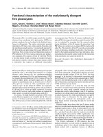
Fig. 5. Caveolin-1 targets ET
B
Rtocaveolae.HEK293 cells stably
expressing ET
B
R were transfected with either Cav.1-pcDNA3.1 or the
empty vector. The transfected cells were incubated with 100 n
M
ET-1
or vehicle and were lysed in 1% Triton X-100 at 4 °C.Thelysateswere
subjected to subcellular fractionation using a 5/30% discontinuous
sucrose gradient, as described in Experimental procedures. (A) A
100-lL aliquot of each fraction prepared from the caveolin-1-trans-
fected cells was precipitated with trichloroacetic acid and resuspended
in 50 lL of Laemmli sample buffer. Aliquots of each fraction (20 lL
for ET
B
Rand2lL for caveolin-1, transferrin receptor and flotillin)
were analysed by immunoblotting with the respective antibodies.
Fractions 2 and 3 correspond to the 5/30% sucrose interface. The
transfected caveolin-1 and endogenous flotillin, which are both
caveolae proteins, were enriched in fractions 2 and 3, whereas the
transferrin receptor, which is distributed in nonlipid raft membranes,
was fractionated to the bottom fractions. The ET
B
R partially
cofractionated with caveolin-1. The two arrowheads represent the
positions of the full-length and N-terminally cleaved ET
B
R. (B) The
ET
B
R in fractions 2 and 3 (DRM) in (A) was concentrated by ultra-
centrifugation following dilution and was compared by immunoblot-
ting with the 2A5 mAb, with (lanes 2 and 3) or without (lane 1)
caveolin-1 expression, and with (lane 3) or without (lanes 1 and 2) ET-1
treatment. Bands I and II indicate the full-length and N-terminally
cleaved ET
B
R, respectively. (C) The band intensities of ET
B
Rrecov-
ered in DRM in (B) are compared. The total (bands I and II) and
N-terminally cleaved ET
B
R (band II) are shown separately. The data
are means ± SE of five independent experiments. The amounts of
total proteins recovered in DRM are more or less constant, as shown
under the columns. The caveolin-1 expression drives the targeting of
ET
B
R to caveolae, and the ET-1 treatment releases a fraction of ET
B
R,
particularly band II ET
B
R, from caveolae.
1824 T. Yamaguchi et al. (Eur. J. Biochem. 270) Ó FEBS 2003
suggest that a fraction of ET
B
R is localized in caveolae,
driven by the interaction with caveolin-1, although the
localization efficiency was only 7% of the cell surface ET
B
R.
In primary astrocytes, substantial amounts of ET
B
Rare
localized in the caveolae fraction [27]. The localization of
ligand-free GPCR to caveolae has been reported for the
adenosine A
1
and b
2
-adrenergic receptors, which interact
with caveolin-3 in cardiomyocytes, while the b
2
-adrenergic
receptor do not require caveolin-3 to target to caveolae,
when expressed in HEK cells containing a functional
homologue of caveolin, flotillin/ESA [20,23]. The Flotillin/
ESA might be able to compensate in the case of b
2
-adren-
ergic receptor, but not in the case of ET
B
R. Therefore, the
molecular mechanisms used in the targeting of b
2
-adrenergic
receptor and ET
B
R to caveolae might be different. Further
studies on the targeting mechanisms to caveolae are
required.
Upon agonist addition, the adenosine A
1
receptors in
ventricular myocytes dissociate from caveolin-3 and trans-
locate out of the caveolae within 15 min at 37 °C [23]. Most
of the b
2
-adrenergic receptors in cardiomyocytes are also
excluded from caveolae upon agonist stimulation by 30 min
at 37 °C [20]. Although such a dramatic decrease in the
abundance of ET
B
R in the caveolae of HEK293 cells was
not observed, an 12% reduction of the total ET
B
Rwas
observed (Fig. 5). This slow reduction could be because
signal transduction of the ET
B
R during the exit from
caveolae might be required, or it may be due to incomplete
ET-1 binding at the cell surface, or to overexpression of
ET
B
R in HEK293 cells. In addition, because most of the
ET
B
R at the cell surface is localized in nonlipid raft
membranes, the constitutive trafficking of ET
B
Rfrom
nonlipid raft membranes or the newly synthesized ET
B
R
moving from inside the cells to the caveolae might mask a
fraction of the ET
B
R exiting from the caveolae. However,
the decrease in the N-terminally cleaved ET
B
R (band II)
was significant (Fig. 5C). The agonist-dependent N-terminal
cleavage of ET
B
R by metalloproteases on the cell surface
has been shown, which yields band II, whose functional
significance is not known [37]. While the full-length ET
B
R
might be supplied from other domains in the cells, the
N-terminally cleaved, agonist-bound ET
B
R may dissociate
from caveolin-1 and be gradually excluded from the
caveolae. The agonist-regulated localization of ET
B
Ron
the cell surface should be studied further using native tissues
or primary cultures of astrocytes, and endothelial cells,
among others. Although a decrease of the ET
B
Rincaveolae
seemed to be slow in HEK293 cells, the transient vasodil-
atation due to the ET
B
R-induced nitric oxide release from
endothelial cells [1,2] might be regulated by interactions with
caveolins, in addition to the rapid desensitization/internal-
ization of ET
B
R [9–11].
The colocalization and coimmunoprecipitation of ET
A
R
with caveolin-1 [21] and the internalization of ligand-bound
ET
A
R via caveolae have been shown [8]. These properties
are explained well by the fact that ET
A
Rinteractswith
caveolin-1, regardless of agonist binding. For ET
B
R, rapid
internalization to a degradative pathway upon agonist
binding in CHO cells [10,11] and constitutive internalization
to lysosomes in HeLa and Clone 9 cells [9] have been
reported. In contrast, the localization to caveolae micro-
domains by interaction with caveolin-1 ensures that a
subfraction of the ET
B
R is present on the cell surface to
transmit ET-1 signals. The reduced ERK signalling via the
ET
B
R in filipin-treated cells might be due to rapid ET
B
R
internalization/degradation or simply due to the lack of
caveolae, where signalling molecules are concentrated, as
filipin reduces the cell-surface caveolin [27]. Both extreme
mechanisms of ET
B
R action may be well balanced,
depending upon the cell and tissue types.
ET
B
R couples to multiple G proteins, mainly G
q
and G
i
in native tissues but also to G
s
and G
o
in a few tissues,
cultured cells, and in vitro [2,28,45,46], and this could be
regulated by the intracellular conditions. While the G
q
a
subunit has been shown to bind caveolin and to be
concentrated in caveolae, the heterotrimeric G
i
and G
s
are
localized in lipid rafts [47]. In the case of b
2
-adrenergic
receptor in neonatal cardiomyocytes, the localization in
caveolae seems to regulate signal transduction by the
limiting access to G
s
[48]. Although caveolae appear to
facilitate the ERK signalling activated by ET
B
RinCHO
cells, other signalling pathways through ET
B
R could exist in
nonlipid rafts, because substantial amounts of ET
B
Rare
localized here. Therefore, localization to specific membrane
microdomains, such as caveolae, lipid rafts and nonlipid
rafts, may also contribute toward specifying the signal
transduction by ET
B
R.
Furthermore, the recent report that EGF receptors in
noncaveolar lipid rafts show lower EGF binding (B
max
),
than those in nonlipid rafts, suggests that the receptor
properties are regulated by the lipid environment [49]. Based
on the pharmacological heterogeneity of ET
B
R, the exist-
ence of the ET
B1
RandET
B2
R (tentatively termed) subtypes
has been suggested; nevertheless, there is no molecular
biological evidence supporting this. The ET
B1
R located on
the vascular endothelium mediates vasodilatation through
the release of nitric oxide, which is sensitive to the
mixed ET
A
R/ET
B
R antagonist, PD 142893, bosentan,
RES-S701-1, BQ788, etc. The other subtype (ET
B2
R),
Fig. 6. Filipin treatment impairs ET
B
R-mediated ERK1/2 signalling.
CHO cells transiently expressing ET
B
R were cultured in FBS-free
medium for 24 h. After a pretreatment with 0, 1 or 2 lgÆmL
)1
filipin
III for 10 min at 37 °C, these cells were stimulated with 100 n
M
ET-1
for 5 min at 37 °C. The cells were lysed in RIPA buffer and were
analysed by immunoblotting with the anti-(phosphorylated ERK)
mAb (upper panel) or the anti-ERK mAb (lower panel). Pretreatment
with filipin attenuated the phosphorylation of ERK1/2 mediated by
the activation of ET
B
R. The bands for ERK1 (44 kDa) and ERK2
(42 kDa) were not separated in these gels.
Ó FEBS 2003 Endothelin-1 dissociates the ETBR–caveolin-1 complex (Eur. J. Biochem. 270) 1825
located on the vascular smooth muscle cells, directly
mediates vasoconstriction and is sensitive to BQ788, but
insensitive to PD 142893 [2,50,51]. Similarly, two subtypes
with different affinities (super-high and high affinity sites) to
endothelins have been reported in the rat brain and atrium
[52]. This pharmacological heterogeneity of ET
B
Rmight
be due to the membrane lipid raft environments.
In conclusion, the present study shows that ET
B
R
interacts with caveolin-1 in an ET-1-sensitive manner,
suggesting that ET
B
R is targeted to caveolae by binding
to caveolin-1, and is excluded from caveolae by agonist
binding (Fig. 7). Although the physiological significance of
ET-1-sensitive dissociation of the ET
B
R/caveolin-1 complex
is not exactly clear at present, we suggest that this
compartmentalization within caveolae ensures signal trans-
duction by ET
B
R, in spite of the rapid internalization/
degradation process. Further studies of ET
B
Rincaveolae
and in nonlipid rafts using native tissues would provide
further insights into the properties of the subtypes, the
promiscuous coupling with G proteins, and the desensiti-
zation mechanisms of ET
B
R.
Acknowledgements
We thank S. Satoh for useful suggestions and discussions through this
work. This work was supported by Grant-in-Aid for Specially
Promoted Research, Japan, and by the Japan New Energy and
Industrial Technology Development Organization.
References
1. Ruffolo, R.R. Jr, ed. (1995) Endothelin Receptors—From the Gene
to the Human. CRC Press, Boca Roaton, FL.
2. Highsmith, R.F., ed. (1998) Endothelin—Molecular Biology,
Physiology, and Pathology. Humana Press, Totowa, NJ.
3. Chun, M., Lin, H.Y., Henis, Y.I. & Lodish, H.F. (1995)
Endothelin-induced endocytosis of cell surface ET
A
receptors.
J. Biol. Chem. 270, 10855–10860.
4. Cyr, C.R., Rudy, B. & Kris, R.M. (1993) Prolonged desensitiza-
tion of the human endothelin A receptor in Xenopus oocytes.
J. Biol. Chem. 268, 26071–26074.
5. Freedman, N.J., Ament, A.S., Oppermann, M., Stoffel, R.H.,
Exum, S.T. & Lefkowitz, R.J. (1997) Phosphorylation and
desensitization of human endothelin A and B receptors. J. Biol.
Chem. 272, 17734–17743.
6. Cramer, H., Mu
¨
ller-Esterl, W. & Schroeder, C. (1997) Subtype-
specific desensitization of human endothelin ET
A
and ET
B
receptors reflects differential receptor phosphorylation. Biochem-
istry 36, 13325–13332.
7. Pitcher, J.A., Freedman, N.J. & Lefkowitz, R.J. (1998) G protein-
coupled receptor kinases. Annu. Rev. Biochem. 67, 653–692.
8. Okamoto, Y., Ninomiya, H., Miwa, S. & Masaki, T. (2000)
Cholesterol oxidation switches the internalization pathway of
endothelin receptor type A from caveolae to clathrin-coated
pits in chinese hamster ovary cells. J. Biol. Chem. 275,
6439–6446.
9. Abe, Y., Nakayama, K., Yamanaka, A., Sakurai, T. & Goto, K.
(2000) Subtype-specific trafficking of endothelin receptors. J. Biol.
Chem. 275, 8664–8671.
10. Bremnes, T., Paasche, J.D., Mehlum, A., Sandberg, C., Bremnes,
B. & Attramadal, H. (2000) Regulation and intracellular traf-
ficking pathways of the endothelin receptors. J. Biol. Chem. 275,
17596–17604.
11. Paasche, J.D., Attramadal, T., Sandberg, C., Johansen, H.K. &
Attramadal, H. (2001) Mechanisms of endothelin receptor sub-
type-specific targeting to distinct intracellular trafficking path-
ways. J. Biol. Chem. 276, 34041–34050.
12. Simons, K. & Toomre, D. (2000) Lipid rafts and signal trans-
duction. Nature Rev. 1, 31–41.
13. Anderson, R.G.W. & Jacobson, K. (2002) A role for lipid shells in
targeting proteins to caveolae, rafts, and other lipid domains.
Science 296, 1821–1825.
14. Galbiati, F., Razani, B. & Lisanti, M.P. (2001) Emerging themes
in lipid rafts and caveolae. Cell 106, 403–411.
15. Anderson, R.G.W. (1998) The caveolae membrane system. Ann.
Rev. Biochem. 67, 199–225.
16. Okamoto, T., Schlegel, A., Scherer, P.E. & Lisanti, M.P. (1998)
Caveolins, a family of scaffolding proteins for organizing Ôpre-
assembled signaling complexesÕ at the plasma membrane. J. Biol.
Chem. 273, 5419–5422.
17. Song, K.S., Tang, Z., Li, S. & Lisanti, M.P. (1997) Mutational
analysis of the properties of caveolin-1. J. Biol. Chem. 272, 4398–
4403.
18. Couet,J.,Li,S.,Okamoto,T.,Ikezu,T.&Lisanti,M.P.(1997)
Identification of peptide and protein ligands for the caveolin-
scaffolding domain. J. Biol. Chem. 272, 6525–6533.
19. de Weerd, W.F. & Leeb-Lundberg, L.M. (1997) Bradykinin
sequesters B2 bradykinin receptors and the receptor-coupled G
subunits G
q
and G
i
in caveolae in DDT
1
MF-2 smooth muscle
cells. J. Biol. Chem. 272, 17858–17866.
20. Rybin, V.O., Xu, X., Lisanti, M.P. & Steinberg, S.F. (2000) Dif-
ferential targeting of adrenergic receptor subtypes and adenylyl
cyclase to cardiomyocyte caveolae. J. Biol. Chem. 275, 41447–
41457.
21. Chun, M., Liyanage, U.K., Lisanti, M.P. & Lodish, H.L. (1994)
Signal transduction of a G protein-coupled receptor in caveolae.
Proc.NatlAcad.Sci.USA91, 11728–11732.
22. Ishizaki, N., Griendling, K.K., Lasse
´
gue, B. & Alexander, R.W.
(1998) Angiotensin II type 1 receptor – relationship with caveolae
and caveolin after initial agonist stimulation. Hypertension. 32,
459–466.
Fig. 7. A model for regulated localization of ET
B
R by caveolin-1 and
agonist stimulation. This figure illustrates the regulated localization of
ET
B
R by caveolin-1 and ET-1, according to our findings in this report.
AfractionofET
B
R bound to caveolin-1 is targeted to caveolae, where
Ca
2+
signalling and other signalling molecules are concentrated. Upon
agonist stimulation, the ET
B
R dissociates from caveolin-1 and exits
from the caveolae. We suggest that the caveolae localization of ET
B
R
is one of the mechanisms to ensure the balance of ET
B
R-mediated
signal transduction with the rapid internalization/degradation mech-
anism of ET
B
R.
1826 T. Yamaguchi et al. (Eur. J. Biochem. 270) Ó FEBS 2003
23. Lasley, R.D., Narayan, P., Uittenbogaard, A. & Smart. E.J.
(2000) Activated cardiac adenosine A
1
receptors translocate out of
caveolae. J. Biol. Chem. 275, 4417–4421.
24. Feron,O.,Smith,T.W.,Michel,T.&Kelly,R.A.(1997)Dynamic
targeting of the agonist-stimulated m2 muscarinic acetylcholine
receptor to caveolae in cardiac myocytes. J. Biol. Chem. 272,
17744–17748.
25. Murthy, K.S. & Makhlouf, G.M. (2000) Heterologous desensiti-
zation mediated by G protein-specific binding to caveolin. J. Biol.
Chem. 275, 30211–30219.
26. Dessy, C., Kelly, R.A., Balligand, J.L. & Feron, O. (2000)
Dynamin mediates caveolar sequestration of muscarinic choli-
nergic receptors and alteration in NO signaling. EMBO J. 19,
4272–4280.
27. Teixeira, A., Chaverot, N., Schro
¨
der, C., Strosberg, A.D., Cou-
raud, P.O. & Cazaubon, S. (1999) Requirement of caveolae
microdomains in extracellular signal-regulated kinase and focal
adhesion kinase activation induced by endothelin-1 in primary
astrocytes. J. Neurochem. 72, 120–128.
28. Doi, T., Sugimoto, H., Arimoto, I., Hiroaki, Y. & Fujiyoshi, Y.
(1999) Interaction of endothelin receptor subtypes A and B with
G
i
,G
o
,andG
q
in reconstituted phospholipid vesicles. Biochem-
istry 38, 3090–3099.
29. Doi, T., Hiroaki, Y., Arimoto, I., Fujiyoshi, Y., Okamoto, T.,
Satoh, M. & Furuichi, Y. (1997) Characterization of human
endothelin B receptor and mutant receptors expressed in insect
cells. Eur. J. Biochem. 248, 139–148.
30. Li, S., Song, K.S., Koh, S.S., Kikuchi, A. & Lisanti, M.P. (1996)
Baculovirus-based expression of mammalian caveolin in Sf21
insect cells. J. Biol. Chem. 271, 28647–28654.
31. Sargiacomo, M., Sudol, M., Tang, Z. & Lisanti, M.P. (1993)
Signal transducing molecules and glycosyl-phosphatidylinositol-
linked proteins form a caveolin-rich insoluble complex in MDCK
cells. J. Cell Biol. 122, 789–807.
32. Li, S., Okamoto, T., Chun, M., Sargiacomo, M., Casanova, J.E.,
Hansen, S.H., Nishimoto, I. & Lisanti, M.P. (1995) Evidence for a
regulated interaction between heterotrimeric G proteins and
caveolin. J. Biol. Chem. 270, 15693–15701.
33. Razani, B., Rubin, C.S. & Lisanti, M.P. (1999) Regulation of
cAMP-mediated signal transduction via interaction of caveolins
with the catalytic subunit of protein kinase A. J. Biol. Chem. 274,
26353–26360.
34. Schubert, A.L., Schubert, W., Spray, D.C. & Lisanti, M.P. (2002)
Connexin family members target to lipid raft domains and interact
with caveolin-1. Biochemistry 41, 5754–5464.
35. Takasuka, T., Adachi, M., Miyamoto, C., Furuichi, Y. &
Watanabe, T. (1992) Characterization of endothelin receptors
ETAandETBexpressedinCOScells.J. Biochem. 112,
396–400.
36. Kozuka,M.,Ito,T.,Hirose,S.,Lodhi,K.M.&Hagiwara,H.
(1991) Purification and characterization of bovine lung endothelin
receptor. J. Biol. Chem. 266, 16892–16896.
37. Grantcharova,E.,Furkert,J.,Reusch,H.P.,Krell,H.W.,Paps-
dorf, G., Beyermann, M., Sch lein, R., Rosenthal, W. & Oksche,
A. (2002) The extracellular N terminus of the endothelin B (ET
B
)
receptor is cleaved by a metalloprotease in an agonist-dependent
process. J. Biol. Chem. 277, 43933–43941.
38. Karaki, H. & Matsuda, Y. (1996) RES-701-1: a novel endothelin
ET
B
receptor antagonist. Cardiovasc. Drug Rev. 14, 17–35.
39. Ishikawa, K., Ihara, M., Noguchi, K., Mase, T., Mino, N., Saeki,
T., Fukuroda, T., Fukami, T., Ozaki, S., Nagase, T., Nishikibe,
M. & Yano, M. (1994) Biochemical and pharmacological profile
of a potent and selective endothelin B-receptor antagonist,
BQ-788. Proc.NatlAcad.Sci.USA91, 4892–4896.
40. Schnitzer, J.E., Oh, P., Pinney, E. & Allard, J. (1994) Filipin-
sensitive caveolae-mediated transport in endothelium. J. Cell Biol.
127, 1217–1232.
41. Orlandi, P.A. & Fishman, P.H. (1998) Filipin-dependent inhibi-
tion of cholera toxin. J. Cell. Biol. 141, 905–915.
42. Cazaubon, S.M., Ramos-Morales, F., Fischer, S., Schweighoffer,
F.,Strosberg,A.D.&Couraud,P.O.(1994)Endothelininduces
tyrosine phosphorylation and GRB2 association of Shc in astro-
cytes. J. Biol. Chem. 269, 24805–24809.
43. Shapiro, P.S., Evans, J.N., Davis, R.J. & Posada, J.A. (1996) The
seven-transmembrane-spanning receptors for endothelin and
thrombin cause proliferation of airway smooth muscle cells and
activation of the extracellular regulated kinase and c-Jun
NH-terminal kinase groups of mitogen-activated protein kinases.
J. Biol. Chem. 271, 5750–5754.
44. Simonson, M.S., Wang, Y. & Herman, W.H. (1996) Nuclear
signaling by endothelin-1 requires Src protein-tyrosine kinases.
J. Biol. Chem. 271, 77–82.
45. Jouneaux, C., Mallat, A., Gal, C.S L., Goldsmith, P., Hanoune,
J. & Lotersztajn, S. (1994) Coupling of endothelin B receptors to
the calcium pump and phospholipase C via G
s
and G
q
in rat liver.
J. Biol. Chem. 269, 1845–1851.
46. Aramori, I. & Nakanishi, S. (1992) Coupling of two endothelin
receptor subtypes to differing signal transduction in transfected
Chinese hamster ovary cells. J. Biol. Chem. 267, 12468–12474.
47. Oh, P. & Schnitzer, J.E. (2001) Segregation of heterotrimeric G
proteins in cell surface microdomains. Mol. Biol. Cell 12, 685–698.
48. Xiang, Y., Rybin, V.O., Steinberg, S.F. & Kobilka, B. (2002)
Caveolar localization dictates physiologic signaling of b-adreno-
ceptors in neonatal cardiac myocytes. J. Biol. Chem. 277, 34280–
34286.
49. Roepstorff, K., Thomasen, P., Sandvig, K. & van Deurs, B. (2002)
Sequestration of epidermal growth factor receptors in non-
caveolar lipid rafts inhibits ligand binding. J. Biol. Chem. 277,
18954–18960.
50. Warner, T.D., Allcock, G.H., Mickley, E.J., Corder, R. & Vane,
J.R. (1993) Comparative studies with the endothelin receptor
antagonists BQ-123 and PD 142893 indicate at least three
endothelin receptors. J. Cardiovasc. Phamacol. 22 (Suppl. 8),
117–120.
51. Sudjarwo, S.A., Hori, M., Takai, M., Urade, Y., Okada, T. &
Karaki, H. (1993) A novel subtype of endothelin B receptor
mediating contraction in swine pulmonary vein. Life Sci. 53,
431–437.
52. Sokolovsky, M., Ambar, I. & Galron, R. (1992) A novel subtype
of endothelin receptors. J. Biol. Chem. 267, 20551–20554.
Ó FEBS 2003 Endothelin-1 dissociates the ETBR–caveolin-1 complex (Eur. J. Biochem. 270) 1827