Báo cáo khoa học: Lep d 2 polymorphisms in wild and cultured Lepidoglyphus destructor mites pdf
Bạn đang xem bản rút gọn của tài liệu. Xem và tải ngay bản đầy đủ của tài liệu tại đây (174.8 KB, 8 trang )
Lep d 2 polymorphisms in wild and cultured
Lepidoglyphus
destructor
mites
Liselotte Kaiser, Guro Gafvelin, Eva Johansson, Marianne van Hage-Hamsten and Omid Rasool
Department of Medicine, Unit of Clinical Immunology and Allergy, Karolinska Hospital and Institute, Stockholm, Sweden
We have previously cloned, expressed and characterized two
variants of the major allergen Lep d 2 from cultured Lepi-
doglyphus destructor mites. These variants, Lep d 2.0101
and Lep d 2.0201, differ at 13 amino acid positions. In this
study we investigated Lep d 2 sequence diversity between
wild and cultured mites. PCR, Southern blot and DNA
sequence analysis revealed the presence of two different
Lep d 2 genes, one with and one without an intron. In
addition, two new variants of Lep d 2, Lep d 2.0102 and
Lep d 2.0202, were found at different frequencies in wild
andculturedmites.WhenweexpressedtheLep d 2variants
and compared their IgE binding properties by ELISA inhi-
bition, we found that Lep d 2.0102 was a more potent
inhibitor than Lep d 2.0101, and to a lesser extent
Lep d 2.0202 was more potent than Lep d 2.0201. Long-
term cultures of peripheral blood mononuclear cells were
used to assess the ability of the expressed Lep d 2 variants to
induce cytokine release. Although cells from different indi-
viduals released different amounts of interferon-c and
interleukin-5, no consistent cytokine release pattern could be
linked to any specific Lep d 2 variant. In conclusion, we
show that both cultured and wild Lepidoglyphus destructor
mites contain the same pattern of polymorphism. Further-
more, this Lep d 2 sequence diversity seems not to have any
significant impact on the allergens IgE binding or its ability
to induce T cell cytokine release.
Keywords: dust mite; Lepidoglyphus destructor; allergen;
Lep d 2; polymorphism.
Traditionally, mites grown for years in culture have been the
source of allergens for use in research, diagnostics and
therapy. However, the actual source of mite sensitization is
wild mites found in our environment. Therefore, it is
important to study the possible differences between cultured
and wild mites to assure adequate diagnosis and therapy.
In this study we have investigated the occurrence of
polymorphism in the dust mite Lepidoglyphus destructor
(L. destructor) derived from different sources.
Polymorphisms in cultured and/or wild house dust mites
Dermatophagoides farinae [1,2] and Dermatophagoides pter-
onyssinus [3,4] have been investigated previously. In these
studies, genomic and cDNA sequences from group 2
allergens from cultured mites have shown the presence of
missense mutations, resulting in three to five amino acid
substitutions, and several silent mutations. Some of these
mutations are also observed in mites taken straight from
their natural environment. Compared to Der p 2, sequences
from Der p 1 clones show fewer base pair substitutions, but
the base pair changes of Der p 1 more often lead to amino
acid changes [4]. A recent study found no difference in the
ability of four variants of Der p 2 to stimulate peripheral
blood mononuclear cells (PBMC) in an in vitro proliferation
assay [4]. On the other hand, peptides representing various
Der p 1 T cell epitopes containing polymorphic residues
differed in their ability to induce T cell proliferation.
L. destructor is a dust mite found both in rural [5–7] and
urban [8] environments and has been shown to cause
sensitization [9–14] and allergic disease [15–18]. Lep d 2, the
major allergen of L. destructor,aswellasothergroup2
allergens from other dust mite species have been extensively
investigated. Despite this, their function remains unknown.
In a recent study of the crystal structure of the group two
allergen Der p 2, a putative lipid binding cavity was found
indicating that Der p 2 is a lipid binding protein [19]. We
have previously cloned Lep d 2, the major allergen in
L. destructor, using mites from a commercial source [20].
Lep d 2 was found as two isoforms, Lep d 2.01 and
Lep d 2.02, differing in 13 amino acids and numerous
nucleotides. Isoform Lep d 2.01 was found as two variants
with identical amino acid sequence and differing only at the
DNA level. In accordance with WHO/IUS allergen
nomenclature [21], these were named Lep d 2.0101a acces-
sion no. X83876, formally Lep d 2.0101, and
Lep d 2.0101b accession no. X89014, formally
Lep d 2.0102. Lep d 2.02 was found only as one variant
and named Lep d 2.0201 accession no. X83875. The two
isoforms were later expressed as recombinant proteins, both
in Escherichia coli and in a baculovirus expression system
[22]. Subsequent studies showed that the IgE binding
Correspondence to L. Kaiser, Unit of Clinical Immunology and
Allergy, Karolinska Hospital, S-171 76 Stockholm, Sweden.
Fax: + 46 8 33 57 24, Tel.: + 46 8517 766 98,
E-mail:
Abbreviations:IFN-c, interferon-c; IL-5, interleukin 5; PBMC,
peripheral blood mononuclear cells; RAST, radioallergosorbent test.
Note: The genomic sequences of Lep d 2.0102 and Lep d 2.0202 have
been submitted to the EMBL nucleotide database and are available
under the accession numbers AJ487972 and AJ487973, respectively.
(Received 30 September 2002, revised 3 December 2002,
accepted 5 December 2002)
Eur. J. Biochem. 270, 646–653 (2003) Ó FEBS 2003 doi:10.1046/j.1432-1033.2003.03412.x
properties of the recombinant Lep d 2 isoforms from both
expression systems were comparable to the native allergen
as assessed by Western blot inhibition analysis [22]. In a
histamine release assay the two expressed recombinant
isoforms induced 70–84% of total histamine release com-
pared to native Lep d 2, which induced close to 100% [22].
In vivo skin prick testing of 41 L. destructor sensitized
individuals showed similar reactivity of Lep d 2.01 and
Lep d 2.02 [23].
The objective of this study was to describe the genomic
organization and polymorphisms of the major allergen
Lepd2inwildandculturedL. destructor mites. In
addition, the effects of polymorphisms on B and T cell
epitopes were evaluated in inhibition ELISA and T cell
cytokine release assays.
Materials and methods
Mites
Wild L. destructor mites were collected from a frozen hay
dust sample from a farm on the Swedish island of Gotland.
L. destructor mites were identified and isolated at the
Swedish Museum of Natural History, Stockholm.
Cultured L. destructor mites were obtained from Aller-
gon AB, A
¨
ngelholm, Sweden.
Human sera
Sera from six L. destructor allergic farmers from Gotland,
who had previously participated in a study about respirat-
ory diseases among farmers [16], were used in immunoblot-
ting and ELISA inhibition experiments. The sera were
analysed for specific IgE antibodies to L. destructor extract
with the radioallergosorbent testÒ (RASTÒ)(Pharmacia&
Upjohn Diagnostics AB, Uppsala, Sweden) (range: 5.0–94
PRUÆmL
)1
) and also to recombinant Lep d 2.0101 with
Pharmacia CAP System
TM
Specific IgE FEIA (range: 6.1–
88 kUÆL
)1
) [24]. One serum sample with a negative RASTÒ
value to L. destructor was used as a negative control.
Peripheral blood mononuclear cells
PBMC were prepared from fresh whole blood drawn from
six farmers from Gotland who had previously participated
in an investigation regarding T cell responses to Lep d 2
[25]. All had positive skin prick test reactions to
Lep d 2.0101, Lep d 2.0201 and to whole extract of
L. destructor [23]. PBMC were isolated by gradient
centrifugation on Ficoll Paque (Amersham Pharmacia
Biotech, Uppsala, Sweden). The cells were kept at
)140 °C until use.
Amplification, cloning and sequencing of genomic
Lep d 2 DNA from wild and cultured mites
Genomic Lep d 2 DNA was amplified using PCR in a
50-lL reaction mixture containing one mite as DNA
template, 20 m
M
Lepd2 forward and Lepd2 reverse
primers (Table 1) (DNA Technologies A/S, Aarhus, Den-
mark), 0.4 m
M
dNTP (Amersham Pharmacia Biotech) and
5 lL10· Pfu PCR-buffer (Stratagene, La Jolla, CA,
USA). After denaturation at 98 °C for 10 min, 2.5 U of
Pfu polymerase (Stratagene) was added. Using a DNA
Thermal Cycler 480 (Perkin Elmer, Foster City, CA, USA),
35 cycles of 94 °C1min,50°C2minand72°C2min30s
were performed, the last cycle had its elongation step
extended by 10 min. The PCR products were analysed by
electrophoresis in 1.8% (w/v) agarose gels from which the
amplified DNA was extracted by using QIAquick Gel
Extration Kit (Qiagen, Hilden, Germany).
The PCR products were cloned into pCR4-TOPO vector
using TOPO TA Cloning Kit for Sequencing (Invitrogen,
Groningen, the Netherlands), according to the manufac-
turer’s protocol, and plasmids were transformed into E. coli
TOP10 chemo-competent cells (Invitrogen). The recombin-
ant clones were identified by restriction enzyme analysis of
plasmid DNA isolated from the bacterial clones by using
QIA spin Miniprep Kit (Qiagen). Sequencing of DNA was
carried out on an ABI 377 Sequencer (Perkin Elmer) using
an ABI Prism dRhodamine Terminator Cycle Sequencing
Ready Reaction Kit (Perkin Elmer), according to the
manufacturer’s instructions. M13 forward and M13 reverse
primers (Table 1) (Invitrogen) were used to sequence both
strands of the PCR products inserted into the pCR4-TOPO
vector.
Genomic DNA extraction, Southern blot analysis
and hybridization
DNA encoding Lep d 2 was amplified by PCR from one
cultured mite as described above and used as a hybridization
probe in Southern blot analysis. The PCR product was
labelled with [
32
P]dCTP using Ready To Go DNA Label-
ling Beads (-dCTP) (Amersham Pharmacia Biotech) and
purified on ProbeQuant G-50 micro columns (Amersham
Table 1. Primers used for cloning, sequencing and mutagenesis.
Purpose Primer Nucleotide sequence in 5¢ to 3¢orientation
PCR Lep d 2 forward ATGATGAAATTCATTGCTCT
Lep d 2 reverse TTCGACTTGTTCGTGGA
Sequencing M13 forward GTAAAACGACGGCCAG
M13 reverse CAGGAAACAGCTATGAC
Site-directed Lep d 2.01 forward A55T CCATCAAGGTTTTGACCAAGGTTGCCGGTACC
mutagenesis Lep d 2.01 reverse A55T GGTACCGGCAACCTTTGGTCAAAACCTTGATGG
Lep d 2.02 forward A102V CCCCAAGATCAAGGTCGACGTCACCGCC
Lep d 2.02 reverse A102V GGCGGTGACGTCGACCTTGATCTTGGGG
Ó FEBS 2003 Lep d 2 polymorphisms in wild and cultured mites (Eur. J. Biochem. 270) 647
Pharmacia Biotech) according to the manufacturer’s pro-
tocols. High molecular mass genomic DNA was extracted
from cultured L. destructor mites (Allergon AB) by using
PUREGENE DNA isolation Kit (Gentra Systems, Min-
neapolis, MN, USA). Five micrograms of DNA were
digested separately with the following restriction enzymes:
EcoRI, TseI, BsaIandBanI (New England Biolab Inc.,
Beverly, MA, USA). The digestions were carried out
overnight in 50 lL digestion mixtures at the temperature
recommended by the manufacturer. DNA fragments were
separated on a 0.8% (w/v) agarose gel containing ethidium
bromide. Southern blot and hybridization were performed
according to standard protocols [26] before autoradio-
graphy.
Site-directed mutagenesis
Site-directed mutagenesis was performed using primers
(Table 1) designed to change one nucleotide in
Lep d 2.0101 and Lep d 2.0201 resulting in codons found
in Lep d 2.0102 and Lep d 2.0202, respectively. pET17b
expression plasmids (Novagen, R&D Systems, Abingdon,
UK) containing the full coding sequence of Lep d 2.0101 or
Lep d 2.0201 cDNA, prepared earlier in our laboratory
[22], were used as templates. Using QuickChange
TM
Site-
Directed Mutagenesis Kit (Stratagene), a PCR amplifica-
tion was performed according to the manufacturer’s
recommendations. The nucleotide exchange was confirmed
by DNA sequencing, and plasmids containing the correct
sequence were transformed into chemo-competent E. coli
BL21(DE3) pLysS (Invitrogen) for expression.
Expression of recombinant proteins and
immunoblotting
Recombinant proteins were expressed as C-terminal tagged
hexahistidine fusion proteins using the pET-expression
system and purified using metal chelate affinity chromato-
graphy as described previously [22], except that the proteins
were eluted in 20 m
M
Tris/HCl pH 8.0, 100 m
M
NaCl
containing 100 m
M
imidazole. The eluted recombinant
proteins were dialysed against phosphate buffered saline
(NaCl/P
i
) and the concentrations were determined by total
amino acid composition analysis. The analyses were carried
out using a Biochrom 20 Plus ninhydrin-based analyser
(Amersham Pharmacia Biotech) after hydrolysis at 110 °C
for 24 h in evacuated tubes with 6.0
M
HCl containing 0.5%
(w/v) phenol. The recombinant proteins were subjected to
SDS/PAGE and electroblotted onto a poly(vinylidene
fluoride) membrane. Immunodetection with human sera
was performed as described previously [27].
ELISA inhibition
IgE binding inhibition analysis was performed in 96-well
ELISA plates. The wells were coated with Lep d 2.0101,
Lep d 2.0102, Lep d 2.0201 or Lep d 2.0202, 10 lgÆmL
)1
in carbonate buffer pH 9.6 overnight at 4 °C. All further
incubations were performed at room temperature with
washes between them with NaCl/P
i
containing 0.05% (v/v)
Tween 20. The wells were blocked for 2 h in 1.0% (w/v)
BSA followed by a 2-h incubation with sera positive in
immuno-blotting to the Lep d 2 variants. The sera were pre-
incubated for 2 h with 10-fold dilutions, 10–0.001 lgÆmL
)1
,
of Lep d 2.0101, Lep d 2.0102, Lep d 2.0201, Lep d 2.0202
or with diluent only. The wells were then sequentially
incubated with rabbit anti-human IgE (MIAB, Uppsala,
Sweden) for 2 h and alkaline phosphatase conjugated goat
anti-rabbit IgG (DAKO, Glostrup, Denmark) for 1 h.
Finally, a 1.5-h incubation with substrate (p-nitrophenyl
phosphate disodium) (Sigma Diagn., St Louis, MO, USA)
was performed in the dark, where after the absorbance was
measured at 405 nm. Inhibition values were calculated by
using the following formula: % inhibition ¼ 100–100(A/B),
where A is the absorbance value obtained for a serum
incubated with allergen and B is the value for the same
serum incubated with diluent.
T cell responses
Cytokine release by PBMC in response to the four Lep d 2
variants Lep d 2.0101, Lep d 2.0102, Lep d 2.0201 and
Lep d 2.0202, was assayed as described elsewhere [25].
Briefly, PBMC were cultured in Iscoves’s modified Dul-
becco’s medium supplemented with 5% pooled heat inac-
tivated AB + serum, 25 lgÆmL
)1
gentamicin, 2 m
M
L
-glutamine, 100 IUÆmL
)1
penicillin, 100 lgÆmL
)1
strepto-
mycin and 50 l
M
2-mercaptoethanol. The allergens were
purified from endotoxins using Affi-Prep Polymyxin Matrix
(Bio-Rad, Richmond, CA, USA). PBMC cultures,
2.5 · 10
6
cells per well in 2.5 mL, were set up in 12-well
culture plates. The cells were stimulated with 5 lgÆmL
)1
allergen at day zero and were further stimulated with
interleukin 2 (20 UÆmL
)1
) at days 5 and 8 of culture.
Supernatants were collected at day 11 and kept at )20 °C
until assessment. Interferon c (IFN-c) and interleukin 5
(IL-5) were measured by ELISA (IFN-c,MabTechAB,
Stockholm, Sweden; IL-5, PharMingen Research Products,
San Diego, CA, USA) as described in detail elsewhere [28]
except that blocking was performed in NaCl/Pi containing
1.0% BSA. As standards IFN-c and IL-5 from MabTech
were used. The detection limit was for IFN-c 0.03 pgÆmL
)1
and for IL-5 0.06 pgÆmL
)1
.
Results
PCR amplification, cloning and sequencing
of
L. destructor
genomic DNA encoding Lep d 2
Eight cultured mites from a commercial source and 10 wild
mites from a hay dust sample were used as templates to
amplify genomic Lep d 2 DNA. In each PCR reaction, a
single mite was used as template to amplify the complete
coding sequence of the Lep d 2 gene. Two PCR products,
400 and 480 bp in size, were obtained from each PCR
reaction using both wild and cultured mites as template
(data not shown). The 400 bp band corresponds well to the
size of the known Lep d 2 cDNA [20]. Both PCR products
were subsequently ligated into a cloning vector and
sequenced. Sequence analysis revealed that the 400 bp
PCR products were similar to the known Lep d 2 cDNA
sequence [20]. Sequence analysis of clones containing the
480 bp PCR products revealed the presence of 76 and 75
nucleotides interrupting the Lep d 2.01 and Lep d 2.02
648 L. Kaiser et al. (Eur. J. Biochem. 270) Ó FEBS 2003
coding sequence, respectively. The sequence was inserted
after base pair 73 in the cDNA-sequence [20] corresponding
toaminoacidnineinthematureprotein(Fig.1).The
inserted sequence most likely corresponds to an intron, as it
begins with GT and ends with AG, which is in agreement
with 5¢ and 3¢ splice junctions surrounding intron sequences
[29].
DNA sequences of Lep d 2 in wild mites
Analysis of sequences from six to eight clones originating
from each of 10 wild mites revealed that the clones are
clustered into two groups with a high degree of sequence
identity within the group. We found clones from all 10 mites
that were identical or similar to Lep d 2.0101, and in all but
one mite to Lep d 2.0201. Fifty percent of the clones
contained nucleotide changes resulting in one to three
amino acid substitutions compared to the known Lep d 2
isoforms. The two most consistent variations were in the
nucleotides encoding the amino acids at positions 55 and
102 in the mature protein of Lep d 2.0101 and
Lep d 2.0201, respectively (Fig. 1). Because these substitu-
tions differed only in one amino acid compared to known
isoforms, they were considered to be variants of the
Lep d 2.01 and Lep d 2.02 isoforms and were designated
Lep d 2.0102 and Lep d 2.0202, respectively, according to
nomenclature rules [21]. Figure 2 shows the relationship
between the different variants. More than one variant was
often identified from a single mite. For the Lep d 2.01
isoform, variant Lep d 2.0101 was found in all the 10 mites
while Lep d 2.0102 was found in seven out of 10 mites
analysed. The distribution of isoform Lep d 2.02 variants
showed that Lep d 2.0201 was present in nine out of the 10
mites and Lep d 2.0202 in six (Table 2). In addition to these
variants with amino acid substitutions, several silent muta-
tions were found in almost all sequences investigated.
DNA sequences of Lep d 2 in cultured mites
Sequences from six to eight clones originating from each of
eight cultured mites from a commercial source resembled
those from wild mites regarding the distribution of
Lep d 2.0101- and Lep d 2.0201-like clones. The same
patterns of base pair changes were seen in the cultured
mites as in the wild mites. However, the frequencies of the
amino acid substitutions were different. The variants
Lep d 2.0101 and Lep d 2.0102 were both found in seven
out of eight mites. Lep d 2.0201 was found in all eight mites
and Lep d 2.0202 in one (Table 2).
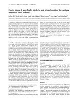
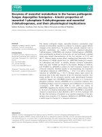
Southern blot hybridization
Data from sequencing, showing the presence of Lep d 2
with and without an intron, indicated that multiple copies of
theLepd2genearepresentintheL. destructor genome.
To investigate this possibility, Southern blot analysis was
performed on genomic L. destructor DNA. The DNA was
digested with each of four enzymes: EcoRI recognizing no
restriction site, TseIandBsaI recognizing single and BanI
recognizing two restriction sites in the Lep d 2 complete
DNA sequence. DNA encoding the full open reading frame
of Lep d 2 was PCR amplified, [
32
P]dCTP labelled and used
as a probe. Hybridization of the probe with EcoRI-cleaved
DNA gave rise to two bands (Fig. 3), and at least three
bands were seen when the DNA was cleaved with TseI, BsaI
or BanI (Fig. 3). These data support the idea that there is
more than one copy of the Lep d 2 gene at different loci in
the L. destructor genome.
Immunoblotting
The four main Lep d 2 variants, Lep d 2.0101,
Lep d 2.0102, Lep d 2.0201 and Lep d 2.0202 were
expressed as recombinant proteins and subjected to immu-
noblotting experiments in order to verify an IgE binding
ability. All variants were recognized by six different sera
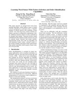
Fig. 1. Amino acid sequence alignment of Lep d 2 variants. Identities
with Lep d 2.0101 are indicated with full stops. Amino acids changed
using site-directed mutagenesis are indicated in bold. Arrowheads
markthesiteoftheintron.
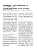
Lep d 2
Lep d 2.0101a/b
Lep d 2.0102
Lep d 2.01
Lep d 2.02
Lep d 2.0201
Lep d 2.0202
Fig. 2. Lep d 2 isoforms and variants. Lep d 2 is present as two distinct
isoforms, Lep d 2.01 and Lep d 2.02 differing in 13 amino acids. The
variants of each isoform differ only in a few amino acids with the
exception of Lep d 2.0101a and Lep d 2.0101b, which have identical
amino acid sequence but differ on the DNA level.
Table 2. Frequencies of Lep d 2 variants. PCR amplification and
sequence analysis of Lep d 2 performed as described in Materials and
methods revealed four variants differing at the amino acid level
between clones from wild and cultured L. destructor mites.
Lep d
2.0101
Lep d
2.0102
Lep d
2.0201
Lep d
2.0202
Wild L. destructor 10796
Cultured L. destructor 7781
Ó FEBS 2003 Lep d 2 polymorphisms in wild and cultured mites (Eur. J. Biochem. 270) 649
from subjects allergic to L. destructor (data not shown). An
L. destructor negative control serum detected none of the
variants.
ELISA inhibition
To investigate if the Lep d 2 variants found in cultured
and wild mites have different IgE binding properties, we
performed ELISA inhibition with the six sera positive to
all variants in immunoblotting. A dose-dependent inhibi-
tion was observed with all variants in all six sera. The IgE
binding capacity for the two isoforms was evaluated
separately. In all experiments 100% inhibition was
reached. Figure 4 shows the inhibition results obtained
with two of the six sera using the two variants of the
isoform Lep d 2.01. For both sera, a lower concentration
of the variant Lep d 2.0102 was needed to reach 50%
inhibition compared to the variant Lep d 2.0101 regardless
of whether homologous or heterologous inhibition was
performed. Similar results were obtained with the isoform
Lep d 2.02, where a lower concentration of the variant
Lep d 2.0202 was needed to reach 50% inhibition com-
pared to the Lep d 2.0201 variant, although the difference
was less pronounced. To evaluate the overall inhibition
results obtained with all six sera, we compared the
concentrations of the different Lep d 2 variants needed
for 50% IgE binding inhibition in each serum (Table 3).
The same pattern of inhibition could be seen with a lower
concentration needed of the variants Lep d 2.0102 and
Lep d 2.0202 compared to the variants Lep d 2.0101 and
Lep d 2.0201, respectively.
T cell responses
Interferon-c and IL-5 were measured in culture superna-
tants from long-term PBMC cultures stimulated with
Lep d 2.0101, Lep d 2.0102, Lep d 2.0201 or Lep d 2.0202.
Although there were differences in the amounts of IFN-c
and IL-5 released by PBMC from individual patients, no
consistent pattern of difference was found between the
variants from either isoform, Lep d 2.0101 vs. Lep d 2.0102
or Lep d 2.0201 vs. Lep d 2.0202 in the six subjects
investigated. However, the IFN-c levels after stimulation
with the variants from the Lep d 2.02 isoform (median
16.3 ngÆmL
)1
; range 7.57–105) were slightly higher com-
pared to after stimulation with the Lep d 2.01 variants
(median 14.1 ngÆmL
)1
; range 3.08–80.0). The levels for IL-5
in response to the Lep d 2.01 isoform (median
2.75 ngÆmL
)1
; range 0.42–6.05) was similar to Lep d 2.02
isoform (median 2.23 ngÆmL
)1
; range 0.53–3.93).
Discussion
It is well known that several dust mite allergens exist as
different isoforms due to polymorphisms in the genes [30].
Differences between allergens from cultured and wild mites
have also been reported [3,4]. In routine diagnostics, extracts
from cultured L. destructor mites are used to detect
sensitized individuals. However, it is not cultured mites that
cause sensitization, but the wild mites found in our
environment. Therefore, we decided to study polymorphism
in the Lep d 2 gene of wild and cultured mites and what
influence polymorphic residues might have on IgE binding
and T cell responses.
In analogy with results obtained from sequence analyses
of Der p 2 [4], sequencing of the Lep d 2 gene revealed
several silent mutations but only a few mutations resulted in
amino acid substitutions. Furthermore, all analysed clones
were highly homologous to the previously published
Lep d 2 cDNA sequences [20], either to Lep d 2.0101 or
Lep d 2.0201. These results indicate the evolutionary
divergence of two sequences corresponding to two main
isoforms, a pattern similar to what has been found earlier
for Der p 2 [4]. Our data show that the two most common
substitutions of each isoform were found at positions 55 and
102 in clones otherwise identical to Lep d 2.0101 and
Lep d 2.0201, respectively. Both substitutions were the
result of single nucleotide exchanges. The possibility that
these substitutions represent PCR artefacts is not likely,
because they were found in several clones from different
mites and different PCR amplifications. Moreover, we used
Pfu polymerase that has a 3¢ to 5¢ exonuclease activity
1
0.5 kb
1kb
2kb
3kb
6kb
5kb
4kb
2
34
Fig. 3. Southern blot analysis of Lep d 2. Genomic L. destructor DNA
digested with each of four enzymes: EcoRI recognizing no restriction
site, TseIandBsaI recognizing single and BanI recognizing two
restriction sites in the Lep d 2 complete DNA. After electrophoresis
theDNAwastransferredtoamembraneandhybridizedwith
[
32
P]dCTP labelled, PCR amplified DNA encoding Lep d 2. Lane 1,
BsaI; lane 2, BanI; lane 3, EcoRI; lane 4, TseI.
650 L. Kaiser et al. (Eur. J. Biochem. 270) Ó FEBS 2003
resulting in a low error rate. The source of the commercially
available mites used in this study are cultured mites that
have been grown isolated for years without introducing new
mites from other sources (A. Anderson, Allergon AB,
A
¨
ngelholm, Sweden, personal communication). This could
explain the disparate frequency of the variants found in
cultured and wild mites.
Two amplified Lep d 2 sequences were obtained in PCR
amplifications with single mites as templates, and sequence
analysis revealed the presence of a small intron in one of the
amplified Lep d 2 PCR products. The size of the intron
correlates well with introns previously reported in Der p 2
(80–83 bp) [3] and Der f 2 (87 bp) [2]. This finding indicates
the presence of more than one copy of the Lep d 2 gene at
different loci in the genome and was further supported by
Southern blot hybridization experiments. In contrast to
Der f 2, for which there is only a single gene in the genome
[2], the substitutions found in the Lep d 2 gene are probably
not only due to polymorphisms within the Lep d 2 gene,
but also to multiple copies of the gene in individual mites.
The new Lep d 2 variants, Lep d 2.0102 and Lep
d 2.0202 identified in the present study, were expressed
as recombinant proteins and their IgE binding capacity
evaluated by ELISA inhibition and compared to those of
Lep d 2.0101 and Lep d 2.0201. The results did not reveal
any major differences in IgE binding capacity between the
variants. However, we found that Lep d 2.0102 inhibited
the binding to Lep d 2.0101 to a somewhat higher degree
than what was obtained with Lep d 2.0101. A possible
explanation for this finding could be that IgE antibodies
show higher avidity to Lep d 2.0102 than to Lep d 2.0101.
Similar results were obtained with isoform Lep d 2.02
where Lep d 2.0202 was found to be slightly more effective
as inhibitor than Lep d 2.0201. Studies of antibody
epitopes in Der p 2, have shown that residues at position
55 and 102 are within the predicted epitopes and could be
important in IgE binding [31]. The crystal structure of
Der p 2 has been used in standard homology modelling to
predict the secondary and tertiary structure of
Lep d 2.0101 [32]. According to this model, amino acids
µg/ml
% inhibition
0
20
40
60
80
100
0.001 0.01 0.1 1 10
A
solid phase
Lep d 2.0101
µg/ml
% inhibition
0
20
40
60
80
100
0.001 0.01 0.1 1 10
B
solid phase
Lep d 2.0102
µg/ml
% inhibition
0
20
40
60
80
100
0.001 0.01 0.1 1 10
C
solid phase
Lep d 2.0101
µg/ml
% inhibition
0
20
40
60
80
100
0.001 0.01 0.1 1 10
D
solid phase
Lep d 2.0102
Fig. 4. ELISA inhibition. ELISA inhibition of IgE-binding to Lep d 2.0101 (A and C), and Lep d 2.0102 (B and D) on solid phase. Inhibition
curves obtained with Lep d 2.0101 (s), Lep d 2.0102 (h) as indicated in the figure. Serum no. 6 was used in A and B and serum no. 4 in C and D.
Table 3. Concentrations (lgÆmL
-1
) of the inhibiting allergen needed to reach 50% inhibition of IgE binding. ELISA inhibition was performed as
described in Materials and methods. Concentrations of the inhibiting allergen needed to reach 50% inhibition of IgE binding to Lep d 2.0101 and
Lep d 2.0102 and to Lep d 2.0201 and Lep d 2.0202 in L. destructor positive sera.
Serum
no.
Lep d 2.0101 solid phase Lep d 2.0102 solid phase Lep d 2.0201 solid phase Lep d 2.0202 solid phase
Lep d 2.0101 Lep d 2.0102 Lep d 2.0101 Lep d 2.0102 Lep d 2.0201 Lep d 2.0202 Lep d 2.0201 Lep d 2.0202
1 0.029 0.006 0.020 0.007 0.014 0.004 0.008 0.004
2 0.100 0.045 0.081 0.048 0.055 0.043 0.051 0.043
3 0.195 0.090 0.206 0.215 0.058 0.047 0.055 0.047
4 0.085 0.074 0.083 0.074 0.079 0.064 0.085 0.070
5 0.238 0.062 0.206 0.064 0.047 0.029 0.047 0.030
6 0.471 0.091 0.424 0.095 0.060 0.049 0.062 0.051
Ó FEBS 2003 Lep d 2 polymorphisms in wild and cultured mites (Eur. J. Biochem. 270) 651
55 and 102 are buried inside the protein core of Lep d 2
and not exposed on the surface of the molecule
(D. Benjamin, The Asthma and Allergic Disease Center,
University of Virginia, VA, USA, personal communica-
tion). The difference in IgE binding can not be caused by
direct antibody interaction of the side chains of the
variable amino acid residues; rather it must be caused by
more subtle changes in the tertiary structure.
The levels of two cytokines were measured in long-term
PBMC cultures after stimulation with the four Lep d 2
variants. The cytokines measured were IFN-c,atypicalTh1
cytokine, and IL-5, a cytokine present in allergic inflam-
mation. No consistent differences were found between the
different variants. Using peptides that represent all parts of
the mature Lep d 2, a previous study has shown that
Lep d 2 contains two immuno-dominant regions spanning
amino acids 11–25 and 61–75 [25]. The fact that we did not
see any difference between the Lep d 2 variants differing at
position 55 and 102 is therefore not surprising. However,
individual differences between patients in cytokine responses
could be detected.
In this study, we discovered new variants of Lep d 2 and
found differences in the frequency of the variants between
wild and cultured mites. In addition, our data suggest the
presence of two genes, one with and one without an intron,
encoding Lep d 2. After analysing the importance of these
differences regarding IgE binding and T cell responses
in vitro, we found that the differences between wild and
cultured mites have no major impact on the allergenicity of
Lep d 2. We can thus conclude that the commercially
available cultured mites used in diagnostics and research
today show the same general pattern of polymorphism in
the Lep d 2 gene as the mites that are found in the
environment and cause sensitization, at least in Sweden.
Whether this holds true for other allergens in L. destructor
and for mites from other geographical locations remains to
be investigated.
Acknowledgements
WethankDrSvenBostro
¨
m at the Swedish Museum of Natural
History, Stockholm, for skilful identification and isolation of
L. destructor mites. This work was supported by grants from the
Swedish Foundation for Health Care Sciences and Allergy Research,
the Swedish Medical Research Council, the Swedish Asthma and
Allergy Association, the Swedish Council for Working Life and Social
Research, the Hesselman Foundation, King Gustaf V 80th Birthday
Foundation, the Magnus Bergvalls Foundation, Konsul Th C Berghs
Foundation, A
˚
ke Wibergs Foundation and the Karolinska Institute.
References
1. Thomas, W.R., Chua, K.Y. & Smith, W.A. (1992) Molecular
polymorphisms of house dust mite allergens. Exp. Appl. Acarol.
16, 153–164.
2. Yuuki, T., Okumura, Y. & Okudaira, H. (1997) Genomic orga-
nization and polymorphisms of the major house dust mite allergen
Der f 2. Int. Arch. Allergy Immunol. 112, 44–48.
3. Chua, K.Y., Huang, C.H., Shen, H.D. & Thomas, W.R. (1996)
Analysis of sequence polymorphism of a major mite allergen, Der
p2.Clin.Exp.Allergy26, 829–837.
4. Smith, W.A., Hales, B.J., Jarnicki, A.G. & Thomas, W.R. (2001)
Allergens of wild house dust mites: environmental Der p 1 and Der
p 2 sequence polymorphisms. J. Allergy Clin. Immunol. 107, 985–
992.
5. Terho, E.O., Leskinen, L., Husman, K. & Karenlampi, L. (1982)
Occurrence of storage mites in Finnish farming environments.
Allergy 37, 15–19.
6. Bostro
¨
m, S.J.E., Ha
¨
rfast, B., Lundqvist, L., Ba
¨
ckman, L., von
Rosen, E. & van Hage-Hamsten, M. (1997) Characterisation of
the mite fauna (Acari) in Swedish barn dust. Int. J. Acarol. 23,
127–132.
7. Franz, J.T., Masuch, G., Musken, H. & Bergmann, K.C. (1997)
Mite fauna of German farms. Allergy 52, 1233–1237.
8. Warner, A., Bostrom, S., Moller, C. & Kjellman, N.I. (1999) Mite
fauna in the home and sensitivity to house-dust and storage mites.
Allergy 54, 681–690.
9. Gaig, P., Botey, J., Pena, M., Marin, A. & Eseverri, J.L. (1993)
Study of the sensitization to storage mites in a pediatric population
in Barcelona. J. Invest. Allergol. Clin. Immunol. 3, 151–155.
10. Ebner, C., Feldner, H., Ebner, H. & Kraft, D. (1994) Sensitization
to storage mites in house dust mite (Dermatophagoides pter-
onyssinus) allergic patients. Comparison of a rural and an urban
population. Clin. Exp. Allergy 24, 347–352.
11. Bernd, L.A., Ambrozio, L.C. & Baggio, D. (1996) Storage mite
allergy in perennial rhinitis patients not sensitized to house dust
mites. J. Invest. Allergol. Clin. Immunol. 6, 94–97.
12. van der Heide, S., Niemeijer, N.R., Hovenga, H., de Monchy,
J.G., Dubois, A.E. & Kauffman, H.F. (1998) Prevalence of
sensitization to the storage mites Acarus siro, Tyrophagus
putrescentiae,andLepidoglyphus destructor in allergic patients
with different degrees of sensitization to the house-dust mite
Dermatophagoides pteronyssinus. Allergy 53, 426–430.
13. Gislason, D. & Gislason, T. (1999) IgE-mediated allergy to
Lepidoglyphus destructor in an urban population – an epidemio-
logic study. Allergy 54, 878–883.
14. Musken, H., Franz, J.T., Wahl, R., Paap, A., Cromwell, O.,
Masuch, G. & Bergmann, K.C. (2000) Sensitization to different
mite species in German farmers: clinical aspects. J. Invest. Allergol.
Clin. Immunol. 10, 346–351.
15. Cuthbert, O.D., Brostoff, J., Wraith, D.G. & Brighton, W.D.
(1979) ÔBarn allergyÕ: asthma and rhinitis due to storage mites.
Clin. Allergy 9, 229–236.
16. van Hage-Hamsten, M., Johansson, S.G., Hoglund, S., Tull, P.,
Wiren, A. & Zetterstrom, O. (1985) Storage mite allergy is com-
mon in a farming population. Clin. Allergy 15, 555–564.
17. Iversen, M. & Pedersen, B. (1990) The prevalence of allergy in
Danish farmers. Allergy 45, 347–353.
18. Kronqvist, M., Johansson, E., Pershagen, G., Johansson, S.G. &
van Hage-Hamsten, M. (1999) Increasing prevalence of asthma
over 12 years among dairy farmers on Gotland, Sweden: storage
mites remain dominant allergens. Clin. Exp. Allergy 29, 35–41.
19. Derewenda, U., Li, J., Derewenda, Z., Dauter, Z., Mueller, G.A.,
Rule, G.S. & Benjamin, D.C. (2002) The crystal structure of a
major dust mite allergen Der p 2, and its biological implications.
J. Mol. Biol. 318, 189–197.
20. Schmidt, M., Olsson, S., van der Ploeg, I. & van Hage-Hamsten,
M. (1995) cDNA analysis of the mite allergen Lep d 1 identifies
two different isoallergens and variants. FEBS Lett. 370, 11–14.
21. WHO/IUS (1995) Allergen nomenclature. WHO/IUS Allergen
Nomenclature Subcommittee World Health Organization,
Geneva, Switzerland. Clin. Exp. Allergy 25, 27–37.
22. Olsson, S., van Hage-Hamsten, M., Whitley, P., Johansson, E.,
Hoffman, D.R., Gafvelin, G. & Schmidt, M. (1998) Expression of
two isoforms of Lep d 2, the major allergen of Lepidoglyphus
destructor, in both prokaryotic and eukaryotic systems. Clin. Exp.
Allergy 28, 984–991.
23. Kronqvist, M., Johansson, E., Magnusson, C.G., Olsson, S.,
Eriksson, T.L., Gafvelin, G. & van Hage-Hamsten, M. (2000)
652 L. Kaiser et al. (Eur. J. Biochem. 270) Ó FEBS 2003
Skin prick test and serological analysis with recombinant group 2
allergens of the dust mites L. destructor and T. putrescentiae. Clin.
Exp. Allergy 30, 670–676.
24. Johansson, E., Eriksson, T.L., Olsson, S., Kronqvist, M., Whitley,
P.,Johansson,S.G.,Gafvelin,G.&vanHage-Hamsten,M.
(1999) Evaluation of specific IgE to the recombinant group 2 mite
allergens Lep d 2 and Tyr p 2. the Pharmacia CAP system. Int.
Arch. Allergy Immunol. 120, 43–49.
25. Eriksson, T.L., Gafvelin, G., Elfman, L.H., Johansson, C., Van
Hage-Hamsten, M. & Olsson, S. (2001) T cell responses to
recombinant isoforms, synthetic peptides and a mutant variant of
Lep d 2, a major allergen from the dust mite Lepidoglyphus
destructor. Clin. Exp. Allergy 31, 1881–1890.
26. Sammbrook, J., Fritsch, E.F. & Maniatis, T. (1989) Molecular
Cloning: a Laboratory Manual, 2nd edn. Cold Spring. Harbour
Laboratory Press, Cold Spring Harbour, New York, USA.
27. Johansson, E., Johansson, S.G. & Van Hage-Hamsten, M. (1994)
Allergenic characterization of Acarus siro and Tyrophagus
putrescentiae and their crossreactivity with Lepidoglyphus
destructor and Dermatophagoides pteronyssinus. Clin.Exp.Allergy
24, 743–751.
28. Tengvall Linder, M., Johansson, C., Bengtsson, A., Holm, L.,
Harfast, B. & Scheynius, A. (1998) Pityrosporum orbiculare-
reactive T-cell lines in atopic dermatitis patients and healthy
individuals. Scand.J.Immunol.47, 152–158.
29. Mount, S.M. (1982) A catalogue of splice junction sequences.
Nucleic Acids Res. 10, 459–472.
30. Thomas, W.R. & Smith, W. (1998) House-dust-mite allergens.
Allergy 53, 821–832.
31. Mueller, G.A., Smith, A.M., Chapman, M.D., Rule, G.S. &
Benjamin, D.C. (2001) Hydrogen exchange nuclear magnetic
resonance spectroscopy mapping of antibody epitopes on the
house dust mite allergen Der p 2. J. Biol. Chem. 276, 9359–9365.
32. Gafvelin, G., Johansson, E., Lundin, A., Smith, A.M., Chapman,
M.D., Benjamin, D.C., Derewenda, U. & van Hage-Hamsten, M.
(2001) Cross-reactivity studies of a new group 2 allergen from the
dust mite Glycyphagus domesticus,Glyd2,andgroup2allergens
from Dermatophagoides pteronyssinus, Lepidoglyphus destructor,
and Tyrophagus putrescentiae with recombinant allergens.
J. Allergy Clin. Immunol. 107, 511–518.
Ó FEBS 2003 Lep d 2 polymorphisms in wild and cultured mites (Eur. J. Biochem. 270) 653