Báo cáo khoa học: Functional expression of the quinoline 2-oxidoreductase genes (qorMSL) in Pseudomonas putida KT2440 pUF1 and in P. putida 86-1 Dqor pUF1 and analysis of the Qor proteins doc
Bạn đang xem bản rút gọn của tài liệu. Xem và tải ngay bản đầy đủ của tài liệu tại đây (288.08 KB, 11 trang )
Functional expression of the quinoline 2-oxidoreductase genes
(
qorMSL
)in
Pseudomonas putida
KT2440 pUF1 and in
P. putida
86-1
D
qor
pUF1 and analysis of the Qor proteins
Ursula Frerichs-Deeken
1
, Birgit Goldenstedt
1,2,
*, Renate Gahl-Janßen
1
, Reinhard Kappl
3
,
Ju¨ rgen Hu¨ ttermann
3
and Susanne Fetzner
1,2,
*
1
AG Mikrobiologie, Institut fu
¨
r Chemie und Biologie des Meeres, Carl von Ossietzky Universita
¨
t Oldenburg, Germany;
2
Institut fu
¨
r Mikrobiologie, Westfa
¨
lische Wilhelms-Universita
¨
tMu
¨
nster, Germany;
3
Fachrichtung Biophysik und
Physikalische Grundlagen der Medizin, Universita
¨
t des Saarlandes, Homburg/Saar, Germany
The availability of a system for the functional expression of
genes coding for molybdenum hydroxylases is a prerequisite
for the construction of enzyme variants by mutagenesis. For
the expression cloning of quinoline 2-oxidoreductase (Qor)
from Pseudomonas putida 86 – that contains the molybdo-
pterin cytosine dinucleotide molybdenum cofactor
(Mo-MCD), two distinct [2Fe)2S] clusters and FAD – the
qorMSL genes were inserted into the broad host range
vector, pJB653, generating pUF1. P. putida KT2440 and
P. putida 86-1 Dqor were used as recipients for pUF1.
Whereas Qor from the wild-type strain showed a specific
activity of 19–23 UÆmg
)1
, the specific activity of Qor purified
from P. putida KT2440 pUF1 was only 0.8–2.5 UÆmg
)1
,
and its apparent k
cat
(quinoline) was about ninefold lower
than that of wild-type Qor. The apparent K
m
values for
quinoline were similar for both proteins. UV/visible and
EPR spectroscopy indicated the presence of the full set of
[2Fe)2S] clusters and FAD in Qor from P. putida
KT2440 pUF1, however, the very low intensity of the
Mo(V)-rapid signal, that occurs in the presence of quinoline,
as well as metal analysis indicated a deficiency of the
molybdenum center. In contrast, the metal content, and the
spectroscopic and catalytic properties of Qor produced by
P. putida 86-1 Dqor pUF1 were essentially like those of
wild-type Qor. Release of CMP upon acidic hydrolysis of the
Qor proteins suggested the presence of the MCD form of the
pyranopterin cofactor; the CMP contents of the three
enzymes were similar.
Keywords: quinoline 2-oxidoreductase; molybdenum hydro-
xylase; expression cloning; molybdopterin cytosine dinucleo-
tide; Pseudomonas sp.
Quinoline 2-oxidoreductase (Qor) from Pseudomonas
putida 86 catalyses the formation of 1H-2-oxoquinoline
(2-hydroxyquinoline) from quinoline [1,2]. Besides quino-
line, some quinoline derivatives and the benzodiazines
quinazoline and quinoxaline are accepted as substrates [1,3].
Like other enzymes catalysing the hydroxylation of
N-heteroaromatic rings at positions that are susceptible to
nucleophilic attack, Qor belongs to the family of molyb-
denum hydroxylases that introduce an oxygen atom
(originating from water) into their substrate according to
the following stoichiometry: R-H + H
2
O fi R-OH +
2[e
–
]+2H
+
. Due to a common structure of their molyb-
denum center and due to significant amino acid sequence
similarity to xanthine oxidases/xanthine dehydrogenases,
the molybdenum hydroxylases have also been classified as
enzymes belonging to the Ôxanthine oxidase familyÕ [4–7].
Molybdenum hydroxylases basically contain the same type
of redox centers constituting an intramolecular electron
transport chain, namely a molybdenum ion, that is the site
of substrate hydroxylation, two distinct [2Fe)2S] clusters,
and – in most cases – FAD [5,8,9]. The molybdenum is
bound to the sulfur atoms of the ene-dithiolate function of a
unique pyranopterin cofactor. Other coordination positions
to the molybdenum are occupied by a sulfido and an oxo
ligand, and a catalytically labile )OH group or H
2
Omole-
cule [5–7,10–12]. Whereas almost all known xanthine dehy-
drogenases contain a pyranopterin derivative, known as
molybdopterin (MPT), as the organic part of the moly-
bdenum cofactor [13], Qor [2,14] as well as isoquinoline
1-oxidoreductase [15], quinaldine 4-oxidase [3], nicotinate
dehydrogenase [16], isonicotinate and 2-hydroxyisonicoti-
nate dehydrogenase [16,17], CO dehydrogenases [18–20]
and the aldehyde oxidoreductases belonging to the xanthine
oxidase family [11,12,21,22], contain Mo-MPT that is
modified by covalent attachment of cytidine monophos-
phate to its terminal phosphate group to form molybdenum
molybdopterin cytosine dinucleotide (Mo-MCD).
Correspondence to S. Fetzner, Institut fu
¨
r Mikrobiologie,
Westfa
¨
lische Wilhelms-Universita
¨
tMu
¨
nster, Corrensstr. 3,
D-48149 Mu
¨
nster, Germany.
Fax: +49 251 83 38388, Tel.: +49 251 83 39824,
E-mail:
Abbreviations: Mo-MCD, molybdopterin cytosine dinucleotide
form of the molybdenum pyranopterin cofactor; Mo-MGD, molyb-
dopterin guanine dinucleotide form of the molybdenum pyranopterin
cofactor; MPT, molybdopterin; Qor, quinoline 2-oxidoreductase.
Enzymes: Quinoline 2-oxidoreductase; quinoline:acceptor 2-oxido-
reductase (hydroxylating) (EC 1.3.99.17).
*Present address: Institut fu
¨
r Mikrobiologie, Westfa
¨
lische Wilhelms-
Universita
¨
tMu
¨
nster, Germany.
(Received 20 November 2002, revised 3 February 2003,
accepted 19 February 2003)
Eur. J. Biochem. 270, 1567–1577 (2003) Ó FEBS 2003 doi:10.1046/j.1432-1033.2003.03526.x
The genes encoding Qor have been cloned and sequenced,
some biochemical properties of Qor have been described,
and its redox-centers have been characterized by EPR
spectroscopy [1,2,23–25]. However, a thorough study of the
catalytic mechanism of Qor and other molybdenum
hydroxylases should also involve the construction of protein
variants carrying distinct amino acid replacements, and
their biochemical, spectroscopic, and – if possible – struc-
tural characterization. A prerequisite for such a mutagenic
approach is the availability of a suitable system for the
manipulation and the regulated, functional expression of
genes coding for molybdenum hydroxylases. Whereas genes
coding for Mo-MPT- or molybdenum molybdopterin
guanine dinucleotide- (Mo-MGD-) containing hydroxylases
have been expressed successfully in Escherichia coli hosts
[26–28], attempts to achieve heterologous functional
expression of Mo-MCD-containing enzymes in E. coli
failed [23,29] (K. Parschat & S. Fetzner, unpublished
results). However, in E. coli, all known molybdoenzymes
contain the MGD form of the molybdenum cofactor.
Synthesis of Mo-MGD from Mo-MPT and Mg
2+
-GTP is
catalyzed by the MobA protein [30–36]. Possibly, E. coli
lacks an enzyme that catalyses the formation of Mo-MCD
from Mo-MPT, and/or it is not able to integrate the
Mo-MCD cofactor into the corresponding apoprotein.
In a first attempt to functionally express genes coding
for a Mo-MCD-containing hydroxylase in heterologous
hosts, the iorAB genes of Brevundimonas diminuta 7,
coding for isoquinoline 1-oxidoreductase, were cloned in
P. putida KT2440 and in the quinoline degrading strain,
P. putida 86. However, the level of Ior synthesis was very
low in both expression clones, and only P. putida
86 pIL1 produced Ior protein that was catalytically
active [37].
As it is highly desirable to obtain an expression system for
molybdenum hydroxylases harboring the MCD cofactor,
we tested whether expression of the qorMSL genes from
P. putida 86 in P. putida host strains results in the forma-
tion of catalytically competent enzyme.
Materials and methods
Plasmids, bacterial strains and growth conditions
Plasmids and bacterial strains used in this work are listed in
Table 1. E. coli XL-1 Blue MRF¢ and E. coli S17-1 were
grown at 37 °C in Luria–Bertani (LB) broth [38]. P. put-
ida 86 was grown in mineral salts medium containing
quinoline as the sole carbon source [2], or in LB broth [38],
at 30 °C. For the preparation of P. putida cells that are
competent for electroporation, TB medium (Terrific broth)
[38] was used. When growing P. putida 86-1, streptomycin
(500 lgÆmL
)1
)wasaddedtotherespectivemedium.
DNA techniques
Standard recombinant DNA techniques were used for
DNA isolation [38,39] and restriction, agarose gel electro-
phoresis and cloning [38]. Random digoxigenin labelling of
probes was performed using the DIG High Prime Labeling
and Detection Kit (Roche Diagnostics). Competent E. coli
and P. putida cells for electroporation were generated as
described by Dower et al.[40]andIwasakiet al.[41],
respectively.
Construction of
P. putida
86-1 D
qor
A DNA segment containing the qorMSL genes and flanking
regions (Ôqor-upÕ,1055bpandÔqor-downÕ, 1898 bp) was
inserted into the SmaI restriction site of pUC18 [42],
forming pBG1. Competent E. coli XL-1 Blue MRF¢ cells
were transformed with pBG1 by electroporation. The
qorMSL genes in pBG1 were removed using XhoI, that
cleaves 364-bp upstream of the start codon of qorM,and
DraIII, that cleaves 8-bp downstream of the stop codon of
qorL. After removing the 3¢ overhang and filling the 5¢
overhang of the plasmid with T4 DNA-polymerase, a PCR
amplificate of nptII [43], that contained flanking XhoIand
DraIII sites, was inserted by blunt-end ligation, resulting in
the two constructs, pBG2a and pBG2b (nptII in the same
and in the opposite orientation with respect to the deleted
qor genes, respectively). E. coli XL-1 Blue MRF¢ was used
as host strain for pBG2a and pBG2b. The nptII inserts
together with the flanking regions (Ôqor-upÕ and Ôqor-downÕ)
were removed from pBG2a and pBG2b using HindIII, and
inserted into the HindIII restriction site of pSUP202 [44],
resulting in pBG3a and pBG3b. Competent E. coli S17-1
cells were transformed with pBG3a and pBG3b. Mating of
E. coli S17-1 pBG3a/3b and P. putida 86-1 was performed
as described by Masepohl et al.[45],exceptthatLBplates
were used instead of PY plates. P. putida 86-1 transconju-
gants were selected for kanamycin resistance and chloram-
phenicol sensitivity, indicating replacement of qorMSL
in P. putida 86-1 by nptII by double cross-over events.
Mutants with nptII in the same orientation (P. putida 86-1
Km-a) as well as mutants with nptII in the opposite
orientation (P. putida 86-1 Km-b) with respect to the
deleted qor genes were obtained. DNA isolated from
these mutants did not hybridize with a DIG-labelled
probe for pSUP202, confirming that nptII actually was
inserted by double cross-over. However, DNA from these
mutants still showed a positive hybridization signal with a
DIG-labelled probe for the qor genes (corresponding to the
nucleotides 1201–4233 of GenBank accession number
X98131), and the P. putida 86-1 kanamycin resistant
mutants still formed Qor. PCR analyses confirmed that
nptII was replacing one copy of qorMSL and that P. putida
86-1 contains more than one copy of the qor genes and their
flanking regions.
The plasmid pBG3a was digested with XhoIandDraIII
to remove nptII. After the removal of the 3¢ overhang and
the filling of the 5¢ overhang of the plasmid with T4 DNA-
polymerase, a PCR amplificate of aacC1 [46] was inserted
by blunt end ligation, resulting in pBG4a and pBG4b
(aacC1 in the same and in the opposite orientation with
respect to the deleted qor genes, respectively), that were used
to transform E. coli S17-1. Mating of E. coli S17-1 pBG4a/
4b and P. putida 86-1 Km-a/P. putida 86-1 Km-b yielded
three Kan
r
and Gen
r
mutants of P. putida 86-1 with a Qor
–
phenotype. DNA isolated from these three P. putida 86-1
Dqor (Kan
r
Gen
r
) mutants did not hybridize with the probes
for pSUP202 and qor. PCR analyses confirmed the
complete deletion of the qor genes and showed that all
three mutants contained nptII in the same orientation with
1568 U. Frerichs-Deeken et al. (Eur. J. Biochem. 270) Ó FEBS 2003
respect to the deleted qorMSL genes, and aacC1 in the
opposite orientation with respect to the deleted second copy
of qorMSL.
Expression cloning of
qorMSL
genes
Using genomic DNA isolated from wild-type P. putida 86
as template, the qorMSL genes, including the preceding
Shine-Dalgarno sequence [23] (GenBank accession
number X98131), were amplified using 5¢-GCAGgaattc
CTGCTGGTTTTTCGCTTG-3¢ as the forward primer
and 5¢-ATAGggatccCTGGTAGACAGGACTCACCC-3¢
as the reverse primer in the Expand Long Template PCR
System (Roche Diagnostics). The nucleotides of the forward
and reverse primer that are set as bold are complementary
to nucleotides 653–670 and 4439–4420 of GenBank acces-
sion number X98131, respectively. The primers included an
EcoRI and a BamHI recognition site in the forward and
reverse primer, respectively (small letters), that allowed the
ligation of the PCR product into the multiple cloning site of
pJB653, generating pUF1. The recipient strains P. putida
KT2440 and P. putida 86-1 Dqor were transformed by
electroporation [40]. Clones containing pUF1 were identi-
fied by colony blotting and hybridization [47] using the qor
probe described above.
Growth of recombinant strains and preparation
of crude extracts
All P. putida pUF1 clones were grown in the presence of
500 lgÆmL
)1
ampicillin in mineral salts medium [2] supple-
mented with 1 gÆL
)1
ammonium sulfate. Induction of
qorMSL expression from the Pm promoter of pUF1 was
achieved by addition to the medium of the XylS effectors,
benzoate and 2-methylbenzoate. For small-scale growth of
P. putida KT2440 pUF1 clones, succinate (10 gÆL
)1
)and
sodium benzoate (8 m
M
) were used as sources of carbon.
Two 4 L glass fermenters were used to generate biomass for
protein purification. Benzoate (8 m
M
) was used as the
carbon and energy source for growth of P. putida KT2440
pUF1; it was added repeatedly to the cultures. 2-Methyl-
benzoate (2 m
M
), as an additional XylS effector, was added
Table 1. Bacterial strains and plasmids used in this study.
Strain/plasmid Genotype and/or relevant properties
Reference
or source
Escherichia coli S17-1 RP4-2 (Tc::Mu) (Km::Tn7) integrated into the chromosome; Tra
+
, recA, pro, thi, hsdR [44]
E. coli XL-1 Blue MRF¢ D(mcrA)183, D(mcrCB-hsdSMR-mrr)173, endA1, supE44, thi-1, rec A1, gyrA96 relA1
lac [F¢ proAB lacI
q
ZDM15 Tn10 (Tet
r
)] Stratagene
Pseudomonas putida KT2440 r
–
derivative of P. putida mt-2 [67]
P. putida 86 Wild-type strain utilizing quinoline as sole source of carbon and energy [68]
P. putida 86-1 Spontaneous Str
r
mutant of P. putida 86 This work
P. putida 86-1 Km-a nptII replacing one copy of qorMSL in P. putida 86-1; nptII in the same orientation
with respect to the deleted qor genes; Str
r
, Kan
r
,Qor
+
This work
P. putida 86-1 Km-b nptII replacing one copy of qorMSL in P. putida 86-1; nptII in the opposite orientation
with respect to the deleted qor genes; Str
r
, Kan
r
,Qor
+
This work
P. putida 86-1 Dqor Two copies of qorMSL replaced by nptII and aacC1, respectively; nptII is in the same
orientation with respect to the deleted qorMSL genes, and aacC1 is in the opposite orientation
with respect to the deleted second copy of qorMSL. Str
r
, Kan
r
, Gen
r
;Qor
–
This work
pUC18 ori
colE1
, lacZ, Amp
r
[42]
pSUP202 RP4-Mob
+
ori
colE1
; Amp
r
, Cam
r
, Tet
r
[44]
pBG1 6678 bp segment of P. putida 86 DNA (qorMSL and flanking regions Ôqor upÕ [1898 bp]
and Ôqor downÕ [1055 bp]) cloned into SmaI site of pUC18 This work
pBG2a derivative of pBG1: 4097 bp XhoI-DraII fragment containing qorMSL replaced by nptII;
nptII in the same orientation with respect to the deleted qor genes This work
pBG2b derivative of pBG1: 4097 bp XhoI-DraII fragment containing qorMSL replaced by nptII;
nptII in the opposite orientation with respect to the deleted qor genes This work
pBG3a HindIII fragment of pBG2a containing nptII together with flanking regions
Ôqor-upÕ (1534 bp) and Ôqor-downÕ (1047 bp) cloned into the HindIII restriction site
of pSUP202 This work
pBG3b HindIII fragment of pBG2b containing nptII together with flanking regions
Ôqor-upÕ (1534 bp) and Ôqor-downÕ (1047 bp) cloned into the HindIII restriction site
of pSUP202 This work
pBG4a nptII in pBG3a replaced by aacC1; aacC1 in the same orientation with respect to the
deleted qor genes This work
pBG4b nptII in pBG3a replaced by aacC1; aacC1 in the opposite orientation with respect to the
deleted qor genes This work
pJB653 Broad-host-range cloning vector; Pm promoter, xylS for transcriptional regulation; Amp
r
[52]
pUF1 qorMSL (3786 bp PCR amplificate from P. putida 86 DNA) inserted into
EcoRI – BamHI sites of pJB653 This work
Ó FEBS 2003 Expression of quinoline 2-oxidoreductase genes (Eur. J. Biochem. 270) 1569
at a D
550
value of 0.7–1.0. P. putida 86-1 Dqor pUF1 was
either grown in benzoate (8 m
M
, fed repeatedly), or in
benzoate (5 m
M
)plus1H-2-oxoquinoline (2.8 m
M
)as
carbon sources (fed repeatedly). 2-Methylbenzoate was
added at a D
550
value of 0.7–1.0. P. putida KT2440 pUF1
as well as P. putida 86-1 Dqor pUF1 cells were harvested by
centrifugation (5 500 g,20min)ataD
550
value ‡ 3.0.
Crude extracts were prepared by French
TM
Press
treatment at 2.1–2.4 · 10
8
Pa of cell suspensions in
100 m
M
Tris/HCl buffer (pH 8.5) containing 10 l
M
phe-
nylmethanesulfonylfluoride and 0.05 lLÆml
)1
Benzon nuc-
lease (Merck, Darmstadt, Germany), subsequent
sonification, and removal of debris by centrifugation
(48 000 g,45min,4°C).
PAGE
Non-denaturing PAGE was performed using the high pH
discontinuous system according to Hames [48], and 10%
and 4% acrylamide (w/v) in the separating and stacking
gels, respectively. SDS/PAGE was performed according to
the method of Laemmli [49]. Proteins were stained in
Coomassie blue R-250 [0.1% (w/v) in 50% (w/v) aqueous
trichloroacetic acid], and de-stained in water/methanol/
acetic acid (60 : 30 : 10, v/v/v).
Purification of Qor from
P. putida
86,
P. putida
KT2440 pUF1 and
P. putida
86-1 D
qor
pUF1
Qor was purified using ammonium sulfate fractionation
(0.8–1.5
M
), hydrophobic interaction chromatography [phe-
nyl Sepharose CL-4B (Amersham Pharmacia, Freiburg,
Germany) packed into a 15 · 113 mm BioScale MT20
column (Bio-Rad, Mu
¨
nchen, Germany)], and anion
exchange chromatography (BioScale DEAE10 column,
Bio-Rad) essentially as described by Tshisuaka et al.[2],
but omitting the heat precipitation step.
Preparation of anti-Qor antisera
Polyclonal rabbit Igs were raised against Qor that was
purified from wild-type P. putida 86. An initial subcuta-
neous injection of Qor protein was followed by boost
injections on days 14, 28 and 56, and the sera were collected
on day 87 (Eurogentec, Belgium).
Western blotting, and immunodetection of Qor protein
Proteins separated in SDS/PAGE were transferred onto
nitrocellulose membranes (Optitran BA-983 reinforced NC,
Schleicher & Schuell, Dassel, Germany) by semidry blotting
for 70 min at 0.9 mAÆcm
)2
using 25 m
M
Tris, 190 m
M
glycine in 20% (v/v) aqueous methanol as continuous
blotting buffer [50]. Antisera diluted 1500-fold in blocking
solution (Roche Diagnostics), digoxigenin-labelled anti-
(rabbit IgG) Igs (diluted 60-fold), and alkaline phospha-
tase-labelled anti-digoxigenin Ig (diluted 5000-fold) were
used to detect Qor. Colorimetric immunodetection with
nitroblue tetrazolium salt and 5-bromo-4-chloro-3-indolyl
phosphate was performed as recommended by the supplier
(The DIG System User’s Guide for Filter Hybridization,
Roche Diagnostics, 1995).
Assays for Qor activity and protein content,
and determination of the apparent
K
m
and
k
cat
values
for quinoline
The activity of Qor was determined spectrophotometrically
by measuring the quinoline-dependent reduction of the
artificial electron acceptor, iodonitrotetrazolium chloride
(INT) [2]. One unit was defined as the amount of enzyme
that reduces 1 lmol INTÆmin
)1
at 25 °C. For activity
staining of Qor in PA gels, gels were immersed in the same
buffer as used for the spectrophotometric assay, containing
substrate and electron acceptor [2]. Protein concentrations
were estimated by the method of Bradford as modified by
Zor and Selinger [51], using bovine serum albumin as
standard protein.
The Qor preparations from P. putida KT2440 pUF1 and
from P. putida 86-1 Dqor pUF1 used for the determination
of K
m app, (quinoline)
and k
cat app, (quinoline)
showed a specific
activity of 2 UÆmg
)1
and 17 UÆmg
)1
, respectively. The
kinetic parameters were estimated from Hanes plots.
Determination of the metal contents of the Qor proteins,
and detection of the nucleotide moiety of the
molybdenum cofactor
The contents of molybdenum and iron were determined by
inductively coupled argon plasma (ICAP) emission spectro-
scopy (Thermo Jarrell-Ash Enviro 36 ICAP) by The
Chemical Analysis Laboratory of the University of Georgia
(Athens, GA, USA). The protein samples used for metal
analyses showed specific activities of 15.5 UÆmg
)1
,1.8–
2.3 UÆmg
)1
and 18 UÆmg
)1
for the Qor proteins from
P. putida 86, P. putida KT2440 pUF1 and P. putida 86-1
Dqor pUF1, respectively. For each protein, two independ-
ent analyses were performed.
For identification of the nucleotide moiety of the
molybdenum cofactor, the enzymes were incubated at
95 °C for 10 min in the presence of sulfuric acid (3%, v/v);
hydrolysis leads to the release of nucleotides from MCD
and FAD. After centrifugation for 10 min at 20 000 g,the
supernatant was analyzed by isocratic HPLC on a
Lichrospher 100 RP-18 EC column, or a Nucleosil 100–
5C18 column (5 lm particle size, 4 · 250mm)ataflow
rate of 1 mLÆmin
)1
with 0.2% acetic acid, 0.5% methanol
(v/v) with water as the eluent. The compounds were
identified by their retention times, as well as the corres-
ponding spectra (obtained with a photodiode array detec-
tor, Waters 996), and by co-chromatography with
authentic reference compounds (CMP, AMP, GMP,
FAD). For quantification of CMP, the system was
calibrated with external standards.
Nucleotides bound loosely to Qor proteins were
extracted by boiling the enzymes for 10 min in 20 m
M
Tris/HCl, pH 7.5, containing 2% SDS (w/v). The extract
was separated from the protein by ultra-filtration and
analysed by HPLC as described above.
Electron paramagnetic resonance (EPR) spectroscopy
The Qor samples from P. putida KT2440 pUF1 and from
P. putida 86-1 Dqor pUF1 used for the EPR analyses
showed a specific activity of 1 UÆmg
)1
and 22 UÆmg
)1
,
1570 U. Frerichs-Deeken et al. (Eur. J. Biochem. 270) Ó FEBS 2003
respectively. The samples (Qor from P. putida KT2440
pUF1: 12.4 nmol; Qor from P. putida 86-1 Dqor pUF1:
9.6 nmol, in 50 m
M
Tris/HCl buffer pH 8.5) were reduced
in a first step by a tenfold excess of quinoline dissolved
in ethanol. For Qor from P. putida KT2440 pUF1, a
subsequent reduction with a tenfold molar excess of
dithionite (Na
2
S
2
O
4
) was performed. The samples were
transferred into quartz EPR-tubes and frozen in liquid
nitrogen within 1 min. EPR spectra at X-band frequencies
were recorded on a Bruker ESP 300 spectrometer equipped
with a continuous helium flow cryostat (ESR 900, Oxford
Instruments) for the temperature range 5–80 K or with a
quartz dewar for measurements at liquid nitrogen temper-
atures. The magnetic field and the microwave frequency
were determined with a NMR gaussmeter and a microwave
counter, respectively. The modulation amplitude for spectra
recording generally was 0.5 mT. Spectra of Qor from both
clones were recorded with identical spectrometer settings.
Due to the low spin concentrations, spectra were accumu-
lated to achieve a reasonable signal-to-noise ratio.
Results and discussion
Qor protein from
P. putida
KT2440 pUF1
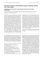
Crude extracts of P. putida KT2440 pUF1 clones when
grown in the presence of benzoate and/or methylbenzoate
contained a prominent protein showing the same electro-
phoretic mobility as Qor from wild-type P. putida 86,
suggesting that P. putida KT2440 pUF1 synthesized signi-
ficant amounts of Qor protein (Fig. 1A). Western blot
analysis confirmed the presence of the three subunits of Qor
in crude extracts of P. putida KT2440 pUF1 clones
(Fig. 1B). Qor from P. putida KT2440 pUF1 was enriched
91-fold with a yield of 43% (Table 2). Whereas the specific
activity of Qor purified to electrophoretic homogeneity
from wild-type P. putida 86 usually varied between 19 and
23 UÆmg
)1
, the specific activity of Qor preparations purified
from P. putida KT2440 pUF1 was only 0.8–2.7 UÆmg
)1
.
P. putida
86-1 D
qor
As the wild-type strain P. putida 86 is known to be able to
synthesize Mo–MCD, a deletion mutant lacking the genes
that code for Qor might be a suitable host for the expression
cloning of genes coding for Mo–MCD-containing molyb-
denum hydroxylases. By replacing two copies of qorMSL in
thegenomeofP. putida 86-1 by nptII and aacC1, the
mutant P. putida 86-1 Dqor was obtained. It had lost the
ability to grow on quinoline, and it did not synthesize Qor
protein. However, it was able to utilize 1H-2-oxoquinoline,
i.e., the product of the Qor-catalyzed reaction, with a
growth rate comparable to that of wild-type P. putida 86.
This indicates that the mutations did not affect any
subsequent step of the quinoline degradation pathway.
Fig. 1. Synthesis of Qor protein by P. putida KT2440 pUF1. (A) Non-
denaturing PAGE. Lane 1, crude extract of P. putida KT2440; lane 2,
crude extract of P. putida KT2440 pJB653; lanes 3–5, crude extracts of
different P. putida KT2440 pUF1 clones; lane 6, Qor purified from
wild-type P. putida 86; lane 7, crude extract of P. putida 86 grown in
mineral salts medium containing quinoline as sole carbon source; lane
8, crude extract of P. putida 86 grown in LB broth. (B) Immuno-
detection of Qor subunits in Western blot of crude extracts separated
by SDS/PAGE. Lane 1, crude extract of P. putida KT2440; lane 2,
crude extract of P. putida KT2440 pJB653; lanes 3–5, crude extracts of
different P. putida KT2440 pUF1 clones; lane 6, Qor purified from
wild-type P. putida 86.
Ó FEBS 2003 Expression of quinoline 2-oxidoreductase genes (Eur. J. Biochem. 270) 1571
Conditions of Qor synthesis in wild-type
P. putida
86,
P. putida
86-1 D
qor
pUF1 and
P. putida
86 pJB653
Qor of the wild-type strain P. putida 86 has been described
as an inducible enzyme [2]. In crude extracts of P. putida 86
cells grown on quinoline, the specific activity of Qor was
about 0.2 UÆmg
)1
of protein, whereas the specific Qor
activity in crude extracts of succinate- or benzoate-grown
cells was below 0.001 UÆmg
)1
(Table 3).
Succinate-grown cells of P. putida 86-1 Dqor pUF1 did
not contain any detectable Qor activity (Table 3), as
expression of the qorMSL genes inserted into the multiple
cloning site of pJB653 from the Pm promoter is controlled
by the plasmid-encoded XylS protein, that is activated by
benzoate effectors [52].
The presence of the expression vector pJB653 in P. put-
ida 86 did not significantly influence the specific Qor
activities in extracts of succinate-grown cells, and quino-
line-grown cells (Table 3). However, in benzoate-grown
cells of P. putida 86 pJB653, the specific Qor activity was
more than 110-fold higher than in benzoate-grown cells of
the wild-type strain P. putida 86. As benzoate as such is not
an inducer of Qor synthesis in P. putida 86, the effect of
benzoate in P. putida 86 pJB653 probably is mediated by
the plasmid-encoded XylS protein. The family of AraC/
XylS proteins comprises positive transcriptional regulators
that are characterized by significant amino acid sequence
homology extending over a 100-residue stretch constituting
the DNA binding domain [53–56]. In P. putida 86, a
putative xylS homologue designated oxoS has been previ-
ously identified upstream of the oxoO gene that codes for
a protein involved in the quinoline degradation pathway;
oxoO is localized about 7 kb upstream of the qorMSL genes
[57]. We may speculate that the degradation pathway is
regulated by the XylS-type transcriptional activator OxoS,
that might bind quinoline as an effector. In P. putida
86 pJB653, the plasmid-encoded XylS protein when activa-
ted by its effector benzoate might recognize the putative
DNA binding site of OxoS and activate transcription of the
catabolic gene cluster.
Qor protein from
P. putida
86-1 D
qor
pUF1
Immunodetection of the subunits of Qor in Western blots
confirmed that the deletion mutant P. putida 86-1 Dqor
containing the expression vector pJB653 did not synthesize
Qor protein, whereas P. putida 86-1 Dqor pUF1 grown on
benzoate or on a mixture of benzoate and 1H-2-oxoqui-
noline formed Qor (not shown). From a 4 L fermenter of
P. putida 86-1 Dqor pUF1 fed repeatedly with benzoate and
1H-2-oxoquinoline as carbon sources, between 16 and 18 g
of wet biomass were obtained after cultivation for 24–28 h.
Table 4 summarizes the enrichment of Qor from P. put-
ida 86-1 Dqor pUF1. The protein preparations showed
specific activities of 20–23 UÆmg
)1
, that is comparable to the
activity of wild-type Qor.
Kinetic properties of the Qor proteins from
P. putida
86,
P. putida
KT2440 pUF1 and
P. putida
86-1 D
qor
pUF1
The apparent K
m
values of the Qor proteins for quinoline
were similar, whereas the apparent k
cat
value for quinoline
of Qor from strain KT2440 pUF1 was eight- to tenfold
lower than that of wild-type Qor and Qor from P. put-
ida 86-1 Dqor pUF1 (Table 5).
Table 2. Purification of Qor protein from P. putida KT2440 pUF1. Starting material was 34 g of wet biomass. In crude extracts, quinoline-
independent INT reduction mediated by unspecific reductases of strain KT2440 impedes accurate measurement of quinoline-dependent INT
reduction catalyzed by Qor. ppt, precipitation.
Fraction Activity (Units) Protein (mg) Specific activity (UÆmg
)1
) Purification (-fold) Yield (%)
Crude extract 85.3 3696 0.023 1 100
Ammonium sulfate ppt 87.4 516 0.17 7 102
Phenyl Sepharose CL-4B 71.9 76 0.95 41 84
BioScale DEAE10 36.5 17.4 2.10 91 43
Table 3. Activity of Qor in crude extracts of wild-type P. putida 86,
P. putida 86 pJB653 and P. putida 86-1 Dqor pUF1 grown on different
carbon sources.
Strain
Specific activity (UÆmg
)1
)ofQor
in crude extracts after growth on:
Succinate Benzoate Quinoline
P. putida 86 < 0.001 < 0.001 0.21
P. putida 86 pJB653 0.001 0.11 0.18
P. putida 86-1 Dqor pUF1 0 0.10 –
a
a
As benzoate is necessary as an XylS effector for expression of
qorMSL from pUF1, P putida 86-1 Dqor pUF1 is not able to grow
on quinoline as a sole source of carbon.
Table 4. Purification of Qor protein from P. putida 86-1 Dqor pUF1. Starting material was 27 g of wet biomass. ppt, precipitation.
Fraction Activity (Units) Protein (mg) Specific activity (UÆmg
)1
) Purification (-fold) Yield (%)
Crude extract 312 3045 0.10 1 100
Ammonium sulfate ppt 271 416 0.65 6.4 87
Phenyl Sepharose CL-4B 299 78 3.83 37.5 96
BioScale DEAE10 206 9.3 22.1 216.7 66
1572 U. Frerichs-Deeken et al. (Eur. J. Biochem. 270) Ó FEBS 2003
Metal content of the Qor proteins and analysis
of nucleotides released from the Qor proteins from
P. putida
86,
P. putida
KT2440 pUF1 and
P. putida
86-1 Dqor pUF1
Native Qor is expected to contain 2 g atom of molybdenum
and8gatomofironpermolofenzyme[1,2].However,
with the analytical method performed (direct analysis
without preceding digestion), only 0.8 g atom of molyb-
denum and 5.5 g atom of iron were detected per mol of
wild-type Qor. The iron content of Qor from P. put-
ida KT2440 pUF1 corresponded to that of wild-type Qor,
however, its molybdenum content was tenfold lower
(Table 5); this could explain the decrease in activity.
The molybdenum cofactor of wild-type Qor has previ-
ously been identified as Mo-MCD [14]. Treatment of Qor
proteins with sulfuric acid and subsequent analysis of the
preparation by reverse-phase HPLC showed the presence of
CMP and AMP (from FAD). GMP was not present in any
Qor extract, indicating that the host strains did not
incorporate Mo-MGD, or free GMP, into the cofactor
binding domain of the Qor protein. Similar amounts of
CMP were released from the three Qor proteins (Table 5).
However, especially in the nearly inactive Qor protein from
P. putida KT2440 pUF1, it may be possible that the
nucleotide is occupying the CMP binding site of the Qor
protein, without being part of an MCD cofactor. To detect
loosely bound CMP, nucleotides were extracted from the
proteins by boiling in aqueous SDS. This method led to the
release of about 0.4 mol of CMP per mol of enzyme,
however, approximately the same amounts of CMP were
released from the different Qor enzymes. Thus, the low
activity observed for the Qor protein from P. putida
KT2440 pUF1 seems to be correlated to a deficiency in
the metal, not to a deficiency in the organic part of the
molybdenum cofactor. However, we cannot exclude that
the pyranopterin part of the cofactor is somehow defective
in Qor from strain KT2440 pUF1.
UV/Visual spectra of the Qor proteins from
P. putida
86,
P. putida
KT2440 pUF1 and
P. putida
86-1 D
qor
pUF1
The UV/Visual spectra of Qor purified from P. putida
KT2440 pUF1 and of wild-type Qor were very similar,
except for the absorption around 305 nm, that was signi-
ficantly decreased in Qor from P. putida KT2440 pUF1.
This decrease might reflect a deficiency in the pyranopterin
cofactor. The ratios A
280nm
/A
450nm
and A
450nm
/A
550nm
of
4.5–5 and 2.8–3, respectively, were identical in both proteins,
indicating the presence of the full set of iron–sulfur clusters
and stoichiometric amounts of FAD. The UV/Visual
spectrum of Qor from P. putida 86-1 Dqor pUF1 was typi-
cal for a molybdo-iron/sulfur flavoprotein; it lacked the
marked decrease at 305 nm observed in the Qor protein
from P. putida KT2440 pUF1 (Fig. 2).
Analysis of redox-active centers in Qor from
P. putida
KT2440 pUF1 and
P. putida
86-1 D
qor
pUF1
by EPR spectroscopy
Mo. Reduction of the Qor protein isolated from wild-type
P. putida 86 with its substrate quinoline led to the forma-
tion of the Mo(V)-rapid species that is readily observable
at 77 K; the Mo(V) rapid species is indicative of the
monooxo-monosulfido-type molybdenum center [2] and is
thought to represent a complex of substrate with enzyme [5].
The typical, almost axial, spectrum in Fig. 3A shows the
splitting of the H-D-exchangeable proton attributed to the
Table 5. Metal content, amount of CMP released by hydrolysis with sulfuric acid and kinetic parameters of the Qor proteins.
Source of Qor
Metal content
(g atom per mol
of enzyme)
a
CMP released by
hydrolysis (mol per
mol of enzyme)
b
Kinetic parameters
Mo Fe CMP K
m app (quinoline)
(m
M
) k
cat app (quinoline)
(s
)1
)
P. putida 86 (grown on quinoline) 0.8 5.5 1.2 0.18
c
74
P. putida KT2440 pUF1
(grown on benzoate)
0.08 5.4 1.3 0.12 8.7
P. putida 86–1Dqor pUF1
(grown on benzoate +
1H-2-oxoquinoline)
0.5 3 1.2 0.12 85.4
a
Average of two determinations;
b
average of three experiments;
c
[1].
Fig. 2. UV/Visual spectra of Qor proteins. Solid line, Qor purified
from wild-type P. putida 86; dotted line, Qor purified from P. putida
KT2440 pUF1; dashed line, Qor from P. putida 86-1 Dqor pUF1. The
increased absorption at 280 nm of the latter is due to contaminating
colourless proteins.
Ó FEBS 2003 Expression of quinoline 2-oxidoreductase genes (Eur. J. Biochem. 270) 1573
sulfhydryl-group of the one electron reduced complex [2,24].
When the Qor protein isolated from P. putida KT2440
pUF1 (specific activity: 1 UÆmg
)1
) was reacted with quino-
line, a rapid-type EPR-signal of rather small intensity was
detected (Fig. 3B). In contrast, the catalytically competent
Qor protein purified from P. putida 86-1 Dqor pUF1
produced the rapid EPR-signal in considerably higher
amounts (Fig. 3C). As both Qor samples were treated and
recorded under identical experimental conditions, the
relative quantities of the Mo(V)-rapid species could be
estimated from the EPR intensities. This comparison
showed that the amount of Mo(V)-species formed in Qor
from P. putida KT2440 pUF1 was approximately 25-fold
lower than in Qor from P. putida 86-1 Dqor pUF1. In
accordance with the results of the metal analyses, the very
low intensity of the Mo(V) rapid EPR signal suggested that
most of the Qor molecules are deficient in molybdenum.
This is in line with the finding that only very weak EPR-
signals of reduced FeS-clusters were detected after addition
of substrate (almost no electron transfer from quinoline via
Mo to FeS), but clearly are formed by direct reduction of
the FeS-clusters with dithionite (see below). Thus, although
P. putida KT2440 pUF1 presumably is able to catalyse the
synthesis and insertion of a cytidine dinucleotide cofactor
into recombinant Qor as suggested by the release of CMP
after hydrolysis of the enzyme, it appears that the assembly
of intact Mo-MCD is a bottleneck in strain KT2440 pUF1,
leading to the incorporation of a defective, molybdenum
deficient cofactor into the maturing Qor protein.
The rapid EPR-signal of Qor from P. putida 86-1
Dqor pUF1 (Fig. 3C) shows some minor differences as
compared to the signal of the wild-type protein. The
distortions marked by arrows in trace C are caused by
signals of the resting species which are associated with
inactive Mo(V)-centers formed during the preparation
process in varying amounts [24].
The finding that in each of the three Qor enzymes the
majority of the Mo(V) species was represented by the rapid
type EPR signal indicates that the molybdenum centers are
predominantly in the correct monooxo-monosulfido form.
The ÔslowÕ type signal, associated with the inactive desulfo
(¼ dioxo) form [2], could not be identified in the spectral
patterns indicating that this species is, if at all, present only
in negligible amounts.
Besides the low intense resonances visible at the high-
and low-field side of the rapid EPR-signal (traces A and C)
and originating from natural Mo-isotopes with nuclear spin
I ¼ 5/2, also small lines of the semiquinone radical form of
FAD were observed at g ¼ 2.004.
Fe/S. When the temperature was lowered to about 20 K
the characteristic rhombic EPR-patterns of two [2Fe)2S]
clusters, FeSI and FeSII, became visible. Their assignment is
given in Fig. 4A for the wild-type Qor reduced with
quinoline. In this case, the g
2
-component of FeSII is
superimposed by the intense and saturation broadened
Mo(V)-signal. An identical spectrum of FeS-clusters and
Mo(V) contribution was found for Qor from P. putida 86-1
Dqor pUF1 reduced with quinoline as shown in Fig. 4C. In
contrast, for Qor from P. putida KT2440 pUF1 only
extremely weak signals of the FeS-centers (not shown) were
present after reduction with substrate. When this sample
was subsequently reduced with a tenfold excess of dithio-
nite, the signals of both FeS-centers appeared in appreciable
intensity as indicated in Fig. 4B. The absence of Mo(V)-
signals reveals the g
2
-component of FeSII. It is noted that
the g-factors of the FeSI and II signals of the Qor proteins
from the wild-type strain and from P. putida 86-1
Dqor pUF1 are identical, whereas the g
1
-components for
Qor from P. putida KT2440 pUF1 are shifted slightly to
lower g-factors. Such spectral differences depending on the
mode of reduction have been reported for wild-type Qor
[24]. In general, the differences in g-factor of the FeS-signals
are less than 0.003 compared to the corresponding signals
of wild-type Qor [24]. An exception is found for the
g
3
-component ofFeSII of Qor from P. putida KT2440pUF1
that is shifted to a lower g-factor of 1.858 as compared to
1.871 for the wild-type Qor (Fig. 4B). The change of the
g
3
-factor of FeSII may indicate that the electronic structure
was influenced probably by an unknown alteration of the
immediate environment of this FeS-cluster.
For completeness, it should be mentioned here that a
weak signal of yet unknown origin is observed in all reduced
Qor samples. Although it is located close to the g-factor of
Fig. 3. EPR spectra of the rapid species in Qor from wild-type P. putida
(A), P. putida KT2440 pUF1 (B) and P. putida 86-1 Dqor pUF1 (C)
formed after reduction with substrate quinoline. Spectra were recorded
at 77 K at a microwave power of 2 mW. Trace B is multiplied by a
factor of six to show the small signals of the rapid species in this
sample. The arrows indicate the position of contribution of the resting
species particularly to spectrum C.
1574 U. Frerichs-Deeken et al. (Eur. J. Biochem. 270) Ó FEBS 2003
the FAD radical signal its saturation and temperature
behaviour points to a metal centered species.
The EPR analyses showed that, apart from some small
contribution of nonfunctional species (resting), the EPR-
signals of wild-type Qor and of Qor from P. putida 86-1
Dqor pUF1 are virtually superimposable, indicating identi-
cal cofactor composition and arrangement.
Conclusions
Assembly of [2Fe)2S] clusters as well as flavin and Mo-MPT
biosynthesis [58] are thought to involve ubiquitously
conserved pathways, but additional reactions that modify
Mo-MPT appear to be restricted to certain organisms. In
E. coli, for example, all known molybdenum enzymes
contain MGD as the organic part of the molybdenum
cofactor, and attempts to express genes encoding Mo-MCD-
containing enzymes in E. coli failed [23,29] (K. Parschat & S.
Fetzner, unpublished results). In this work, we tested
whether expression of the qorMSL genes from P. putida 86
in P. putida KT2440 and in a qorMSL deletion mutant of
P. putida 86-1 results in the formation of catalytically active
enzyme.
The expression clone P. putida 86-1 Dqor pUF1 synthes-
ized catalytically competent Qor protein that in its kinetic
and spectroscopic properties seemed identical to wild-type
Qor. This clone did not allow overproduction of Qor,
however, as about 6–8 mg of Qor protein can be purified
from 10 g of wild-type P. putida 86 biomass, protein
production was not the primary goal of this work. This
expression system will allow the genetic manipulation of the
qor genes by mutagenic approaches, and the synthesis of
enzyme variants, that after purification by the established
protocol, will be available for further biochemical and
spectroscopic characterization. The mutant P. putida 86-1
Dqor may also be a suitable recipient for the expression
cloning of genes coding for other Mo-MCD-containing
hydroxylases.
Bacterial strains synthesizing molybdenum hydroxylases,
or isolated molybdenum hydroxylases catalyzing regio-
specific hydroxylation reactions, are useful biocatalysts
for industrial processes to manufacture hydroxy-substi-
tuted N-heteroaromatic compounds [59–63]. Enzyme
engineering may be used to improve the stability or
catalytic efficiency of the enzymes, or to alter their
substrate specificity [64–66]. Most of the molybdenum
hydroxylases catalyzing the hydroxylation of N-hetero-
aromatic compounds contain the Mo-MCD cofactor
[5,8,9]. Thus, a system enabling the genetic manipulation
and regulated expression of genes coding for Mo-MCD-
containing hydroxylases might also be of biotechnological
importance.
Acknowledgements
We thank S. Valla, Norwegian University of Science and Technology,
Trondheim, Norway, for kindly providing pJB653 and M. Sohni,
Oldenburg, for selecting the streptomycin resistant mutant of P. put-
ida 86. We thank W. Wackernagel, Oldenburg, and the late W. Klipp,
Bochum, for the generous gift of plasmids and strains. This work was
supported by the Deutsche Forschungsgemeinschaft (Fe 383/4-4), the
European Commission within the ÔXanthine Oxidase NetworkÕ
(Contract No. HPRN-CT-1999-00084), and the Fonds der Chemischen
Industrie.
References
1. Bauder, R., Tshisuaka, B. & Lingens, F. (1990) Quinoline
oxidoreductase from Pseudomonas putida: a molybdenum-
containing enzyme. Biol. Chem. Hoppe-Seyler 371, 1137–1144.
2. Tshisuaka, B., Kappl, R., Hu
¨
ttermann, J. & Lingens, F. (1993)
Quinoline oxidoreductase from Pseudomonas putida 86: an
improved purification procedure and electron paramagnetic
resonance spectroscopy. Biochemistry 32, 12928–12934.
3. Stephan, I., Tshisuaka, B., Fetzner, S. & Lingens, F. (1996)
Quinaldine 4-oxidase from Arthrobacter sp. Ru
¨
61a, a versatile
procaryotic molybdenum-containing hydroxylase active towards
N-containing heterocyclic compounds and aromatic aldehydes.
Eur. J. Biochem. 236, 155–162.
4. Fetzner, S. (2000) Enzymes involved in the aerobic bacterial
degradation of N-heteroaromatic compounds: molybdenum
hydroxylases and ring-opening 2,4-dioxygenases. Naturwissens-
chaften 87, 59–69.
Fig. 4. EPR spectra of the two [2Fe2S]-centers FeSI and FeSII in
Qor from wild-type P. putida (A), P. putida KT2440 pUF1 (B) and
P. putida 86-1 Dqor pUF1 (C). Spectra A and C were obtained upon
reduction with substrate quinoline, spectrum B after subsequent
reduction with dithionite. The spectra were recorded at 20 K at a
microwave power of 12 mW. The stick diagram (top) indicates the
rhombic g-components of both FeS-centers. The asterisk marks the
position of a low intensity unknown signal.
Ó FEBS 2003 Expression of quinoline 2-oxidoreductase genes (Eur. J. Biochem. 270) 1575
5. Hille, R. (1996) The mononuclear molybdenum enzymes. Chem.
Rev. 96, 2757–2816.
6. Hille, R. (1999) Molybdenum enzymes. Essays Biochem. 34, 125–
137.
7. Hille, R. (2002) Molybdenum enzymes containing the pyrano-
pterin cofactor: an overview. In Molybdenum and Tungsten.
Their Roles in Biological Processes (Sigel,A.&Sigel,H.,eds),
pp. 187–226, Vol. 39 of Met. Ions Biol. Syst.MarcelDekker,
New York.
8. Andreesen, J.R. & Fetzner, S. (2002) The molybdenum-containing
hydroxylases of nicotinate, isonicotinate, and nicotine. In
Molybdenum and Tungsten. Their Roles in Biological Processes
(Sigel, A. & Sigel, H., eds), pp. 405–430, Vol. 39 of Met. Ions Biol.
Syst. Marcel Dekker, New York.
9. Kappl, R., Hu
¨
ttermann, J. & Fetzner, S. (2002) The molybdenum-
containing hydroxylases of quinoline, isoquinoline, and qui-
naldine. In Molybdenum and Tungsten. Their Roles in Biological
Processes (Sigel, A. & Sigel, H., eds), pp. 481–537, Vol. 39 of Met.
Ions Biol. Syst. Marcel Dekker, New York.
10. Bray, R.C. & Lowe, D.J. (1997) Towards the reaction mechanism
of xanthine oxidase from EPR studies. Biochem. Soc. Trans. 25,
762–768.
11. Roma
˜
o,M.J.,Archer,M.,Moura,I.,Moura,J.J.G.,LeGall,J.,
Engh, R., Schneider, M., Hof, P. & Huber, R. (1995) Crystal
structure of the xanthine oxidase-related aldehyde oxido-reductase
from D. gigas. Science 270, 1170–1176.
12. Roma
˜
o, M.J. & Huber, R. (1998) Structure and function of the
xanthine-oxidase family of molybdenum enzymes. Structure
Bonding 90, 69–95.
13. Parschat, K., Canne, C., Hu
¨
ttermann, J., Kappl, R. & Fetzner, S.
(2001) Xanthine dehydrogenase from Pseudomonas putida 86:
Specificity, oxidation-reduction potentials of its redox-active
centers, and first EPR characterization. Biochim. Biophys. Acta
1544, 151–165.
14. Hettrich, D., Peschke, B., Tshisuaka, B. & Lingens, F. (1991) The
molybdopterin cofactors of quinoline oxidoreductases from
Pseudomonas putida 86 and Rhodococcus spec. B1 and of xanthine
dehydrogenase from Pseudomonas putida 86. Biol. Chem. Hoppe-
Seyler 372, 513–517.
15. Lehmann, M., Tshisuaka, B., Fetzner, S., Ro
¨
ger, P. & Lingens, F.
(1994) Purification and characterization of isoquinoline 1-oxi-
doreductase from Pseudomonas diminuta 7, a novel molybdenum-
containing hydroxylase. J. Biol. Chem. 269, 11254–11260.
16. Kretzer,A.,Frunzke,K.&Andreesen,J.R.(1993)Catabolismof
isonicotinate by Mycobacterium sp. INA1: extended description of
the pathway and purification of the molybdoenzyme isonicotinate
dehydrogenase. J. Gen. Microbiol. 139, 2763–2772.
17. Schra
¨
der, T., Hillebrand, C. & Andreesen, J.R. (1998) 2-Hydro-
xyisonicotinate dehydrogenase isolated from Mycobacterium sp.
INA1. FEMS Microbiol. Lett. 164, 311–316.
18. Meyer, O., Jacobitz, S. & Kru
¨
ger, B. (1986) Biochemistry and
physiology of aerobic carbon monoxide-utilizing bacteria. FEMS
Microbiol. Rev. 39, 161–179.
19. Meyer, O., Frunzke, K. & Mo
¨
rsdorf, G. (1993) Biochemistry of
the aerobic utilization of carbon monoxide. In: Microbial Growth
on C
1
Compounds (Murrell, J.C. & Kelly, D.P., eds), pp. 433–459.
Intercept Ltd, Andover, Hampshire, UK.
20. Dobbek,H.,Gremer,L.,Kiefersauer,R.,Huber,R.&Meyer,O.
(2002) Catalysis at a dinuclear [CuSMo (¼O) OH] cluster in a CO
dehydrogenase resolved at 1.1-A
˚
resolution. Proc.NatlAcad.Sci.
USA 99, 15971–15976.
21. Rebelo, J., Macieira, S., Dias, J.M., Huber, R., Ascenso, C.S.,
Rusnak,F.,Moura,J.J.G.,Moura,I.&Roma
˜
o, M.J. (2000)
Gene sequence and crystal structure of the aldehyde oxido-
reductase from Desulfovibrio desulfuricans ATCC 27774. J. Mol.
Biol. 297, 135–146.
22. Rebelo,J.M.,Dias,J.M.,Huber,R.,Moura,J.J.G.&Roma
˜
o,
M.J. (2001) Structure refinement of the aldehyde oxidoreductase
from Desulfovibrio gigas (MOP)at1.28A
˚
. J. Biol. Inorg. Chem. 6,
791–800.
23. Bla
¨
se,M.,Bruntner,C.,Tshisuaka,B.,Fetzner,S.&Lingens,F.
(1996) Cloning, expression, and sequence analysis of the three
genes encoding quinoline 2-oxidoreductase, a molybdenum-
containing hydroxylase from Pseudomonas putida 86. J. Biol.
Chem. 271, 23068–23079.
24. Canne, C., Stephan, I., Finsterbusch, J., Lingens, F., Kappl, R.,
Fetzner, S. & Hu
¨
ttermann, J. (1997) Comparative EPR and redox
studies of three prokaryotic enzymes of the xanthine oxidase
family: quinoline 2-oxidoreductase, quinaldine 4-oxidase, and
isoquinoline 1-oxidoreductase. Biochemistry 36, 9780–9790.
25. Canne, C., Lowe, D.J., Fetzner, S., Adams, B., Smith, A.T.,
Kappl, R., Bray, R.C. & Hu
¨
ttermann, J. (1999) Kinetics and
interactions of molybdenum and iron-sulfur centers in bacterial
enzymes of the xanthine oxidase family: mechanistic implications.
Biochemistry 38, 14077–14087.
26. Garrett, R.M. & Rajagopalan, K.V. (1994) Molecular cloning
of rat liver sulfite oxidase. Expression of a eukaryotic
Mo-pterin-containing enzyme in Escherichia coli. J. Biol. Chem.
269, 272–276.
27. Pollock, V.V. & Barber, M.J. (1997) Biotin sulfoxide reductase.
Heterologous expression and characterization of a functional
molybdopterin guanine dinucleotide-containing enzyme. J. Biol.
Chem. 272, 3355–3362.
28. Temple, C.A., Graf, T.N. & Rajagopalan, K.V. (2000) Optimi-
zation of expression of human sulfite oxidase and its molybdenum
domain. Arch. Biochem. Biophys. 383, 281–287.
29. Black, G.W., Lyons, C.M., Williams, E., Colby, J., Kehoe, M. &
O’Reilly, C. (1990) Cloning and expression of the carbon mon-
oxide dehydrogenase genes from Pseudomonas thermocarboxyd-
ovorans strain C2. FEMS Microbiol. Lett. 70, 249–254.
30. Johnson, J.L., Indermaur, L.W. & Rajagopalan, K.V. (1991)
Molybdenum cofactor biosynthesis in Escherichia coli.Require-
ment of the chlB gene product for the formation of molybdopterin
guanine dinucleotide. J. Biol. Chem. 266, 12140–12145.
31. Palmer, T., Vasishta, A., Whitty, P.W. & Boxer, D.H. (1994)
IsolationofproteinFA,aproductofthemob locus required for
molybdenum cofactor biosynthesis in Escherichia coli. Eur. J.
Biochem. 222, 687–692.
32. Leimku
¨
hler, S. & Klipp, W. (1999) The molybdenum cofactor
biosynthesis protein MobA from Rhodobacter capsulatus is
required for the activity of molybdenum enzymes containing
MGD, but not for xanthine dehydrogenase harboring the MPT
cofactor. FEMS Microbiol. Lett. 174, 239–246.
33.Stevenson,C.E.M.,Sargent,F.,Buchanan,G.,Palmer,T.&
Lawson, D.M. (2000) Crystal structure of the molybdenum
cofactor biosynthesis protein MobA from Escherichia coli at near-
atomic resolution. Structure Fold. Res. 8, 1115–1125.
34. Lake, M.W., Temple, C.A., Rajagopalan, K.V. & Schindelin, H.
(2000) The crystal structure of the Escherichia coli MobA protein
provides insight into molybdopterin guanine dinucleotide bio-
synthesis. J. Biol. Chem. 275, 40211–40217.
35. Temple, C.A. & Rajagopalan, K.V. (2000) Mechanism of assem-
bly of the bis (molybdopterin guanine dinucleotide) molybdenum
cofactor in Rhodobacter sphaeroides dimethyl sulfoxide reductase.
J. Biol. Chem. 275, 40202–40210.
36.Buchanan,G.,Kuper,J.,Mendel,R.R.,Schwarz,G.&
Palmer, T. (2001) Characterization of the mob locus of Rhodo-
bacter sphaeroides WS8: mobA istheonlygenerequiredfor
molybdopterin guanine dinucleotide synthesis. Arch. Microbiol.
176, 62–68.
37. Israel, I., Sohni, M. & Fetzner, S. (2002) Expression of the iorAB
genes from Brevundimonas diminuta 7 encoding the molybdenum
1576 U. Frerichs-Deeken et al. (Eur. J. Biochem. 270) Ó FEBS 2003
hydroxylase isoquinoline 1-oxidoreductase in Pseudomonas putida.
FEMS Microbiol. Lett. 210, 123–127.
38. Sambrook, J., Fritsch, E.F. & Maniatis, T. (1989) Molecular
Cloning: a Laboratory Manual, 2nd edn. Cold Spring Laboratory
Press, Cold Spring Harbor, NY, USA.
39. Davis, R.W., Botstein, D. & Roth, J.R. (1980) A Manual for
Genetic Engineering. Advanced Bacterial Genetics. Cold Spring
Harbor Laboratory Press, Cold Spring Harbor, NY, USA.
40. Dower, W.J., Miller, J.F. & Ragsdale, C.W. (1988) High efficiency
transformation of E. coli by high voltage electroporation. Nucleic
Acids Res. 16, 6127–6145.
41. Iwasaki, K., Uchiyama, H., Yagi, O., Kurabayashi, T., Ishizuka,
K. & Takamura, Y. (1994) Transformation of Pseudomonas
putida by electroporation. Biosci. Biotechnol. Biochem. 58,
851–854.
42. Vieira, J. & Messing, J. (1982) The pUC plasmids, an M13mp7-
derived system for insertion mutagenesis and sequencing with
synthetic universal primers. Gene 19, 259–268.
43.Beck,E.,Ludwig,G.,Auerswald,E.A.,Reiss,B.&Schaller,
H. (1982) Nucleotide sequence and exact localization of the
neomycin phosphotransferase gene from transposon Tn5. Gene
19, 327–336.
44. Simon,R.,Priefer,U.&Pu
¨
hler, A. (1983) A broad host range
mobilization system for in vivo genetic engineering: transposon
mutagenesis in Gram negative bacteria. Biol/Technology 1,784–
791.
45. Masepohl, B., Klipp, W. & Pu
¨
hler, A. (1988) Genetic char-
acterization and sequence analysis of the duplicated nifA/nifB
gene region of Rhodobacter capsulatus. Mol. Gen. Genet. 212,
27–37.
46. Hirsch, P.R. & Beringer, J.E. (1984) A physical map of pPH1JI
and pJB4JI. Plasmid 12, 39–141.
47. Grunstein, M. & Hogness, D.S. (1975) Colony hybridization: a
method for the isolation of cloned DNAs that contain a specific
gene. Proc. Natl Acad. Sci. USA 72, 3961–3965.
48. Hames, B.D. (1990) One-dimensional polyacrylamide gel electro-
phoresis. In Gel Electrophoresis of Proteins – a Practical Approach
(Hames, B.D. & Rickwood, D., eds), 2nd edn, pp. 1–147. IRL
Press (Oxford University Press), Oxford.
49. Laemmli, U.K. (1970) Cleavage of structural proteins during
the assembly of the head of bacteriophage T4. Nature 227,
680–685.
50. Caponi, L. & Migliorini, P. (1999) Immunoblotting. In Antibody
Usage in the Laboratory (Caponi, L. & Migliorini, P., eds), p. 42.
Springer, Berlin, Heidelberg, New York.
51. Zor, T. & Selinger, Z. (1996) Linearization of the Bradford protein
assay increases its sensitivity: theoretical and experimental studies.
Anal. Biochem. 236, 302–308.
52. Blatny, J.M., Brautaset, T., Winther-Larsen, H.C., Haugan, K. &
Valla, S. (1997) Construction and use of a versatile set of broad-
host-range cloning and expression vectors based on the RK2
replicon. Appl. Environ. Microbiol. 63, 370–379.
53. Gallegos, M T., Schleif, R., Bairoch, A., Hofmann, K. & Ramos,
J.L. (1997) AraC/XylS family of transcriptional regulators.
Microbiol. Mol. Biol. Rev. 61, 393–410.
54. Martin, R.G. & Rosner, J.L. (2001) The AraC transcriptional
activators. Curr. Opin. Microbiol. 4, 132–137.
55. Tobes, R. & Ramos, J.L. (2002) AraC-XylS database: a family of
positive transcriptional regulators in bacteria. Nucleic Acids Res.
30, 318–321.
56. Gerischer, U. (2002) Specific and global regulation of genes
associated with the degradation of aromatic compounds in bac-
teria. J. Mol. Microbiol. Biotechnol. 4, 111–121.
57. Rosche, B., Tshisuaka, B., Hauer, B., Lingens, F. & Fetzner, S.
(1997) 2-Oxo-1,2-dihydroquinoline 8-monooxygenase: phyloge-
netic relationship to other multicomponent nonheme iron oxyge-
nases. J. Bacteriol. 179, 3549–3554.
58. Mendel, R.R. & Schwarz, G. (2002) Biosynthesis and molecular
biology of the molybdenum cofactor (Moco). In: Molybdenum and
Tungsten. Their Roles in Biological Processes (Sigel, A. & Sigel, H.,
eds), pp. 317–368, Vol. 39 of Met. Ions Biol. Syst.MarcelDekker,
NY, USA.
59. Kulla, H.G. (1991) Enzymatic hydroxylations in industrial appli-
cation. Chimia 45, 81–85.
60. Kawashima, H., Sueyoshi, H. & (Nippon Steel Corp, Japan)
(1993) Manufacture of carbostyril and/or 6-hydroxycarbostyril
with Pseudomonas species. Japanese Patent 05 304 973 [93 304
973], Chem. Abstract. (1994), 120, 132464K.
61. Tinschert, A., Kiener, A., Heinzmann, K. & Tschech, A. (1997)
Isolation of new 6-methylnicotinic-acid-degrading bacteria, one of
which catalyses the regioselective hydroxylation of nicotinic acid
at position C2. Arch. Microbiol. 168, 355–361.
62. Tinschert, A., Tschech, A., Heinzmann, K. & Kiener, A. (2000)
Novel regioselective hydroxylations of pyridine carboxylic acids at
position C2 and pyrazine carboxylic acids at position C3. Appl.
Microbiol. Biotechnol. 53, 185–195.
63. Ueda, M. & Sashida, R. (1998) Microbial production of
2-hydroxynicotinic acid from nicotinic acid by intact cells of
MCI3289. J. Mol. Catal. B: Enzym. 4, 199–204.
64. Arnold, F.H. (1998) Design by directed evolution. Acc. Chem.
Res. 31, 125–131.
65. Arnold, F.H. & Volkov, A.A. (1999) Directed evolution of bio-
catalysts. Curr. Opin. Chem. Biol. 3, 54–59.
66. Reetz, M.T. & Jaeger, K E. (1999) Superior biocatalysts by
directed evolution. Topics Curr. Chem. 200, 31–57.
67. Bagdasarian, M., Lurz, R., Ru
¨
ckert, B., Franklin, F.C.H.,
Bagdasarian, M.M., Frey, J. & Timmis, K.N. (1981) Specific-
purpose plasmid cloning vectors. II. Broad host range, high copy
number, RSF1010-derived vectors, and a host-vector system for
gene cloning in Pseudomonas. Gene 16, 237–247.
68. Schwarz, G., Senghas, E., Erben, A., Scha
¨
fer,B.,Lingens,F.&
Ho
¨
ke, H. (1988) Microbial metabolism of quinoline and related
compounds. I. Isolation and characterization of quinoline-
degrading bacteria. Syst. Appl. Microbiol. 10, 185–190.
Ó FEBS 2003 Expression of quinoline 2-oxidoreductase genes (Eur. J. Biochem. 270) 1577