Báo cáo khoa học: Key substrate recognition residues in the active site of a plant cytochrome P450, CYP73A1 ppt
Bạn đang xem bản rút gọn của tài liệu. Xem và tải ngay bản đầy đủ của tài liệu tại đây (403.54 KB, 12 trang )
Key substrate recognition residues in the active site of a plant
cytochrome P450, CYP73A1
Homology model guided site-directed mutagenesis
Guillaume A. Schoch
1
, Roger Attias
2
, Monique Le Ret
1
and Danie
`
le Werck-Reichhart
1
1
Department of Plant Stress Response, Institute of Plant Molecular Biology, Universite
´
Louis Pasteur, Strasbourg, France;
2
Laboratoire de Chimie et Biochimie Pharmacologiques et Toxicologiques, Universite
´
Paris V, 45 Paris, France
CYP73 enzymes are highly conserved cytochromes P450 in
plant species that catalyse the regiospecific 4-hydroxylation
of cinnamic acid to form precursors of lignin and many other
phenolic compounds. A CYP73A1 homology model based
on P450 experimentally solved structures was used to iden-
tify active site residues likely to govern substrate binding and
regio-specific catalysis. The functional significance of these
residues was assessed using site-directed mutagenesis. Active
site modelling predicted that N302 and I371 form a hydro-
gen bond and hydrophobic contacts with the anionic site or
aromatic ring of the substrate. Modification of these residues
led to a drastic decrease in substrate binding and metabolism
without major perturbation of protein structure. Changes to
residue K484, which is located too far in the active site model
to form a direct contact with cinnamic acid in the oxidized
enzyme, did not influence initial substrate binding. However,
the K484M substitution led to a 50% loss in catalytic
activity. K484 may affect positioning of the substrate in the
reduced enzyme during the catalytic cycle, or product
release. Catalytic analysis of the mutants with structural
analogues of cinnamic acid, in particular indole-2-carboxylic
acid that can be hydroxylated with different regioselectivi-
ties, supports the involvement of N302, I371 and K484 in
substrate docking and orientation.
Keywords: active site; cinnamate 4-hydroxylase; homology
modeling; plant cytochrome P450; site-directed muta-
genesis.
CYP73 designates a family of plant cytochromes P450 that
evolved with or before the evolution of vascular plants. Up
to 20% of the woody plant biomass is processed by CYP73
enzymes to form lignin monomers, UV-shielding or insect
attracting pigments, and defensive compounds [1,2]. CYP73
enzymes belong to the same subfamily, i.e. share more than
55% amino acid identity, and catalyse the regiospecific
4-hydroxylation of trans-cinnamic acid into p-coumaric acid
[3–5]. The importance of this reaction in plant biology seems
to have precluded further evolution and diversification of
the CYP73A P450 subfamily to the processing of other
endogenous metabolites.CYP73A1was one of the first
plant P450 genes isolated [6]. Expression in yeast indicated
that the cinnamate 4-hydroxylase (C4H) activity proceeds
with a perfect coupling of oxygen consumption and
reducing equivalents to produce hydroxylated substrates
[3]. CYP73A1 provides a good model for determining the
residues that control catalytic efficiency and optimal
substrate positioning in a typical plant P450 enzyme
contributing to a high throughput anabolic pathway.
CYP73A1 is one of the most extensively studied plant
P450 enzymes. It has a quite high substrate specificity but
can accommodate a diverse array of compounds, as far as
they are structural analogues of the natural substrate.
Structural requirements for such analogues include a planar,
aromatic structure, a small size of about two adjacent
aromatic rings, and an anionic site opposite (i.e. at about
8.5 A
˚
) to the position of oxidative attack [7,8]. A recent site-
directed mutagenesis study that investigated the role of
unusual residues in the most conserved regions involved in
haem binding and oxygen activation [9], suggested that
some are likely to contribute to the optimal coupling of the
C4H reaction. The protein residues that govern substrate
recognition and orientation have not yet been identified.
In order to obtain information on the orientation and
positioning of the substrates in the active site, we have
recently engineered a stable and water-soluble form of
CYP73A1 that is suitable for
1
H-NMR paramagnetic
relaxation experiments [10]. The results of the NMR
analysis indicated that the average initial orientation of
the substrates in the catalytic site of the resting Fe(III)
protein is roughly parallel to the haem. We decided to use a
structure-based approach to site-directed mutagenesis in
order to identify residues that affect substrate binding and
turnover. However, only one structure for a membrane-
bound P450 protein was available [11]. We thus had to rely
on a homology model based on soluble P450 structures
to predict residues that might participate in the substrate
recognition and docking. In this paper, we report the
Correspondence to D. Werck-Reichhart, Department of Plant Stress
Response, Institute of Plant Molecular Biology, CNRS-UPR2357,
Universite
´
Louis Pasteur, 28 rue Goethe, F-67000 Strasbourg, France.
Fax: +33 3 90 24 18 84, Tel.: + 33 3 90 24 18 54,
E-mail:
Abbreviations:CA,trans-cinnamic acid; C4H, cinnamate 4-hydroxy-
lase; IAA, indole-3-acetic acid; I2C, indole-2-carboxylic acid;
I3C, indole-3-carboxylic acid; 7MC, 7-methoxycoumarin;
NA, 2-naphthoic acid; SRS, substrate recognition site.
(Received 24 March 2003, revised 28 May 2003, accepted 2 July 2003)
Eur. J. Biochem. 270, 3684–3695 (2003) Ó FEBS 2003 doi:10.1046/j.1432-1033.2003.03739.x
construction of a CYP73A1 model, the identification of
residues likely to form contacts with the substrate, and the
confirmation by site-directed mutagenesis of the involve-
ment of some of these residues in the docking and catalysis
of cinnamic acid. The impact of the active site mutations on
the binding and metabolism of cinnamic acid analogues is
also described.
Experimental procedures
Chemicals
Trans-cinnamic acid (CA), trans-cinnamaldehyde, indole-
3-acetic acid (IAA), indole-2-carboxylic acid (I2C), indole-
3-carboxylic acid (I3C), 7-methoxycoumarin (7MC),
2-naphthoic acid (NA), phenylpyruvic acid, NADPH and
umbelliferone were from Sigma-Aldrich (l’Isle d’Abeau
Chesnes, France). trans-Cinnamylic alcohol and 6-hydroxy-
2-naphthoic acid were from Lancaster Synthesis (Stras-
bourg, France).
L
(–)-Phenylalanine and naphthalene-1-acetic
acid were from Merck (Schuchardt, Germany). 2-Amino-
quinoline and 2-phenoxyacetamidine were from Maybridge
(Tintagel, UK), 5-hydroxy-2-indolecarboxylic acid was
from Acros Organics (Noisy-Le-Grand, France), trans-
[3-
14
C]cinnamate was from Isotopchim (Ganagobie,
France). 4-Propynyl-oxybenzoic acid was a gift from
W. Alworth (Tulane University, New Orleans).
Mutagenesis
The modified CYP73A1 cDNAs were generated using
QuickChange
TM
Site-Directed Mutagenesis (Stratagene)
using as a template the double-stranded wild-type
CYP73A1 cDNA from Helianthus tuberosus (GenBank
Z17369) subcloned as an EcoRI–BamHI fragment into the
shuttle vector pYeDP60 [12] and the primers listed in
Table 1. PCR mixtures (40 lL) contained 250 l
M
of each
dNTP, 0.5 l
M
each primer, 30 ng template DNA, 2.5 U
Pfu DNA polymerase (Stratagene), 20 m
M
Tris/HCl
pH 8.75, 10 m
M
KCl, 6 m
M
(NH
4
)
2
SO
4
,2m
M
MgSO
4
,
0.1% Triton X-100 and 10 lgÆmL
)1
BSA. The polymerase
was added after preheating for 2 min at 95 °C. Thirteen
cycles of amplification (90 °C, 1 min; specific annealing
temperatures for each set of primers given in Table 1, 90 s
and 72 °C, 22 min) followed by 10 min extension at 72 °C.
Parental methylated DNA was selectively digested with
DpnI before transformation of Escherichia coli. The inserts
of the selected neosynthetized vectors were fully sequenced.
As neosynthetized DNA is not a template for the reaction,
the amplification is linear, which is expected to keep the
error frequency low in the final PCR product. Two
problems were, however, encountered in our experiments:
additional mutations around the site of mutagenesis and a
large proportion of wild-type vectors were frequently
obtained. As controls showed that the parental DNA was
digested, this was attributed to poor primer synthesis or
correcting properties of the polymerase.
Yeast expression and microsome preparation
The pYeDP60 vector [12] and the modified strain of
Saccharomyces cerevisae W(R) over-expressing its own
NADPH-P450 reductase were used for the expression of
the constructs [13]. Yeast transformation was performed as
described in [14], growth and induction were based on the
high density procedure described in [15]. To achieve optimal
expression, a yeast colony grown on an SGI plate was
tooth-picked into 50 mL SGI and grown for 18 h at 30 °C
to a density of 6 · 10
7
cellsÆmL
)1
. This preculture was
diluted in YPGE to a density of 2 · 10
5
cellsÆmL
)1
,and
grown for 30–31 h until it reached a density of 8 · 10
7
cellsÆmL
)1
. Protein expression was induced by addition of
10% aqueous solution of galactose at 200 gÆL
)1
.Final
density after 17 h of induction at 28 °C was routinely
around 2 · 10
8
cellsÆmL
)1
. Microsomal membranes were
isolated by ultracentrifugation after mechanical disruption
of the yeast cells with glass beads [15]. Microsomes from
W(R)transformedwithvoidpYeDP60wereusedasa
negative control.
Spectrophotometric measurements and catalytic activity
P450 content was calculated from CO-reduced vs. reduced
difference spectra [16]. Low to high-spin conversion and
Table 1. PCR primers used for site-directed mutagenesis. The DKR primer, meant to generate K248T/R249M double mutants, actually produced
the D247E/K248T/R249M and K248T/R249M/I371K triple mutants.
Mutant Sense Primer Antisense Primer T
m
R101M 5¢-GAGTTTGGTTCGATGACAAGGAATGTTG-3¢ 5¢-CAACATTCCTTGTCATCGAACCAAACTC-3¢ 58
R103M 5¢-GTTCGAGAACAATGAATGTTGTGTTC-3¢ 5¢-GAACACAACATTCATTGTTCTCGAAC-3¢ 55
R103E 5¢-GAACACAACATTCTCTGTTCTCGAACC-3¢ 5¢-GGTTCGAGAACAGAGAATGTTGTGTTC-3¢ 55
DKR 5¢-GAAGTTAAAGATACAATGATTCAGCTC 5¢-GAGCTGAATCATTGTAACTTTAACTTC-3¢ 48
N302D 5¢-CATTGTTGAAGACATCAATGTTG-3¢ 5¢-CAACATTGATGTCTTCAACAATG-3¢ 43
N302F 5¢-CTTTACATTGTTGAATTCATCAATGTTGCAGC-3¢ 5¢-GCTGCAACATTGATGAATTCAACAATGTAAAG-3¢ 43
I303A 5¢-CATTGTTGAAAACGCTAATGTTGCAG-3¢ 5¢-CTGCAACATTAGCGTTTTCAACAATG-3¢ 52
R366M 5¢-CAAGGAAACCCTCATGCTCCGTATG-3¢ 5¢-CATACGGAGCATGAGGGTTTCCTTG-3¢ 55
R368K 5¢-CCCTCCGTCTCGAAATGGCGATCCG-3¢ 5¢-CGGGATCGCCATTTCGAGACGGAGGG-3¢ 50
R368F 5¢-CCCTCCGTCTCTTTATGGCGATCCG-3¢ 5¢-CGGGATCGCCATAAAGAGACGGAGGG-3¢ 50
I371F 5¢-TCCGTATGGCGTTCCCGCTTCTAGTC-3¢ 5¢-GACTAGAAGCGGGAACGCCATACGGA-3¢ 58
I371A 5¢-TCCGTATGGCGGCTCCGCTTCTAGTC-3¢ 5¢-GACTAGAAGCGGAGCCGCCATACGGA-3¢ 58
I371K 5¢-TCCGTATGGCGAAACCGCTTCTAGTC-3¢ 5¢-GACTAGAAGCGGTTTCGCCATACGGA-3¢ 58
K484M 5¢-GATACCGATGAGATGGGTGGGCAGTTTAG-3¢ 5¢-CTAAACTGCCCACCCATCTCATCGGTATC-3¢ 58
Ó FEBS 2003 Key residues for substrate recognition in CYP73A1 (Eur. J. Biochem. 270) 3685
dissociation constants of enzyme–ligand complexes were
evaluated from Type I ligand binding spectra using the
e
peak-trough
¼ 125Æm
M
)1
Æcm
)1
[7]. Integrity of the enzyme
was checked at the end of each titration experiment by
recording a difference spectrum of the CO-reduced protein.
Cytochrome c reductase activity of the NADPH-cyto-
chrome P450 reductase was assayed as in [17]. Trans-CA
hydroxylation was assayed using radiolabelled trans-
[3-
14
C]CA and TLC analysis of the metabolites [18]. For
determination of the kinetic constants, data were fitted using
the nonlinear regression program
DNRPEASY
derived from
DNRP53 [19].
I2C and I3C hydroxylations were assayed in a total
volume of 200 lL 100 m
M
sodium phosphate pH 7.4
containing 600 l
M
NADPH, 100 l
M
substrate and 70 lg
yeast microsomal protein. Incubations, at 27 °Cfor20min
for measurement of catalytic activity and for 90 min for
products identification, were stopped by the addition of
20 lL 4 N HCl. Reaction products were extracted three
times with two vols ethyl acetate, the organic phases were
pooled and evaporated under argon. The residue was
dissolved in acetonitrile, water, acetic acid (10 : 90 : 0.2,
v/v/v) and analysed by reverse-phase HPLC (LiChrosorb
RP-18 Merck, 4 · 125 mm, 5 lm); flow rate 1 mLÆmin
)1
;
5 min isocratic, then 20 min linear gradient from 10 to 52%
acetonitrile. Negative controls incubated with W(R) yeast
microsomes were used to test for CYP73-independent
reactions and to evaluate extraction yields. Product elution
was monitored by photodiode array detection. Retention
times of I3C and its oxygenated product were 13 and
6.5 min, respectively. Products of I2C incubation were
collected, evaporated and submitted to MS analysis on a
BioQ triple quadrupole (Micromass).
Phenylalanine, which is insoluble at pH 7.4, was dissolved
in sodium borate 100 m
M
pH 8.3. Phenylalanine and
2-phenoxyacetamidine hydroxylations were assayed by
HPLC under similar conditions as I2C and I3C, excepted
for phenylalanine mobile phase (isocratic 5% acetonitrile,
7.5 m
M
(NH
4
)
2
PO
4
,7.5m
M
HCl). NA hydroxylation was
assayed by fluorometry [7] in 2 mL 100 m
M
sodium
phosphate pH 7.4 containing 0.2, 0.5 or 1 mg yeast micro-
somal protein, 600 l
M
NADPH, and 100 l
M
substrate.
Product formation was monitored for 10 min at 30 °C.
7MC hydroxylation was assayed as in Werck-Reichhart
et al. [20] with 1 mg microsomal protein in the assay.
Modelling programs and calculations
Calculations were carried out on an Indy Silicon Graphics
computer. Common structural blocks were determined
previously using the
GOK
interactive program [21]. Side
chain atoms determination, distance and dihedral con-
straints calculation, rotamer selection, and data analysis
were performed by writing macros in
BCL
language from
Accelrys (MSI), in
AWK
language, in
UNIX
macros, and by
using the functionalities of
INSIGHTII
and
BIOPOLYMER
modules from Accelrys. The program
DYANA
that calculates
the initial minimized model, was designed for NMR
applications. It was modified (mainly the array sizes) in
order to handle the large number of constraints generated
by this method (about 35 000 constraints were kept for the
present application). The input data to the modified
DYANA
program are then no longer NMR constraints, but
geometrical distances and torsions derived statistically from
the templates.
DYANA
minimization includes Van de Waals’
interaction calculations, and proposes its best solution from
a starting conformation.
Structures were analysed by using the
PROCHECK
package
and Accelrys
INSIGHTII
. Model minimization was further
refined with the functionalities of Accelrys
DISCOVER
3
(version 97.0, Force Field CVFF and ESFF when including
the haem iron atom). At this stage, electrostatic interactions
are included in the minimization process. At each of the
modelling steps, models are selected on the basis of quality
scores supplied by the related program (f factor in
DYANA
,
or
PROCHECK
G-factors scoring ideally above )0.5 for
instance).
Construction of the models
Homology models of cytochrome P450 CYP73A1 were
constructed using building blocks corresponding to com-
mon P450 three-dimensional substructures (or common
structural blocks) of the four structures (P450
BM3
,
P450
CAM
,P450
TERP
,andP450
eryF
) available from the
Brookhaven PDB at the start of this work as entries 2HPD,
3CPP, 1CPT and 1OXA, respectively. Common structural
blocks were determined for the four structures by Jean et al.
[21] using the program
GOK
and the related strategy. For
specified tolerance parameters, this program performs a
multiple structure comparison from internal coordinates
(we used Alpha, Tau). Consensus sequence of the blocks
were then independently located in CYP73A1 using a
multiple alignment of the available CYP73 sequences. For
assigning three-dimensional coordinates to the common
structural blocks in CYP73A1, we used a procedure
implemented for modelling the CYP2Cs [22], and based
on the adaptation of a technique designed for deriving
structures from NMR data [23].
The atoms of the side chains showing identical spatial
location when superimposing each set of residues were
considered as conserved atoms. They were identified and
added to the list of the block backbone conserved atoms.
These side chain atoms also provide the resulting rotamer
value for the related target residue. Other rotamers, for
residues with no conserved side chain atom, were attributed
by using a rotamer library [24].
From the three-dimensional coordinates of the common
structural blocks, we derived a set of geometrical constraints
(mean distances between two atoms, mean Phi and Psi
values), and their standard deviations. The distance cutoff
between two atoms was set to 5 A
˚
, except for interblock CB
atoms where no cutoff was given in order to reflect the more
flexible relative location of the blocks. These constraints
constitute, within a tolerance interval, the spatial informa-
tion that was used to build the model. The
DYANA
program
was then used to calculate initial random coordinates of the
target protein and performed minimization under this set of
distance and dihedral constraints [25]. The loops between
the blocks were built with no constraints. From each model,
Phi and Psi additional constraints for nonconserved residues
were derived in order to restrain them in an allowed region
of the Ramachandran region.
DYANA
was then rerun and
proposed a family of models. Minimization refinements and
3686 G. A. Schoch et al. (Eur. J. Biochem. 270) Ó FEBS 2003
docking were performed for a set of selected models. The
PROCHECK
program [26] was applied as a help to the
selection of the models.
Results
Modelling the active site of CYP73A1
CYP73A1 does not show strong identity with any of the
P450 proteins that have been crystallized. Also, it does not
reliably align with the sequences of known structures in
areas other than the most conserved regions common to all
P450 enzymes. A CYP73A1 homology model was built
using the computational strategy, previously described by
Jean et al. [21] and further improved by Minoletti [22], that
identifies substructures, or structurally conserved blocks, in
the crystal structures of related proteins, and then locates
similar blocks in the target sequence. Common structural
blocks of the four P450 structures (P450
BM3
,P450
CAM
,
P450
TERP
, and P450
eryF
) available from the Brookhaven
PDB at the start of this work were located in the CYP73A1
sequence as represented in Fig. 1. Common structural
blocks were used to assign three-dimensional coordinates to
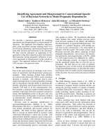
the corresponding blocks in CYP73A1. The resulting model
of the CYP73A1 core structure and active site region is
represented in Fig. 2. The advantage of this approach is that
it merges structural information from several known
structures into the target protein rather than producing a
model that is based on a single structure. All techniques are
limited by the prediction of the protein alignments, but
integration of information from multiple structures has
some chances to be better when, as in our case, protein
identities are very low.
The 6–8 A
˚
distances between the substrate protons and
the haem iron were recently deduced from
1
H paramagnetic
relaxation experiments [10] indicate that CA initially binds
roughly parallel to the haem in the oxidized CYP73A1. The
carboxylic function, which can be replaced by other anionic
groups, was previously shown to be an essential determinant
of substrate docking in the active site [7,8]. An ionic or
hydrogen bond is likely to anchor CA to a cationic or
hydrophilic residue of the protein. These data suggest that a
set of residues within 5–9 A
˚
above haem iron could be
considered putative active site contacts and tested by site-
directed mutagenesis. A search of the model for hydrophilic
residues likely to form a hydrogen bond with the substrate
pointed to N302 in the I helix as a good carboxylate binding
candidate as it is one turn away from the so-called oxygen
groove. A set of cationic residues that were predicted to
reside in the substrate-binding regions (substrate recognition
sites or SRS [27]), in particular SRS 1, 3 and 5, on the basis
of a multiple alignments with bacterial and mammalian
enzymes, were also chosen for mutagenesis to circumvent
model-prediction inaccuracy. Based on SRS predictions, the
modified cationic residues included R101, R103, K248,
R249, R366, R368 and K484. Only K484 was predicted in
the substrate pocket in our model. However, its distance to
the haem seemed too large to allow direct interaction with
the substrate anionic site.
Hydrophobic contacts with the aromatic ring of the
substrate were also investigated. A306 modification was
previously shown to adversely affect the binding of cinna-
mate and the coupling of the hydroxylation reaction [9].
This effect was probably due to a direct interaction of its
side-chain with the aromatic ring of the substrate. Our
model supports this hypothesis. The model predicts that
I371 is another residue in close proximity to the substrate.
I371 aligns with F361 in the limonene 6-hydroxylase, a
residue that was shown to control the regioselectivity of
limonene hydroxylation by CYP71Ds [28]. Finally, I303 is
located close enough to the putative substrate pocket to
form a hydrophobic contact. However, such a contact
would be precluded in the hypothesis of a van der Waals’
interaction with I371 and a hydrogen bond to N302.
Substitute residues were chosen to alter charge and
hydrophilicity with minimal change alteration to side chain
Fig. 1. Predicted location of the conserved structural blocks and SRSs
on the primary sequence of CYP73A1. Sequence alignments of
CYP73A1 with the common structural blocks of four bacterial crystal
structures (P450
BM3
, P450
CAM
, P450
TERP
,andP450
eryF
)predicted
some of the substrate recognition sites regions. SRS locations were
corroborated on the basis of a multiple alignment with the four bac-
terial enzymes also including some members of the CYP2 and CYP73
families. CYP73A1 putative SRSs determined on the basis of this
alignment are underlined (numbered 1–6 from N to C terminal) and
residues selected for directed mutagenesis are indicated by stars. The
region interblocks in CYP73A1 are displayed in grey. For the bacterial
sequences only the common structural blocks are represented, the
identity between sequences is shaded in black, similarity is shaded in
grey (threshold of 70%).
Ó FEBS 2003 Key residues for substrate recognition in CYP73A1 (Eur. J. Biochem. 270) 3687
bulk, except in the case of hydrophobic contacts for which
the influence of side-chain size was investigated. The
consecutive residues K248 and R249 were modified simul-
taneously to avoid charge compensation. As the desired
double mutations were not obtained, we analysed the triple
mutants D247E/K248T/R249M (DKR) and K248T/
R249M/I371K (KRI).
Impact of mutations on the structure and stability
of the protein
The impact of amino acid substitutions on protein stability
was investigated using the initial CO-difference spectra of
the reduced enzyme to quantify amounts of properly folded
protein with correct incorporation of haem. The time- and
temperature-dependent disappearance of the peak at
450 nm was monitored as well as any conversion of P450
into P420 that would reflect disruption of the haem–thiolate
bond [29], to test the stability of the core structure. High and
fast P450 disappearance usually correlated with decreased
yeast expression and indicated a link between improper
folding or stability loss and expression levels of the mutant
protein.
Immunoblot quantification of the apoprotein content in
yeast microsomes using antibodies raised against purified
CYP73A1 [30] revealed decreases in polypeptide expression
of the mutants that did not exceed 40% compared to the
wild-type construct. Carbon monoxide difference spectra
detected the presence of haem in all of the mutants,
although very low CO-binding was obtained with R366M,
R101M or for the triple mutants (Table 2). The modifica-
tions to residues I303, I371, R103 and K484 did not appear
to affect the production of haem protein.
P450 disappearance followed pseudo-first order kinetics.
Under standard conditions, i.e. when P450 spectra were
recorded in the presence of 0.5 mgÆmL
)1
sodium dithionite
and 30% glycerol, the half-life (t
1/2
) of the wild-type
CYP73A1 was around 3 h. In the presence of a higher
sodium dithionite concentration (4.5 mgÆmL
)1
), the t
1/2
of
CYP73A1 was 45 min when the buffer contained 3%
glycerol, and 60 min with 30% glycerol. Stability tests were
performed at 4.5 mgÆmL
)1
dithionite, using different con-
centrations of glycerol depending on the stability of each
mutant (Table 2). The results identified three classes of
mutants. The first group consisted of N302D, I371F and
I371K, that had a stability at least equal to that of the wild-
type. The second group included K484M, with a stability
that was slightly decreased compared to the wild-type, and
R103M, N302F and I371A that displayed a more pro-
nounced decrease with a t
1/2
shift from 45 to approximately
15 min. The third group, included all other mutants in
particular R366M and R101M, which demonstrated a
drastic loss in protein stability. The R366M, R368F/K,
R101M, R103M/E, DRK and KRI modifications resulted
in a very significant disruption of the tertiary structure of
the CYP73A1 protein.
Effect of mutations on cinnamic acid recognition
and metabolism
The impact of the mutations on CA binding and metabo-
lism was investigated (Table 2). The K484M mutation that
Fig. 2. A preliminary model of the active site of
CYP73A1. Construction of this first model
was based on four bacterial crystallized
structures. Only part of the active site is
shown. Based on the
1
H-NMR data [10], the
substrate is expected to be located between the
spheres. Generated by using
SWISS
-
PDB
viewer
andrenderedwith
POV
-
RAY
.
3688 G. A. Schoch et al. (Eur. J. Biochem. 270) Ó FEBS 2003
Table 2. Impact of the mutations on protein stability and CA recognition and metabolism. Expression levels were calculated from CO-difference spectra. The initial proportion of P420 was estimated from these
spectra. Stability of the haem protein was assayed by monitoring the disappearance of the 450 nm peak from the CO difference spectra in recombinant yeast microsomes reduced with high sodium dithionite
(4.5 mgÆmL
)1
). The half-lives (t
1/2
) calculated from the pseudo-first order kinetics of P450 decrease are reported. Spectra were recorded every 0.5 or 1 min during 30 min. (1) Microsomes were incubated at
30 °C in sodium phosphate 100 m
M
pH 7.4 containing 3% glycerol. (2) Low stability mutants were tested in buffer containing 30% glycerol to underline the differences between them. C4H activity was
measured using a concentration of cinnamate (150 l
M
) expected to be saturating for most of the mutants. The binding constants were calculated from the amplitude of the type I difference spectra induced by
increasing concentrations of substrate, e
type I
being the molar absorption coefficient of the saturated P450-substrate complex (DA
max
/P450 concentration) and K
s
the dissociation constant. Expression and
activity values are relative to the wild-type (100%): P450 expression, 847 pmolÆmg
)1
microsomal protein; C4H activity, 287 pkatÆmg
)1
; cytochrome c reductase activity, 1520 pkatÆmg
)1
. Cytochrome c
reductase activity is used as a control for protein induced expression and integrity. Values ± SD are the mean of three or more experiments. n.m. not measurable.
Hydrophobic and hydrogen bonding residues Positively charged residues
I helix (SRS 4) Loop 3 (SRS 5) B helix (SRS 1) (SRS 3) K helix (SRS 5) (SRS 6)
Wild-type N302F N302D I303A I371F I371A I371K Wild-type R101M R103M R103E DKR KRI R366M R368K R368F K484M
Yeast
expression
level (%)
100 ± 4.8 30 ± 0.3 71 ± 7.2 96 ± 5.1 95 ± 9.9 93 ± 2.8 105 ± 2.4 100 ± 4.8 11.2 ± 0.7 60 ± 3.5 78 ± 1.7 7.8 ± 0.2 11.6 ± 0.8 <5 81 ± 3.3 54 ± 4.3 89 ± 1.4
Initial P420
(%)
– 15 – <5 – – – – 50 – 5 65 <5 >80 – – –
t
1/2
(min) (1) 46 ± 7.5 13.8 ± 3.9 45.1 ± 5.7 5 ± 1 53.2 ± 7.1 12.7 ± 0.8 53.9 ± 7.6 46 ± 7.5 2 15 ± 4.7 2<1 2 n.m. 2 2 30.7 ± 0.6
t
1/2
(min) (2) 59.8 ± 7 10.2 ± 4.8 – 6.7 ± 0.7 3 ± 2 8.9 ± 0.9 n.m. 26± 2.4 13 ± 1.2 –
C4H activity
(%)
100 ± 1.0 0.5 ± 0.2 10 ± 0.9 75 ± 4.6 0.09 ± 0.02 11.3 ± 1.5 1.1 ± 0.2 100 ± 1.0 0.2 ± 0.3 44 ± 4.5 36 ± 5.8 1.0 ± 0.1 0.1 ± 0.05 0.1 ± 0.1 60 ± 3.5 48 ± 4.1 55 ± 6.1
Cinnamate
binding
K
s
(l
M
) 7.1 ± 1.0 13.7 ± 2.6 45 ± 9.0 3.9 ± 0.3 >100 25 ± 3.0 11.1 ± 2.2 7.1 ± 1.0 no type I 16.7 ± 2.1 >50 5.2 ± 1.6 11.9 ± 0.7 >100 11 ± 0.5 11 ± 0.6 5.9 ± 0.2
e
type I
(m
M
)1
Æcm
)1
)
128 ± 7.5 23 ± 4.7 7.5 ± 1.0 106 ± 3.0 1.0 ± 0.5 25 ± 1.5 15.6 ± 2.3 128 ± 7.5 – 120 ± 9.9 103± 2.2 35 ± 3.8 23 ± 9.2 n.m. 133 ± 6.6 126 ± 5.7 112 ± 0.9
Cyt c reductase
activity (%)
100 ± 5.0 98 ± 6.9 166 ± 5.9 107 ± 4.9 101 ± 7.2 146 ± 13 106 ± 12 100 ± 5.0 91.1 ± 21 136 ± 14 119 ± 17 83 ± 1.7 100 ± 13 118 ± 6.0 102 ± 7.3 98 ± 6.4 125 ± 9.8
Ó FEBS 2003 Key residues for substrate recognition in CYP73A1 (Eur. J. Biochem. 270) 3689
did not significantly affect protein expression or stability
had no significant impact on the binding of CA; however, it
did result in a 45% decrease in catalytic activity. All other
modifications of positively charged amino acids adversely
affected expression and/or stability of the enzyme but had a
comparatively minor affect on substrate recognition and
metabolism. Exceptions included R366M, R101M and the
triple mutations for which drastic decreases in stable haem
protein were paralleled by dramatic losses in activity.
Despite the loss of activity and structural integrity, the
DKR mutation rather unexpectedly seemed to retain an
intact affinity for substrate binding.
Modifications of N302 and I371 resulted in limited or no
apparent perturbation of protein folding and stability but
led to dramatic decreases in CA binding and hydroxylation.
N302 is likely to provide a hydrogen bonding side chain for
anchoring the carboxylate of CA. The conversion of aspa-
ragine into negatively charged aspartic acid (N302D)
resulted in a drastic effect on substrate binding affinity.
Whereas replacement with a bulky hydrophobic residue
(N302F) compromised overall protein structure and cata-
lysis.
I371 is predicted to form a van der Waals’ contact with
the aromatic ring of CA. In the I371 mutants, I371A opens
more space in the active site and thus should allow for
increased substrate mobility. Conversely, I371F and I371K
should create a steric hindrance to the binding of the
substrate above the haem iron. As expected, the I371A
mutation substantially decreases CA affinity and the ability
to desolvate the active site. Around 10% of the catalytic
activity is conserved, which would be in agreement with the
conservation of the carboxylate anchoring function of the
protein. The I371F and I371K mutations lead to an almost
complete loss in C4H activity. This activity loss is correlated
with impaired substrate binding. A complete loss of binding
was also observed upon substitution of I371 with the bulky
phenylalanine. The insertion of a positive charge in the 371
position does not completely prevent the binding of the
substrate, but almost totally hinders catalysis. This probably
results from improper positioning of the substrate’s aro-
matic ring above the haem iron.
Mutation of I303, adjacent to N302, into alanine slightly
increased affinity but modified substrate positioning and
decreased catalytic activity. This data is concordant with a
model where I303 is not a direct contact residue, but rather
contributes to optimal CA orientation in the active site.
Binding of alternate ligands to CYP73A1 mutants
The mutants that showed strongly impaired CA binding
and metabolism, but that did not display a major structural
alteration in terms of protein stability and expression, were
further tested for their ability to recognize a set of structural
analogues of CA. This set included CA precursors, plus
other natural and synthetic compounds. Some of these
compounds present a quite high intrinsic affinity for wild-
type CYP73A1, such as phenylpyruvic acid (K
s
¼ 3.1 l
M
),
phenylalanine, indole-2-carboxylic acid or cinnamyl alcohol
(K
s
¼ 12 l
M
), 2-aminoquinoline (K
s
¼ 17 l
M
) and indole-
3-carboxylic acid (the natural auxin, K
s
¼ 18 l
M
). These
compounds are ordered from gain to loss of binding to the
mutant proteins in Table 3.
As shown in Table 3, the analogues investigated were
better ligands for the mutants than the physiological
substrate CA. Relative to wild-type CYP73A1, the binding
efficiency for CA decreases 10-fold in the mutant I371K,
50-fold in I371F and 100-fold in N302D. In contrast,
increases in binding efficiency are observed for a few ligands
after modification of the protein. The most notable increases
are 15-fold for N302D with phenylalanine, 12-fold for
I371K with 2-phenoxyacetamidine, and 10-fold for I371F
with phenylalanine or cinnamylic alcohol.
The I371F modification is likely to block access to the
active centre for most of the potential substrates. Only
compounds with increased side chain flexibility or reduced
bulkiness in the CA ring region are expected to have
increased binding efficiencies compared to CA. This is
actually the case, with a gain in binding efficiency being
observed only for phenylalanine, 2-phenoxyacetamidine,
cinnamylic alcohol or 4-propynyl-oxybenzoic acid. More
relevant are the N320D and I371K mutations that could
provide a new salt-bridge or hydrogen bonding opportu-
nities in the active site region. Increased affinity of several
ligands indicates that new bonds are formed in the mutants
and may reflect a reversed orientation of the ligands or
occupation of different subpockets in the active site.
Noteworthy are the increased binding of phenylalanine
and 2-phenoxyacetamidine, which are highly polar mole-
cules. However, none of the compounds that displayed an
increased affinity produced a large spin transition of the
ferrous haem, which would be indicative of effective
desolvation of the active site and appropriate positioning
for an efficient oxidative attack. Some of the analogues
listed at the top of Table 3 that showing better binding
efficiencies than CA with the modified proteins, were
analysed in catalytic assays.
Metabolism of alternate substrates
The metabolism of CA analogues was assayed with the
N302, I371 and K484 mutants (Table 4). Microsomes from
yeast transformed with the empty expression plasmid, and
also incubations without NADPH were used to control for
CYP73-independent reactions. No metabolism of phenyl-
alanine and 2-phenoxyacetamidine was detected with the
wild-type or any of the mutants. The sensitivity of the tests
was low, due to high detection thresholds and the need to
test phenylalanine metabolism at pH 8.3, which decreases
C4H activity of the wild-type by 80%.
NA was previously shown to be the best structural mimic
and alternate substrate for wild-type CYP73A1 [7]. NA was
metabolized by all mutants with an efficiency very compar-
able to that observed with CA. This suggests that both
compounds have a very similar positioning in the active site
and validates use of NA for fluorometric quantification of
the enzyme activity [7]. Metabolism of I2C, I3C and 7MC
does not parallel that of CA in the different mutants. For
example the I371A and I371K mutations have less influence
on demethylation of 7MC than on CA hydroxylation. Also
noteworthy is the opposite effect of several amino acid
substitutions on I3C and I2C hydroxylations. Most muta-
tions have less impact on I2C than on I3C and CA
metabolism, probably due to the symmetry axis of I2C and
to the possible attack on two different carbon atoms.
3690 G. A. Schoch et al. (Eur. J. Biochem. 270) Ó FEBS 2003
Unexpectedly, the K484M substitution, which results in
close to 50% loss in C4H activity, does not affect I2C
hydroxylation. As initial cinnamate binding is not influ-
enced by this mutation (Table 2) and binding kinetics are
first-order (indicating a single binding-site), this suggests
that K484 does not directly affect catalysis but might have a
selective role in substrate position adjustment during the
catalytic cycle.
Modified regiospecificity of indole-2-carboxylic acid
hydroxylation
I2C metabolism by wild-type CYP73A1 was previously
shown to result in the formation of two products that were
not further characterized [7]. On the basis of its HPLC
retention time, UV spectrum, and monoisotopic mass, the
most polar product P1 (RT 9.8 min) was unambiguously
identified as 5-hydroxy-I2C (Fig. 3). P2 presents the same
mass as P1 and is thus a monohydroxylated product.
Superimposition of the NA and CA structures, and of their
positions of attack on those of I2C, indicates that P2 is most
likely 6-hydroxy-I2C, although an authentic standard was
not commercially available for verification. In favour of the
latter hypothesis, 5-hydroxy-I2C was tested as a substrate of
CYP73A1 and was not further metabolized.
As preliminary experiments indicated that the ratio
between the two products varies upon metabolism by the
different mutants, this ratio was used as a reporter of the
influence of the mutations on substrate docking (Table 4).
In the wild-type CYP73A1, the formation of P2 is five times
more frequent than that of P1.
The K484M mutation does not significantly affect the
P2/P1 ratio. This is not surprising considering that it does
not affect the global rate of I2C metabolism. As the length
of the I2C molecule and the distance between its carboxylate
and the positions of attack are slightly shorter than for CA
or NA, it is possible that the carboxylate of I2C is beyond
the area of influence of K484.
The N302 mutations, in particular N302D, significantly
increased the proportion of P1 so that the P2/P1 ratio
dropped closer to 1. This loss in regiospecificity in the
mutant is concordant with the increased mobility of the
Table 3. Alternate ligand binding to the mutant protein. 4-Propynyl-oxybenzoic acid and wild-type CYP73A1 is the only complex for which data
fitting with the Michaelis–Menten equation indicated second order kinetics. Binding efficiency is the e
type I
/K
s
ratio calculated for each complex. The
values listed are relative to the wild-type for each ligand. Standard deviations (not shown) are less than 12% of these values.
Ó FEBS 2003 Key residues for substrate recognition in CYP73A1 (Eur. J. Biochem. 270) 3691
molecule in the active site that is reflected by a low e
type I
(Table 3). Taken together, the data support a role for N302
in controlling of substrate orientation in the active site.
The I371 mutations to K and A have opposite effects.
The I371K mutation increases the preferential attack at the
putative 6-position, most likely by increasing steric hin-
drance near C5 of the indole ring. In contrast, the I371A
mutation appears to remove the steric constraint existing in
the wild-type CYP73A1 and favours a P2/P1 ratio closer
to 1. The observed effects of both of these mutations
support the assumption of a direct contact of I371 with the
aromatic ring of I2C or CA.
Discussion
The computational homology modelling strategy des-
cribed by Jean et al. [21] allows a reasonable prediction
of the most conserved P450 substructures, although
hypervariable regions cannot be predicted. Our present
model was based on four crystallized bacterial enzymes
(Fig. 2) and seems to correctly predict several residues
forming contacts with CA.
The model predicts that N302, which resides in the I helix
and SRS 4, is likely to form a hydrogen bond with the
carboxylate of the substrate. Mutations of this residue lead to
a dramatic loss in CA binding efficiency (10-fold for the
N302F and 100-fold for the N302D substitution) together
with a very strong decrease in catalytic activity. This confirms
a critical role for this residue in the initial binding and correct
positioning of CA during catalysis. A role of N302 in
anchoring the side-chain carboxylate of CA is further
supported by the enhanced binding of amine substituted
ligands and also by the loss of regiospecificity of I2C
hydroxylation when N302 is replaced by an aspartic acid.
Together with A306, I371 is predicted to form a
hydrophobic pocket that positions the aromatic ring of
the substrate in close proximity to the haem iron. The
adverse impact of the A306G substitution on substrate
binding and metabolism as well as coupling of the reaction
was described previously [9]. Modifications of I371, espe-
cially I371F, produced a dramatic loss in binding and
activity with CA and all other substrates. The less
detrimental effect of these substitutions on the binding of
analogues, which are less rigid or bulky than CA, and
differential impact on the regiospecificity of I2C ring-
hydroxylation support the hypothesis that the side chain of
I371 is an essential element ensuring correct positioning and
orientation of the aromatic ring in the active site.
Our model predicts that K484 is in the substrate pocket.
Its distance to the CA carboxylate in the oxidized enzyme
model does not allow for any direct interaction and, as
expected, modification of K484 has no impact on the initial
binding of CA. However, the K484M substitution leads to a
50% decrease in catalytic activity with both CA and NA. A
possible explanation is that K484 plays some role in the
electron transfer from the P450 reductase to the haem iron.
However, this residue is located on the distal side of the
haem, while interaction with the reductase and electron
transfer should involve residues on the proximal side of the
protein [31]. The unchanged I2C hydroxylase activity in
K484M when compared to that of the wild-type confirms
that the mutant is not impaired in electron transfer. Thus,
K484 must exert some control on CA/NA positioning or
product release during the catalytic cycle. Although the
K484 effect might be indirect and the interaction with the
carboxylate of CA might occur via a molecule of solvent, it
can also be postulated that the reduction of the protein or
binding of oxygen results in a conformational change of the
Fig. 3. Analysis of the products of I2C hydroxylation. Upper panel:
HPLC analysis of the products of the metabolism of 10 nmol I2C by
30 pmol recombinant CYP73A1 in 60 min and in a 100 lL assay.
Absorbance was monitored at 290 nm. Lower panel: UV spectra
corresponding to the centre of the peaks. P1 and P2 collected after
90 min incubation of 120 nmol of I2C were analysed by negative ESI-
MS. Monoisotopic mass of both compounds was 176 Da. P1 retention
time and UV spectrum was identical to that of commercial 5-hydroxy-
2-indolecarboxylic acid.
Table 4. Metabolism of alternate substrates by mutant CYP73A1s. Activities are expressed relative to wild-type CYP73A1. 100% activity is
287 pkatÆmg
)1
microsomal protein for CA, 311 pkatÆmg
)1
for NA, 38.8 pkat mg
)1
for I2C, 20.4 pkat mg
)1
for I3C, 6.6 pkat mg
)1
for 7MC. n.d.
not determined.
Mutant CA NA I2C P1/P2 I3C 7MC
73A1 % 100 % 100 ± 2.3 % 100 ± 1.1 (5.7) % 100 ± 2.0 % 100 ± 8.2
N302D 10 9.7 ± 7.0 14.6 ± 0.8 (1.5) 3.0 ± 0.4 8 ± 1.5
N302F 0.5 2.3 ± 1.2 £ 1 (2.3) n.d. n.d.
I371F 0.09 < 0.6 < 0.1 n.d. n.d. n.d.
I371A 11 12 ± 0.8 32.1 ± 2.2 (0.7) 9.3 ± 0.3 27 ± 3.4
I371K 1.1 < 0.6 5.9 ± 0.5 (8.0) £ 1 6 ± 0.9
K484M 55 50 ± 3.8 94.3 ± 3.8 (4.9) n.d. n.d.
3692 G. A. Schoch et al. (Eur. J. Biochem. 270) Ó FEBS 2003
protein, similar to that observed for P450
BM3
or P450
CAM
substrate complexes [32–34]. Such a change could bring
K484 much closer to CA. In this case, ion pairing or
hydrogen bond between K484 and the CA carboxylate
could control the optimal positioning and orientation of the
substrate for catalysis. NMR measurement of the distances
of the substrate protons to the haem iron indicate an initial
positioning of CA approximately 6–8 A
˚
from the iron in the
oxidized enzyme, which might not be optimal for catalysis
and would not particularly favour ring 4-hydroxylation. If
these measurements are correct then a structural change
that brings CA closer to the iron and adjusts substrate
position, possibly tilting the substrate so as to favour attack
at the 4 position or on the 3–4 bond, would be needed for
efficient and regiospecific catalysis. The K484M mutation
has no impact on I2C metabolism or the regioselectivity of
attack. This observation is compatible with a role of K484 in
CA reorientation as the slightly smaller size and different
shape of I2C compared to that of CA might prevent
interaction between its anionic site and K484.
N302 and I371 align with residues that have been
shown to confer substrate specificity or regioselectivity to
many other plant or mammalian P450 enzymes. Residues
corresponding to I371 govern the regiospecificity of the
hydroxylation of 4S-limonene in CYP71D18 from spear-
mint and CYP71D15 from peppermint for the synthesis
of carvone and menthol, respectively [28]. In the
mammalian CYP2B family, residues 294 and 363 are
equivalent as N302 and I371, respectively. The CYP2B
mutations were shown to affect steroid regioselectivity.
At position 363, a CYP2B1 mutant (V363L) exhibited a
twofold decrease in androgen activity [35], whereas in
CYP2B11 the reverse mutant shows a fivefold increase in
androgen activity [36]. The same residue was identified as
a determinant of substrate specificity in CYP2B2 [37],
CYP2B5 [38] and CYP2B6 [39]. Likewise, residue 294
was shown to play a key role in androgen metabolism by
CYP2B1 [40] and CYP2B4 [38]. A similar affect on
catalysis by these residue positions has been reported for
other mammalian enzymes. For example in CYP2A5,
mutation of M365, the equivalent of I371, decreased the
metabolism of aflatoxin B1 [41], while modification of
the corresponding residue (A370) in human CYP3A4
enhanced the hydroxylation of steroids [42,43].
A significant portion of the protein, which was not
reliably predicted in the model, is not shown in Fig. 2 and
was not thoroughly investigated in our site-directed experi-
ments. It is therefore likely that additional residues, such as
R or K that can form an ion pair with the carboxylate of
CA, may contribute to substrate recognition or docking.
Mutation of positively charged residues found in the
putative SRSs (Fig. 1), based on a multiple alignment did
not lead to the identification of a residue that would be
critical for the recognition or positioning of CA. Mutation
of all arginines led to a significant loss in protein stability
suggesting that they are involved in protein fold structure
rather than binding of the substrate.
The overall picture of the CYP73A1 active site provided
by our data is reminiscent of P450
BM3
[44,45], as it involves
a hydrogen bond and possibly an ion pair for the anchoring
of the carboxylate on the substrate, and also a major
hydrophobic region for the docking of the aromatic ring. As
in P450
BM3
, a substantial protein rearrangement must occur
during the catalytic cycle [32,33], probably upon reduction,
to ensure an optimal positioning of the substrate relative to
the ferryl-oxo intermediate for coupled, regiospecific attack
of the ring at the 4 position. While mutant analysis was in
progress, the first X-ray structure was described for a
membrane-bound mammalian P450, CYP2C5 [11]. This
new structure confirmed the conservation of the P450
spatial organization in eukaryotic microsomal enzymes. The
position of SRS 4 that is located in the centre of the I helix,
which includes N302 in CYP73A1, was highly conserved
relative to the haem. However, significant local changes
were detected, particularly in all other SRSs. For example,
SRS 5, facing the I helix, shows a double bend due to two
proline residues (P360 and P364). The resulting topology
orients three leucine side chains toward the active site (L358,
L359 and L363). In P450
CAM
[46] and P450
TERP
[47], SRS 5
is a b-strand partially involved in b-sheet formation with
SRS 6. In P450
BM3
[44], the first bend found in CYP2C5 is
present and the C-terminal part of SRS 5 is a b-strand not
involved in a b-sheet with SRS 6. The alignment of SRS 5
of CYP2C5 and the whole CYP2B family with those of
CYP73A1 and CYP71Ds is not ambiguous. The two
prolines and the adjacent positive charge (H365) that bind
the haem propionate in CYP2C5 are conserved. This
suggests that the double bend structure is present and
confirms I371 as a central residue of SRS 5 in CYP73A1. If
the position of the SRS relative to the haem is conserved, the
phenyl side chain in the I371F mutant should stack over the
haem, which would explain the complete impairment of
substrate binding and the increased stability of the mutant
protein. The orientation and size of SRS 6 is quite variable
between the different structures and reliable alignment of
K484 with the crystallized sequences is not possible.
Consequently, the role of K484 could not be correlated
with the mammalian structure.
CYP73A1 is more closely related to CYP2C5 (47%
similarity) than to any of the bacterial proteins (36%,
P450
BM3
;28%,P450
CAM
; 27%; P450
TERP
;30%,P450
eryF
),
and the structure of SRS 5 seems to be conserved between
CYP2C5 and CYP73A1. In order to refine our understand-
ing of CYP73A1 and to gain structural information on
SRS 5 topology, a new model was built based on the
CYP2C5 structure exclusively (1DT6). CA was positioned
in the active site of this new model, taking into account the
results of the previous NMR measurements [10] and
information obtained from mutagenesis (Fig. 4). In this
new model, N302 easily forms a hydrogen bond with the
carboxylate of CA and I371 is well positioned for hydro-
phobic contact with the substrate aromatic ring. A306 was
shown to be critical for substrate recognition [9]. In this new
model, its methyl group is 4.8 A
˚
from the haem iron and
3.5 A
˚
from the substrate. K484 is still too far away to form a
direct contact with the cinnamate.
In conclusion, a combination of homology modelling
and site-directed mutagenesis of CYP73A1 has identified
N302 and I371 as key determinants of substrate binding
and orientation for catalysis. K484 is not involved in
initial substrate binding, but seems to play a significant
role in catalysis, possibly by contributing to substrate
reorientation during the catalytic cycle. Modification
of active site residues improved affinity for substrate
Ó FEBS 2003 Key residues for substrate recognition in CYP73A1 (Eur. J. Biochem. 270) 3693
analogues, but correct positioning allowing for a gain of
function could not be achieved. Indole 2-carboxylic acid,
which is regiospecifically attacked at the 5 and 6
positions, is a very useful probe for investigating the
topology of the CYP73A1 active site.
Acknowledgments
We thank P. Ullmann for help and support, M. Bergdoll for helpful
discussion, D. Little and K. Griffin for critical readings of the
manuscript. The W(R) and WAT11 yeast strains and the pYeDP60
expression vector were kindly provided by Drs D. Pompon and
P. Urban (CNRS, Gif-sur-Yvette). This work was supported by the
CNRS Program Chimie-Physique du Vivant, and a fellowship from the
French Ministry of Research to G.A.S.
References
1. Weisshaar, B. & Jenkins, G.I. (1998) Phenylpropanoid biosyn-
thesis and its regulation. Curr. Opin. Plant Biol. 1, 251–257.
2. Lewis, N.G. (1999) A 20(th) century roller coaster ride: a short
account of lignification. Curr. Opin. Plant Biol. 2, 153–162.
3.Pierrel,M.A.,Batard,Y.,Kazmaier,M.,Mignotte-Vieux,C.,
Durst, F. & Werck-Reichhart, D. (1994) Catalytic properties of
the plant cytochrome P450 CYP73 expressed in yeast. Substrate
specificity of a cinnamate hydroxylase. Eur. J. Biochem. 224, 835–844.
4. Mizutani,M.,Ohta,D.&Sato,R.(1997)IsolationofacDNA
and a genomic clone encoding cinnamate 4-hydroxylase from
Arabidopsis and its expression manner in planta. Plant Physiol.
113, 755–763.
5. Nedelkina, S., Jupe, S.C., Blee, K.A., Schalk, M., Werck-Reich-
hart, D. & Bolwell, G. (1999) Novel characteristics and regulation
of a divergent cinnamate 4-hydroxylase (CYP73A15) from French
bean: engineering expression in yeast. Plant Mol. Biol. 39,
1079–1090.
6. Teutsch, G.H., Hasenfratz, M.P., Lesot, A., Stoltz, C., Garnier,
J.M.,Jeltsch,J.M.,Durst,F.&Werck-Reichhart,D.(1993)
Isolation and sequence of a cDNA encoding the Jerusalem arti-
choke cinnamate 4-hydroxylase, a major plant cytochrome P450
involved in the general phenylpropanoid pathway. Proc. Natl
Acad. Sci. USA 90, 4102–4106.
7. Schalk, M., Batard, Y., Seyer, A., Nedelkina, S., Durst, F. &
Werck-Reichhart, D. (1997) Design of fluorescent substrates and
potent inhibitors of CYP73As, P450s that catalyze 4-hydroxyla-
tion of cinnamic acid in higher plants. Biochemistry 36, 15253–
15261.
8. Schalk, M., Cabello-Hurtado, F., Pierrel, M.A., Atanossova, R.,
Saindrenan, P. & Werck-Reichhart, D. (1998) Piperonylic acid, a
selective, mechanism-based inactivator of the trans-
cinnamate 4-hydroxylase: a new tool to control the flux of
metabolites in the phenylpropanoid pathway. Plant Physiol. 118,
209–218.
9. Schalk, M., Nedelkina, M., Schoch, G., Batard, Y. & Werck-
Reichhart, D. (1999) Role of unusual amino acid residues in the
proximal and distal heme regions of a plant P450, CYP73A1.
Biochemistry 38, 6093–6103.
10. Schoch, G.A., Attias, R., Belghazi, M., Dansette, P.M. & Werck-
Reichhart, D. (2003) Engineering of a water-soluble plant cyto-
chrome P450, CYP73A1, and NMR based orientation of natural
and alternate substrates in the active site. Plant Physiol., in press.
11. Williams, P.A., Cosme, J., Sridhar, V., Johnson, E. & McRee,
D.E. (2000) Mammalian microsomal cytochrome P450 mono-
oxygenase: structural adaptations for membrane binding and
functional diversity. Mol. Cell 5, 121–131.
12. Urban, P., Cullin, C. & Pompon, D. (1990) Maximizing the
expression of mammalian cytochrome P-450 monooxygenase
activities in yeast cells. Biochimie 72, 463–472.
13. Truan, G., Cullin, C., Reisdorf, P., Urban, P. & Pompon, D.
(1993) Enhanced in vivo monooxygenase activities of mammalian
P450s in engineered yeast cells producing high levels of
NADPH-P450 reductase and human cytochrome b
5
. Gene 125,
49–55.
14. Urban, P., Werck-Reichhart, D., Teutsch, H.G., Durst, F., Reg-
nier, S., Kazmaier, M. & Pompon, D. (1994) Characterization of
recombinant plant cinnamate 4-hydroxylase produced in yeast.
Kinetic and spectral properties of the major plant P450 of the
phenylpropanoid pathway. Eur. J. Biochem. 222, 843–850.
15. Pompon,D.,Louerat,B.,Bronine,A.&Urban,P.(1996)Yeast
expression of animal and plant P450s in optimized redox
environments. Methods Enzymol. 272, 51–64.
16. Omura, T. & Sato, R. (1964) The carbon monoxide-binding pig-
ment of liver microsomes. I. Evidence for its hemoprotein nature.
J. Biol. Chem. 239, 2370–2378.
17. Benveniste, I., Gabriac, B. & Durst, F. (1986) Purification and
characterization of the NADPH-cytochrome P-450 (cytochrome
c) reductase from higher-plant microsomal fraction. Biochem.
J. 235, 365–373.
18. Reichhart, D., Salau
¨
n, J.P., Benveniste, I. & Durst, F. (1980)
Time-course of induction of cytochrome P450,
Fig. 4. Predicted orientation of cinnamic acid
in the active site of CYP73A1 model was built
on the crystal structure of the membrane bound
eukaryotic CYP2C5. Substrate positioning
was based on the combination of distances
calculated from
1
H-NMR measurements [10]
and site-directed mutagenesis data.
3694 G. A. Schoch et al. (Eur. J. Biochem. 270) Ó FEBS 2003
NADPH-cytochrome c reductase, and cinnamic acid hydro-
droxylase by phenobarbital, ethanol, herbicides, and manganese in
higher plant microsomes. Plant Physiol. 66, 600–604.
19. Duggleby, R.G. (1984) Regression analysis of nonlinear Arrhenius
plots: an empirical model and a computer program. Comput. Biol.
Med. 14, 447–455.
20. Werck-Reichhart,D.,Gabriac,B.,Teutsch,H.&Durst,F.(1990)
Two cytochrome P-450 isoforms catalysing O-de-ethylation of
ethoxycoumarin and ethoxyresorufin in higher plants. Biochem.
J. 270, 729–735.
21.Jean,P.,Pothier,J.,Dansette,P.M.,Mansuy,D.&Viari,A.
(1997) Automated multiple analysis of protein structures: appli-
cation to homology modeling of cytochromes P450. Proteins 28,
388–404.
22. Minoletti, C. (1998) PhD Thesis. University Paris VI Rene
´
Descartes, Paris, France.
23. Patard, L., Stoven, V., Gharib, B., Bontems, F., Lallemand, J.Y.
& De Reggi, M. (1996) What function for human lithostathine?:
structural investigations by three-dimensional structure modeling
and high-resolution NMR spectroscopy. Protein Eng. 9, 949–957.
24. Dunbrack, R.L. Jr & Karplus, M. (1993) Backbone-dependent
rotamer library for proteins. Application to side-chain prediction.
J. Mol. Biol. 230, 543–574.
25.Guntert,P.,Mumenthaler,C.&Wuthrich,K.(1997)Torsion
angle dynamics for NMR structure calculation with the new
program DYANA. J. Mol. Biol. 273, 283–298.
26. Laskowski, R.A., MacArthur, M.W., Moss, D.S. & Thornton,
J.M. (1993) PROCHECK: a program to check the streochemical
quality of protein structures. J. Appl. Cryst. 26, 283–291.
27. Gotoh, O. (1992) Substrate recognition sites in cytochrome P450
family 2 (CYP2) proteins inferred from comparative analyses of
amino acid and coding nucleotide sequences. J. Biol. Chem. 267,
83–90.
28. Schalk, M. & Croteau, R. (2000) A single amino acid substitution
(F363I) converts the regiochemistry of the spearmint (–)-limonene
hydroxylase from a C6- to a C3-hydroxylase. Proc.NatlAcad.Sci.
USA 97, 11948–11953.
29. Martinis, S.A., Blanke, S.R., Hager, L.P., Sligar, S.G., Hoa, G.H.,
Rux, J.J. & Dawson, J.H. (1996) Probing the heme iron co-
ordination structure of pressure-induced cytochrome P420cam.
Biochemistry 35, 14530–14536.
30. Werck-Reichhart, D., Batard, Y., Kochs, G., Lesot, A. & Durst,
F. (1993) Monospecific polyclonal antibodies directed against
purified cinnamate 4-hydroxylase from Helianthus tuberosus.
Immunopurification, immunoquantitation, and interspecies cross-
reactivity. Plant Physiol. 102, 1291–1298.
31. Graham, S.E. & Peterson, J.A. (1999) How similar are P450s and
what can their differences teach us? Arch. Biochem. Biophys. 369,
24–29.
32. Modi, S., Primrose, W.U., Boyle, J.M., Gibson, C.F., Lian, L.Y.
& Roberts, G.C. (1995) NMR studies of substrate binding to
cytochrome P450 BM3: comparisons to cytochrome P450 cam.
Biochemistry 34, 8982–8988.
33. Modi, S., Sutcliffe, M.J., Primrose, W.U., Lian, L.Y. & Roberts,
G.C. (1996) The catalytic mechanism of cytochrome P450 BM3
involves a 6 A movement of the bound substrate on reduction.
Nat. Struct. Biol. 3, 414–417.
34. Schlichting, I., Berendzen, J., Chu, K., Stock, A.M., Maves, S.A.,
Benson, D.E., Sweet, R.M., Ringe, D., Petsko, G.A. & Sligar,
S.G. (2000) The catalytic pathway of cytochrome p450cam at
atomic resolution. Science 287, 1615–1622.
35. He, Y.Q., He, Y.A. & Halpert, J.R. (1995) Escherichia coli
expression of site-directed mutants of cytochrome P450 2B1 from
six substrate recognition sites: substrate specificity and inhibitor
selectivity studies. Chem. Res. Toxicol. 8, 574–579.
36. Hasler, J.A., Harlow, G.R., Szklarz, G.D., John, G.H., Kedzie,
K.M.,Burnett,V.L.,He,Y.A.,Kaminsky,L.S.&Halpert,J.R.
(1994) Site-directed mutagenesis of putative substrate recognition
sites in cytochrome P450 2B11: importance of amino acid residues
114, 290, and 363 for substrate specificity. Mol. Pharmacol. 46,
338–345.
37. Strobel, S.M. & Halpert, J.R. (1997) Reassessment of cytochrome
P450 2B2: catalytic specificity and identification of four active site
residues. Biochemistry 36, 11697–11706.
38. Szklarz,G.D.,He,Y.Q.,Kedzie,K.M.,Halpert,J.R.&Burnett,
V.L. (1996) Elucidation of amino acid residues critical for unique
activities of rabbit cytochrome P450 2B5 using hybrid enzymes
and reciprocal site-directed mutagenesis with rabbit cytochrome
P450 2B4. Arch. Biochem. Biophys. 327, 308–318.
39. Domanski, T.L., Schultz, K.M., Roussel, F., Stevens, J.C. &
Halpert, J.R. (1999) Structure–function analysis of human cyto-
chrome P-450 2B6 using a novel substrate, site-directed muta-
genesis, and molecular modeling. J. Pharmacol. Exp. Ther. 290,
1141–1147.
40. Domanski, T.L., He, Y.Q., Scott, E.E., Wang, Q. & Halpert, J.R.
(2001) The role of cytochrome 2B1 substrate recognition site
residues 115, 294, 297, 298, and 362 in the oxidation of steroids and
7-alkoxycoumarins. Arch. Biochem. Biophys. 394, 21–28.
41. Pelkonen, P., Lang, M.A., Negishi, M., Wild, C.P. & Juvonen,
R.O. (1997) Interaction of aflatoxin B1 with cytochrome P450 2A5
and its mutants: correlation with metabolic activation and toxicity.
Chem. Res. Toxicol. 10, 85–90.
42. He, Y.A., He, Y.Q., Szklarz, G.D. & Halpert, J.R. (1997) Iden-
tification of three key residues in substrate recognition site 5
of human cytochrome P450 3A4 by cassette and site-directed
mutagenesis. Biochemistry 36, 8831–8839.
43. Domanski, T.L., He, Y.A., Khan, K.K., Roussel, F., Wang, Q. &
Halpert, J.R. (2001) Phenylalanine and tryptophan scanning
mutagenesis of CYP3A4 substrate recognition site residues and
effect on substrate oxidation and cooperativity. Biochemistry 40,
10150–10160.
44. Li, H. & Poulos, T.L. (1997) The structure of the cytochrome
p450BM-3 haem domain complexed with the fatty acid substrate,
palmitoleic acid. Nat. Struct. Biol. 4, 140–146.
45. Ost, T.W., Miles, C.S., Murdoch, J., Cheung, Y., Reid, G.A.,
Chapman, S.K. & Munro, A.W. (2000) Rational re-design of the
substrate binding site of flavocytochrome P450 BM3. FEBS Lett.
486, 173–177.
46. Raag, R. & Poulos, T.L. (1991) Crystal structures of cytochrome
P-450CAM complexed with camphane, thiocamphor, and
adamantane: factors controlling P-450 substrate hydroxylation.
Biochemistry 30, 2674–2684.
47. Hasemann, C.A., Ravichandran, K.G., Peterson, J.A. &
Deisenhofer, J. (1994) Crystal structure and refinement of
cytochrome P450terp at 2.3 A resolution. J. Mol. Biol. 236,
1169–1185.
Ó FEBS 2003 Key residues for substrate recognition in CYP73A1 (Eur. J. Biochem. 270) 3695