Báo cáo khoa học: A new paradigm for oxygen binding involving two types of ab contacts docx
Bạn đang xem bản rút gọn của tài liệu. Xem và tải ngay bản đầy đủ của tài liệu tại đây (493.55 KB, 11 trang )
REVIEW ARTICLE
Human haemoglobin
A new paradigm for oxygen binding involving two types of ab contacts
Keiji Shikama
1,2
and Ariki Matsuoka
3
1
Biological Institute, Graduate School of Life Sciences, Tohoku University, Sendai, Japan,
2
PHP Laboratory for Molecular Biology,
Sendai, Japan;
3
Department of Biology, Fukushima Medical University, Fukushima, Japan
This review summarizes the most recent state of haemo-
globin (Hb) research based on the literature and our own
results. In particular, an attempt is made to form a unified
picture for haemoglobin function by reconciling the
cooperative oxygen binding with the stabilization of the
bound dioxygen in aqueous solvent. The HbA molecule
contains two types of ab contacts. One type is the a1b2or
a2b1 contacts, called sliding contacts, and these are strongly
associated with the cooperative binding of O
2
to the a
2
b
2
tetramer. The other type is the a1b1ora2b2 contacts, called
packing contacts, but whose role in Hb function was not
clear until quite recently. However, detailed pH-dependence
studies of the autoxidation rate of HbO
2
have revealed that
the a1b1anda2b2 interfaces are used for controlling the
stability of the bound O
2
. When the a1b1ora2b2contactis
formed, the b chain is subjected to a conformational con-
straint which causes the distal (E7) histidine to be tilted
slightly away from the bound dioxygen, preventing the
proton-catalysed nucleophilic displacement of O
2
–
from the
FeO
2
by an entering water molecule. This is one of the most
characteristic features of HbO
2
stability. Finally we discuss
the role of the a1b1ora2b2 contacts by providing some
examples of unstable haemoglobin mutants. These patho-
logical mutations are found mostly on the b chain, especially
in the a1b1 contact regions. In this way, HbA seems to
differentiate two types of ab contacts for its functional
properties.
Keywords: ab contacts; distal (E7) histidine; HbA; heme
oxidation; oxygen binding.
Two types of ab contacts in HbA
In haemoglobin (Hb) research, the central problem is
understanding the mechanism for the cooperative oxygen
binding to the a
2
b
2
tetramer. For human HbA, the a and b
chains contain 141 and 146 amino acid residues, respect-
ively, and a representative set of the successive oxygen-
binding constants is given in terms of Torr
)1
as follows:
K
1
¼ 0.0188, K
2
¼ 0.0566, K
3
¼ 0.407 and K
4
¼ 4.28 in
0.1
M
Bis/Tris buffer containing 0.1
M
KCl at pH 7.4 and
25 °C [1]. In this reaction, major differences have been
found between deoxyhaemoglobin and oxyhaemoglobin by
comparing their X-ray crystal structures (e.g. [2–6]). These
include a movement of the iron atom into the haem plane
with a simultaneous change in the orientation of the
proximal (F8) histidine, a rotation of the a1b1 dimer relative
to the other a2b2 dimer about an axis P by 12–15 degrees,
and a translation of the one dimer relative to the other along
the P axis by approximately 1 A
˚
. The latter two changes are
accompanied by sequential breaking of the so-called salt
bridges by C-terminal residues. Incidentally, the P is taken
as an axis which is perpendicular to the dyads of both the
liganded and unliganded Hb molecules.
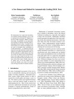
As illustrated in Fig. 1, there are two types of ab contacts
in the Hb molecule. One is the a1b1(ora2b2) contact
involving B, G, and H helices and the GH corner, and the
other is the a1b2(ora2b1) contact involving mainly helices
C and G and the FG corner [3,7]. When HbA goes from the
deoxy to the oxy form, the a1b2anda2b1 contacts undergo
the principal changes associated with cooperative oxygen
binding, so that these are named the sliding contacts. As a
result of the relative rotation of the a1b1anda2b2dimers,
the gap between the b chains becomes too small to
accommodate 2,3-diphosphoglyceric acid (DPG) that serves
to reduce the oxygen affinity of HbA. At the a1b1anda2b2
interfaces, on the other hand, negligible changes are found
insofar as the crystal structure has been examined. These are
called the packing contacts accordingly, but their role in
haemoglobin function was not clear for a very long time.
To the packing contacts, we have recently assigned a key
role for stabilizing the HbO
2
tetramer, as the formation of
the a1b1ora2b2 contact greatly suppresses the haem
oxidation, particularly of the b chain at acidic pH values
[8,9]. Based on a nucleophilic displacement of O
2
–
from the
FeO
2
centre, kinetic analyses of HbO
2
oxidation were
carried out with special focus on the proton-catalysed
Correspondence to K. Shikama, PHP Laboratory for Molecular
Biology, Nakayama-Yoshinari 1-16-8, Sendai 989-3203, Japan.
E-mail:
Abbreviations: Hb, haemoglobin; DPG, 2,3-diphosphoglyceric acid.
Dedication:ThisreviewisdedicatedtoMaxF.Perutz
(19 May 1914–6 February 2002), who laid the foundation for an entire
field of haemoglobin research. According to a kind suggestion made
by one of the referees, it should be added that Perutz once called
haemoglobin a Ôhonorary enzymeÕ. Both haemoglobin and myoglobin
are actually antienzymes, because they prevent the undesired
electron transfer from Fe(II) to the bound O
2
as far as possible in
aqueous solution.
(Received 5 June 2003, revised 29 July 2003,
accepted 13 August 2003)
Eur. J. Biochem. 270, 4041–4051 (2003) Ó FEBS 2003 doi:10.1046/j.1432-1033.2003.03791.x
process performed by the distal (E7) histidine residue. Such
examinations seem to be of primary importance, not only
for a full understanding of the molecular mechanism of
haemoglobin autoxidation, but also for planning new
molecular designs for synthetic oxygen carriers that are
highly resistant to haem oxidation under physiological
conditions. Finally, we revisit haemoglobin function as seen
from the two different types of ab contacts, and try to
reconcile cooperative oxygen binding with stabilization of
the bound dioxygen. With respect to this, we also give
possible implications for the unstable haemoglobin mutants
leading to the formation of Heinz bodies in red blood cells,
resulting in haemolytic anaemia.
Autoxidation reaction of HbO
2
and its
constituent chains
Biphasic nature of the autoxidation reaction
The reversible and stable binding of molecular oxygen with
the haem iron(II) is the basis of haemoglobin function. Even
in air-saturated buffers, however, HbA is oxidized easily
from the oxygenated form (HbO
2
) to the ferric(III) met-
form (metHb) with generation of the superoxide anion
[10,11] as follows:
HbO
2
!
k
obs
metHb þ O
À
2
ð1Þ
where k
obs
represents the first-order rate constant
observed at a given pH value in terms of the constituent
chains. This autoxidation reaction can be monitored by
the spectral changes with time, after fresh HbO
2
was
placed in 0.1
M
buffer containing 1 m
M
EDTA at 35 °C.
The spectra evolved to the final state of each run, which
was identified as the usual ferric met-form, with a set of
isosbestic points. Consequently, the process was fol-
lowed by a plot of experimental data as
_
ln([HbO
2
]
t
/
[HbO
2
]
0
) vs. time t, where the ratio of HbO
2
concentra-
tion after time t to that at time t ¼ 0 can be obtained by
the absorbance changes at 576 nm for the a-peak of
human HbO
2
.
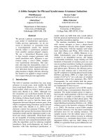
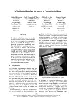
Fig. 2 shows such examples of the first-order plot for the
autoxidation reaction of human HbO
2
at two different pH
values. At pH 6.2, HbA exhibited a biphasic curve that can
be described by the first-order kinetics containing two rate
constants as follows:
½HbO
2
t
½HbO
2
0
¼ P  expðÀk
f
 tÞþð1 À PÞÂexpðÀk
s
 tÞ
ð2Þ
In this equation, a fast first-order rate constant k
f
is
attributed to the a chains and a slow rate constant k
s
is for
the b chains in the HbO
2
tetramer. P is the molar fraction
of the rapidly reacting haems. This conclusion is based on
the rapid chain separation experiment of partially (30%)
oxidized HbO
2
on polyacrylamide gel [8,12].
By iterative least-squares procedures inserting various
values for k
f
and k
s
into Eqn (2), the best fit to the
experimental data was obtained as a function of time t.In
these computations, the value of P was also allowed to vary
across a large range from 0.40 to 0.60 [8,9]. In this way, the
following parameters were established at pH 6.2: k
f
¼ 0.82
(± 0.03) · 10
)1
h
)1
, k
s
¼ 0.13 (± 0.01) · 10
)1
h
)1
,and
P ¼ 0.52 (± 0.04) in 0.1
M
Mes buffer at 35 °C. At pH 9.2,
on the other hand, the reaction was described completely by
a single first-order rate constant of 0.99 (± 0.02) · 10
)2
h
)1
(i.e. k
f
¼ k
s
with P ¼ 0.50) in 0.1
M
Caps buffer at 35 °C.
We have also studied the effect of DPG on the autoxidation
rate of HbA at 35 °C. DPG was added to stripped HbO
2
(0.13 m
M
)atmolarexcessesof5,14and24,butthis
allosteric effector offered no significant effect on either k
f
or
k
s
values at pH 6.5 and 8.5 [13].
Fig. 2. First-order plots for the autoxidation reaction of human HbO
2
in
0.1
M
buffer at 35 °C. Each curve was obtained by a least-squares
fitting to the experimental points, based on Eqn (2). At pH 6.2, HbA
showed a biphasic autoxidation curve containing two rate constants, k
f
and k
s
, respectively. At pH 9.2, however, the reaction was mono-
phasic. Redrawn from Yasuda et al.[9].
Fig. 1. Schematic diagram of HbA tetramer showing the two different
types of ab contacts. HbA has a molecular dyad axis (which is per-
pendicular to the plane of the figure) relating the a1b1 dimer to the
a2b2dimer.
4042 K. Shikama and A. Matsuoka (Eur. J. Biochem. 270) Ó FEBS 2003
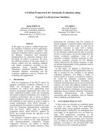
pH-Dependencies of the autoxidation rate
If the values of k
f
and k
s
are plotted against the pH of the
solution, we can obtain a pH profile for the stability of
HbO
2
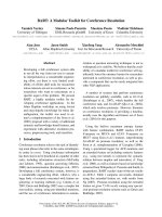
. Fig. 3 shows such profiles for both of the a and b
chains in the HbO
2
tetramer over the range pH 5–11, under
air-saturated conditions in 0.1
M
buffer at 35 °C. In the
acidic range of pH 7–5, the logarithmic values of k
f
increased very rapidly with increasing hydrogen ion con-
centration. The values of k
s
also increased with increasing
proton concentration but much less so than for k
f
.Rather,
the k
s
values exhibited a rate saturation behaviour on the
acidic extreme. In a plot of log(k
obs
) vs. pH, its slope showed
a value of n ¼ )1fork
f
, whereas a value close to n ¼ )0.6
was for k
s
. In the basic side higher than pH 8, on the other
hand, practically no difference was observed between the
k
f
and k
s
values, indicative of the oxidation curve being
monophasic. Nevertheless, it is also true that both graphs
depend strongly upon the pH of the solution, having a
parabolic part with a minimum rate appearing at pH 8.5.
At this point, the most important questions have arisen as
to whether the constituent a and b chains each has its own
different stability, and, if not, what the origin is of
nonequivalence of the chains in haem oxidation. In this
regard, it should be noted that such a chain heterogeneity of
HbO
2
oxidation can be retained even in very diluted
concentrations of haemoglobin [13]. When human HbO
2
is
placed in dilution, the tetrameric species is known to
dissociate into ab dimers along the a1b2ora2b1 interface,
so that the dimers produced are of the a1b1ora2b2type
[14,15]. Accordingly, these results strongly suggest that the
formation of the a1b1ora2b2 contact must be responsible
for the remarkable stability of the b chain against the acidic
autoxidation. This was the next step to be clarified.
Stability property of the separated a and b chains
In separated a and b chain solutions, the protein is known
to exist in an equilibrium of a
ÀÀ*
)ÀÀ a
2
and b
ÀÀ*
)ÀÀ b
4
respectively. Under our experimental conditions, the mono-
meric form (87%) was predominant in the a chain, while
the tetrameric form (99%) was predominant in the b chain.
This estimation was made on the basis of the results of
McDonald et al. [16]. As for the tetrameric form of the b
chain, Borgstahl et al. [7] have reported the 1.8 A
˚
structure
with carbonmonoxy-b
4
(COb
4
) derivative, and compared
subunit–subunit contacts between three types of interfaces
(a1b1, a1b2, and a1a2) of HbO
2
and the corresponding
COb
4
interfaces. As a result, they found that the b1b2
interface of the COb
4
tetramer is less stable and more
loosely packed than its a1b1 counterpart in HbO
2
.In
particular, there are significant packing differences at the
end of the B helix between these homologous interfaces; the
B helix–H helix contact region is spread apart by approxi-
mately 1 A
˚
in COb
4
relative to oxyHb. Specifically, the b1b2
interface of the COb
4
tetramer does not include close
contacts between residues Pro-125 (H3) and Val-33 (B15),
Gln-127 (H5) and Val-34 (B16), and Ala-128 (H6) and Val-
34 (B16). The side chain disorder also makes the centre of
the b1b2 interface packed less tightly in the COb
4
tetramer.
Therefore, the b1b2 contact sites in the b
4
tetramer are
indeed different from the corresponding a1b1 contact sites
in the HbA tetramer.
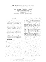
Anyway, we have revealed that over the wide range of
pH 5–10, the separated a and b chains are both oxidized
much more rapidly than in the parent HbO
2
tetramer.
Fig. 4 represents such pH-dependencies of the observed
rate constants, k
a
obs
and k
b
obs
, for autoxidation of the isolated
a and b chains in 0.1
M
buffer at 35 °C. It thus becomes
evident that the b chain, when separated from the HbO
2
tetramer, does not show any rate saturation behaviour at
low pH. Rather, its rate increased very rapidly with
increasing hydrogen ion concentration, exhibiting a value
close to n ¼ )1 for the slope against the acidic pH. We can
therefore conclude that the intrinsic oxidation rate is almost
thesamewiththeseparateda and b chains, completely freed
from the remarkable differences between them in the
autoxidation reaction of the parent HbO
2
tetramer.
Mechanism of the haem oxidation for HbO
2
FeO
2
bonding and its nucleophilic displacement of O
2
–
It has been widely accepted that HbA is much more resistant
to autoxidation than myoglobin. However, it is now evident
that the constituent a and b chains, once separated from
the parent HbO
2
, are oxidized more rapidly than most
Fig. 3. Differential pH-dependencies of k
f
and k
s
for the autoxidation
reaction of human HbO
2
in 0.1
M
buffer at 35 °C. Apairofthe
observed first-order rate constants, k
f
(s)andk
s
(d), was obtained by
a least-squares fitting to each of the oxidation curves at different pH
values. In the acidic range of pH 7–5, the logarithmic plots of k
f
give a
slope of n ¼ )1againstthepH,butn¼ )0.6 for k
s
.Redrawnfrom
Tsuruga et al.[8].
Ó FEBS 2003 A unified picture for Hb function (Eur. J. Biochem. 270) 4043
mammalian oxymyoglobins. Such enhancements in the
oxidation rate have been frequently attributed to the
increased concentration of the deoxygenated species in
HbO
2
or MbO
2
solution, since the deoxy form is certainly
the preferred target for many kinds of oxidants. This simple
mechanism, however, cannot explain the above-mentioned
results for the separated a and b chains, because it has been
definitively established that both chains have a much higher
oxygen affinity with fewer deoxygenated species than the
parent HbO
2
tetramer. In 0.1
M
phosphate buffer at pH 7.0
and 30 °C, indeed, Tyuma et al. [17] reported the P
50
values
of 1.00 Torr for the a chains and 0.45 Torr for the b chains,
whereas HbA showed P
50
¼ 16.59 Torr in the absence of
DPG.
Certainly, dioxygen is a powerful oxidizing agent in
a triplet ground state,
3
P
À
g
, whose biradical electronic
configuration is given by the following notation:
O
2
ðr1sÞ
2
ðr
Ã
1sÞ
2
ðr2sÞ
2
ðr
Ã
2sÞ
2
ðr2p
z
Þ
2
ðp2p
x
Þ
2
ðp2p
y
Þ
2
ðp
Ã
2p
x
Þ
1
ðp
Ã
2p
y
Þ
1
ðr
Ã
2p
z
Þ
0
ð3Þ
Dioxygen therefore has a very strong tendency to take
electrons from other substances and to make the com-
plete electron-pairing in its unoccupied orbitals. This
property leads to the sequential production of the so-
called active oxygen species such as superoxide anion
(O
2
–
), peroxide anion (O
¼
2
Þ and hydroxyl radical (HO
•
).
For O
2
at 760 Torr
1
, pH 7 and 25 °C, its midpoint
oxidation-reduction potential is + 0.81 V for the com-
plete, four-equivalent reduction to water, showing a total
free energy change of )74.7 kcalÆmol
)1
()312 kJÆmol
)1
).
Nevertheless, the addition of the first electron to O
2
is an
unfavourable, uphill process with a low redox potential
of e°¢(O
2
/O
2
–
) ¼ )0.33 V [18]. All of the steps subsequent
to water are downhill. In this sense, molecular oxygen is
a rather poor one-electron acceptor, and this thermo-
dynamic barrier to the first step seems to be the crucial
ridge located between the stabilization and the activation
of dioxygen bound to the haemoproteins [19].
Using a value of + 0.150 V for the oxidation–reduction
potential of human Hb at pH 7 and 30 °C[20],wemay
write the primary step for the autoxidation reaction of
HbO
2
as follows:
In this scheme, the reaction from left to right is associated
with a change in redox potential (De°¢)of)0.48 V, which
corresponds to a positive free energy change of + 11.0 kcalÆ
mol
)1
(+ 4 6 . 0 k J Æmol
)1
). Accordingly, a considerable energy
barrier accompanies the reduction of O
2
to O
2
–
by deoxy-
Hb, so this one-electron transfer cannot occur spontane-
ously. In many respects, the spontaneous dissociation of O
2
–
from the FeO
2
centre is an energetically unfavourable
process, so that there must be involved some specific
mechanism that causes very rapid generation of O
2
–
from
HbO
2
, as formulated in Eqn (1), in aqueous solution.
Recently, Shikama [21] has carefully evaluated various
mechanisms proposed so far for the autoxidation reaction
of myoglobin and haemoglobin, including the effects of pH,
oxygen pressure, and subsequent side reactions with the
H
2
O
2
produced by the spontaneous dismutation of O
2
–
.As
a result, he concluded that a displacement mechanism is
needed to make it possible to yield O
2
–
so readily from the
FeO
2
centre. In essence, kinetic and thermodynamic studies
of the stability of mammalian oxymyoglobins have shown
that the autoxidation reaction is not a simple, dissociative
loss of O
2
–
from MbO
2
but is due to a nucleophilic
displacement of O
2
–
from MbO
2
by a water molecule or a
hydroxyl ion that can enter the haem pocket from the
surrounding solvent. The iron is thus converted to the ferric
met-form, and the water molecule or the hydroxyl ion
remains bound to the Fe(III) at the sixth coordinate
position so as to form aqua- or hydroxide-metMb. Even the
complicated pH-dependence for the autoxidation rate can
thereby be explained primarily in terms of the following
three types of displacement processes [19,21–24]:
Mb(II)(O
2
ÞþH
2
O
!
k
0
Mb(III)(OH
2
ÞþO
À
2
ð5Þ
Mb(II)(O
2
ÞþH
2
O þ H
þ
À!
k
H
Mb(III)(OH
2
ÞþHO
2
ð6Þ
Mb(II)(O
2
ÞþOH
À
À!
k
OH
Mb(III)(OH
À
ÞþO
À
2
ð7Þ
In these equations, k
0
is the rate constant for the basal
displacement by H
2
O, k
H
is the rate constant for the
Fig. 4. pH profiles for the autoxidation rate of the separated a and b
chains in 0.1
M
buffer at 35 °C. Both of the computed curves were
obtained by a least-squares fitting to the experimental points over the
whole range of pH studied, based on Eqn (8). Redrawn from Tsuruga
et al.[8].
ð4Þ
4044 K. Shikama and A. Matsuoka (Eur. J. Biochem. 270) Ó FEBS 2003
proton-catalysed displacement by H
2
O, and k
OH
is the
rate constant for the displacement by OH
–
. The extent of
contribution of these elementary processes to the
observed or overall autoxidation rate, k
obs
in Eqn (1),
can vary with the concentrations of H
+
or OH
–
ion.
Consequently, the autoxidation rate exhibits a very
strong parabolic dependence on pH. The reductive
displacement of the bound dioxygen as O
2
–
byH
2
O
can proceed without any protonation, but it has been
clearly shown that the rate is enormously accelerated
with the proton assistance by a factor of 10
6
per mole, as
formulated by Eqn (6). In this proton catalysis, the
distal histidine, which forms a hydrogen bond to the
bound dioxygen [25], appears to facilitate the effective
movement of a catalytic proton from the solvent to the
bound, polarized dioxygen via its imidazole ring and by
a proton-relay mechanism [21,24]. In this way, such a
nucleophilic displacement mechanism has successfully
been applied to detailed pH-dependence studies of the k
f
and k
s
values, both for the HbO
2
tetramer and the
separated chains, over the wide range of pH 5–11 in
0.1
M
buffer at 35 °C [8].
Numerical analyses of the pH-dependence curves
In the autoxidation reaction, pH can affect the rate in many
different ways. To work out definitely the kinetic and
thermodynamic parameters contributing to each k
obs
vs. pH
profile, we have proposed some mechanistic models for each
case. The rate equations derived therefrom were tested for
their fit to the experimental data with the aid of a computer.
As a result, the pH-dependence curves for the autoxidation
rate of the separated a and b chains have been analysed
completely in terms of an Ôacid-catalysed two-state modelÕ
[8]. In this kinetic formulation, it is assumed that a single,
dissociable group, XH with pK
1
, is involved in the reaction.
Consequently, there are two forms of the oxygenated chain,
represented by A and B, at molar fractions of F and Y
(¼ 1–F), respectively, which are in equilibrium with each
other but which differ in dissociation state for the group
XH. These forms can be oxidized to the ferric met-form by
a nucleophilic displacement of O
2
–
from the FeO
2
centre
by an entering water molecule or hydroxyl ion.
By using the rate constants defined in the preceding
section, the observed first-order rate constant, k
a
obs
or k
b
obs
in
Eqn (1), can be reduced to:
k
a
obs
ðor
k
b
obs
Þ¼f
k
A
0
½H
2
Oþ
k
A
H
½H
2
O½H
þ
gðUÞ
þf
k
B
0
½H
2
Oþ
k
B
H
½H
2
O½H
þ
þ
k
B
OH
½OH
À
gðWÞð8Þ
where
U ¼
½H
þ
½H
þ
þK
1
and W ¼ð1 À UÞ¼
K
1
½H
þ
þK
1
ð9Þ
By iterative least-squares procedures inserting various
values for K
1
, the adjustable parameter in Eqn (9), the best
fit to more than 60 experimental points was obtained for
each of k
a
obs
and k
b
obs
as a function of pH (see Fig. 4). In this
way, the rate constants and the acid dissociation constant
involved in the autoxidation reaction of the separated a and
b chains were established in 0.1
M
buffer at 35 °C, as
summarized in Table 1.
These results clearly indicate that both a and b chains are
inherently quite susceptible to haem oxidation over the
whole range of pH studied. For example, their k
B
0
values are
even higher (by 2.5–4.5-fold) than that of bovine MbO
2
(k
B
0
¼ 0.17 · 10
)3
h
)1
Æ
M
)1
)in0.1
M
buffer at 35 °C [26]. It
becomes also evident that the proton-catalysed processes
with the rate constants k
A
H
and k
B
H
promote most of the
autoxidation reaction of each chain, above the basal
processes in water with the rate constants k
A
0
and k
B
0
.In
fact, the catalytic proton enhances the rate dramatically
both in the separated a and b chains, by a factor of more
than 10
6
per mole for state A and state B as well. In this
proton catalysis, the distal histidine (the dissociable group
XH with pK
1
¼ 6.1), which is located at position 58 for the
a chain and at position 63 for the b chain, appears to
participate by a proton-relay mechanism the same as in
mammalian oxymyoglobins [21,24]. Indeed, random and
undirected access of a proton to the bound dioxygen cannot
yield such an enzyme-like, catalytic effect on the acidic
autoxidation of MbO
2
and HbO
2
as well.
In the HbO
2
tetramer, on the other hand, a marked
difference was found between the a and b chains in the
oxidation rate. As seen in Fig. 3, the values of k
f
(due to the
a chain) were suppressed considerably over the wide range
of pH 7–11, but its pH-dependence was quite similar in
shape to that of the separated a chain. By the same
mechanism as described in Eqn (8) therefore, we can obtain
the best fit to more than 75 experimental points of k
f
over
thewholepHrangeasfollows:
Table 1. Rate constants and acid dissociation constants obtained from the pH-dependence curves for the autoxidation rate of the separated a and b
chains in 0.1
M
buffer at 35 °C. Taken from Tsuruga et al. [8].
Ó FEBS 2003 A unified picture for Hb function (Eur. J. Biochem. 270) 4045
k
f
¼f
k
A
0
½H
2
Oþ
k
A
H
½H
2
O½H
þ
gðUÞ
þf
k
B
0
½H
2
Oþ
k
B
H
½H
2
O½H
þ
þ
k
B
OH
½OH
À
gðWÞð10Þ
Table 2 summarizes the rate constants and the acid
dissociation constant involved in the autoxidation reac-
tion of the a chain in the HbO
2
tetramer [8]. From these
results, it is quite clear that the proton-catalysed
processes with the rate constants k
A
H
and k
B
H
are mainly
responsible for the acidic oxidation of human HbO
2
.In
this proton catalysis, the distal histidine at position 58
should also participate as the dissociable group XH with
pK
1
¼ 6.2.
In sharp contrast to the a chain, the autoxidation of the b
chain in the HbO
2
tetramer exhibited a rate-saturation
behaviour below pH 5. Unfortunately, at more acidic pH
data points could not be obtained due to denaturation of the
protein. By a simple Ôtwo-state modelÕ, however, we have
reached the best fit to more than 80 values of k
s
over the
whole range of pH studied, in a quite acceptable way as seen
in Fig. 3. In this mechanism, we assumed that a single,
dissociable group (XH with pK
1
)isalsoinvolvedinthe
reaction, but the proton-catalysed processes (with the rate
constants k
A
H
and k
B
H
) were totally omitted from Eqn (10) as
follows:
k
s
¼f
k
A
0
½H
2
OgðUÞþf
k
B
0
½H
2
Oþ
k
B
OH
½OH
À
gðWÞ
ð11Þ
where the molar fractions of F and Y for the states A
and B can be given by Eqn (9). According to the same
fitting procedures, the rate constants and the acid
dissociation constant involved in the autoxidation of
the b chain in the HbO
2
tetramer were established in
0.1
M
buffer at 35 °C, as summarized in Table 2 also.
In these kinetic analyses, one of the most remarkable
features is that in the HbO
2
tetramer, the b chain does not
show any proton-catalysed process that has the term of
k
H
[H
2
O][H
+
] containing the distal histidine as its catalytic
residue. Instead, the b chain shows the involvement of a
dissociable group (XH) with pK
1
¼ 5.1 in 0.1
M
buffer at
35 °C. For this group the most probable candidate would
also be the distal histidine at position 63. This residue
however, if compared to the corresponding His58 (with
pK
1
¼ 6.2) of the a chain, seems to be less accessible to
solvent protons, titrating at a lower pH by almost one
pH unit. Moreover, this residue in the b chain would
probably be located a little more apart from the bound
O
2
so as to lose its catalytic effect on the acidic autoxi-
dation.
Key role of the a1b1 contact in stabilizing
the HbO
2
tetramer
Tilting of the distal histidine residue in the b chain
As is evident from Fig. 3, the remarkable stability of human
HbO
2
can be ascribed mostly to the delayed oxidation of the
b chain in acidic pH range. It is also evident that the b chain
has obtained this stability by blocking out the proton
catalysis (Eqn 6) from the acidic oxidation. At this point, it
should be emphasized that such a stability characteristic of
the HbO
2
tetramercanberetainedeveninthelow
concentrations of haemoglobin corresponding to appreci-
able dissociation into a1b1ora2b2 dimers [13]. The
mechanism whereby the b chain acquires the enhanced
stability in the HbO
2
tetramer must therefore be associated
with the formation of the a1b1ora2b2 contact. These
recent findings have led us to conclude that the packing
contact produces in the b chain a conformational constraint
whereby the distal (E7) histidine at position 63 is tilted away
from the bound dioxygen, so as to prevent the acid-
catalysed displacement of O
2
–
from the FeO
2
centre by an
entering water molecule.
Similarly, Shaanan [27] reported the stereochemistry of
the iron-dioxygen bond in human HbO
2
bysingle-crystal
X-ray analysis. In the a chain, the distance between N
e
of
His (E7) and the terminal oxygen atom (O-2) is found to
be2.7A
˚
, and the geometry favours a similar hydrogen
bond as in the case of sperm whale MbO
2
[25]. In the
b chain, however, N
e
(or N
e2
relative to C
e1
)ofHis(E7)
is located further away from both O-2 and O-1 (3.4 and
3.2 A
˚
, respectively), indicating that the hydrogen bond,
even if formed, must be very weak. Recently, Lukin et al.
[28] claimed that a hydrogen bond is formed between O
2
and the distal histidine in both a and b chains of human
HbO
2
, as revealed by heteronuclear NMR spectra of the
chain-selectively labelled samples. In 0.1
M
phosphate
buffer at pH 8.0 and 29 °C, the (H
e2
,N
e2
) cross-peaks of
the distal histidyl residues were clearly observed as
doublets in the (
1
H,
15
N) spectrum of HbO
2
,at
1
H
chemical shifts of 4.79 p.p.m. for b63His and 5.42 p.p.m.
for a58His. These were taken as an indication that the
Table 2. Rate constants and acid dissociation constants obtained from the pH-dependence curves for the autoxidation rate of HbO
2
tetramer in 0.1
M
buffer at 35 °C. Taken from Tsuruga et al. [8].
4046 K. Shikama and A. Matsuoka (Eur. J. Biochem. 270) Ó FEBS 2003
H
e2
proton is stabilized against solvent–water exchange by
a hydrogen bond between the distal His and the O
2
ligand
in both a and b chains. At the same time, they reported
that much wider separation of 1.17 p.p.m. appears on the
H
e1
resonances of the two distal histidine residues,
showing that b63His is different from a58His in either
the orientation or distance or both, with respect to the
haem-bound dioxygen. Such marked differences between
the two distal haem pockets must also be responsible for
our kinetic results of the a and b chains in the HbO
2
tetramer.
Figure 5 illustrates in a very schematic way the structure
of human HbO
2
, as seen in the a1b1(ora2b2) contact
leading to the nonequivalence of the a and b chains. The
four haem pockets are all exposed at the surface of
the molecule, so that each FeO
2
centre is always subject
to the nucleophilic attack of an entering water molecule
or hydroxyl ion. In the a chain, the distal histidine at
position 58 can stabilize the bound O
2
by hydrogen bond
formation. Nevertheless, it is also true that this residue
participates, via its imidazole ring and by a proton-relay
mechanism, in facilitating the effective movement of a
catalytic proton from the solvent to the bound, polarized
dioxygen. This proton-assisted nucleophilic displacement
of O
2
–
from the FeO
2
centre by an entering water molecule,
that is an S
N
-2 type process with proton assistance [21,24],
can account for most of the autoxidation reaction at acidic
pH side. In the b chain, on the other hand, the remarkable
stability is produced by the formation of the a1b1and
a2b2 contacts, which give rise to a conformational
constraint whereby the distal histidine at position 63 is
tilted away from the bound O
2
. As a result, the constituent
b chains lose a proton-catalysed process and thus provide
the HbO
2
tetramer with the enhanced stability against the
acidic oxidation.
To understand more quantitatively the effect of the a1b1
or a2b2 contact on the haem oxidation, the next step was to
construct the iron valency hybrid tetramers containing
either the a or b chains in the ferric met-form, and to test
their stability as compared with the native HbO
2
tetramer as
well as the separated a and b chains.
Further evidence from the iron valency hybrid
haemoglobins
By mixing equivalent amounts of the separated a and b
chains whose sulfhydryl groups were completely recovered,
we can prepare the reconstructed HbO
2
and its valency
hybrid tetramers such as (a
3+
)
2
(bO
2
)
2
and (aO
2
)
2
(b
3+
)
2
.To
obtain the ferric met-form for each chain, the oxygenated
species was oxidized by the addition of potassium ferri-
cyanide. The mixed chain solution containing either the a
or b chain in the ferric met-form was then applied to a
CM-cellulose column to separate each hybrid tetramer from
its unassociated chains [9].
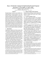
When the iron valency hybrids are placed in air-saturated
buffers, the oxygenated chains of each tetramer are oxi-
dized easily to the ferric met-form. Fig. 6 represents such
first-order plots to show wide differences in the oxidation
rate of the b chain, when it exists as the separated (bO
2
)
4
,
valency hybrid (a
3+
)
2
(bO
2
)
2
, and reconstructed HbO
2
tetramers in 0.1
M
Mes buffer at pH 6.2 and 35 °C. In this
way, the resulting rate constants for the a and b chains
are compared between the native, separated, reconstructed,
and valency hybrid haemoglobins at several pH values [9].
At pH 6.2, for instance, native HbO
2
gives the rate
constants of k
f
¼ 0.82 · 10
)1
h
)1
and k
s
¼ 0.13 · 10
)1
h
)1
in its biphasic curve. As listed in Table 3, almost the
same oxidation rates were obtained for the reconstructed
HbO
2
with a biphasic ratio of k
f
/k
s
¼ 6.1. Among those, the
most remarkable effect was found on the b chain. The
separated b chain in itself undergoes quite rapid oxidation
with a rate constant of k
obs
¼ 0.10 h
)1
,butthisratewas
dramatically suppressed up to k
s
¼ 0.14 · 10
)1
h
)1
(by
sevenfold) in the reconstructed HbO
2
,asisinnativeHbO
2
.
More importantly, such a retarded k
s
value could be
maintained totally in the valency hybrid (a
3+
)
2
(bO
2
)
2
tetramer.
All of these features were essentially the same at other
pH values. Certainly, the biphasic nature of the autoxi-
dation rate of HbO
2
became much slower at pH 7.5, and
even disappeared at pH 9.0. Nevertheless, the rate of
oxidation of the separated b chain was markedly reduced
by up to 15-fold at pH 7.5 and up to 23-fold at pH 9.0 in
the tetrameric haemoglobin, either it is native or recon-
structed or even valency hybrid species. The similar
situation was also found in the a chain, but its effect on
the stability of human HbO
2
wasmuchlesscrucialthan
the b chain.
It thus becomes evident that the b chain has acquired a
remarkable resistance against the acidic oxidation in a
manner of contacting with the a chain, no matter which
valency the latter partner is in, the ferrous or the ferric state.
From these recent findings, we conclude that the packing
contact produces a conformational constraint in the
Fig. 5. Schematic representation of human oxyhaemoglobin as seen in
the a1b1 contact to produce tilting of the distal histidine in the b chain. In
HbO
2
, the four haem pockets are all exposed at the surface of the
molecule. By the formation of the a1b1contact,theb chain is subject
to a structural constraint whereby the distal histidine at position 63 is
tilted slightly away from the bound O
2
.
Ó FEBS 2003 A unified picture for Hb function (Eur. J. Biochem. 270) 4047
b chain, so that the proton-catalysed process performed by
the distal histidine residue disappears from its acidic
autoxidation. Furthermore, spectral examinations have
disclosed that the formation of the a1b1ora2b2 contact
also protects the b chain from its haemichrome conversion.
As a matter of fact, the oxidation product of the isolated
b chain was not for the usual ferric met-form but for its
admixture with haemichrome. In this way, the noticeable
stability of human HbO
2
depends largely upon the very
unique property of the b chain on the a1b1ora2b2
interface.
Concluding remarks: a unified picture
for Hb function
In HbA, the four haem pockets are all exposed at the
surface of the molecule. From the X-ray crystal structures
(e.g. [2–6]), however, it becomes apparent that the
ligands ) including O
2
andCOtotheferrousformand
H
2
O, OH
–
,N
3
–
and CN
–
to the ferric form ) cannot gain
access to the closed haem pockets of haemoglobin as in
the case of myoglobin. Karplus and McCammon [29]
expressed this situation by the following passage in a
satirical way. If the structure of sperm whale myoglobin
was so rigid that the rotations of side chains were
impossible, an oxygen molecule might take many billions
of years to enter or leave the haem pocket across high
energy kinetic barriers: the time would be much longer
than a whale’s lifetime. Consequently, the thermal fluctu-
ations of side chain amino acid residues are essential for
the penetration of ligands from the surrounding solvent
through the globin matrix to the haem pocket [29–32]. In
this respect, much attention has been paid to the possible
roles of the distal (E7) histidine residue in myoglobin and
haemoglobin functions. It has been suggested that it acts
as a gate [29] or a swinging door [33,34] for ligand entry
into the haem pocket, and that it stabilizes the bound
dioxygen by hydrogen-bond formation [25], as well as it
stabilizes the axial water molecule of the ferric, high-spin
species [35–37]. Furthermore, the distal histidine via its
imidazole ring participates in a proton-relay mechanism as
a catalytic residue for the acidic oxidation of MbO
2
and
HbO
2
[8,21,24].
Fig. 6. First-order plots to compare the autoxidation rate of the b chain
between three different haemoglobin derivatives in 0.1
M
maleate buffer
at pH 6.2 and 35 °C. Each curve was obtained by a least-squares fitting
to the experimental points, based on Eqn (2). The oxidation of the
separated b chains could be described by a single rate constant of
k
obs
¼ 0.10 h
)1
in the presence of 20% (v/v) glycerol. This inherent
rate was dramatically suppressed not only in the reconstructed HbO
2
but also in the valency hybrid (a
3+
)
2
(bO
2
)
2
as well. Redrawn from
Yasuda et al.[9].
Table 3. Comparison of the autoxidation rate constants between the whole, separated, reconstructed, and hybrid haemoglobins in 0.1
M
buffer at
pH 6.2 and 35 °C. Taken from Yasuda et al. [9].
4048 K. Shikama and A. Matsuoka (Eur. J. Biochem. 270) Ó FEBS 2003
To make clear the functional role of the distal histidine
residue in the autoxidation reaction, Brantley et al.[38]
were the first to use systematically the site-directed
mutagenesis of sperm whale myoglobin. They showed
that mutations of the distal His at position 64, such as
those of H64G, H64V, H64L and H64Q, caused dramatic
increases in the autoxidation rate. At pH 7.0, for instance,
the H64V mutant MbO
2
was oxidized 400 times more
rapidly than the wild-type (H64H) MbO
2
.Usingthese
mutant myoglobins, we have also carried out detailed
pH-dependence studies of the autoxidation rate over the
wide range of pH 5–12 in 0.1
M
buffer at 25 °C[39].The
resulting pH-profiles were then compared with those of the
corresponding myoglobins occurring in nature. As a result,
if the distal (E7) histidine was replaced by other amino
acid residues, all such mutant oxymyoglobins were found
to contain no proton-catalysis in the autoxidation reaction.
Their pH profiles could be formulated by the kinetic
equations lacking in the rate constants k
A
H
and k
B
H
accordingly.
Along with these lines of evidence, we have recently
proposed that the distal histidine can play a dual role in the
nucleophilic displacement of O
2
–
from MbO
2
or HbO
2
[39].
One is in a proton-relay mechanism via the imidazole ring
at acidic pH. Insofar as we have examined, such a proton-
catalysed process could never be observed in the autoxi-
dation reaction of myoglobins lacking the usual distal
histidine residue, no matter what the protein is, the
naturally occurring or the distal His mutant [39]. As a
matter of fact, even if the distal residue is a histidine, it
cannot manifest any proton-catalysis when the residue is
tilted away from the precise E7 position. This is just the
case we have described here for the b chainintheHbO
2
tetramer. The other role of the distal histidine would be in
the maximum protection of the FeO
2
centre against a
water molecule or a hydroxyl ion that can enter the haem
pocket from the surrounding solvent [38]. This is relevant
to the considerable stability of MbO
2
and HbO
2
in the
neutral pH range. In this way, the distal histidine provides
the delicate balance of catalytic and steric factors necessary
for controlling the reversible oxygen binding to myoglobin
and haemoglobin in aqueous solution.
It is now clear that the constituent a and b chains, once
separated from the HbO
2
, are oxidized much more easily
than in the parent tetramer over the whole range of
pH 5–10. Moreover, their rates come to be almost equal to
each other and exhibit a very strong acid catalysis. This
inherently high oxidation rate of each chain can be
suppressed dramatically by the formation of a1b1(or
a2b2) contact. In particular, the b chain provides a further
effect on the stability of HbO
2
by preventing the proton-
catalysed oxidation at acidic pH. In order to explain such
unique properties of human HbO
2
, a nucleophilic displace-
ment mechanism has successfully been applied to detailed
pH-dependence studies of the autoxidation rate.
As for the dimer and tetramer effects on haem
oxidation, probable explanations are as follows. At basic
pH, the separated a and b chains are both quite susceptible
to autoxidation. Each haem pocket seems to be consid-
erably open to allow easier attack of the solvent hydroxyl
ion on the FeO
2
centre. As a result, there occurs a very
rapid formation of hydroxide-met species, its rate being
dependent directly upon the concentration of OH
–
ion.
When the a1b1(ora2b2) contact is formed, accessibility of
OH
–
ion to the haem pocket would be greatly reduced by
conformational constraints. As OH
–
ion is one of the
strongest nucleophiles in vivo, practically no rate difference
could be observed between the a and b chains on the basic
pH side, so that the autoxidation curve would become
monophasic regardless of the ab dimer and the HbO
2
tetramer.
On the acidic side from neutral pH, the displacing
nucleophile is an entering water molecule and its concen-
tration is always taken as 55.5
M
in aqueous solution.
Participation of the catalytic proton via the distal histidine
residue should therefore be a most decisive factor in
accelerating the displacing rate of O
2
–
from FeO
2
with
H
2
O. This is just the case with the separated a and b chains,
both exhibiting a very strong acid catalysis in their oxidation
rate. Once the a1b1(ora2b2) contact is established, the
b chain is subjected to a conformational constraint whereby
the distal histidine at position 63 is tilted away from the
bound dioxygen so as to be free from the proton-catalysed
displacement. In this way, the b chain can acquire a
remarkable resistance against the acidic autoxidation, and
this is one of the most characteristic features of the HbO
2
stability.
In relevance to a clinical aspect, it is interesting to note
that a quite large number of unstable haemoglobins have
been reported so far in the medical literature [3,4,40]. Many
of the mutants which occur at the a1b2 interface have
altered oxygen affinity, but bulk of evidence suggests that
the a1b1 interface is much more important in maintaining
normal haemoglobin stability than is the a1b2 interface. In
fact, haemolytic anaemia is known to result from substitu-
tions affecting the a1b1 interface or the haem pocket. If such
mutations occur, the haem iron will be more easily oxidized,
and a sequence of events leads to the globin precipitation or
Heinz body formation in red blood cells. Typical examples
of such variants are: Tacoma [b30(B12)Arg fi Ser],
Abraham Lincoln [b32(B14)Leu fi Pro], Castilla [b32
(B14)Leu fi Arg], Philly [b35(C1)Tyr fi Phe], Peterbor-
ough [b111(G13)Val fi Phe], Madrid [b115(G17)Ala fi
Pro], Khartoum [b124(H2)Pro fi Arg],J.Guantanamo
[b128(H6)Ala fi Asp], Leslie [b131(H9)Gln fi deleted]
and so on. Surprisingly, most of the pathological mutations
are found on the b chain, especially in the a1b1 contact
regions. In these unstable haemoglobins, the a1b1 contact
would become loose or disruptive due to many different
causes including: the insertion of proline (Abraham Lincoln,
Madrid), the substitution with a too-small amino acid side
chain (Tacoma) or a too-large side chain (Peterborough),
the introduction of a charged or very polar group (Castilla,
Khartoum, J. Guantanamo), and the deletion of amino acid
residue (Leslie).
The transport and storage of molecular oxygen by
haemoglobin and by myoglobin are essential to life. The
iron(II)-dioxygen bond in these haem proteins plays a
vital role in their physiology. It is in the ferrous form that
haemoglobin or myoglobin can bind molecular oxygen
reversibly and carry out its physiological function. From
known changes in valency of the haem iron, one can write
the functional cycle of the haemoglobin molecule as
follows:
Ó FEBS 2003 A unified picture for Hb function (Eur. J. Biochem. 270) 4049
During reversible oxygen binding, the oxygenated form of
haemoglobin, as well as of myoglobin, is oxidized easily to
the ferric met-form with generation of the superoxide
anion. The met-haemoglobin or met-myoglobin thus
produced cannot bind molecular oxygen and is therefore
physiologically inactive.
In red blood cells and muscle tissues, however, an
NADH-cytochrome b
5
oxidoreductase is present which can
reduce metHb or metMb to the ferrous deoxy-species again
and thus prevent the continued accumulation of the ferric
met-form in situ. The enzyme is called methaemoglobin
reductase [41] and metmyoglobin reductase [42], respec-
tively, and is known to have a FAD group that can accept
electrons from NADH. As a matter of fact, a strong and
cyclic reduction of the iron(III) species by these enzymes is a
basis for the continuity of haemoglobin and myoglobin
functions in vivo, since the autoxidation reaction is inevitable
in nature for all oxygen-binding haem proteins [21,23,24], as
well as for all synthetic dioxygen carriers [43,44]. In fact, it
is a matter of our experience that the metMb content in
myoglobin extracts from various muscle tissues is com-
monly about 40%, while the metHb content of freshly
drawn blood is usually maintained within 1–2% but by a
very strong reductive environment.
In conclusion, human haemoglobin seems to differentiate
two types of ab contacts quite properly for its functional
properties. The a1b2ora2b1 contact is associated with the
cooperative oxygen binding, whereas the a1b1ora2b2
contact is used for controlling the stability of the bound O
2
.
We can thus form a unified picture for haemoglobin function
by closely integrating the cooperative and the stable binding
of molecular oxygen with iron(II) in aqueous solvent.
Acknowledgements
The materials of our previous publications were used with permission
from Publishers including: American Society for Biochemistry and
Molecular Biology, Inc. (J. Biol. Chem.) and Blackwell Publishing Ltd.
(Eur. J. Biochem.).
References
1. Imai, K. (1994) Adair fitting to oxygen equilibrium curves of
hemoglobin. Methods Enzymol. 232, 559–576.
2. Baldwin, J. & Chothia, C. (1979) Haemoglobin: the structural
changes related to ligand binding and its allosteric mechanism.
J. Mol. Biol. 129, 175–220.
3. Dickerson, R.E. & Geis, I. (1983) Hemoglobin: Structure, Func-
tion, Evolution and Pathology. Benjamin Cummings Publishing
Co.Inc.MenloPark,CA,USA.
4. Fermi, G. & Perutz, M.F. (1981) Haemoglobin and myoglobin. In
Atlas of Molecular Structure in Biology, Vol. 2 (Phillips, D.C. &
Richards, F.M., eds), Clarendon Press, Oxford, UK.
5. Perutz, M. (1990) Mechanisms of Cooperativity and Allosteric Regu-
lation in Proteins. Cambridge University Press, Cambridge, UK.
6. Perutz, M.F., Wilkinson, A.J., Paoli, M. & Dodson, G.G. (1998)
The stereochemical mechanism of the cooperative effects in
hemoglobin revisited. Annu. Rev. Biophys. Biomol. Struct. 27, 1–34.
7. Borgstahl, G.E.O., Rogers, P.H. & Arnone, A. (1994) The 1.8 A
˚
structure of carbonmonoxy-b
4
hemoglobin. J. Mol. Biol. 236,
817–830.
8. Tsuruga, M., Matsuoka, A., Hachimori, A., Sugawara, Y. &
Shikama, K. (1998) The molecular mechanism of autoxidation for
human oxyhemoglobin: Tilting of the distal histidine causes
nonequivalent oxidation in the b chain. J. Biol. Chem. 273,
8607–8615.
9. Yasuda, J., Ichikawa, T., Tsuruga, M., Matsuoka, A., Sugawara,
Y. & Shikama, K. (2002) The a1b1 contact of human hemoglobin
plays a key role in stabilizing the bound dioxygen. Further
evidence from the iron valency hybrids. Eur. J. Biochem. 269,
202–211.
10. Brunori, M., Falcioni, G., Fioretti, E., Giardina, B. & Rotilio, G.
(1975) Formation of superoxide in the autoxidation of the isolated
a and b chains of human hemoglobin and its involvement in
hemichrome precipitation. Eur. J. Biochem. 53, 99–104.
11. Gotoh, T. & Shikama, K. (1976) Generation of the superoxide
radical during autoxidation of oxymyoglobin. J. Biochem.
(Tokyo) 80, 397–399.
12. Mansouri, A. & Winterhalter, K.H. (1973) Nonequivalence of
chains in hemoglobin oxidation. Biochemistry 12, 4946–4949.
13. Tsuruga, M. & Shikama, K. (1997) Biphasic nature in the autox-
idation reaction of human oxyhemoglobin. Biochim. Biophys. Acta
1337, 96–104.
14. Rosemeyer, M.A. & Huehns, E.R. (1967) On the mechanism of
the dissociation of haemoglobin. J. Mol. Biol. 25, 253–273.
15. Edelstein, S.J., Rehmar, M.J., Olson, J.S. & Gibson, Q.H. (1970)
Functional aspects of the subunit association-dissociation equili-
bria of hemoglobin. J. Biol. Chem. 245, 4372–4381.
16. McDonald, M.J., Turci, S.M., Mrabet, N.T., Himelstein, B.P. &
Bunn, H.F. (1987) The kinetics of assembly of normal and variant
human oxyhemoglobins. J. Biol. Chem. 262, 5951–5956.
17. Tyuma, I., Benesch, R.E. & Benesch, R. (1966) The preparation
and properties of the isolated a and b subunits of hemoglobin A.
Biochemistry 5, 2957–2962.
18. Sawada, Y., Iyanagi, T. & Yamazaki, I. (1975) Relation between
redox potentials and rate constants in reactions coupled with the
system oxygen-superoxide. Biochemistry 14, 3761–3764.
19. Shikama, K. (1990) Autoxidation of oxymyoglobin: a meeting
point of the stabilization and the activation of molecular oxygen.
Biol. Rev. (Cambridge) 65, 517–527.
20. Antonini, E., Wyman, J., Brunori, M., Taylor, J.F., Rossi-Fanelli,
A. & Caputo, A. (1964) Studies on the oxidation-reduction
potentials of heme proteins. I. Human hemoglobin. J. Biol. Chem.
239, 907–912.
21. Shikama, K. (1998) The molecular mechanism of autoxidation for
myoglobin and hemoglobin. A venerable puzzle. Chem. Rev. 98,
1357–1373.
22. Satoh, Y. & Shikama, K. (1981) Autoxidation of oxymyoglobin.
A nucleophilic displacement mechanism. J. Biol. Chem. 56, 10272–
10275.
23. Shikama, K. (1985) Nature of the FeO
2
bonding in myoglobin. An
overview from physical to clinical biochemistry. Experientia 41,
701–706.
24. Shikama, K. (1988) Stability properties of dioxygen-iron (II)
porphyrins: an overview from simple complexes to myoglobin.
Coordination Chem. Rev. 83, 73–91.
ð12Þ
4050 K. Shikama and A. Matsuoka (Eur. J. Biochem. 270) Ó FEBS 2003
25. Phillips, S.E.V. & Schoenborn, B.P. (1981) Neutron diffraction
reveals oxygen-histidine hydrogen bond in oxymyoglobin. Nature
(London) 292, 81–82.
26. Sugawara, Y. & Shikama, K. (1980) Autoxidation of native
oxymyoglobin. Thermodynamic analysis of the pH profile. Eur.
J. Biochem. 110, 241–246.
27. Shaanan, B. (1982) The iron-oxygen bond in human oxyhaemo-
globin. Nature (London) 296, 683–684.
28. Lukin, J.A., Simplaceanu, V., Zou, M., Ho, N.T. & Ho, C. (2000)
NMR reveals hydrogen bonds between oxygen and distal histi-
dines in oxyhemoglobin. Proc. Natl Acad. Sci. USA 97, 10354–
10358.
29. Karplus, M. & McCammon, J.A. (1986) The dynamics of pro-
teins. Sci. Am. 254, 30–39.
30. Karplus, M. & McCammon, J.A. (1983) Dynamics of proteins:
elements and function. Annu. Rev. Biochem. 52, 263–300.
31. Tian, W.D., Sage, J.T. & Champion, P.M. (1993) Investigations of
ligand association and dissociation rates in the ÔopenÕ and ÔclosedÕ
states of myoglobin. J. Mol. Biol. 233, 155–166.
32. Ramadas, N. & Rifkind, J.M. (1999) Molecular dynamics of
human methemoglobin: the transmission of conformational
information between subunits in an ab dimer. Biophys. J. 76,
1796–1811.
33. Johnson, K.A., Olson, J.S. & Phillips, G.N. Jr (1989) Structure
of myoglobin-ethylisocyanide: Histidine as a swinging door for
ligand entry. J. Mol. Biol. 207, 459–463.
34. Scott, E.E., Gibson, Q.H. & Olson, J.S. (2001) Mapping the
pathways for O
2
entry into and exit from myoglobin. J. Biol.
Chem. 276, 5177–5188.
35. Matsuoka, A., Kobayashi, N. & Shikama, K. (1992) The Soret
magnetic circular dichroism of ferric high-spin myoglobins. A
probe for the distal histidine residue. Eur. J. Biochem. 210, 337–
341.
36. Shikama, K. & Matsuoka, A. (1994) Aplysia myoglobin with
unusual properties: another prototype in myoglobin and haemo-
globin biochemistry. Biol. Rev. (Cambridge) 69, 233–251.
37. Quillin, M.L., Arduini, R.M., Olson, J.S. & Phillips, G.N. Jr
(1993) High-resolution crystal structures of distal histidine
mutants of sperm whale myoglobin. J. Mol. Biol. 234, 140–155.
38. Brantley, R.E. Jr, Smerdon, S.J., Wilkinson, A.J., Singleton, E.W.
& Olson, J.S. (1993) The mechanism of autooxidation of myo-
globin. J. Biol. Chem. 268, 6995–7010.
39. Suzuki, T., Watanabe, Y H., Nagasawa, M., Matsuoka, A. &
Shikama, K. (2000) Dual nature of the distal histidine residue in
the autoxidation reaction of myoglobin and hemoglobin. Com-
parison of the H64 mutants. Eur. J. Biochem. 267, 6166–6174.
40. Winslow, R.M. & Anderson, W.F. (1978) The hemoglobino-
pathies. In The Metabolic Basis of Inherited Disease 4th edn
(Stanbury, J.B., Wyngaarden, J.B. & Fredrickson, D.S., eds).
pp. 1465–1507. McGraw-Hill, Inc., New York, NY, USA.
41. Yubisui, T., Miyata, T., Iwanaga, S., Tamura, M. & Takeshita, M.
(1986) Complete amino acid sequence of NADH-cytochrome b
5
reductase purified from human erythrocytes. J. Biochem. (Tokyo)
99, 407–422.
42. Livingston, D.J., McLachlan, S.J., La Mar, G.N. & Brown, W.D.
(1985) Myoglobin: Cytochrome b
5
interactions and the kinetic
mechanism of metmyoglobin reductase. J. Biol. Chem. 260,
15699–15707.
43. Momenteau, M. & Reed, C.A. (1994) Synthetic heme dioxygen
complexes. Chem. Rev. 94, 659–698.
44. Busch, D.H. & Alcock, N.W. (1994) Iron and cobalt ÔlacunarÕ
complexes as dioxygen carriers. Chem. Rev. 94, 585–623.
Ó FEBS 2003 A unified picture for Hb function (Eur. J. Biochem. 270) 4051