Báo cáo khoa học: A pathway through interferon-c is the main pathway for induction of nitric oxide upon stimulation with bacterial lipopolysaccharide in mouse peritoneal cells pot
Bạn đang xem bản rút gọn của tài liệu. Xem và tải ngay bản đầy đủ của tài liệu tại đây (466.47 KB, 10 trang )
A pathway through interferon-c is the main pathway for induction
of nitric oxide upon stimulation with bacterial lipopolysaccharide
in mouse peritoneal cells
Motohiro Matsuura
1
, Shinji Saito
1
, Yoshikazu Hirai
1
and Haruki Okamura
2
1
Department of Microbiology, Jichi Medical School, Tochigi, Japan;
2
Institute for Advanced Medical Sciences,
Hyogo College of Medicine, Nishinomiya, Hyogo, Japan
Production of nitric oxide (NO) in response to bacterial
lipopolysaccharide (LPS) was investigated using cultures of
mouse peritoneal exudate cells (PEC) and the macrophage
cell line RAW264.7. In the presence of anti-(interferon-c)
(IFN-c), NO production was markedly suppressed in the
PEC culture but not in the RAW264.7 culture. In the PEC
culture, LPS induced both IFN-c production and activation
of IFN response factor-1, which leads to the gene expression
of inducible NO synthase, but neither was induced in the
culture of RAW264.7 cells. In addition to anti-(IFN-c),
antibodies against interleukin (IL)-12 and IL-18 showed a
suppressive effect on LPS-induced NO production in the
PEC culture, and these antibodies in synergy showed strong
suppression. Stimulation of the PEC culture with IL-12 or
IL-18 induced production of IFN-c and NO, and these
cytokines, in combination, exhibited marked synergism.
Stimulation of the culture with IFN-c induced production of
NO, but not IL-12. The macrophage population in the PEC,
prepared as adherent cells, responded well to LPS for IL-12
production, but weakly for production of IFN-c and NO.
The macrophages also responded well to IFN-c for NO
production. For production of IFN-c by stimulation with
LPS or IL-12 + IL-18, nonadherent cells were required in
the PEC culture. Considering these results overall, the indi-
rect pathway, through the production of intermediates (such
as IFN-c-inducing cytokines and IFN-c) by the cooperation
of macrophages with nonadherent cells, was revealed to play
the main role in the LPS-induced NO production pathway,
as opposed to the direct pathway requiring only a macro-
phage population.
Keywords: lipopolysaccharide; nitric oxide; interferon-c;
interleukin-12; interleukin-18.
The ability of bacterial lipopolysaccharide (LPS) to stimu-
late the mammalian immune system is mediated by the
action of LPS-induced mediators such as cytokines, chemo-
kines and lipid mediators [1,2]. Nitric oxide (NO) is also
regarded as an important mediator, with its unique
characteristic as a liquid-soluble gas. Multiple physiological
functions of this molecule, in relation to LPS activity, have
been reported, including enhancement of resistance against
microbial infections and tumor cells as well as induction of
tissue damage, hypotension and shock [3–6]. Macrophages
have been shown to be the primary cellular source to
recognize LPS by CD14 [7] and transduce its signals
through the Toll-like receptor 4 (TLR4)–MD-2 complex [8]
for the production of mediators, including NO. Production
of NO is governed by the activity of three NO synthase
(NOS) isoforms in which neuronal NOS and endothelial
NOS are constitutively expressed, while another macro-
phage-inducible NOS (iNOS or mac-NOS), is transcrip-
tionally induced in response to immune stimuli such as LPS
and produces large amounts of NO [9,10]. Expression of the
iNOS gene is therefore an intracellular event that must
occur before NO production in macrophages. In the
promoter of this gene, two important regions, region 1
containing nuclear factor-jB(NF-jB)-binding sequence
[11,12] and region 2 containing interferon response factor-1
(IRF-1)-binding sequence [13–15], have been defined. Acti-
vation of region 1 is essential for expression of the iNOS
gene in murine macrophages stimulated with LPS. This
indicates that LPS can directly induce NO production by
macrophages through a pathway activating NF-jBandits
binding to region 1 without the help of cofactors such as
cytokines. In the case of region 2 activation, stimulation of
the macrophages with interferon-c (IFN-c), but not with
LPS, was found to be effective for inducing the binding of
IRF-1toregion2,leadingtoiNOS gene expression.
The important role of IFN-c in the pathogenesis of LPS-
induced shock was confirmed using mice deficient for the
IFN-c receptor [16,17]. IFN-c is produced primarily by
natural killer (NK) cells and a certain subpopulation of T
lymphocytes (T helper 1 cells) [18] upon stimulation with
Correspondence to M. Matsuura, Department of Microbiology,
Jichi Medical School, 3311-1, Yakushiji,
Minamikawachi-machi, Tochigi, 329-0498, Japan.
Fax: + 81 285 44 1175, Tel.: + 81 285 58 7332,
E-mail:
Abbreviations:IFN-c, interferon-c; IL, interleukin; iNOS, inducible
nitric oxide synthase; IRF-1, interferon response factor-1; LPS,
lipopolysaccharide; NF-jB, nuclear factor-jB; NK, natural killer;
NO, nitric oxide; PEC, peritoneal exudate cells; TLR4,
Toll-like receptor 4; TNF-a, tumor necrosis factor-a.
(Received 27 June 2003, revised 31 July 2003,
accepted 14 August 2003)
Eur. J. Biochem. 270, 4016–4025 (2003) Ó FEBS 2003 doi:10.1046/j.1432-1033.2003.03792.x
interleukin (IL)-12 [19], IL-18 [20,21] and some others, but
not with LPS. A single report described the detection of
IFN-c mRNA, without secreted protein, in murine macro-
phages upon LPS stimulation [22]. This mechanism is,
however, hard to consider as the main underlying mechan-
ism of LPS-induced IFN-c production in vivo,inviewofthe
large amount of IFN-c (detectable as protein level) secreted
in the serum of mice after LPS challenge. Recently, LPS-
induced production of IFN-c in large amounts in in vitro
systems was reported by our group using murine peritoneal
cells [23] and by other groups using murine spleen cells
[24,25]. These results indicate that additional cellular
populations, besides the macrophage population, are
required for effective production of IFN-c in response to
LPS, unlike the production of the usual LPS mediators such
as IL-1, IL-6 and tumor necrosis factor-a (TNF-a), for
which the macrophage population alone is sufficient.
In the course of our study concerning the mechanism of
LPS-induced NO production, participation of IFN-c was
not suggested when murine macrophage cell lines such as
RAW264.7 [26] and J774.1 [27] were used. However, in a
study using murine peritoneal cells [23], participation of
endogenously produced IFN-c in LPS-induced NO pro-
duction was thought to occur. In the present study, we
aimedtoclarifytheroleofIFN-c in LPS-induced NO
production and revealed the important role of IFN-c as a
key mediator in the main pathway for LPS-induced NO
production in murine peritoneal cell culture. The underlying
mechanisms in the IFN-c-mediated pathway were also
elucidated.
Materials and methods
Animals and cells
Mice of the C3H/HeN and C3H/HeJ strains (mutants with
a defect in the function of TLR4) [28], were obtained from
Japan Charles River (Tokyo, Japan) and Clea Japan, Inc.
(Tokyo, Japan), respectively, and used at the age of
7–10 weeks. All animal experiments in the present study
were conducted according to the guidelines of the Labor-
atory Animal Center, Jichi Medical School. Peritoneal
exudate cells (PEC) were obtained from mice that had been
injected intraperitoneally with 2 mL of thioglycollate broth
(Difco Laboratories, Detroit, MI, USA) 4 days previously.
The PEC usually contained 87–92% macrophages and
6–9% lymphocytes, as determined by morphological
criteria. The murine macrophage cell line RAW264.7,
originally from the American Type Culture Collection
(Manassas, VA, USA) and maintained in our laboratory,
was also used. For cell culture, RPMI-1640 (Dainippon
Pharmaceutical Co. Ltd, Osaka, Japan) supplemented with
10 m
M
Hepes, 2 m
ML
-glutamine, 100 UÆmL
)1
penicillin,
100 lgÆmL
)1
streptomycin, 0.2% NaHCO
3
, and 5% heat-
inactivated fetal bovine serum (Flow Laboratories Inc.,
Rockville, MD, USA) was used. All cells were suspended in
the culture medium, plated onto a 24-well culture plate
(Corning Inc., Corning, NY, USA) at a dose of 6 · 10
5
cells
per well (final volume, 0.6 mL) and cultured in a humidified
chamber at 37 °Cwith5%CO
2
and 95% air. Cells were
cultured for 5 h before stimulation with LPS and cytokines.
In some experiments, the 5 h cultures were washed three
times with Hanks’ balanced salt solution (Gibco-BRL,
Gaithersburg, MD, USA) to remove nonadherent cells and
the adherent cells only were used as the macrophage culture.
Reagents
The LPS used was a kind gift from C. Galanos (MIP fu
¨
r
Immunbiologie, Freiburg, Germany), prepared from Sal-
monella enterica serovar Abortus-equi, as described previ-
ously [29]. The antibodies used were anti-(mouse IL-1b)
(R&D Systems, Minneapolis, MN, USA), anti-(mouse
IL-6) and anti-(mouse IL-12) (C17.8) (Genzyme Inc.,
Cambridge, MA, USA), anti-(mouse IL-15) (Torrey Pines
Biolabs Inc., Houston, TX, USA), anti-(mouse IFN-b)
(MB-7) (Seikagaku Co., Tokyo, Japan), anti-(mouse
IFN-c) (R4-6A2) (Endogen, Woburn, MA, USA), anti-
(mouse TNF-a) (a kind gift from Suntory Co. Ltd, Osaka,
Japan) [26], anti-(mouse CD14) (4C1) (kindly donated by
Y. Adachi, Tokyo University of Pharmacy and Life Science,
Tokyo, Japan) [30] and anti-(mouse IL-18), prepared as
described previously [20]. The cytokines used were recom-
binant murine IFN-c (Pepro Tech EC, London, UK),
recombinant mouse IL-12 (Genzyme Inc.) and recombinant
mouse IL-18, prepared as described previously [20].
Assay of NO and cytokines
Production of NO was determined as the amount of nitrite,
a stable end-product of NO, in the culture supernatant
obtained 48 h after stimulation with LPS or cytokine.
Nitrite was measured by a colorimetric assay using the
Griess reagent (1% sulfanilamide and 0.1% N-1-naphtyl-
ethylendiamine dihydrochloride in 2.5% H
3
PO
4
solution)
[31]. The absorbance at 540 nm was measured by the
Biomek 1000 spectrophotometer and the nitrite concentra-
tion was quantified (in l
M
) using sodium nitrite as the
standard in each assay.
Production of IFN-c and IL-12p70 (active form) was
determined as the amount in the culture supernatant
obtained 24 h after stimulation. The concentration of each
cytokine was measured using a specific sandwich ELISA,
according to the manufacturer’s instructions (Endogen)
using matched antibody pairs. A 96-well EIA/RIA plate
(Corning Inc.), coated with a coating antibody and masked
with skim milk, was incubated with test samples and
standard solutions. A biotin-labeled detecting antibody
was then added to the plate followed by reaction with a
streptavidin–horseradish peroxidase conjugate. Enzyme
reaction of the peroxidase was performed with a substrate
solution (0.01% 3,3¢,5,5¢-tetramethylbenzidine and 0.03%
H
2
O
2
in 0.11-
M
acetate buffer, pH 5.5). The color reaction
was stopped by adding 0.18
M
H
2
SO
4
and the absorbance
was measured at 450 nm. Quantification of each cytokine
(in ngÆmL
)1
for IFN-c andinpgÆmL
)1
for IL-12p70) was
performed based on the standard curve in each assay.
Preparation of nuclear extract
The peritoneal cells and RAW264.7 cells were stimulated
with LPS or IFN-c, and nuclear proteins of the cells were
prepared as described previously [27]. Briefly, the cells were
washed with NaCl/Pi and resuspended by vortexing for 10 s
Ó FEBS 2003 LPS-induced NO production through IFN-c (Eur. J. Biochem. 270) 4017
in hypotonic buffer A [10 m
M
Hepes (pH 7.8), 10 m
M
KCl, 0.1 m
M
EDTA, 0.5% Nonidet P-40 (NP-40), 1 m
M
dithiothreitol, 0.5 m
M
phenylmethanesulfonyl fluoride,
5 lgÆmL
)1
aprotinin, 5 lgÆmL
)1
pepstatin and 5 lgÆmL
)1
leupeptin). Nuclei were separated from the cytosol by
centrifugation at 1600 g for 1 min, resuspended in buffer C
(50 m
M
Hepes pH 7.8, 0.42
M
KCl, 0.1 m
M
EDTA, 5 m
M
MgCl
2
, 20% glycerol, 1 m
M
dithiothreitol, 0.5 m
M
phenyl-
methanesulfonyl fluoride, 5 lgÆmL
)1
aprotinin, 5 lgÆmL
)1
pepstatin and 5 lgÆmL
)1
leupeptin), and incubated on ice
for 30 min with occasional vortexing. Nuclear extracts were
obtained from the suspension by centrifugation at 15 000 g
for 15 min and stored in small aliquots at )80 °C.
EMSA
Oligonucleotide containing the downstream NF-jB-binding
site (nts )85 to )76) of the mouse iNOS promoter plus the
downstream 47 base pairs, designated NF-jBd (5¢-CAT
GGG GAC TCT CCC TTT GGG AAC AGT TAT GCA
AAA TAG CTC TGC AGA GCC TGG AGG GGT
CGA-3¢) [12] and the IRF-1 consensus sequence oligo-
nucleotide (5¢-GGA AGC GAA AAT GAA ATT GAC T-3¢)
were constructed as probes for EMSA. The oligonucleotides
were annealed and labeled with [
32
P]dCTP[aP]. Binding
reactions were performed (20 lL of the total volume) by
incubating these probes ( 20 000 counts per minute) with
nuclear extracts (5 lg of protein content) at room tempera-
ture for 30 min in binding buffer. The binding buffer
consists of 10 m
M
Hepes (pH 7.8), 50 m
M
KCl, 1 m
M
EDTA, 5 m
M
MgCl
2
, 10% glycerol, 5 m
M
dithiothreitol,
0.7 m
M
phenylmethanesulfonyl fluoride and 2 lgÆmL
)1
poly(dI-dC). Reaction products were electrophoresed on
5% polyacrylamide with 0.25 · TBE (Tris/borate/EDTA;
22.5 m
M
/22.2 m
M
/0.5 m
M
) for analysis by autoradiogra-
phy. Before starting the binding reaction, nuclear extracts
were incubated for 20 min on ice in the presence or absence
of competitors and antibodies. As competitors, a 100-fold
excess of unlabelled oligonucleotide probes was used for
analysis of binding specificities. As antibodies, anti-IRF-1,
anti-p50, anti-p65 and anti-(c-Rel) Igs (1 lg; all from Santa
Cruz Biotechnology Inc., Santa Cruz, CA, USA) were used
for supershift assays.
Results
Suppression, by anti-IFN-c Ig, of LPS-induced NO
production in murine PEC but not in the RAW264.7
murine macrophage cell line
Cells of murine PEC and of the RAW264.7 murine
macrophage cell line were cultured and stimulated with
LPS in the presence or absence of anti-(IFN-c) Ig. In both
cultures, a large amount of NO was produced upon LPS
stimulation at doses of > 1 ngÆmL
)1
, as shown in Fig. 1. In
the presence of anti-(IFN-c) Ig, production of NO was
suppressed markedly in the PEC culture but not at all in the
RAW264.7 culture. Another murine macrophage cell line,
J774.1, was also used for the experiments and results similar
to those for RAW264.7 were obtained. Namely, no
suppression of LPS-induced NO production by anti-IFN-
c Ig was observed (data not shown). These results indicate
that LPS-induced NO production in PEC depends largely
on IFN-c, which is probably produced upon LPS stimula-
tion, but that IFN-c does not participate at all in LPS-
induced NO production in macrophage cell lines such as
RAW264.7 and J774.1.
Production of IFN-c and IL-12 by murine PEC
upon LPS stimulation
We then investigated the production of IFN-c and related
cytokines upon LPS stimulation of PEC and RAW264.7
cells. As shown in Fig. 2A, IFN-c was produced dose
dependently upon stimulation with LPS in PEC, but not in
RAW264.7cells,atdosesofLPSupto10ngÆmL
)1
.Asfor
the production of IL-12p70, results similar to those for IFN-
c production were obtained; namely, a dose-dependent
production of IL-12p70 in PEC and no production in
RAW264.7 cells (Fig. 2B). An attempt to determine the
LPS-induced production of IL-18 was also made as IL-18 is
known to be a representative IFN-c-inducing cytokine, in
addition to IL-12. Production of IL-18 in PEC culture was
detected, although at a very low level (<70 pgÆmL
)1
at
1ngmL
)1
of LPS); IL-18 was also detected in RAW264.7
cells, but at a somewhat higher level (data not shown).
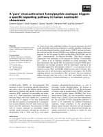
Fig. 1. Effect of anti-(IFN-c)IgonLPS-inducedNOproductionby
peritoneal cells of C3H/HeN mice (A) and by RAW264.7 mouse
macrophage line cells (B). Peritoneal exudate cells (PEC) were obtained
from C3H/HeN mice which had been injected with thioglycollate
broth intraperitoneally 4 days previously. The PEC and RAW264.7
cells were cultured with 5% fetal bovine serum–RPMI in the presence
(d)orabsence(s)of10lgÆmL
)1
anti-(IFN-c).LPSwasaddedtothe
cultures at the indicated concentrations 1 h later. Culture super-
natant obtained 48 h after LPS stimulation was assayed to determine
the concentration of NO. Data represent mean ± SEM of triplicate
samples. A representative result from three independent experiments is
shown.
4018 M. Matsuura et al. (Eur. J. Biochem. 270) Ó FEBS 2003
Improvement of the ELISA sensitivity may help to deter-
mine more precisely the level of IL-18 produced in response
to LPS stimulation.
Effect of cytokine antibodies on LPS-induced
production of NO and IFN-c in PEC
Various cytokines other than IFN-c, IL-12 and IL-18 are
produced by PEC upon LPS stimulation. To estimate the
participation of those cytokines, known as LPS media-
tors, in LPS-induced NO production, PEC were cultured
in the presence of antibodies to those cytokines and
stimulated with LPS. Even at the highest concentration
of antibodies tested (10 lgÆmL
)1
) most showed no
significant suppressive effect of NO production (Fig. 3).
Only anti-(IL-12) and anti-(IL-18) Igs exhibited a signi-
ficant suppressive effect, although the effect was weaker
than that of anti-(IFN-c) Ig. When anti-(IL-12) and anti-
(IL-18) Igs were combined at the same concentration
(5 lgÆmL
)1
each, giving a total protein concentration of
10 lgÆmL
)1
), the NO production was strongly suppressed
to a level similar to that achieved using the anti-(IFN-c)
Ig, and more effective than the suppression achived using
either anti-(IL-12) or anti-(IL-18) alone at 10 lgÆmL
)1
.
Neither of these antibodies showed cross-reactivity to
neutralize the antigenicity of murine IFN-c (data not
shown). These results, together with those presented in
Fig. 2, indicate that IFN-c is produced by PEC upon
stimulation with LPS, and that both IL-12 and IL-18
participate in the process of the LPS-induced IFN-c
production. Besides these cytokine antibodies, anti-CD14
Ig (4C1) was also used in this experiment. Strong
suppression of the LPS-induced production of NO and
IFN-c indicates that the signals of LPS for the produc-
tion of these mediators are transduced through CD14.
Stimulatory effect of IFN-c and IL-12 + IL-18 on NO
production by PEC of C3H/HeN and C3H/HeJ mice
TheroleofIFN-c,aswellasofIL-12andIL-18,as
intermediates in the pathway of LPS-induced NO produc-
tion, was indicated from the results obtained above.
Preparations of recombinant IFN-c, IL-12 and IL-18 were
added to PEC cultures of HeN and HeJ mice, and
production of NO in the culture supernatant was measured.
As shown in Fig. 4A, a significant effect of IFN-c on NO
production was observed at concentrations of > 1 ngÆmL
)1
in the cultures of both mouse strains. Concerning IL-12p70,
no significant production was observed in response to
IFN-c in both cultures (data not shown), unlike the case of
LPS stimulation. No response of the HeJ culture to LPS
was observed for the production of NO, IFN-c and
IL-12p70 (data not shown).
We also examined the effect of IL-12 and IL-18 on NO
production and found that the combined use of these
cytokines was more effective than when either was used
alone. Both PEC cultures of HeN and HeJ mice responded
to these cytokines for production of NO (Fig. 4B). At the
same time, the production of IFN-c was also determined.
As shown in Fig. 4C, both cultures responded to a
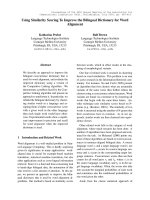
Fig. 2. Production of IFN-c (A) and IL-12 (B) in response to stimulation
with LPS by peritoneal cells of C3H/HeN mice and by RAW264.7 cells.
Peritoneal exudate cells (PEC) of C3H/HeN mice (d) and RAW264.7
cells (s) were cultured in the presence of LPS at the indicated con-
centrations. Culture supernatant obtained 24 h after LPS stimulation
was assayed, by ELISA, to determine the concentrations of interferon-
c (IFN-c) and IL-12p70 (active form). Data represent mean ± SEM of
triplicate samples. A representative result from three independent
experimentsisshown.
Fig. 3. Effect of cytokine antibodies on LPS-induced production of NO
and IFN-c by mouse peritoneal cells (PEC). PEC of C3H/HeN mice
were cultured in the presence or absence of cytokine antibodies and
anti-CD14. The cultures were stimulated with 1 ngÆmL
)1
LPS 1h
later. Culture supernatants obtained at 24 h and 48 h time-points were
assayed to determine the concentration of IFN-c and NO, respectively.
A representative result with the highest concentration of antibodies
tested (10 lgÆmL
)1
) is shown. In the case of anti-(IL-12) + anti-(IL-18)
Igs, the antibodies were mixed at the same concentration, i.e.
5 lgÆmL
)1
of each antibody was included. Data represent mean ±
SEM of triplicate samples. Similar results were obtained in two other
independent experiments.
Ó FEBS 2003 LPS-induced NO production through IFN-c (Eur. J. Biochem. 270) 4019
combination of these cytokines for the production of IFN-c,
similarly to NO production. The responses, of the HeN
culture, were not suppressed by anti-CD14 Ig (data not
shown). These results suggest that NO production by
IL-12 + IL-18 is mediated by IFN-c via signaling path-
ways that do not depend on either CD14 or TLR4, unlike
LPS-induced signals. This is further supported by the
observation that production of NO upon stimulation
with IL-12 + IL-18 was suppressed by anti-(IFN-c)Ig,
but that upon IFN-c stimulation was not suppressed by
anti-(IL-12) + anti-(IL-18) (data not shown).
Effect of nonadherent cells in the PEC of HeN mice
on the production of NO and cytokines in response
to LPS and its mediators
In the preparation of the PEC used in this study, non-
adherent cells were usually present at 10%, the remaining
90% of cells (almost all of which were macrophages) being
adherent. To investigate the role of nonadherent cells, PEC
were cultured for 5 h to permit adhesion, then the non-
adherent cells were washed off. The adherent cells were
cultured as macrophage culture and stimulated with LPS,
IFN-c and IL-12 + IL-18. Production of NO, IFN-c and
IL-12p70 by the adherent cell culture was compared with
that of the whole-cell PEC culture containing nonadherent
cells. The production of NO in response to LPS and
IL-12 + IL-18 was markedly reduced in the adherent cell
culture compared with that of the whole cell culture, while
the production in response to IFN-c was only slightly
reduced (Fig. 5A). The adherent cell culture showed a
marked reduction in the production of IFN-c in response to
LPS and IL-12 + IL-18 (Fig. 5B), similarly to that found
for NO production. Production of IL-12p70 in response to
LPS was reduced only slightly in the adherent cell culture
compared with that in the whole-cell culture (Fig. 5C), and
the rate of the reduction was far less than that observed in
the LPS-induced production of NO and IFN-c. Represen-
tative cytokines produced from macrophages upon LPS
stimulation, e.g. IL-6 and TNF-a, were also measured in
the culture supernatant. Similarly to the production of
IL-12p70, the production of these cytokines in response to
LPS was only marginally reduced in the adherent cell
culture compared with that in the whole cell culture (data
not shown). The IFN-c response of the adherent cells and
RAW264.7 cells to produce NO was strong, but the
response to produce IL-6 and TNF-a was not observed
(data not shown), similarly to the case of IL-12p70. These
results indicate that nonadherent cells participate largely in
the production of NO and IFN-c in response to LPS and
IL-12 + IL-18, but very little in the production of NO in
response to IFN-c and weakly in the production of
IL-12p70 in response to LPS.
In another experiment, nonadherent cells recovered from
the PEC culture of C3H/HeJ mice were added to the
adherent cell culture of HeN-PEC and the mixed population
culture was stimulated with LPS. Production of both NO
and IFN-c in the mixed population culture was remarkably
enhanced compared with that in the adherent cell culture
alone and reached the level observed in the whole cell
culture of HeN-PEC (data not shown). This indicates that
the production of IFN-c by nonadherent cells occurs
independently of LPS, as neither adherent cells nor non-
adherent cells of HeJ mice respond to LPS.
Effect of LPS stimulation on the activation of IRF-1
and NF-jB in PEC and RAW264.7 cells
Activation of transcription factors relating to iNOS gene
expression, such as IRF-1 and NF-jB, was then investi-
gated by EMSA using PEC and RAW264.7 cells stimulated
with LPS. Activation of IRF-1 in the PEC of HeN mice was
observed 18 h after LPS stimulation, but not at 2 or 6 h,
while activation after IFN-c stimulation was already evident
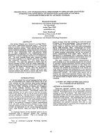
Fig. 4. Stimulation effect IFN-c and IL-12 + IL-18 on peritoneal cells
of C3H/HeN and C3H/HeJ mice for production of NO. Peritoneal
exudate cells (PEC) of HeN mice (d)andHeJmice(s), which have a
defect in the function of Toll-like receptor 4 (TLR4), were stimulated
with the indicated concentrations of IFN-c (A) and IL-12 + IL-18
(mixed at the equal concentrations indicated) and the culture super-
natant was assayed to determine the concentrations of NO (B) and
IFN-c (C). Data represent mean ± SEM of triplicate samples. A
representative result from three independent experiments is shown.
4020 M. Matsuura et al. (Eur. J. Biochem. 270) Ó FEBS 2003
at the 2-h time-point and maintained up to the 18-h time-
point (Fig. 6A). Specificity of IRF-1 binding to the DNA
probe was confirmed from the result that the strong bands
observed at 6 h as a result of stimulation with IFN-c were
remarkably reduced in intensity by the addition of excess
nonradiolabeled cold probe (lane cp in competition), but
not by the addition of a mutant probe (lane mp), which
lacks the binding ability owing to substitution of two AA
residues in the IRF-1 binding motif to GG, and also because
the bands were supershifted out of the range by preincu-
bation of the nuclear extract with anti-(IRF-1) Ig (lane Ab).
Production of IFN-c in the PEC upon LPS stimulation was
first detectable at the 8-h time-point, becoming maximal at
20 h (data not shown). These results support the idea that
IRF-1 is activated by IFN-c, but not by LPS, and that the
IFN-c induced by LPS is the direct activator of IRF-1 in the
LPS-stimulated PEC. In RAW264.7 cells, activation of
IRF-1 was observed (to some extent) without stimulation,
and enhancement from that level of activation was obscure
by LPS stimulation up to 18 h while that by IFN-c
stimulationwasclearfrom2hupto18h(Fig.6B).This
result is understandable considering the above result that
IFN-c was not induced by LPS during the culture of
RAW264.7 cells (Fig. 2A).
When the NF-jB consensus sequence oligonucleotide was
constructed as a probe for EMSA, activation of NF-jBwas
clearly observed in both the PEC and RAW264.7 cells in
response to LPS and also to IFN-c (data not shown). We
determined that the specificity of this probe was too low for
our experiments to detect activation of NF-jB leading to
expression of the iNOS gene and selected a probe designated
NF-jBd with higher specificity (as described in the Materials
and methods) for further experiments. In the PEC of HeN
mice, activation of NF-jB upon LPS stimulation was weak
and that upon IFN-c stimulation was no higher than
observed in the negative control (none) without stimulation
(Fig. 7A). In RAW264.7 cells, the activation was clearly
observed at 1 and 2 h after LPS stimulation but no more
clearly at 4 h, and the activation after IFN-c stimulation was
not observed at any of the time-points examined (Fig. 7B).
Specificity of NF-jB binding to the DNA probe was
confirmed from the result that the appearance of the strong
bands at 1 h with LPS stimulation was completely sup-
pressed by the addition of excess nonradiolabeled cold probe
(Fig. 7B, lane cold probe). These results support the idea that
activation of NF-jB participates in the direct induction of
NO by LPS (as seen in RAW264.7 cells), but not in the
induction by IFN-c (as seen in both types of cells) or in the
indirect induction by LPS via IFN-c (as seen in the PEC).
Attempts were made to characterize, by supershift assay,
the components of the activated NF-jB members. Nuclear
extracts obtained from RAW264.7 cells at 1 h after LPS
stimulation were incubated with antibodies against p50, p65
and c-Rel subunits of NF-jB followed by addition of the
NF-jBd probe for binding to NF-jB. Both the upper and
Fig. 5. Removal of nonadherent cells from peritoneal exudate cells and the subsequent effect on the production of NO (A), IFN-c (B) and IL-12 (C) in
response to stimulation with LPS, IFN-c and IL-12 + IL-18. PEC of C3H/HeN mice were cultured for 5 h and the nonadherent cells washed off to
obtain macrophages as adherent cells. The whole-cell culture (grey column) without washing and the adherent cell culture (white column) were
stimulated with the indicated stimulants. Culture supernatant obtained at 24 h was assayed to determine the concentrations of IFN-c and IL-12p70,
and culture supernatant obtained at 48 h was assayed to determine the concentration of NO. Data represent mean ± SEM of triplicate samples.
Similar results were obtained in two other independent experiments. N.D., not determined.
Fig. 6. Effect of LPS stimulation on the activation of IRF-1 in peritoneal
cells of HeN mice and in RAW264.7 cells. Peritoneal exudate cells of
HeN mice (A) and RAW264.7 cells (B) were stimulated with LPS
(1 ngÆmL
)1
)orIFN-c (10 ngÆmL
)1
) for the indicated times. Nuclear
extracts of the cells were prepared and EMSA performed using an
IRF-1 probe. Part of the nuclear extract from PEC, prepared 6 h after
IFN-c stimulation, was incubated with a 100-fold excess of unlabeled
cold probe (lane cp) or mutant probe (lane mp), or with 1 lgofanti-
(IRF-1) Ig (lane Ab) before the EMSA. Lanes labeled as ÔprobeÕ and
ÔnoneÕ indicate free probe without nuclear extract and nuclear extract
prepared from unstimulated cells, respectively. Similar results were
obtained from three independent experiments.
Ó FEBS 2003 LPS-induced NO production through IFN-c (Eur. J. Biochem. 270) 4021
the lower bands, clearly observed without antibody
(Fig. 7B, lane 1 in LPS), supershifted dramatically with
anti-p50 Ig (lane p50 in Ab) and less extensively with anti-
p65 Ig. Supershift of the upper band alone was clearly
observed with anti-c-Rel Ig, and the resulting weak upper
band seemed to supershift completely by the addition of
anti-p65 Ig (lane p65 + c-Rel in Ab). It is assumed, from
these results, that the lower band consists predominantly of
p50 homodimer and that the upper band consists of the
mixed heterodimers of p50/c-Rel and p50/p65.
Discussion
Most of the LPS mediators studied, to date, have been
shown to be produced directly from macrophages upon
stimulation with LPS. NO is thought to be one such
mediator. It is known that there is a direct pathway for the
production of NO in macrophages stimulated with LPS, via
activation of NF-jB and its binding to region 1 in the
promoter of the iNOS gene [11,12]. In the present study,
LPS-induced NO production in the RAW264.7 macro-
phage cell line was not suppressed by anti-(IFN-c)Ig
(Fig. 1) and activation of NF-jB was clearly observed in the
LPS-stimulated RAW264.7 cells (Fig. 7), indicating NO
production through this direct pathway. On the other hand,
the NO production in murine PEC was strongly suppressed
by anti-(IFN-c)Ig.IFN-c is known to be an inducing factor
of NO from macrophages through the activation of IRF-1
and its binding to region 2 in the promoter of iNOS gene
[13–15]. This indicates that NO is produced from LPS not
only through a direct pathway but also through an indirect
pathway via IFN-c. Activation of IRF-1 in relation to
production of IFN-c in the PEC upon LPS stimulation was
observed (Fig. 6A), and the amount of NO was reduced to
< 10% by the anti-(IFN-c), indicating that the indirect
pathway is mainly used for production of NO in the PEC
culture, which contains varied cellular populations and is
closer to in vivo situations than cultures with a single cellular
population of macrophage. This is the first report demon-
strating that the indirect pathway through IFN-c partici-
pates in LPS-induced NO production and, moreover, that it
playsamajorrole.
The role of IFN-c as a mediator of LPS has been indi-
cated in in vivo experiments [32], but in vitro investigations to
elucidate the role have found little. The present study is one
such in vitro study and clarifies the role of IFN-c as a
mediator of LPS in NO production. A synergistic effect of
exogenous IFN-c + LPS on the induction of LPS medi-
ators in macrophage cultures is well known. The effect is,
however, influenced profoundly by the sequence of stimu-
lation with IFN-c and LPS, as reported previously [33]. It
was indicated that exposure of macrophages to IFN-c,
before or simultaneously with LPS stimulation, induced
significant levels of NO release, but that exposure to LPS
prior to IFN-c resulted in poor induction of NO release. We
examined the effect of the sequence of stimulation with
IFN-c and LPS on the production of NO using the adherent
cell culture of HeN-PEC prepared under our experimental
conditions. We found that addition of IFN-c to the culture,
before or simultaneously with LPS stimulation, induced a
synergistic effect on the production of NO, addition of
IFN-c 3 h later still showed some synergistic effect, but
addition of IFN-c 8 h later showed no synergistic effect
(data not shown). In the whole-cell culture of HeN-PEC,
IFN-c was detectable only 8 h after LPS stimulation. These
results indicate that the endogenously induced IFN-c
contributes little to enhance LPS-induced NO production
but largely to the induction of NO by itself under the
experimental conditions of the present study. In addition to
these activities, IFN-c has diverse activities participating in
both innate and acquired immunity [34], and such activities
should also be taken into consideration in further investi-
gations of the role of this cytokine as an LPS mediator. The
present PEC culture also showed the advantage of using
mixed-cell cultures for the investigation of LPS mediators
induced indirectly after interaction with different cellular
populations. Using such mixed-cell cultures, new LPS
mediators are expected to be found.
IL-12 and IL-18 have been shown to play important roles
as IFN-c-inducing cytokines and exhibit marked synergism
in combination [35,36]. Production of IL-12 by PEC in
response to LPS was clearly detected (Fig. 2B) but that of
IL-18 was not so clear. Production of IL-18 was detected;
however, the amount was too small to determine accurately
using our current ELISA system. Anti-(IL-18) Ig showed a
suppressive effect, similar to that of anti-(IL-12) Ig, against
the production of IFN-c and NO, and combined treatment
using these two antibodies exhibited a synergistic effect
(Fig. 3), suggesting the participation of IL-18 in concert
with IL-12 in the indirect pathway for NO production. We
Fig. 7. Effect of LPS stimulation on the activation of nuclear factor (NF)-jB. Nuclear extracts prepared from peritoneal exudate cells of HeN mice
(A) and RAW264.7 cells (B), stimulated as indicated in the legend to Fig. 6, were analysed by EMSA using an NF-jBd probe. Part of the nuclear
extract from RAW264.7 cells, prepared 1 h after LPS stimulation, was incubated with a 100-fold excess of unlabeled cold probe (lane cold probe)
before the EMSA. Another part of this nuclear extract was also used for supershift experiments by incubating with 1 lg of antibodies against
NF-jB subunits, such as anti-p50 Ig (lane p50), anti-p65 Ig (lane p65), anti-(c-Rel) Ig, or a combination of anti-p65 and anti-(c-Rel) Igs (lane
p65 + c-Rel) before the EMSA. Lanes of probe and none are as indicated in the legend to Fig. 6. Similar results were obtained from three
independent experiments.
4022 M. Matsuura et al. (Eur. J. Biochem. 270) Ó FEBS 2003
confirmed that the anti-(IL-18) Ig used in the present study
does not inhibit the antigenicity of IL-12 by ELISA and that
the same results as shown in the present study are obtained
even when another anti-mouse IL-18 mAb (clone 93-10C;
MBL Co. Ltd, Nagoya, Japan) is used instead of the present
antibody (data not shown). It has been reported that the
active form of IL-18 is secreted from cells after processing
the precursor form of IL-18 by caspase-1 [37,38]. In
preliminary experiments, we observed the suppressive effect
of caspase-1 inhibitor on LPS-induced NO production by
the PEC, supporting further the requirement for active
IL-18. It is possible that only a small quantity of this
cytokine is produced, as detected using our ELISA, or it
may be rapidly degraded under the culture conditions;
further investigations are therefore required to elucidate
more fully how IL-18 contributes in the production of
IFN-c and NO.
Besides IL-12 and IL-18, IL-15 has also been reported
to play a role as an LPS-induced mediator for IFN-c
production and participate in the exhibition of an LPS-
induced general Shwartzman reaction [39]. The suppressive
effect of anti-(IL-15) Ig on the LPS-induced production of
IFN-c and NO was not clear in the present study (Fig. 3).
The effect of recombinant IL-15 on the induction of IFN-c
in the PEC was not evident, even in combination with IL-12
or IL-18 (data not shown). These results indicate that IL-15
contributes far less to the indirect pathway of NO produc-
tion than IL-12 and IL-18.
Induction of IL-12 by LPS was not observed in the
PEC of HeJ mice (Fig. 2) and the strong induction
observed in the PEC of HeN mice was suppressed by
anti-CD14 (Fig. 3). These results indicate that LPS
activates macrophages initially through CD14 and the
TLR4–MD-2 complex on their cellular surface to trans-
duce the signals inside the cells for production of IL-12, in
a manner recently revealed as the usual way for LPS
to induce a macrophage response [2,8]. The cellular
populations contributing to a synergistic effect of IL-12
and IL-18 on IFN-c production have been shown to be
T helper 1 cells [40,41] and NK cells [42], and these
populations are known to be nonadherent cells. The
response of HeJ cells to IL-12 + IL-18 for the production
of IFN-c (Fig. 4), and no suppressive effect of anti-CD14
in this response of HeN cells, indicates that CD14 and
TLR4–MD-2 are not required in this step. For the final
step to produce NO in response to IFN-c, macrophages
obtained as adherent cells were sufficient (Fig. 5) without
the help of nonadherent cells. The response of HeJ (Fig. 4),
and the effect of anti-CD14 on HeN, indicates that this
final step is also independent of CD14 and TLR4–MD-2,
although the macrophage population is the target.
These processes, participating in the pathways of LPS-
induced NO production, are schematically summarized in
Fig. 8. In the present study, the important role of the
indirect pathway through IFN-c in LPS-induced NO
production in murine peritoneal cells was clarified, for the
first time, by in vitro experiments.
Acknowledgement
This work was supported, in part, by Grant 13670280 (to M.M.) from
the Ministry of Education, Culture, Sports, Science and Technology of
Japan.
References
1. Ulevitch, R.J. & Tobias, P.S. (1995) Receptor-dependent
mechanisms of cell stimulation by bacterial endotoxin. Annu. Rev.
Immunol. 13, 437–457.
2. Alexander, C. & Rietschel, E.T. (2001) Bacterial lipopoly-
saccharide and innate immunity. J. Endotoxin Res. 7, 167–202.
3. Culotta, E. & Koshland, D.E.J. (1992) NO news is good news.
Science 258, 1862–1865.
4. Stamler, J.S., Single, D.J. & Loscalzo, J. (1992) Biochemistry of
nitric oxide and its redox-activated forms. Science 258, 1898–1902.
5. Schumidt, H.H.H.W. & Walter, U. (1994) NO at work. Cell 78,
919–925.
Fig. 8. Pathways for LPS-induced NO production in mouse peritoneal cells. The direct pathway through activation of nuclear factor (NF)-jBis
considered to be the sole pathway for LPS-induced NO production in murine macrophage cell line culture, but a minor pathway in peritoneal cell
culture. The indirect pathway, through IFN-c, was indicated to play the major role in the peritoneal cell culture in the present study.
Ó FEBS 2003 LPS-induced NO production through IFN-c (Eur. J. Biochem. 270) 4023
6. MacMicking, J.D., Nathan, C., Hom, G., Chartrain, N., Fletcher,
D.S.,Trumbauer,M.,Stevens,K.,Xie,Q W.,Sokol,K.,
Hutchinson, N., Chen, H. & Mudgett, J.S. (1995) Altered response
to bacterial infection and endotoxin shock in mice lacking
inducible nitric oxide synthase. Cell 81, 641–650.
7. Wright, S.D., Romos, R.A., Tobias, P.S., Ulevitch, R.J. &
Mathison, J.C. (1990) CD14, a receptor for complexes of lipo-
polysaccharide (LPS) and LPS binding protein. Science 249,
1431–1433.
8. Akashi, S., Shimazu, R., Ogata, H., Nagai, Y., Takeda, K.,
Kimoto, M. & Miyake, K. (2000) Cutting edge: cell surface
expression and lipopolysaccharide signaling via the Toll-like
receptor 4–MD-2 complex on mouse peritoneal macrophages.
J. Immunol. 164, 3471–3475.
9.Lowenstein,C.J.,Glatt,C.S.,Bredt,D.S.&Snyder,S.H.
(1992) Cloned and expressed macrophage nitric oxide synthase
contrasts with the brain enzyme. Proc. Natl Acad. Sci. USA 89,
6711–6715.
10. Xie, Q W., Cho, H.J., Calaycay, J., Mumford, R.A., Swiderek,
K.M., Lee, T.D., Ding, A., Troso, T. & Nathan, C. (1992) Cloning
and characterization of inducible nitric oxide synthase from mouse
macrophages. Science 256, 225–228.
11. Lowenstein, C.J., Alley, E.W., Raval, P., Snowman, A.M.,
Snyder, S.H., Russell, S.W. & Murphy, W.J. (1993) Macrophage
nitric oxide synthase gene: two upstream regions mediate induc-
tion by interferon c and lipopolysaccharide. Proc. Natl Acad. Sci.
USA 90, 9730–9734.
12. Xie, Q W., Kashiwabara, Y. & Nathan, C. (1994) Role of tran-
scription factor NF-jB/Rel in induction of nitric oxide synthase.
J. Biol. Chem. 269, 4705–4708.
13. Kamijo, R., Harada, H., Matsuyama, T., Bosland, M., Gerecit-
ano, J., Shapiro, D., Le, J., Koh, S.I., Kimura, T., Green, S.J.,
Mak,T.W.,Taniguchi,T.&Vilcek,J.(1994)Requirementfor
transcription factor IRF-1 in NO synthase induction in macro-
phages. Science 263, 1612–1615.
14. Martin, E., Nathan, C. & Xie, Q W. (1994) Role of interferon
regulatory factor 1 in induction of nitric oxide synthase. J. Exp.
Med. 180, 977–984.
15. Weisz, A., Cicatiello, L. & Esumi, H. (1996) Regulation of the
mouse inducible-type nitric oxide synthase gene promoter by
interferon-c, bacterial lipopolysaccharide and N
G
-monomethyl-
L-arginine. Biochem. J. 316, 209–215.
16. Kamijo, R., Le, J., Shapiro, D., Havell, E.A., Huang, S., Aguet,
M., Bosland, M. & Vilcek, J. (1993) Mice that lack the interferon-c
receptor have profoundly altered responses to infection with
bacillus Calmette–Gue
´
rin and subsequent challenge with lipo-
polysaccharide. J. Exp. Med. 178, 1435–1440.
17.Car,B.D.,Eng,V.M.,Schnyder,B.,Ozmen,L.,Huang,S.,
Gallay, P., Heumann, D., Aguet, M. & Ryffel, B. (1994) Interferon
c receptor deficient mice are resistant to endotoxic shock. J. Exp.
Med. 179, 1437–1444.
18. Billiau, A. (1996) Interferon-c: biology and role in pathogenesis.
Adv. Immunol. 62, 61–130.
19. Trinchieri, G. (1995) Interleukin-12: a proinflammatory cytokine
with immunoregulatory functions that bridge innate resistance
and antigen-specific adaptive immunity. Annu. Rev. Immunol. 13,
251–276.
20. Okamura, H., Tsutsui, H., Komatsu, T., Yutsudo, M., Hakura,
A.,Tanimoto,T.,Torigoe,K.,Okura,T.,Nukada,Y.,Hattori,
K., Akita, K., Namba, M., Tanabe, F., Konishi, K., Fukuda, S. &
Kurimoto, M. (1995) Cloning of a new cytokine that induces IFN-
c production by T cells. Nature 378, 88–91.
21. Dinarello, C., Novick, D., Puren, A.J., Fantuzzi, G., Shapiro, L.,
Mu
¨
hl, H., Yoon, D Y., Rezinikov, L.L., Kim, S H. & Rubin-
stein, M. (1998) Overview of interleukin-18: more than an inter-
feron-c inducing factor. J. Leukoc. Biol. 63, 658–664.
22. Fultz, M.J., Barber, S.A., Dieffenbach, C.W. & Vogel, S.N. (1993)
Induction of IFN-c in macrophages by lipopolysaccharide. Int.
Immunol. 5, 1383–1392.
23. Tominaga, K., Saito, S., Matsuura, M., Funatogawa, K.,
Matsumura, H. & Nakano, M. (1999) Role of IFN-c on dissoci-
ation between nitric oxide and TNF/IL-6 production by murine
pertoneal cells after restimulation with bacterial lipopolysaccha-
ride. J. Leukoc. Biol. 66, 974–980.
24. Balkhy, H.H. & Heinzel, F.P. (1999) Endotoxin fails to induce
IFN-c in endotoxin-tolerant mice: deficiencies in both IL-12 het-
erodimer production and IL-12 responsiveness. J. Immunol. 162,
3633–3638.
25. Varma, T.K., Lin, C.Y., Toliver-Kinsky, T.E. & Sherwood, E.R.
(2002) Endotoxin-induced c-interferon production: contributing
cell types and key regulatory factors. Clin. Diagn. Lab. Immunol. 9,
530–543.
26. Funatogawa, K., Matsuura, M., Nakano, M., Kiso, M. & Hase-
gawa, A. (1998) Relationship of structure and biological activity of
monosaccharide lipid A analogues to induction of nitric oxide
production by murine macrophage RAW264.7 cells. Infect.
Immun. 66, 5792–6798.
27. Saito, S., Matsuura, M., Tominaga, K., Kirikae, T. & Nakano, M.
(2000) Important role of membrane-associated CD14 in the
induction of IFN-b and subsequent nitric oxide production by
murine macrophages to bacterial lipopolysaccharide. Eur. J.
Biochem. 267, 37–45.
28. Poltorak, A., He, X., Smilnova, I., Liu, M Y., Van Huffel, C.X.,
Birdwell, D., Alejos, E., Silva, M., Galanos, C., Freudenberg, M.,
Ricciardi-Castagnoli, P., Layton, B. & Beutler, B. (1998) Defective
LPS signaling in C3H/HeJ and C57BL/ScCr mice: mutation in the
Tlr4 gene. Science 282, 2085–2088.
29. Galanos, C., Lu
¨
deritz, O. & Westphal, O. (1979) Preparation and
properties of a standardized lipopolysaccharide from Salmonella
abortus equi. Zentbl. Bakteriol. Mikrobiol. Hyg. Abt. 1 Orig. A 243,
226–244.
30. Adachi, Y., Satokawa, C., Ohno, N., Tamura, H., Tanaka, S. &
Yadomae, T. (1999) Inhibition by a CD14 monoclonal antibody
of lipopolysaccharide binding to murine macrophages. J. Endo-
toxin Res. 5, 139–146.
31. Green, L.C., Wagner, D.A., Glogowski, J., Skipper, P.L., Wish-
nok, J.S. & Tannenbaum, S.R. (1982) Analysis of nitrate, nitrite,
and [
15
N]nitrate in biological fluids. Anal. Biochem. 126, 131–138.
32. Heinzel, F.P. (1990) The role of IFN-c in the pathology of
experimental endotoxemia. J. Immunol. 145, 2920–2924.
33. Lorsbach, R.B. & Russell, S.W. (1992) A specific sequence of
stimulation is required to induce synthesis of the antimicrobial
molecule nitric oxide by mouse macrophages. Infect. Immun. 60,
2133–2135.
34. Bochem, U., Klamp, T., Groot, M. & Howard, J.C. (1997)
Cellular responses to interferon-c. Annu. Rev. Immunol. 15,
749–795.
35. Okamura, H., Kashiwamura, S., Tsutsui, H., Yoshimoto, T. &
Nakanishi, K. (1998) Regulation of interferon-c production by
IL-12 and IL-18. Curr. Opin. Immunol. 10, 259–264.
36. Nakahira,M.,Ahn,H J.,Park,W R.,Gao,P.,Tomura,M.,
Park,C S.,Hamaoka,T.,Ohta,T.,Kurimoto,M.&Fujiwara,
H. (2002) Synergy of IL-12 and IL-18 for IFN-c gene expression:
IL-12-induced STAT4 contributes to IFN-c promoter activation
by up-regulating the binding activity of IL-18-induced activator
protein 1. J. Immunol. 168, 1146–1153.
37. Gu, Y., Kuida, K., Tsutsui, H., Ku, G., Hsiao, K., Fleming, M.A.,
Hayashi, N., Higashino, K., Okamura, H., Nakanishi, K., Kuri-
moto, M., Tanimoto, T., Flavell, R.A., Sato, V., Harding, M.W.,
Livingston, D.J. & Su, M.S S. (1997) Activation of interferon-c
inducing factor mediated by interleukin-1b converting enzyme.
Science 275, 206–209.
4024 M. Matsuura et al. (Eur. J. Biochem. 270) Ó FEBS 2003
38. Ghayur, T., Banerjee, S., Hugunin, M., Butler, D., Herzog, L.,
Carter, A., Quintal, L., Sekut, L., Talanian, R., Paskind, M.,
Wong, W., Kamen, R., Tracey, D. & Allen, H. (1997) Caspase-1
processes IFN-c-inducing factor and regulates LPS-induced
IFN-c production. Nature 386, 619–623.
39. Fehninger, T.A., Yu, H., Cooper, A., Suzuki, K., Shah, M.H. &
Caligiuri, M.A. (2000) Cutting edge: IL-15 costimulates the gen-
eralized Shwartzman reaction and innate immune IFN-c pro-
duction in vivo. J. Immunol. 164, 1643–1647.
40. Robinson, D., Shibuya, K., Mui, A., Zonin, F., Murphy, E.,
Sanna,T.,Hartley,S.B.,Menon,S.,Kasteline,R.,Bazan,F.&
O’Garra, A. (1997) IGIF does not drive Th1 development but
synergizes with IL-12 for interferon-c production and activates
IRAK and NFjB. Immunity 7, 571–581.
41. Ahn, H J., Maruo, S., Tomura, M., Mu, J., Hamaoka, T.,
Nakanishi, K., Clark, S., Kurimoto, M., Okamura, H. & Fuji-
wara, H. (1997) A mechanism underlying synergy between IL-12
and IFN-c-inducing factor in enhanced production of IFN-c.
J. Immunol. 159, 2125–2131.
42. Tomura,M.,Zhou,X.Y.,Maruo,S.,Ahn,H J.,Hamaoka,T.,
Okamura, H., Nakanishi, K., Tanimoto, T., Kurimoto, M. &
Fujiwara, H. (1998) A critical role for IL-18 in the proliferation
and activation of NK1.1
+
CD3
–
cells. J. Immunol. 160, 4738–
4746.
Ó FEBS 2003 LPS-induced NO production through IFN-c (Eur. J. Biochem. 270) 4025