Báo cáo Y học: Domain V of m-calpain shows the potential to form an oblique-orientated a-helix, which may modulate the enzyme’s activity via interactions with anionic lipid potx
Bạn đang xem bản rút gọn của tài liệu. Xem và tải ngay bản đầy đủ của tài liệu tại đây (446.83 KB, 9 trang )
Domain V of m-calpain shows the potential to form
an oblique-orientated a-helix, which may modulate the enzyme’s
activity via interactions with anionic lipid
Klaus Brandenburg
1
, Frederick Harris
2
, Sarah Dennison
2
, Ulrich Seydel
1
and David Phoenix
2
1
Division of Biophysics, Forschunginstitute Borstel, Germany;
2
Department of Forensic and Investigative Science, University of
Central Lancashire, Preston, UK
The activity of m-calpain, a heterodimeric, Ca
2+
-dependent
cysteine protease appears to be modulated by membrane
interactions involving oblique-orientated a-helix formation
by a segment, GTAMRILGGVI, in the protein’s smaller
subunit. Here, graphical and hydrophobic moment-based
analyses predicted that this segment may form an a-helix
with strong structural resemblance to the influenza virus
peptide, HA2, a known oblique-orientated a-helix former.
Fourier transform infrared spectroscopy showed that a
peptide homologue of the GTAMRILGGVI segment, VP1,
adopted low levels of a-helical structure ( 20%) in the
presence of zwitterionic lipid and induced a minor decrease
(3 °C) in the gel to liquid-crystalline phase transition tem-
perature, T
C
, of the hydrocarbon chains of zwitterionic
membranes, suggesting interaction with the lipid headgroup
region. In contrast, VP1 adopted high levels of a-helical
structure (65%) in the presence of anionic lipid, induced a
large increase (10 °C) in the T
C
of anionic membranes, and
showed high levels of anionic lipid monolayer penetration
(DSP ¼ 5.5 mNÆm
)1
), suggesting deep levels of membrane
penetration. VP1 showed strong haemolytic ability
(LD
50
¼ 1.45 m
M
), but in the presence of ionic agents, this
ability, and that of VP1 to penetrate anionic lipid mono-
layers, was greatly reduced. In combination, our results
suggest that m-calpain domain V may penetrate membranes
via the adoption of an oblique-orientated a-helix and elec-
trostatic interactions. We speculate that these interactions
may involve snorkelling by an arginine residue located in the
polar face of this a-helix.
Keywords: domain V; hydrophobicity gradient; m-calpain;
membrane; oblique-orientated a-helix.
Calpains are a growing family [1] of structurally related
intracellular Ca
2+
-dependent cysteine proteases [2,3], with
calpain 10 the most recently characterized member [4]. The
physiological functions of calpains are not fully understood
but they are believed to play important roles in such
processes as cytoskeletal remodelling, cell differentiation,
apoptosis and signal transduction [5–7]. Calpains are also of
medical importance, having been implicated in a number of
pathological conditions including: cataract formation [8],
type 2 diabetes [9], muscular dystrophy, rheumatoid arth-
ritis, ischaemic tissue damage, and neurodegenerative con-
ditions such as Alzheimer’s and Parkinson’s disease [10,11].
The major calpains are l-calpain (calpain 1) and
m-calpain (calpain 2), which are ubiquitous in mammalian
cells [10]. These enzymes are heterodimeric and possess
larger, 80-kDa, subunits, which show high levels of homo-
logy, and smaller, 30-kDa, subunits, which show lower
levels of homology [2,3]. Originally based on sequence
comparisons [12], these calpains were assigned a domainal
organization, with the larger subunit divided into domains I
to IV and the smaller subunit divided into domains V and
VI. Domain II possesses the active site and is a papain-like
cysteine protease domain, and domain IV contains a
calmodulin-like Ca
2+
-binding domain with multiple
EF-hand motifs. The smaller subunit is divided into domain
VI, which also possesses a calmodulin-like Ca
2+
-binding
domain, and domain V [2,3,13]. The recently solved crystal
structures of calcium-free rat m-calpain [14] and human
m-calpain [15] confirmed that this domain structure is
essentially correct.
l-Calpain and m-calpain possess similar substrate spe-
cificity and show an absolute requirement for Ca
2+
for
activation, although they differ in the level of this require-
ment: 5–50 l
M
and 250–1000 l
M
, respectively [2,3]. These
observations suggested that other factors may be involved in
the activation of m-calpain because, clearly, the millimolar
levels of Ca
2+
needed to activate the enzyme in vivo far
exceed normal intracellular levels. Calpains are known to
exist in a membrane-associated form [16,17], and it has been
shown that the presence of membrane lipid mixtures can
lower the Ca
2+
requirement for m-calpain activation to
near physiological levels [18]. On the basis of these and other
results [19–21], it has been suggested that lipid or membrane
interaction may modulate the activity of the enzyme [3].
Consistent with this suggestion, it has been shown that
m-calpain domain III folds into an antiparallel b-sandwich,
which is structurally related to C2 domains [14,15,22]. These
domains bind phospholipid in a Ca
2+
-dependent manner
and are believed to be responsible for orchestrating the
Correspondence to D. A. Phoenix, Department of Forensic and
Investigative Science, University of Central Lancashire, Preston
PR1 2HE, UK. Fax: + 1772 894981, Tel.: + 1772 894381,
E-mail:
Abbreviations:Myr
2
PtdCho, dimyristoylphosphatidylcholine;
Myr
2
PtdEtn, dimyristoylphosphatidylethanolamine; Myr
2
PtdSer,
dimyristoylphosphatidylserine; FTIR, Fourier transform infrared;
SUV, small unilamellar vesicle.
(Received 20 May 2002, accepted 2 September 2002)
Eur. J. Biochem. 269, 5414–5422 (2002) Ó FEBS 2002 doi:10.1046/j.1432-1033.2002.03225.x
membrane–Ca
2+
regulation of enzyme activity of a number
of proteins [23]. A number of studies have suggested that
interaction of m-calpain domain V with lipid or membranes
may also be involved in the modulation of the enzyme’s
activity. Earlier investigations showed that a C-terminal
segmentofdomainV,G
17
TAMRILGG, is required for
lipid interaction of the enzyme [24]. More recent investiga-
tions, using peptides homologous to various regions of
m-calpain’s domain V, showed that, although the presence
of the TAMRIL sequence was required for m-calpain–lipid
interaction, the presence of a polyglycine sequence was also
necessary for such interaction [25].
The recently determined crystal structures of m-calpain
did not include domain V [15,16] and no three-dimensional
structure for this domain is currently available. Further-
more, the primary structure of domain V shows no apparent
homology with that of any other known protein [22].
However, a recent theoretical analysis of domain V derived
from four different mammalian m-calpains showed that
each possessed a common segment, GTAMRILGGVI,
which was a candidate for formation of a lipid-interactive
oblique-orientated a-helix [26]. These a-helices show a
highly specialized structure/function relationship and pene-
trate membranes at a shallow angle because of a hydro-
phobicity gradient along the a-helical long axis [27,28]. It
was suggested by Daman et al.[26]thattheformationof
such an a-helix may feature in the membrane interactions of
m-calpain domain V. Here, to investigate this suggestion, we
have undertaken theoretical analysis and studied the lipid–
membrane interactions of a peptide homologue of the
GTAMRILGGVI segment using haemolytic analysis,
monolayer studies, Fourier transform infrared (FTIR)
conformational analysis and FTIR lipid-phase transition
analysis. Our results are discussed in relation to the
influenza viral fusion peptide, HA2 [29,30], a known
oblique-orientated a-helix former [31–35].
MATERIALS AND METHODS
Reagents
The peptide VP1 was supplied by Pepsyn, University of
Liverpool, UK, produced by solid-state synthesis and
purified by HPLC to a purity of greater than 99%. The
peptide was stored as a stock solution (10 m
M
) in 10% (v/v)
ethanol at 4 °C. Packed human red blood cells were
supplied by the Royal Preston Hospital, UK. The phos-
pholipids dimyristoylphosphatidylcholine (Myr
2
PtdCho),
dimyristoylphosphatidylethanolamine (Myr
2
PtdEtn) and
dimyristoylphosphatidylserine (Myr
2
PtdSer) and all sol-
vents, which were of spectroscopic grade, were purchased
from Sigma (UK). For FTIR spectroscopy, deuterated
Myr
2
PtdSer, purchased from Avanti, was used.
Theoretical analyses of candidate oblique-orientated
a-helix-forming segments
The sequences of the influenza viral fusion peptide, HA2, a
known oblique-orientated a-helix former [31–35], and that
of the putative oblique-orientated a-helix of m-calpain
domain V (Table 1) were analysed by conventional hydro-
phobic moment methods [36]. The hydrophobicity of
successive amino acids in these sequences are treated as
vectors and summed in two dimensions, assuming an
amino-acid side chain periodicity of 100 °. The resultant of
this summation, the hydrophobic moment, l
H
,providesa
measure of a-helix amphiphilicity. Our analysis used a
moving window of 11 residues, and, for each sequence
under investigation (Table 1), the window with the highest
hydrophobic moment was identified (Table 1). For these
windows, the mean hydrophobic moment, Æl
H
æ,andthe
corresponding mean hydrophobicity, ÆH
0
æ (Table 1), were
computed using the normalized consensus hydrophobicity
scale of Eisenberg et al. [37] and plotted on the hydrophobic
moment plot diagram of Eisenberg et al. [38] as modified by
Harris et al.[32](Fig.1).
WINGEN
/
WINPEG
software [39] was used to perform
hydropathy plot analysis (Fig. 2) of the GTAMRILGGVI
sequence using the hydrophobicity scale of Kyte & Dolittle
[40] and a seven-residue window. The software was also
used to represent both this sequence and that of HA2 as
two-dimensional axial projections assuming an angular
periodicity of 100 ° (Fig. 3).
Haemolytic assay of VP1
Haemolytic assay was conducted as described by Harris &
Phoenix [41]. Essentially, packed red blood cells were
washed three times in Tris-buffered sucrose (0.25
M
sucrose, 10 m
M
Tris/HCl, pH 7.5) and resuspended in
the same medium to give an initial blood cell concentra-
tion of 0.05%. For haemolytic assay, this concentration
was adjusted such that incubation with 0.1% (v/v) Triton
X-100 for 1 h produced a supernatant with A
416
¼ 1.0,
and this was taken as 100% haemolysis. Aliquots (1 mL)
of blood cells at assay concentration were then used to
solubilize various amounts of stock peptide solution,
each of which had been added to a test-tube and dried
under nitrogen gas. The resulting mixtures were incu-
bated at room temperature with gentle shaking. After 1 h,
the suspensions were centrifuged at low speed (1500 g,
15 min, 25 °C), and the A
416
of the supernatants
Table 1. Hydrophobic moment analysis of protein structure. Primary structure of the putative oblique-orientated a-helix-forming segment identified
in m-calpain, domain V [26] and that of the influenza peptide, HA2, a known oblique-orientated a-helix former, obtained from Peuvot et al. [47].
Values of Æl
H
æ and ÆH
0
æ for each sequence were determined by the method of Eisenberg et al.[36].
Source protein Segment sequence Æl
H
æÆH
0
æ
+
m-calpain, domain V
GTAMRILGGVI 0.46 0.47
)) )
HA2 fusion peptide GLFGAIAGFIENGWEGMIDG 0.65 0.23
Ó FEBS 2002 Lipid-interactive a-helical structure in m-calpain (Eur. J. Biochem. 269) 5415
determined. Similar experiments were also performed
except that Tris-buffered sucrose was replaced with Tris-
buffered saline (100 m
M
NaCl, 10 m
M
Tris/HCl, pH 7.5).
In all cases, levels of haemolysis were determined as the
percentage haemolysis relative to that of Triton X-100 and
the results recorded (Fig. 4). Basal lysis was less than 3%
in all cases.
Preparation of phospholipid small unilamellar
vesicles (SUVs)
SUVs were prepared by the method of Keller et al.[42].
Essentially, lipid/chloroform mixtures were dried with
nitrogen gas and hydrated with aqueous Hepes at pH 7.5
to give final phospholipid concentrations of 50 m
M
.The
resulting cloudy suspensions were sonicated at 4 °Cwitha
Soniprep 150 sonicator (amplitude 10 lm) until clear
suspensions resulted (30 cycles of 30 s), which were then
centrifuged (15 min, 3000 g,4°C).
FTIR conformational analyses of VP1
To give a final peptide concentration of 1 m
M
,VP1was
solubilized in 50 m
M
aqueous Hepes (pH 7.5) or suspen-
sions of SUVs, which were formed from Myr
2
PtdSer,
Myr
2
PtdCho or Myr
2
PtdEtn, prepared as described
above. Samples of solubilized peptide were spread on a
CaF
2
crystal, and the free excess water was evaporated at
room temperature. The single-band components of the
VP1 amide I vibrational band (predominantly C¼O
stretch) was monitored using an FTIR Ô5-DXÕ spectro-
meter (Nicolet Instruments, Madison, WI, USA), and,
for each sample, absorbance spectra were produced
(Fig. 5). These spectra were analysed, and, for those
with strong absorption bands, the band parameters (peak
position, band width, and intensity) were evaluated with
the original spectra, if necessary after the subtraction of
strong water bands. In the case of spectra with weak
absorption bands, resolution-enhancement techniques
such as Fourier self-deconvolution [43] were applied after
baseline subtraction with the parameters: bandwidth, 22–
28 cm
)1
; resolution-enhancement factor, 1.2–1.4; Gauss/
Lorentz ratio, 0.55. In the case of overlapping bands,
curve fitting was applied using a modified version of the
CURFIT
procedure written by D. Moffat (National
Research Council, Ottowa, Ontario, Canada). An esti-
mation of the number of band components was obtained
from deconvolution of the spectra; curve fitting was then
applied within the original spectra after the subtraction
of baselines resulting from neighbouring bands. Similar
to the deconvolution technique, the bandshapes of the
single components are superpositions of Gaussian and
Lorentzian bandshapes. Best fits were obtained by
assuming a Gauss fraction of 0.55–0.6. The
CURFIT
procedure measures the peak areas of single band
components and, after statistical evaluation, determines
the relative percentages of primary structure involved in
secondary-structure formation. For VP1, relative levels of
a-helical structure (1650–1655 cm
)1
)andb-sheet struc-
tures (1625–1640 cm
)1
) were computed and are shown in
Table 2.
FTIR analysis of phospholipid phase transition properties
Using FTIR spectroscopy, the effects of VP1 on the phase-
transition properties of phospholipid were investigated. To
give a final peptide concentration of 1 m
M
,VP1was
solubilized in suspensions of SUVs formed from Myr
2
Ptd-
Ser, Myr
2
PtdCho or Myr
2
PtdEtn, prepared as described
above. As controls, SUVs formed from Myr
2
PtdSer,
Myr
2
PtdCho or Myr
2
PtdEtn alone were prepared as
described above. These samples were then subjected to
automatic temperature scans with a heating rate of
3 °CÆ(5 min)
)1
and within the temperature range 0–60 °C.
For every 3 °C interval, 50 interferograms were accumu-
lated, apodized, Fourier transformed, and converted into
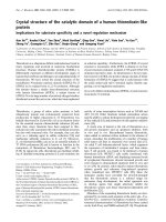
Fig. 1. Hydrophobic moment plot analysis of protein segments. Acon-
ventional hydrophobic moment plot diagram of Eisenberg et al. [38] is
shown with an overlaid grey region delineating candidate oblique-
orientated a-helices [32]. The sequences shown in Table 1 were plotted
on the diagram according to their Æl
H
æ and corresponding ÆH
0
æ values
(Table 1). The data point representing the m-calpain domain V seg-
ment, GTAMRILGGVI (1), can be seen to lie in the grey region,
proximal to that representing the HA2 peptide (2), indicating that the
segment may be a candidate for oblique-orientated a-helix formation.
Fig. 2. Hydropathy plot analysis of protein segments. Hydropathy plot
analysis of the m-calpain domain V segment, GTAMRILGGVI, was
performed using a seven-residue window and the software of Hennig
[39]. It can be seen that hydrophobicty progressively increases along
the length of the segment with a maximal value centred on the
C-terminal glycine, residue 8.
5416 K. Brandenburg et al.(Eur. J. Biochem. 269) Ó FEBS 2002
absorbance spectra [44] (Fig. 6). These spectra monitored
changes in the b fi a acyl chain melting behaviour of
phospholipids, with these changes determined as shifts in
the peak position of the symmetric stretching vibration of
the methylene groups, m
s
(CH
2
), which is known to be a
sensitive marker of lipid order. The peak position of m
s
(CH
2
)
lies at 2850 cm
)1
in the gel phase and shifts at a lipid-specific
temperature T
c
to 2852.0–2852.5 cm
)1
in the liquid-crystal-
line state. For deuterated Myr
2
PtdSer, the values for
the peak position of m
s
(CD
2
) are at 2089–2093 cm
)1
,
respectively.
Monolayer studies on VP1
All monolayer equipment was supplied by NIMA, Coven-
try, UK. Surface tension was monitiored by the Wilhelmy
plate method using a microbalance [45]. Studies were
conducted using a 5 · 15 cm Teflon trough containing
80 mL buffer subphase (10 m
M
Tris/HCl, pH 7.5). The
trough was equipped with moveable barriers, which
responded to the microbalance and could be adjusted to
maintain monolayers at either constant surface pressure or
constant surface area. Monolayers were formed by spread-
ing pure phospholipids (10 m
M
) in chloroform, compressed
to give a surface pressure of 30 mNÆm
)1
,andthen
maintained at constant area. Stock VP1 was added to the
subphase via a reservoir extending into the subphase, which
was contiuously stirred by a magnetic bar, and VP1–
monolayer interactions were recorded as changes in mono-
layer surface pressure (Fig. 7).
RESULTS
Theoretical analyses of candidate oblique-orientated
a-helix-forming segments
The sequences of the influenza viral fusion peptide, HA2,
and m-calpain domain V were analysed. The resulting
values of Æl
H
æ and ÆH
0
æ (Table 1) were plotted on the
hydrophobic moment plot diagram (Fig. 1). The data
points representing these sequences lie proximal in the area
delineating candidate oblique-orientated a-helix-forming
segments, indicating that m-calpain domain V may contain
an oblique-orientated a-helix comparable to that of HA2.
Consistent with these results, hydropathy plot analysis of
the GTAMRILGGVI sequence showed a progressive
increase in hydrophobicity in moving from the N-terminus
to the C-terminus (Fig. 2), suggesting the ability to form an
a-helix with an asymmetric distribution of hydrophobicty
along the a-helical axis. Furthermore, when the sequences of
HA2 and m-calpain domain V were modelled as a-helices
[39], each formed an amphiphilic a-helix with similar
structural properties (Fig. 3). Each a-helix possesses a
glycine-rich hydrophilic face and a wide hydrophobic face,
which includes the bulky amino-acid residues tryptophan,
phenylalanine, leucine and isoleucine.
Fig. 3. Two-dimensional axial projections of protein sequences. Primary structures of (A) the influenza peptide, HA2, a known oblique-orientated
a-helix former and (B) the putative oblique-orientated a-helix-forming segment identified in m-calpain, domain V (Table 1), represented as two-
dimensional axial projections using the software of Hennig [39]. Annotated numbers represent the relative locations of amino-acid residues within
protein primary structure, and hydrophobic residues are circled. It can be seen that each a-helix possesses a glycine-rich polar face and a wide
hydrophobic face rich in bulky amino-acid residues. In the case of the GTAMRILGGVI segment, these residues, isoleucine (6) and leucine (7 and
11), can be seen to be localized in the C-terminal region of the a-helix.
Fig. 4. Haemolytic analysis of VP1. Haemolytic curve of VP1, deter-
mined by the method of Harris & Phoenix [41]. The peptide was
incubated with either human erythrocytes (r) or these erythrocytes in
thepresenceof100m
M
NaCl (j). Percentage haemolysis was deter-
mined (n ¼ 3) and plotted as a function of VP1 concentration. It can
be seen that at a concentration of 2.4 m
M
, VP1 showed 100% lysis of
erythrocytes (LD
50
¼ 1.45 m
M
), but, in the presence of 100 m
M
NaCl,
this ability was reduced by 60%.
Ó FEBS 2002 Lipid-interactive a-helical structure in m-calpain (Eur. J. Biochem. 269) 5417
Haemolytic assay of VP1
It can be seen from Fig. 4 that VP1 is strongly haemolytic
with a sigmoidal relationship between VP1 concentration
and percentage haemolysis. The peptide achieved 100%
lysis of erthrocyte membranes at 2.4 m
M
(LD
50
¼
1.45 m
M
), but in the presence of 100 m
M
NaCl, this ability
of VP1 was reduced the order of 60% (LD
50
¼ 1.85 m
M
).
FTIR conformational analysis of VP1
FTIR spectroscopy was used for conformational analysis of
VP1, either in aqueous solution or in the presence of SUVs
formed from Myr
2
PtdSer, Myr
2
PtdCho or Myr
2
PtdEtn
(Fig. 5). In each case, the relative percentages of a-helical
secondary structure (1650–1655 cm
)1
)andb-sheet secon-
dary structure (1625–1640 cm
)1
) were computed and are
shown in Table 2. In aqueous solution, VP1 showed no
evidence of a-helical structure and was primarily formed
from b-sheet structures (> 90%) (data not shown). In the
presence of Myr
2
PtdEtn and Myr
2
PtdCho, VP1 showed
some evidence of a-helical structure ( 20%) but remained
predominantly formed from b-sheet structures (48% and
61%, respectively) (Fig. 5A,B). In contrast, VP1 showed
high levels of a-helical structure (65%) in the presence of
Myr
2
PtdSer and reduced levels of b-sheet structures (32%)
(Fig. 5C).
FTIR analysis of phospholipid phase-transition
properties
Using FTIR spectroscopy, absorbance spectra representing
the effects of VP1 on the phase-transition temperature and
membrane fluidity of membranes formed from Myr
2
PtdEtn,
Myr
2
PtdCho or Myr
2
PtdSer were derived as a function of
temperature (Fig. 6). Control experiments recorded the lipid
phase-transition temperature, T
c
,ofMyr
2
PtdCho mem-
branes as 27 °C, Myr
2
PtdEtn membranes as 55 °C, and
Myr
2
PtdSer membranes as 37 °C (Figs 6A-6C). The pres-
ence of VP1 had a minor effect on the T
c
and membrane
fluidity of both Myr
2
PtdCho and Myr
2
PtdEtn membranes,
with T
c
being recorded as 24 °Cand52°C, respectively, and
accompanied in each case by a minor increase in membrane
fluidity (Fig. 6A,B). In contrast, the presence of VP1 led to a
large decrease in the fluidity of Myr
2
PtdSer membrane,
accompanied by a large increase in the T
c
of the membranes,
with T
c
being recorded as 47 °C (Fig. 6C).
Monolayer studies on VP1
The interactions of VP1 with Myr
2
PtdSer monolayers were
studied as described above. Myr
2
PtdSer was found to form
Fig. 5. FTIR conformational analysis of VP1 in the presence of lipid.
(A)–(C) Spectra representing FTIR conformational analyses of VP1 in
the presence of lipid with annotated numbers indicating band peak
absorbances. For each spectrum, the relative percentages of a-helical
secondary structure and b-sheet secondary structure were computed,
as described in Materials and Methods, and are shown in Table 2. (A)
and (B) show that, in the presence of Myr
2
PtdEtn and Myr
2
PtdCho,
the major secondary structural contribution to VP1 came from b-sheet
structures (1627 cm
)1
and 1628 cm
)1
, respectively). In contrast, (C)
shows that, in the presence of Myr
2
PtdSer, a-helical structure made the
major contribution to VP1 secondary structure (1650 cm
)1
).
Table 2. VP1 secondary-structural contributions. Relative levels of
a-helical structure and b-sheet structure determined in VP1, a peptide
homologue of the putative oblique-orientated a-helix-forming segment
identified in m-calpain domain V (Table 1). The peptide was either in
aqueous solution (–) or in the presence of lipid. Conformational ana-
lysis of VP1 was performed using FTIR spectroscopy, and the resulting
spectra (Fig. 5) were used to determine relative levels of secondary
structure as described in Materials and methods.
Lipid % a-helix % b-sheet
––92
Myr
2
PtdSer 65 32
Myr
2
PtdEtn 18 61
Myr
2
PtdCho 19 48
5418 K. Brandenburg et al.(Eur. J. Biochem. 269) Ó FEBS 2002
stable monolayers at a surface pressure of 30 mNÆm
)1
,
which was taken to represent that of naturally occurring
membranes. At a final subphase concentration of 20 l
M
,
VP1 showed maximal levels of Myr
2
PtdSer monolayer
penentration, which led to a change in monolayer surface
pressure of 5.5 mNÆm
)1
(Fig. 7). This ability was reduced to
negligible levels in the presence of 100 m
M
NaCl (data not
shown).
DISCUSSION
There is evidence to suggest that the enzymatic activity of
m-calpain is modulated by the membrane interaction of a
segment, GTAMRILGGVI, located in domain V of the
protein’s smaller subunit [24,25]. It has been predicted that
this segment may interact with membrane via the formation
of an oblique-orientated a-helix [26], a class of a-helices [29–
35] that penetrate membranes at a shallow angle because of
a hydrophobicity gradient along the a-helical long axis
[27,28]. Here we have used theoretical techniques to examine
the structural characteristics of the putative domain V
a-helix. In addition, the ability of the GTAMRILGGV
segment to adopt a-helical structure and to interact with
membranes has been investigated using VP1, a peptide
homologue of this segment, in conjunction with haemolytic
analysis, monolayer studies, and FTIR spectroscopy. Our
results are discussed in relation to HA2, a viral fusion
peptide known to penetrate membranes via oblique-orien-
tated a-helix formation [29–35].
The sequences of the GTAMRILGGVI segment and
HA2 were analysed, and data points representing their
a-helices were found to lay proximal in the region of the
hydrophobic moment plot diagram delineating candidate
oblique-orientated a-helices (Fig. 1). This observation sug-
gests that the GTAMRILGGVI segment may form such an
a-helix and therefore may possess a hydrophobicity gradi-
ent. Consistent with this suggestion, hydropathy plot
analysis showed the GTAMRILGGVI segment to become
progressively more hydrophobic in moving from the
Fig. 6. FTIR phase-transition analysis of lipid in the presence of VP1.
Spectra representing FTIR phase-transition analysis of lipid in the
presence of VP1. In the absence of the peptide, the phase-transition
temperature (T
c
)ofMyr
2
PtdEtn was recorded as 55 °C(j;A),of
Myr
2
PtdCho as 27 °C(j; B) and of Myr
2
PtdSeras37°C(j;C).The
presence of VP1 led to a minor increase in the fluidity of both
Myr
2
PtdEtn and Myr
2
PtdCho membranes, which in each case was
accompanied by a minor decrease in T
c
,withT
c
recorded as 52 °Cfor
Myr
2
PtdEtn membranes (h;A)and24°CforMyr
2
PtdCho mem-
branes (h; B). In contrast, the presence of VP1 led to a large decrease
in the fluidity of Myr
2
PtdSer membranes, accompanied by a large
increase in the T
c
of the membranes (h;C)withT
c
being recorded as
47 °C.
Fig. 7. Monolayer interactions of VP1. Time course for interactions of
VP1 with Myr
2
PtdSer monolayers at a surface pressure of 30 mNÆm
)1
,
taken to represent that of naturally occurring membranes. VP1 (final
concentration 20 l
M
) was introduced into the monolayer subphase at
time zero and after 100 s showed rapid penetration of Myr
2
PtdSer
monolayers. Maximal levels of penentration were reached after 600 s,
with a concomitant change in monolayer surface pressure of
5.5 mNÆm
)1
.
Ó FEBS 2002 Lipid-interactive a-helical structure in m-calpain (Eur. J. Biochem. 269) 5419
N-terminus to the C-terminus (Fig. 2), and graphical
analysis showed the GTAMRILGGVI a-helix to possess
a number of structural resemblances to the HA2 a-helix
(Fig. 3). It can be seen that each a-helix shown in Fig. 3
possesses a glycine-rich polar face, which studies on HA2
and a number of other oblique-orientated a-helices have
shown to be critical for maintaining their hydrophobicity
gradients [27,28,46]. It can also be seen from Fig. 3 that each
a-helix possesses a wide hydrophobic face rich in bulky
amino-acid residues. In the case of the GTAMRILGGVI
segment (Fig. 3), leucine and isoleucine can be seen to be
preponderant in the C-terminal region of the a-helix. This
localization of strongly hydrophobic amino-acid residues is
structurally consistent with the higher levels of hydropho-
bicity predicted for the C-terminal region of the segment
(Fig. 2) and possession of a hydrophobicity gradient [47].
Furthermore, when these wide hydrophobic faces, rich in
bulky residues, are combined with narrow polar faces, rich
in glycine residues (Fig. 3), a-helices are given an effective
wedge shape, which appears to assist HA2 and other
oblique-orientated a-helix-forming peptides, to destabilize
membranes in the promotion of their biological activity
[46,48].
It is clear from our theoretical analyses that the
GTAMRILGGVI segment has the potential to form an
a-helix with strong structural similarities to the oblique-
orientated a-helix formed by HA2 and other membrane-
interactive peptides. Consistent with these observations,
FTIR spectroscopic analysis showed that VP1 was able to
adopt a-helical structure in the presence of lipid membranes
(Fig. 5) and to affect the lipid-phase transition properties of
these membranes (Fig. 6). In addition, haemolytic analysis
showed that the peptide is able to lyse erythrocyte
membranes (Fig. 4), and monolayer studies showed that it
is able to penetrate lipid monolayers mimetic of naturally
occurring membranes (Fig. 7). In combination, these results
clearly show that the GTAMRILGGVI segment is able to
form a membrane-interactive a-helix.
In aqueous solution, VP1 showed no evidence of a-helical
structure (data not shown) but was found to adopt such
structure in the presence of Myr
2
PtdCho, Myr
2
PtdEtn and
Myr
2
PtdSer (Fig. 6). This suggests that VP1 requires the
amphiphilic environment of a lipid interface to adopt
a-helical structure, a requirement also observed for HA2
[49]. In the presence of Myr
2
PtdEtn and Myr
2
PtdCho
membranes, VP1 adopted relatively low levels of a-helical
structure ( 20%; Fig. 5A,B) and induced only minor
decreases in the T
c
of these membranes ( 3 °C), accom-
panied by minor increases in liquid-crystalline phase fluidity
(Fig. 6,AB). Such changes in lipid-phase transition proper-
ties are consistent with VP1 binding to the headgroup
regions of Myr
2
PtdCho and Myr
2
PtdEtn membranes and
suggest that the peptide penetrates the surface regions of
these membranes. In contrast, VP1 adopted high levels of
a-helical structure in the presence of Myr
2
PtdSer (65%;
Fig. 5C) and induced a 10 °CriseintheT
c
of these
membranes accompanied by a large decrease in its liquid-
crystalline phase fluidity (Fig. 6C). Such changes are
consistent with VP1 penetration of the Myr
2
PtdSer mem-
brane hydrophobic core region and suggest that the peptide
shows high levels of interaction with these membranes.
Strongly supporting this suggestion, VP1 was found to show
high levels of Myr
2
PtdSer monolayer penetration (Fig. 7),
inducing surface pressure changes of 5.5 mNÆm
)1
.Com-
parable levels of monolayer penetration have been observed
for the C-terminal a-helices of Escherichia coli penicillin-
binding proteins 5 and 6 [50], which function as membrane
anchors for these proteins [51]. Interestingly, a recent study
has suggested that these anchors may form oblique-
orientated a-helices (D. Phoenix & F. Harris, unpublished
work). Taken in combination, these results suggest that VP1
has a low affinity for zwitterionic lipid but a high affinity for
Myr
2
PtdSer and may have a requirement for either this
specific lipid, or anionic lipid in general, to achieve higher
levels of membrane penetration. These results contrast with
HA2, which shows an affinity for zwitterionic lipid [52,53]
and requires the low pH of the endosomal membrane
surface for both oblique-orientated a-helix formation and
enhanced levels of membrane penetration via the deproto-
nation of its negatively charged residues [35,54].
VP1 was found to be strongly haemolytic, but, in the
presence of 100 m
M
NaCl, this ability was reduced by the
order of 60% (Fig. 4) and the ability of VP1 to interact with
Myr
2
PtdSer monolayers was reduced to negligible levels
(data not shown). In combination, these results suggest a
strong electrostatic contribution to VP1–membrane inter-
action and further support a VP1 requirement for anionic
lipid. Indeed, the relatively high LD
50
(1.45 m
M
) shown by
VP1 for haemolytic action could reflect the fact that
erythrocyte membranes possess an asymmetric distribution
of anionic lipids with the extracytoplasmic leaflet depleted of
such lipids [55]. It is interesting to note that the HA2 a-helix
is also strongly haemolytic and that the mutation of polar
face glycine residues, essential for maintaining the HA2
a-helix hydrophobicity gradient, results in the loss of
haemolytic and fusogenic ability [49,56].
In conclusion, based on structural similarities to HA2, we
have suggested that the segment, GTAMRILGGVI, of
m-calpain domain V forms an a-helix, which possesses a
hydrophobicity gradient and penetrates membranes in an
oblique orientation. We speculate that glycine residues in
the polar face of this a-helix could play an important role in
facilitating this mechanism of membrane penetration. This
a-helix has a preference for anionic lipid, which leads to
higher levels of a-helicity and membrane penetration via
electrostatic interactions. These results are consistent with
those of previous authors, which have shown that m-calpain
activity is modulated by the presence of anionic lipid [25].
To satisfy a VP1 requirement for anionic lipid, it seems
probable that the peptide’s single positively charged amino-
acid residue, arginine (Table 1; Fig. 3) would engage in
charge–charge interaction with negatively charged mem-
brane lipid. Furthermore, to achieve the deeper levels of
membrane penetration indicated for Myr
2
PtdSer mem-
branes, it is possible that these interactions may involve the
snorkelling mechanism that features in the membrane
interactions of a number of lipid-interactive a-helices
[28,57]. According to this mechanism, the VP1 arginine
residue would extend or snorkel its long hydrophobic alkyl
chain, facilitating penetration of the membrane hydropho-
bic core region yet still allowing its positively charged moiety
to interact with negatively charged moieties in the mem-
brane lipid headgroup region. We speculate that utilization
of this mechanism by the m-calpain domain V a-helix could
result in enhanced levels of membrane interaction by
domain V, which would support work indicating lipid
5420 K. Brandenburg et al.(Eur. J. Biochem. 269) Ó FEBS 2002
involvement in the reduction of Ca
2+
levels necessary for
the efficient activation of m-calpain.
REFERENCES
1. Sorimachi, H. & Suzuki, K. (2001) The structure of calpain. J. Biol.
Chem. 129, 653–664.
2.Sorimachi,H.,Ishiura,S.&Suzuki,K.(1997)Structureand
physiological function of calpains. Biochem. J. 328, 721–732.
3. Johnson, G.V.W. & Guttmann, R.P. (1997) Calpains: intact and
active? Bioessays 19, 1011–1018.
4. Ma, H., Fukiage, C., Kim, Y.H., Duncan, M.K., Reed, N.A.,
Shih, M., Azuma, M. & Shearer, T.R. (2001) Characterization and
expression of calpain 10. A novel ubiquitous calpain with nuclear
localization. J. Biol. Chem. 276, 28525–28531.
5. Sato, K. & Kawashima, S. (2001) Calpain function in the mod-
ulation of signal transduction molecules. Biol. Chem. 382, 743–751.
6. Calafoli, E. & Molinari, M. (1998) Calpain: a protease in search of
afunction?Biochem. Biophys. Res. Commun. 247, 193–203.
7. Ono, Y., Sorimachi, H. & Suzuki, K. (1998) Structure and phy-
siology of calpain, an enigmatic protease. Biochem. Biophys. Res.
Commun. 245, 289–294.
8. Azuma, M., Fukiage, C., David, L. & Shearer, T.R. (1997) Acti-
vation of calpain in lens: a review and proposed mechanism. Lens.
Exp. Eye Res. 64, 529–538.
9. Horikawa, H. et al. (2000) Genetic variation in the gene encoding
calpain-10 is associated with type 2 diabetes mellitus. Nat. Genet.
26, 163–175.
10. Huang, Y.H. & Wang, K.K.W. (2001) The calpain family and
human disease. Trends Mol. Med. 7, 355–362.
11. Wang, K.K.W. (2000) Calpain and caspase: Can you tell the dif-
ference. Trends Neurosci. 23, 20–26.
12. Ohno,S.,Emori,Y.,Imajoh,S.,Kawasaki,H.,Kisaragi,M.&
Suzuki, K. (1984) Evolutionary origin of a calcium-dependent
protease by fusion of genes for a thiol protease and a calcium-
binding protein? Nature (London) 312, 566–570.
13. Croall, D.E. & DeMartino, G.N. (1991) Calcium-activated neu-
tral protease (calpain) system: structure, function, and regulation.
Physiol. Rev. 71, 813–847.
14. Hosfield, C.M., Elce, J.S., Davies, P.L. & Jia, Z.C. (1999) Crystal
structure of calpain reveals the structural basis for Ca
2+
dependent protease activity and a novel mode of enzyme activa-
tion. EMBO J. 18, 6880–6889.
15. Strobl, S., Fernandez-Catalan, C., Braun, M., Huber, R., Masu-
moto, H., Nakagawa, K., Irie, A., Sorimachi, H., Bourenkow, G.,
Bartunik,H.,Suzuki,K.&Bode,W.(2000)Thecrystalstructure
of calcium-free human m-calpain suggests an electrostatic switch
mechanism for activation by calcium. Proc. Natl. Acad. Sci. USA
97, 588–592.
16. Molinari, M. & Carafoli, E. (1997) Calpain: a cytosolic proteinase
active at the membranes. J. Membr. Biol. 156, 1–8.
17. Kawasaki, H. & Kawashima, S. (1996) Regulation of the calpain-
calpastatin system by membranes (review). Mol. Membr. Biol. 13,
217–224.
18. Chakrabarti, A.K., Dasgupta, S., Gadsden, R.H., Hogan, E.L. &
Banik, N. (1996) Regulation of brain m-calpain Ca
2+
sensitivity
by mixtures of membrane lipids: activation at intracellular Ca
2+
level. J. Neurosci. Res. 44, 374–380.
19. Saido, T.C., Shibata, M., Takenawa, T., Murofushi, H. & Suzuki,
K. (1992) Positive regulation of mu-calpain action by polypho-
sphoinositides. J. Biol. Chem. 267, 24585–24590.
20. Melloni, E. & Pontremoli, S. (1989) The calpains. Trends Neurosci.
12, 438–444.
21. Imajoh, S., Kawasaki, H. & Suzuki, K. (1986) The amino-terminal
hydrophobic region of the small subunit of calcium-activated
neutral protease (CANP) is essential for its activation by phos-
phatidylinositol. J. Biochem. (Tokyo) 99, 1281–1284.
22. Tompa, P., Emori, Y., Sorimachi, H., Suzuki, K. & Friedrich, P.
(2001) Domain III of calpain is a Ca
2+
-regulated phospholipid-
binding domain. Biochem. Biophys. Res. Commun. 280, 1333–
1339.
23. Rizo, J. & Su
¨
dhof, T.C. (1998) C2-domains, structure and func-
tion of a universal Ca
2+
-binding domain. J. Biol. Chem. 273,
15879–15882.
24. Crawford, C., Brown, N.R. & Willis, A.C. (1990) Investigation of
the structural basis of interaction of calpain II with phospholipid
and with carbohydrate. Biochem. J. 265, 575–579.
25. Arthur, J.S.C. & Crawford, C. (1996) Investigation of the inter-
action of m-calpain with phospholipids: calpain–phospholipid
interactions. Biochim. Biophys. Acta. 1293, 201–206.
26. Daman,O.A.,Biswas,S.,Harris,F.,Wallace,J.&Phoenix,D.A.
(2001) Theoretical investigation into the lipid interaction of
m-calpain. Mol. Cell. Biochem. 223, 159–163.
27. Decout, A., Labeur, C., Vanloo, B., Goethals, M., Vandekerck-
hove, J., Brasseur, R. & Rosseneu, M. (1999) Contribution of the
hydrophobicity gradient to the secondary structure and activity of
fusogenic peptides. Mol. Membr. Biol. 16, 37–246.
28. Phoenix, D.A., Harris, F., Daman, O.A. & Wallace, J. (2002) The
prediction of amphiphilic a-helices. Curr. Protein Peptide Sci. in
press.
29. Bentz, J. & Mittal, A. (2000) Deployment of membrane fusion
protein domains during fusion. Cell Biol. Int. 24, 819–838.
30. Luneberg, J., Martin, I., Nussler, F., Ruysschaert, J M. & Herr-
mann, A. (1995) Structure and topology of the influenza virus
fusion peptide in lipid bilayers. J. Biol. Chem. 270, 27606–27614.
31. Brasseur, R. (2000) Tilted peptides: a motif for membrane desta-
bilisation (Hypothesis). Mol. Membr. Biol. 17, 31–40.
32. Harris, F., Wallace, J. & Phoenix, D.A. (2000) Use of the
hydrophobic moment plot to aid the identification of oblique
orientated a-helices. Mol. Membr. Biol. 17, 201–207.
33. Chang, D.K., Cheng, S.F., Trivedi, V.D., Yang, S.H. (2000) The
amino-terminal region of the fusion peptide of influenza virus
hemagglutinin, HA2, inserts into sodium dodecyl sulfate micelle
with residues 16–18 at the aqueous boundary at acidic pH:
oligomerization and the conformational flexibility. J. Biol. Chem.
275, 19150–19158.
34. Macosko, J.C., Kim, C.H. & Shin, Y.K. (1997) The membrane
topology of the fusion peptide region of influenza hemagglutinin
determined by spin-labeling EPR. J. Mol. Biol. 267, 1139–1148.
35. Han, X., Bushweller, J.H., Cafiso, D.S. & Tamm, L.K. (2001)
Membrane structure and fusion-triggering conformational change
of the fusion domain from influenza hemagluttinin. Nat. Struct.
Biol. 8, 715–720.
36. Eisenberg, D., Weiss, R.M. & Terwilliger, T.C. (1982) The helical
hydrophobic moment: a measure of the amphiphilicity of a-helix.
Nature (London) 299, 371–374.
37. Eisenberg, D., Weiss, R.M., Terwilliger, T.C. & Wilcox, W. (1982)
Hydrophobic moment and protein structure. Faraday Symp.
Chem. Soc. 17, 109–120.
38. Eisenberg, D., Schwarz, E., Komaromy, M. & Wall, R. (1984)
Analysis of membrane and surface protein sequences with the
hydrophobic moment plot. J. Mol. Biol. 179, 125–142.
39. Hennig, L. (1999) Wingen/Winpeg: user friendly software for the
analysis of amino acid sequences. Biotechniques 26, 1170–1172.
40. Kyte, J. & Dolittle, R.F. (1982) A simple method for displaying
the hydropathic character of a protein. J. Mol. Biol. 157, 105–132.
41. Harris, F. & Phoenix, D.A. (1997) An investigation into the ability
of C-terminal homologues of the Escherichia coli low molecular
mass penicillin-binding proteins 4, 5 and 6 to undergo membrane
interaction. Biochemie 79, 171–174.
42. Keller, R.C., Killian, J.A. & De Kruijff, B. (1992) Anionic phos-
pholipids are essential for alpha-helix formation of the signal
peptide of prePhoE upon interaction with phospholipid vesicles.
Biochemistry 31, 1672–1677.
Ó FEBS 2002 Lipid-interactive a-helical structure in m-calpain (Eur. J. Biochem. 269) 5421
43. Kauppinen, J.K., Moffat, D.J., Mantsch, H.H. & Cameron, D.G.
(1981) Fourier self-deconvolution: a method for resolving
intrinsically overlapped bands. Appl. Spectrosc. 35, 271–276.
44. Brandenburg, K., Kusomoto, S. & Seydel, U. (1997) Conforma-
tional studies of synthetic lipid A analogues and partial structures
by infrared spectroscopy. Biochim. Biophys. Acta 1329, 183–201.
45. Demel, R.A. (1974) Model membrane monolayers: description of
use and interaction. Methods Enzymol. 32, 539–545.
46. Fujii, G. (1999) To fuse or not to fuse: the effects of electrostatic
interactions, hydrophobic forces and structural amphiphilicity on
protein-mediated membrane destabilisation. Advanced Drug
Delivery Review 38, 257–277.
47. Peuvot, J., Schank, A., Lins, L. & Brasseur, R. (1999) Are the
fusion processes involved in birth, life and death of the cell
depending on tilted insertion of peptides into membranes?
J. Theor. Biol. 198, 173–181.
48. White, J.M. (1990) Viral and cellular membrane fusion proteins.
Annu. Rev. Physiol. 52, 675–697.
49. Plank, C., Zauner, W. & Wagner, E. (1999) Application of
membrane-active peptides for drug and gene delivery across cel-
lular membranes. Advanced Drug Delivery Review 34, 21–35.
50. Harris, F., Demel, R.A., Phoenix, D.A. & De Kruijff, B. (1998) An
investigation into the lipid interactions of peptides corresponding
to the C-terminal anchoring domains of Escherichia coli penicillin-
binding proteins 4, 5 and 6. Biochim. Biophys. Acta 1415, 10–22.
51. Phoenix, D.A. & Harris, F. (1998) Amphiphilic a-helices and lipid
interactions. In Protein Targeting and Translocation (Phoenix,
D.A., ed.), pp. 19–36. Portland Press, London.
52. Zhou, Z., Macosko, J.C., Hughes, D.W., Sayer, B.G., Hawes, J. &
Epand, R.M. (2000) N-15 NMR study of the ionization properties
of the influenza virus fusion peptide in zwitterionic phospholipid
dispersions. Biophys. J. 78, 2418–2425.
53. Duzgunes, N. & Shavnin, S.A. (1992) Membrane destabilization
by N-terminal peptides of viral envelope proteins. J. Membr. Biol.
128, 71–80.
54. Stegmann, T. & Helenius, A. (1993) Influenza virus fusion: from
models toward a mechanism. In Viral Fusion Mechanisms (Bentz,
J., ed.). CRC Press, Boca Raton, FL.
55. Op den Kamp, J.A.F. (1979) Lipid asymmetry in membranes.
Annu. Rev. Biochem. 48, 47–71.
56. Tamm, L.K. & Han, X. (2000) Viral fusion peptides: a tool to
disrupt and connect biological membranes. Biosci. Rep. 29,501–
518.
57. Segrest, J.P., Venkatachalapathi, Y.V., Srinivas, S.K., Gupta,
K.B., De Loof, H. & Anatharamaiah, G.M. (1992) Role of basic
amino acid residues in the amphipathic helix: the snorkel
hypothesis. In Molecular Conformation and Biological Interactions
(Balaram, P. & Ramaseshan, S., eds), pp. 597–635. Indian
Academy of Sciences, Bangalore.
5422 K. Brandenburg et al.(Eur. J. Biochem. 269) Ó FEBS 2002