Báo cáo Y học: A polymer with a backbone of 3-deoxy-D-glycero -D-galacto -non-2ulopyranosonic acid, a teichuronic acid, and a b-glucosylated ribitol teichoic acid in the cell wall of plant pathogenic Streptomyces sp. VKM Ac-2124 pdf
Bạn đang xem bản rút gọn của tài liệu. Xem và tải ngay bản đầy đủ của tài liệu tại đây (175.31 KB, 6 trang )
A polymer with a backbone of 3-deoxy-
D
-
glycero
-
D
-
galacto
-non-2-
ulopyranosonic acid, a teichuronic acid, and a b-glucosylated ribitol
teichoic acid in the cell wall of plant pathogenic
Streptomyces
sp.
VKM Ac-2124
Alexander S. Shashkov
1
, Larisa N. Kosmachevskaya
2
, Galina M. Streshinskaya
2
, Lyudmila I. Evtushenko
3
,
Olga V. Bueva
3
, Viktor A. Denisenko
4
, Irina B. Naumova
2
and Erko Stackebrandt
5
1
N.D. Zelinsky Institute of Organic Chemistry, Russian Academy of Sciences, Moscow, Russia;
2
School of Biology,
M.V. Lomonosov Moscow State University, Moscow, Russia;
3
Institute of Biochemistry and Physiology of Microorganisms,
Russian Academy of Sciences, Pushchino, Moscow Region, Russia;
4
Belarussian Research Institute for Potato Growing,
Samokhvalovitchi, Minsk Region, Belarus;
5
DSMZ-Deutsche Sammlung von Mikroorganismen und Zellkulturen GmbH,
Braunschweig, Germany
Structures of cell wall anionic polymers of the strain Strep-
tomyces sp. VKM Ac-2124, a causative agent of potato scab,
which is phylogenetically the closest to plant pathogenic
species S. setonii and S. caviscabies, were studied. The strain
contains three anionic glycopolymers, viz., a teichuronic acid
with a disaccharide repeating unit fi6)-a-
D
-Glcp-(1fi4)-b-
D
-ManpNAc3NAcA-(1fi,ab-glucosylated polymer of
3-deoxy-
D
-glycero-
D
-galacto-non-2-ulopyranosonic acid (Kdn),
and a b-glucosylated 1,5-poly(ribitol phosphate). The strain
studied is the second representative of plant pathogenic
streptomycetes inducing potato scab disease, the cell wall
anionic polymers of which were shown to contain a Kdn-
polymer. Presumably, the presence of Kdn-containing
structures in the surface regions of pathogens is essential for
their efficient attachment to host plant cells.
Keywords: NMR spectroscopy; teichuronic acid; teichoic
acid; Kdn; Streptomyces.
Cell walls of the majority of Gram-positive bacteria
belonging to the genus Streptomyces (the order Actino-
mycetales) contain teichoic acids, the anionic glycopolymers
which are covalently bound to peptidoglycan and are
situated between other cell wall layers and at the cell surface.
They impart a negative charge to the cell surface, which is
essential for the physiological functioning of the cells and
cell coaggregation [1]. In addition to teichoic acids, other
anionic polymers have been found in the cell wall of
streptomycetes. A teichuronic acid with a disaccharide
repeating unit fi4)-b-
D
-ManpNAc3NAcA-(1fi3)-a-
D
-
GalpNAc-(1fi was identified in the cell wall of Streptom-
yces lavendulocolor VKM Ac-215
T
[2]. Recently, a polymer
of 3-deoxy-
D
-glycero-
D
-galacto-non-2-ulopyranosonic acid
(Kdn), along with small amount of glycerol teichoic acid,
has been found in the cell wall of the plant pathogen
Streptomyces sp. VKM Ac-2090 [3]. This nine-carbon sugar,
which may be regarded as a modification of sialic acid, is
abundant in animal tissues [4] and, presumably, plays a role
in intercell interactions [5].
In the present work, we investigated cell wall polymers
of yet another representative of streptomycetes, viz., of
the strain VKM Ac-2124, a causative agent of potato
scab, which is the closest to Streptomyces setonii ATCC
25497
T
based on the analysis of 16S rRNA gene
sequence.
MATERIALS AND METHODS
The strain VKM Ac-2124 was isolated from common scab
lesions of potatoes, Solanum tuberosum, cultivar ÔIzoraÕ
(Leningrad region, Russia) on ISP2 agar [6] as reported by
Loria & Davis [7]. For studying phenotypical characteris-
tics, the methods and media described by Schirling and
Gottlieb [6] were used. Extraction and purification of DNA
was carried out as reported [8]. The 16S rRNA gene was
amplified by PCR using prokaryotic 16S rDNA universal
primers 27f (5¢-AGAGTTTGATCCTGGCTCAG-3¢)and
1522r (5¢-AAGGAGGTGATCCARCCGCA-3¢) and puri-
fied as described [8]. 16S rDNA was sequenced using a Big
Dye Terminator Kit (Perkin Elmer) with an a model ABI-
310 automatic DNA Sequencer (Perkin Elmer) according to
the manufacturer’s protocol. The sequences of the highest
scores were chosen from NCIB database using
BLAST
search
[10]. Other 16S rDNA sequences of the plant pathogenic
streptomycetes and related strains used in the analysis
were selected from NCIB database. The sequence of
Brevibacterium linens DSM 20425
T
(X77451) was used as
an outgroup. Nucleotide substitution rates were calculated
as described by Kimura & Ohta [11] and the phylogenetic
Correspondence to I. B. Naumova, School of Biology,
M.V. Lomonosov Moscow State University, Moscow 119899, Russia.
E-mail:
Abbreviations: PME, phosphomonoesterase; Kdn, 2-keto-3-deoxy-
nononic acid.
Enzyme: phosphomonoesterase (EC 3.1.3.1).
Note: Kdn is the abbreviation of 2-keto-3-deoxy-nononic acid, named
according to the earlier nomenclature [9].
(Received 5 July 2002, revised 11 September 2002,
accepted 20 September 2002)
Eur. J. Biochem. 269, 6020–6025 (2002) Ó FEBS 2002 doi:10.1046/j.1432-1033.2002.03274.x
tree was constructed by the neighbour-joining method [12]
with
CLUSTAL W
software [13]. Three topologies were
evaluated by bootstrap analysis of the sequence data with
thesamesoftware.
To evaluate the pathogenic activity of the strain, the
aseptically cultured potato microtubers in vitro were used as
described by Lawrence et al. [14]. The microtubers were
immersed for 5–10 min in a suspension of 14-day-old agar
culture (mainly, spore mass) grown on Czapek’s agar [6]
followed by incubation at 100% relative humidity for
5 days at 22–24 °C in the darkness.
To obtain cell wall, the culture was grown on a peptone/
yeast medium [15] on a shaker at 28 °C and harvested by
centrifugation in the middle of the exponential growth
phase (24–30 h). The cells were washed with 0.95% (v/v)
NaClandstoredfrozenat)20 °C before use. The native cell
walls were obtained from crude mycelium by fractional
centrifugation after preliminary disruption by sonication,
and purified using 2% (w/v) SDS to avoid possible
contamination with membrane compounds, including
lipoteichoic acids, washed several times with water, and
freeze-dried. To isolate polymers, cell walls were extracted
twice with 10% (v/v) trichloroacetic acid at 2–4 °Cfor24h
each time; with constant stirring. The extracts were separ-
ated from cell debris, combined, dialyzed against distilled
water and freeze-dried.
Descending chromatography and electrophoresis were
performed on Filtrak FN-13 paper. Electrophoresis was
performed in pyridinium acetate buffer (pH 5.6) to separate
phosphate esters and to purify ribitol teichoic acid
(20 VÆcm
)1
, 5 h). Paper chromatography was performed in
a pyridine-benzene-butanol-water (3 : 1 : 5 : 3, v/v/v/v) sol-
vent system to separate ribitol and glucose. Phosphoric esters
were detected with the molybdate reagent, reducing sugars,
with aniline hydrogenphthalate; and ribitol and monosac-
charides, with 5% (w/v) AgNO
3
in aqueous ammonia.
Acid hydrolysis was carried out with 2
M
HCl for 3 h at
100 °C; alkaline hydrolysis was performed with 1
M
NaOH
for 3 h at 100 °C; enzymatic hydrolysis with phospho-
monoesterase (PME) from calf intestine (EC 3.1.3.1; Sigma)
was conducted in ammonium acetate buffer, pH 9.8 at 37°
for 18–20 h.
Analytical methods used and the scheme of identification
of a glucosylribitol were the same as described previously
[16,17].
NMR spectra were recorded with a DRX-500 (Bruker,
Germany) spectrometer for 2–3% solutions in D
2
Oat30 °C
with acetone (d
H
2.225 d
C
231.45) as the internal standard,
and 80% H
3
PO
4
as the external standard for
31
PNMR.Pre-
saturation of the HDO signal (1 s) was used in the accumu-
lation of the
1
H NMR spectra. Two-dimensional spectra
were obtained using standard pulse sequences from the
Bruker software. Mixing times of 100 and 200 ms were used
in TOCSY and ROESY experiments, respectively. A 60-ms
delay was used for the evolution of long-range connectivities
in
1
H,
13
CHMBCand
1
H,
31
PHMQCexperiments.
RESULTS AND DISCUSSION
To identify the strain VKM Ac-2124 isolated from common
potato scab, an almost complete 16S rRNA gene sequence
(1470 nucleotides) was determined. Phylogenetic analysis
indicated it to be the closest (99.6% 16S rDNA binary
sequence similarity) to S. setonii ACTT 25497
T
(D63872)
and S. caviscabies ATCC 51928
T
(AF112160), which are
also causative agents of potato scab. These three strains and
S. griseus ISP 5236
T
(AY094371) formed a tight cluster with
a 100% bootstrap replication value (not presented), which is
significantly distant from other validly described plant
pathogenic streptomycete species [18–21]. At the pheno-
typical level, the strain was most similar to S. setonii in
accordance to characteristics of S. setonii described previ-
ously [18,22,23]. Spore mass of VKM Ac-2124 was usually
grey or yellowish grey on glycerol-asparagine agar [6], the
spores were smooth, and borne in mature fexuous chains.
Substrate mycelium was yellow or brownish-yellow on most
tested media. Melanoid pigment was not produced on
tyrosine or peptone iron agar while pale or greyish to light
yellowish brown diffusible pigment was formed on some
media. Testing of plant pathogenicity of the strain VKM
Ac-2124 showed that it induced rough, corky lesions such as
those resulting from natural infections, and the lesions
covered about 70% of the tuber surface.
The anionic polymers were isolated from the cell wall and
investigated. Glucosylribitol monophosphate and small
amounts of ribitol mono- and bisphosphates were identified
as alkaline hydrolysis products. Acid hydrolysis afforded
ribitol monophosphates and bisphosphates, anhydroribitol
phosphate, anhydroribitol, ribitol, inorganic phosphate and
glucose. The amount of the latter exceeded considerably
that bound to ribitol phosphate. An unidentified ninhydrin-
positive compound was also detected, which migrated to the
cathode in the electrophoresis, but was absent from
phosphates produced upon alkaline hydrolysis.
Ribitol mono- and bisphosphates were subjected to the
action of phosphomonoesterase; these were identified based
on the ribitol/phosphate ratio. Glucosylribitol phosphate
was identified based on its electrophoretic mobility (pyridi-
nium acetate buffer) in comparison with an analogous ester
obtained upon alkaline hydrolysis of glucosylated ribitol
teichoic acid from the cell wall of S. azureus RIA 1009
[24] and based on the analysis of the products formed upon
acid and enzymatic hydrolysis. Acid hydrolysis afforded
glucose and ribitol monophosphate, while a glycoside
containing glucose and ribitol (1 : 1 molar ratio) was
produced under the action of phosphomonoesterase. Low
content of teichoic acid-linked phosphorus (0.8%) in the cell
wall as well as high percentage of glucose and the presence
of an unidentified ninhydrin-positive component suggest
that the cell wall contains other polymer(s) in addition to
ribitol teichoic acid.
The polymers present in the cell wall were
investigated using NMR spectroscopy
The
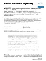
13
C NMR spectrum of the preparation revealed the
presence, in the region typical of anomeric carbon atoms
of carbohydrates, of five signals of unequal intensities at
d103.6, 102.8, 101.2, 100.2, and 97.6 (Table 1). As
followed from the APT spectrum (Fig. 1), four signals
at d100.2–103.6 belonged to the protonated anomeric
carbon atoms, while the fifth signal of low intensity at
d97.6 belonged to the nonprotonated carbon atom,
presumably, to the anomeric atom C(2) of an ulosonic
acid. The presence of 3-deoxyulosonic acid was also
suggested based on the identification of a signal for a
Ó FEBS 2002 Kdn-polymer of plant pathogenic streptomycete (Eur. J. Biochem. 269) 6021
CH
2
-group at d40.4. The spectrum contained also two
signals in the region of resonances of carbon atoms bound
to nitrogen at d52.45 and 54.05, a signal at d23.3
(CH
3
CON), and three signals for CO groups at d174.5–
176.0. The resonances for the CH
2
O groups were found at
d61.9, 62.0, 65.8, 68,0, and 69.4. Other signals of the
spectrum were found at d67.9–80.4, i.e. in the region of
resonances of CH groups bound to one oxygen atom.
The region of resonances of the anomeric protons in the
1
H NMR spectrum (Fig. 1) contained two abundant signals
at d4.90 (J
1,2
<2Hz)and5.07(J
1,2
3.6 Hz) and two signals
of lower intensities at d4.55 (J
1,2
7.9 Hz) and 4.66 (J
1,2
7.9 Hz) (Table 2). Two signals at d1.93 and 2.07 were
observed in the region of resonances of the CH
3
CO– groups.
The presence of 3-deoxynonulosonic acid with the
b-configuration of the glycosidic bond followed from two
doublets of doublets at d2.20 (
2
J
3,3¢
13.0 Hz;
3
J
3,4
4.9 Hz)
and 1.78 (J
3¢,4
12.4 Hz).
The 1D NMR spectra could be interpreted from the
analysis of 2D homonuclear
1
H,
1
HCOSY,TOCSYand
ROESY spectra and 2D heteronuclear
1
H,
13
CHSQC
(Fig. 1) and HMBC and
1
H,
31
P HMQC spectra. The
spectroscopic data obtained suggested the presence of three
different types of anionic glycopolymers (Tables 1,2).
Teichuronic acid (polymer I) with the repeating unit fi6)-
a-
D
-Glcp-(1fi4)-b-
D
-ManpNAc3NAcA-(1fi was the
major component of the cell wall preparation. The absolute
configuration of glucose (
D
-) isolated after hydrolysis of the
total cell wall preparation was determined by its transfor-
mation in 2-octyl glycoside and by comparison of the
derivative obtained with standard samples of (S+)-
and (R-)-2-octyl glucopyranosides using gas-liquid
Table 1.
13
CNMRchemicalshifts(d, p.p.m) for the teichuronic acid (polymer I), the Kdn-containing polymer (polymer II), and the ribitol teichoic acid
(polymer III) from cell wall of Streptomyces sp. VKM Ac-2124.
Carbon
Residue C-1 C-2 C-3 C-4 C-5 C-6 C-7 C-8 C-9
Polymer I
fi6)-a-
D
-Glcp-(1fi (A) 100.2 72.5 73.9 70.05 72.3 69.4
fi4)-b-
D
-ManpNAc3NAcA-(1fi (B) 101.2 52.45 54.05 73.2 78.8 175.1
Polymer II
(C) 176.0 97.6 40.4 70.35 71.6 72.6 68.0 79.4 61.9
(D) 102.8 74.6 77.0 71.0 77.1 61.9
Polymer III
(E) 68.0 71.8 72.5 80.4 65.8
(F) 103.6 74.6 77.0 70.9 77.1 62.0
* or 2);
CH
3
CON, d 23.3; CH
3
CON, d 174.8 and 174.
Fig. 1. Part of the HSQC spectrum of anionic
cell wall polymers from Stre ptomyces sp. VKM
Ac-2124. The signal at d97.6 is marked with an
arrow.
6022 A. S. Shashkov et al. (Eur. J. Biochem. 269) Ó FEBS 2002
chromatography [25]. The absolute configuration of
ManpNAc3NAcA(
D
-) in the polymer I was inferred from
the glycosylation effect on C-3 of this manno-monosacchar-
ide. The small absolute magnitude of the b-effect
(< 0.5 p.p.m) suggests identical absolute configurations
of the glycosylating sugar (glucose) and the 4-substituted
ManpNAc3NAcA residue [26,27]. The signals for a-
D
-Glcp
and b-
D
-ManpNAc3NAcA were identified in the
1
HCOSY
and TOCSY spectra. The anomeric configuration of the
Glcp residue was a, followed from the coupling constant
value (
3
J
H-1,H-2
¼ 3Hz).Theb-anomeric configuration of
the
D
-Manp NAc3NAcA unit was established from both
the presence of the intraresidue correlation peak (H-1/H-5)
in the ROESY spectrum and the low-field chemical shift of
C-5 of this residue (HSQC spectrum). The C-2 and C-3
atoms resonated in the region typical of carbon atoms
bound to nitrogen (HSQC spectroscopic data, see Table 1),
which proves the position of the acetamido groups at C-2
and C-3 of this sugar. The signal of the H-5 of this sugar
appeared as a doublet, which suggests the absence of
protons at H-6. In addition, the HMBC spectrum has
shown a correlation of H-4 and H-5 with a low-field signal
at d175.1 corresponding to the carboxy group.
The interresidue cross-peak H-1(B)/H-6(A)andH-1(B)/
H-6¢(A) in the ROESY spectrum at 4.90/3.98 and
3.88 p.p.m and the correlation H-1(B)/C-6(A)at4.90/
69.40 p.p.m. in the HMBC spectrum suggest that the
b-
D
-ManNAc3NAcA residue is 1fi6-linked to the a-
D
-
Glcp residue. In turn, that the b-
D
-ManNAc3NAcA
residue is substituted at position 4 with the a-
D
-Glcp
residue, followed from the presence of the correlation
peaks H-1(A)/H-4(B) at 5.07/3.93 p.p.m. in the ROESY
spectrum and H-4(B)/C-1(A) at 3.93/100.20 p.p.m. in the
HMBC spectrum.
Two other polymers were present in nearly equal
amounts. One of them was shown to be a Kdn-containing
polymer (polymer II). The structure of its repeating unit was
identified with that found earlier in the Streptomyces sp.
VKM Ac-2090 cell wall [3] based on the coincidence of the
1
Hand
13
C chemical shifts in the NMR spectra of both
these polymers. This was confirmed additionally by the
observation of the correlation peaks H-1(D)/H-8(C)and
H-1(D)/H-9(C)andH-1(D)/H-9¢(C) at 4.55/3.96 p.p.m
and at 4.55/3.93 and 3.83 p.p.m. in the ROESY spectrum
and the correlation peak H-1(D)/C-8(C)at4.55/
79.40 p.p.m. in the HMBC spectrum. The downfied shift
of the C-4 resonance of the b-Kdn residue in the
13
CNMR
spectrum of this polymer equal to 2 p.p.m. as compared to
that of nonsubstituted b-Kdn [28] revealed the 2fi4 linkage
between the Kdn units in the polysaccharide. The anomeric
configuration of the glucose residue was b,whichwas
concluded in particular from the coupling constant value
(
3
J
H-1, H-2
¼ 8Hz).
Thus, the polymer II has the following repeating unit:
The signals of the terminal monosaccharide residues were
not detected. This fact allows one to suggest that the
polymer contains no less than 20 repeating units.
The third cell wall polymer was identified as 1,5-
poly(ribitol phosphate) partially substituted with b-glucose
(
3
J
H-1, H-2
¼ 8 Hz) at position 4(2) (polymer III)basedon
1
H,
13
C, and
31
P NMR spectroscopic data. The structure of
this polymer followed from the coincidence of the chemical
shifts in the respective NMR spectra with those in
the spectra of glucosylated ribitol teichoic acid from
Table 2.
1
H NMR chemical shifts (d, p.p.m) for the teichuronic acid (polymer I), the Kdn-containing polymer (polymer II), and the ribitol teichoic acid
(polymer III) from cell wall of Streptomyces sp. VKM Ac-2124.
Carbon
Residue H-1 H-1¢ H-2 H-3 H-3
ax
H-3
eq
H-4 H-5 H-5¢ H-6 H-6¢ H-7 H-8 H-9 H-9¢
Polymer I
fi6)-a-
D
-Glcp-(1fi (A) 5.07 3.36 3.58 3.40 3.69 3.98 3.88
fi4)-b-
D
-ManpNAc3NAcA-(1fi (B) 4.90 4.40 4.33 3.93 4.03
Polymer II
(C) 1.78 2.20 3.95 3.55 4.01 4.08 3.96 3.93 3.83
(D) 4.55 3.31 3.50 3.38 3.39 3.80 3.66
Polymer III
(E) 4.09 3.97 3.96 3.81 4.18 4.17 4.09
(F) 4.66 3.32 3.50 3.40 3.43 3.90 3.72
* or 2);
CH
3
CON, d1.93 and 2.07.
Ó FEBS 2002 Kdn-polymer of plant pathogenic streptomycete (Eur. J. Biochem. 269) 6023
Streptomyces azureus RIA 1009 [24] and from the presence
of the correlation peaks H-1(F)/H-4(E) at 4.66/4.18 p.p.m.
in the ROESY spectrum and H-1(F)/C-4(E) at 4.66/
80.40 p.p.m. in the HMBC spectrum.
Thus, the cell wall of Streptomyces sp. VKM Ac-2124
contains three anionic glycopolymers, viz., the teichuronic
acid with the repeating unit fi6)-a-
D
-Glcp-(1fi4)-b-
D
-
ManpNAc3NAcA-(1fi,(I)theb-glucosylated Kdn-based
polymer (II), and b-glucosylated ribitol teichoic acid (III).
The percentage of the teichoic acid ( 10 % mass of the cell
wall) was calculated from the content of the teichoic acid-
linked phosphorus (0.8%) and taking into account the
structure of the polymer (the phosphate:glucose molar ratio
in the poly(ribitol phosphate) purified by electrophoresis
was equal to 1 : 0.9).
The ratio of the cell wall glycopolymers I : II : III was
calculated as 1 : 0.33 : 0.33 based on the integral intensities
of the signals in the
1
H NMR spectrum. It is likely that the
percentages of the teichuronic acid, the Kdn-containing
polymer, and the ribitol teichoic acid are 30 %, 10 %, and
10 % of the mass of the cell wall, respectively. The three
polymers altogether constitute 50 % of the mass of dry
cell wall.
Thus, the present study shows that the Kdn-containing
polymer, along with teichuronic and teichoic acids is a
constituent of the cell wall of plant pathogenic strain
Streptomyces sp. VKM Ac-2124, which is phylogenetically
the closest to S. setonii and S. caviscabies. As mentioned
above, the Kdn-containing polymer was also revealed in the
cell wall of a streptomycete strain isolated from common
scab lesions of the potatoes [3], which induced scab disease
in potato tubers, while such polymers have been never
reported in other numerous Streptomyces spp. [29].
It is known, that the virulence of Gram-negative bac-
teria is often correlated with the structures of surface
polysaccharides [30]. An acidic polysaccharide containing
3-deoxy-
D
-manno-octulosonic acid (formerly, 2-keto-3-
deoxy-octonic acid, Kdo), belonging to the same family of
higher 3-deoxyulosonic acids to which Kdn belongs too,
from the plant pathogen Agrobacterium tumefaciens has
been shown to be involved in the attachment of the
microorganism to carrot (host) cells, this being an early step
in crown gall tumor formation [31]. A lipopolysaccharide
from Pseudomonas corrugata, a plant pathogenic bacterium,
contains 5,7-diamino-5,7,9-trideoxynon-2-ulosonic acid
[32], yet another derivative of sialic acid. Probably, the
localization of Kdn-containing structures in the near-
surface regions of actinomycete hyphae is essential for their
growth taxis and their attachment to potato tuber. The
presence of Kdn might be characteristic of plant pathogenic
streptomycete strains causing scab diseases of potatoes and
root crops. Further studies of the cell wall anionic polymers
in the plant pathogenic streptomycetes, including S. scabies,
S. acidiscabies, S. caviscabies, S. setonii, S. turingiscabies,
S. europaeiscabiei and S. reticuliscabiei, where cell wall
anionic polymers have not been analysed yet, will testify
to or against our suppositions.
ACKNOWLEDGEMENTS
This work was supported in part by INTAS no. 01–2040 (Brussels,
Belgium) and the Russian Foundation for Basic Research (Project no.
01-04-48769).
REFERENCES
1. Baddiley, J. (1988) The function of teichoic acids in walls and
membranes of bacteria. In The Roots of Modern Biochemistry
(Kleinkauf, von Dohren & Jaenicke, ed.), pp. 223–229. Walter de
Gruyter, Berlin, Germany.
2. Shashkov, A.S., Kozlova, Yu.I., Streshinskaya, G.M., Kosma-
chevskaya, L.N., Bueva, O.V., Evtushenko, L.I. & Naumova, I.B.
(2001) The carbohydrate-containing cell-wall polymers of certain
species from the cluster Streptomyces lavendulae. Microbiology
(Russia) 70, 477–486.
3. Shashkov, A.S., Streshinskaya, G.M., Kosmachevskaya, L.N.,
Evtushenko, L.I. & Naumova, I.B. (2000) A polymer of 8-O-
glucosylated 2-keto-3-deoxy-
D
-glycero-
D
-galacto-nonulosonic
acid (Kdn) in the cell wall of Streptomyces sp. VKM Ac-2090.
Mendeleev Commun. 5, 167–168.
4. Inoue, Y. & Inoue, S. (1999) Diversity in sialic and polysialic acid
residues and related enzymes. Pure Appl. Chem. 71, 789–800.
5. Mu
¨
hlenhoff, M., Eckhardt, M. & Gerardy-Schahn, R. (1998)
Polysialic acid: three-dimensional structure, biosynthesis and
function. Curr. Opin. Struct. Biol. 8, 558–564.
6. Shirling, E.B. & Gottlieb, D. (1966) Methods for characterization
of Streptomyces species. Int. J. Syst. Bacteriol. 16, 313–340.
7. Loria, R. & Davis, J.R. (1988) III. Gram-positive bacteria, B.
Streptomyces scabies.InLaboratory Guide for Identification of
Plant Pathogenic Bacteria (Schaad, N.W., ed.), pp. 114–119.
American Phytopathological Society, St Paul, MN, USA.
8. Evtushenko,L.I.,Dorofeeva,L.V.,Dobrovolskaya,T.G.,Stre-
shinskaya, G.M., Subbotin, S.A. & Tiedje, J.M. (2001) Agreia
bicolorata gen nov., sp. nov. to accommodate actinobacteria iso-
lated from narrow reed grass infected by nematode Heteroanguina
graminophila. Int. J. Syst. Evol. Microbiol. 51, 2073–2079.
9. Nadano,D.,Iwasaki,M.,Endo,S.,Kitajima,K.,Inoue,S.&
Inoue, Y. (1986) A naturally occurring deaminated neuraminic
acid, 3-deoxy-
D
-glycero-
D
-galacto-nonulosonic acid (KDN). Its
unique occurrence at the non-reducing ends of oligosialyl chains in
polysialoglycoprotein of rainbow trout eggs. J. Biol. Chem. 261,
11550–11557.
10. Altschul, S.F., Madden, T.L., Scha
¨
ffer, A.A., Zhang, J., Zhang,
Z., Miller, W. & Lipman, D.J. (1997) Gapped BLAST and
PSI-BLAST: a new generation of protein database search
programs. Nucleic Acids Res. 25, 3389–3402.
11. Kimura, M. & Ohta, T. (1972) On the stochastic model for esti-
mation of mutation distance between homologous proteins.
J. Mol. Evol. 2, 87–90.
12. Saitou, N. & Nei, M. (1987) The neighbour-joining method: a new
method for reconstructing phylogenetic trees. Mol. Biol. Evol. 4,
406–425.
13. Tompson, J.D., Higgins, D.G. & Gibson, T.J. (1994) CLUSTAL
W: improving the sensitivity of progressive multiple sequence
alignment through sequence weighting, position specific gap
penalties and weight matrix choice. Nucleic Acids Res. 22,
4673–4680.
14. Lawrence, C.H., Clark, M.C. & King, R.R. (1990) Induction of
common scab symptoms in aseptically cultured potato tubers by
the vivotoxin, thaxtomin. Phytopathology 80, 606–608.
15. Naumova, I.B., Kuznetsov, V.D., Kudrina, K.S. & Bezzubenko-
va, A.P. (1980) The occurrence of teichoic acids in streptomycetes.
Arch. Microbiol. 126, 71–75.
16. Naumova, I.B., Yanushkene, N.A., Streshinskaya, G.M. &
Shashkov, A.S. (1990) Cell wall anionic polymers and pepti-
doglycan of Actinoplanes philippinensis VKM Ac-647. Arch.
Microbiol. 154, 483–488.
17. Tul’skaya, E.M., Vylegzhanina, K.S., Streshinskaya, G.M.,
Shashkov, A.S. & Naumova, I.B. (1991) 1,3-Poly(glycerol phos-
phate) chains in the cell wall of Streptomyces rutgersensis var.
castelarense VKM Ac-238. Biochim. Biophys. Acta 1074, 237–242.
6024 A. S. Shashkov et al. (Eur. J. Biochem. 269) Ó FEBS 2002
18. Miyajima, K., Tanaka, F., Takeuchi, T. & Kuninaga, S. (1998)
Streptomyces turgidiscabies sp. nov. Int. J. Syst. Bacteriol. 48, 495–
502.
19. Bouchek-Mechiche, K., Gardan, L., Normand, P. & Jouan, B.
(2000) DNA relatedness among strains of Streptomyces patho-
genic to potato in France: description of three new species,
S. europaeiscabiei sp.nov.&S. stelliscabiei sp.nov.associatedwith
common scab, and S. reticuliscabiei sp. nov. associated with netted
scab. Int. J. Syst. Evol. Microbiol. 50, 91–99.
20. Skerman, V.B.D., McGowan, V. & Sneath, P.H.A. (1980)
Approved Lists of Bacterial Names. Int. J. Syst. Bacteriol. 30,
225–420.
21. Goyer, C., Faucher, E. & Beaulieu, C. (1996) Streptomyces
caviscabies sp. nov., from deep-pitted lesions in potatoes in Que-
bec. Int. J. Syst. Bacteriol. 46, 635–639.
22. Shirling, E.B. & Gottlieb, D. (1969) Cooperative description of
type strains of Streptomyces. IV. Species description from the
second, third and fourth Series. Int. J. Syst. Bacteriol. 19, 391–512.
23. Krasilnikov, N.A. (1970) Ray fungi. Higher Forms (Kuchaeva,
A.G., ed.), p. 210. Nauka, Moscow (in Russian).
24. Streshinskaya, G.M., Naumova, I.B., Romanov, V.V. & Shash-
kov, A.S. (1981) Structure of ribitol teichoic acid from the cell wall
of Streptomyces azureus RIA 1009. Bioorg. Khim. (Russia) 7,
1409–1418.
25. Leontein, K., Lindberg, B. & Lonngren, J. (1978) Assignment of
absolute configuration of sugars by g.1.c. of their acetylated
glycosides formed from chiral alcoholes. Carbohydr. Res. 62,
359–362.
26. Lipkind, G.M., Shashkov, A.S., Knirel, Yu.A., Vinogradov, E.V.
& Kochetkov, N.K. (1988) A computer-assisted structural ana-
lysis of regular polysaccharides on the basis of
13
C-NMR data.
Carbohydr. Res. 175, 59–75.
27. Shashkov, A.S., Lipkind, G.M., Knirel, Yu.A. & Kochetkov,
N.K. (1988) Stereochemical factors determining the effects of
glycosylation on the
13
C chemical shifts in carbohydrates. Magn.
Reson. Chem. 26, 735–747.
28. Auge
´
, C. & Gautheron, Ch. (1987) The use of an immobilized
aldolase in the first synthesis of a natural deaminated neuraminic
acid. J. Chem. Soc., Chem. Commun. 859–860.
29. Naumova, I.B., Shashkov, A.S., Tul’skaya, E.M., Streshinskaya,
G.M., Kozlova, Yu.I., Potekhina, N.V., Evtushenko, L.I. &
Stackebrandt, E. (2001) Cell wall teichoic acids: structural
diversity, species specificity in the genus Nocardiopsis, and
chemotaxonomic perspective. FEMS Microb. Rev. 25, 269–
283.
30. Denny, T.P. (1995) Involvement of bacterial polysaccharides in
plant pathogens. Ann. Rev. Phytopathol 33, 173–197.
31. Reuhs, B.L., Kim, J.S. & Matthysse, A.G. (1997) Attachment of
Agrobacterium tumefaciens to carrot cells and Arabidopsis wound
sites is correlated with the presence of a cell-associated, acidic
polysaccharide. J. Bacteriol. 179, 5372–5379.
32. Corsaro, M.M., Evidente, A., Lanzeta, R., Lavermicocca, P.,
Parrilli, M. & Ummarino, S. (2002) 5,7-Diamino-5,7,9-tri-
deoxynon-2-ulosonic acid: a novel sugar from a phytopathogenic
Pseudomonas lipopolysaccaride. Carbohydr. Res. 337, 955–
959.
Ó FEBS 2002 Kdn-polymer of plant pathogenic streptomycete (Eur. J. Biochem. 269) 6025