Báo cáo Y học: Enzymic properties of recombinant BACE2 pdf
Bạn đang xem bản rút gọn của tài liệu. Xem và tải ngay bản đầy đủ của tài liệu tại đây (381.41 KB, 10 trang )
Enzymic properties of recombinant BACE2
Yong-Tae Kim
1
, Deborah Downs
1,2
, Shili Wu
1,2
, Azar Dashti
1,2
, Yujun Pan
1
, Peng Zhai
1,2
,
Xinjuan Wang
1,2,3
, Xuejun C. Zhang
1
and Xinli Lin
1,2,4
1
Functional Proteomics Laboratory and Crystallography Program, Oklahoma Medical Research Foundation, Oklahoma City,
USA;
2
ProteomTech, Inc., Oklahoma City, USA;
3
Department of Biochemistry and Molecular Biology, Peking University
Health Science Center, Beijing, China;
4
Department of Pathology, University of Oklahoma Medical Center, Oklahoma City, USA
BACE2 (Memapsin 1) is a membrane-bound aspartic pro-
tease that is highly homologous with BACE1 (Memapsin 2).
While BACE1 processes the amyloid precursor protein
(APP) at a key step in generating the b-amyloid peptide and
presumably causes Alzheimer’s disease (AD), BACE2 has
not been demonstrated to be directly involved in APP pro-
cessing, and its physiological functions remain to be deter-
mined. In vivo, BACE2 is expressed as a precursor protein
containing pre-, pro-, protease, transmembrane, and cyto-
solic domains/peptides. To determine the enzymatic prop-
erties of BACE2, two variants of its pro-protease domain,
pro-BACE2-T1 (PB2-T1) and pro-BACE2-T2 (PB2-T2),
were constructed. They have been expressed in Escherichia
coli as inclusion bodies, refolded and purified. These two
recombinant proteins have the same N terminus but differ at
their C-terminal ends: PB2-T1 ends at Pro466, on the
boundary of the postulated transmembrane domain, and
PB2-T2 ends at Ser431, close to the homologous ends of
other aspartic proteases such as pepsin. While PB2-T1 shares
similar substrate specificities with BACE1 and other ÔgeneralÕ
aspartic proteases, the specificity of PB2-T2 is more con-
strained, apparently preferring to cleave at the NH
2
-terminal
side of paired basic residues. Unlike other ÔtypicalÕ aspartic
proteases, which are active only under acidic conditions, the
recombinant BACE2, PB2-T1, was active at a broad pH
range. In addition, pro-BACE2 can be processed at its in vivo
maturation site by BACE1.
Keywords: Alzheimer’s disease; b-amyloid precursor protein;
BACE2; propeptide processing enzyme; b-secretase.
Most genetic and pathological evidence indicates that the
formation of b-amyloid plaques in the brain is a major
pathological event in Alzheimer’s disease (AD) [1,2]. The
plaques are formed by aggregated b-amyloid peptides (Ab),
which are produced from proteolytic cleavages of the
b-amyloid precursor protein (APP) by two proteases known
as b-andc-secretases. The activity of c-secretase is believed
to be either a protease regulated by presenilin-1 (PS1) or PS1
itself [3,4]. APP cleavage by b-secretase is believed to be the
rate-limiting step in Ab production and therefore one of the
most promising pharmaceutical targets for treating AD
[5,6]. Recently, b-secretase has been positively identified as a
new transmembrane aspartic protease, BACE1 (Memap-
sin 2), by several laboratories [6–10]. Its three-dimensional
structure complexed with an inhibitor has also been
determined [11]. These findings provide new opportunities
to design inhibitor drugs against this enzyme for the
prevention and treatment of AD. Newly published results
on BACE1-deficient mice [12,13] demonstrate two facts:
first, no detectable Ab peptide has been produced in the
brain of the BACE1
–/–
mice, and second, the BACE1
–/–
mice appear normal in the observation period of more than
1 year [12]. These results further support the contention that
BACE1 is a strong candidate as a therapeutic target for AD
treatments.
Successful development of inhibitory drugs against a
given target usually requires a good understanding of the
physiological and pathological functions of the target and
related enzymes. BACE2 (Memapsin 1), another human
aspartic protease (AP), was simultaneously identified with
BACE1 [8,10,14–16] because of the high sequence homo-
logy between them and the characteristic sequences around
the two catalytic aspartic acid residues. Currently, there are
five human APs of well-characterized physiological func-
tions: pepsin and gastricsin (food digestion), cathepsin D
and cathepsin E (intracellular protein catabolism), and
renin (blood pressure regulation) [17]. Eukaryotic APs are
homologous at both the gene and protein levels. A typical
AP is usually synthesized as a single-chain zymogen and is
directed to intracellular compartments. It is generally
activated by a self-catalyzed process, by which an
N-terminal pro-segment of 45 residues is cleaved off,
resulting in a mature enzyme [17]. However, few pro-APs,
including pro-renin and pro-BACE1, are activated by other
proteases in vivo [18–21]. The catalytic domains of APs share
the same overall folding in their three-dimensional struc-
tures [17]. A typical structure contains two subdomains with
a substrate-binding cleft located between them, which can
accommodate six to eight residues from the substrate. Four
new human APs have been identified in recent years, namely
BACE1, BACE2, Napsin1, and Napsin2 [6–10,22,23].
Correspondence to Y T. Kim, Oklahoma Medical Research
Foundation, 825 NE 13th St., Oklahoma City, OK73104, USA.
Fax: + 1 405 271 1795, Tel.: + 1 405 271 7641,
E-mail: , and X. Lin, Oklahoma Medical
Research Foundation, 825 NE 13th St, Oklahoma City, OK73104,
USA. Fax: + 1 405 271 7544, Tel.: + 1 405 271 1368,
E-mail:
Abbreviations: AD, Alzheimer’s disease; Ab, b-amyloid peptides;
APP, b-amyloid precursor protein; AP, aspartic protease; BACE,
beta-site APP cleaving enzyme; NCH-c,Notchc-secretase cleavage
site; PB1-T1, pro-BACE1-T1; PB2-T1, pro-BACE2-T1.
(Received 11 July 2002, revised 12 September 2002,
accepted 23 September 2002)
Eur. J. Biochem. 269, 5668–5677 (2002) Ó FEBS 2002 doi:10.1046/j.1432-1033.2002.03277.x
Although the pathological function of BACE1 in AD has
been clearly demonstrated, the physiological functions of
these newly identified APs remain unknown. There is
widespread interest in these human APs because of their
possible important physiological and pathological roles in
general.
The BACE2 gene was mapped to human chromosome
21, where the Down’s Syndrome-associated genes are
located [14–16], suggesting that the corresponding enzyme
may function as a second b-secretase involved in the
pathology of Down’s Syndrome as well as AD. Such a gene
location is consistent with an early prediction that BACE2
may not only be structurally but also functionally homo-
logous to BACE1. Furthermore, both BACE1 and BACE2
are expressed in all parts of the brain [24]. Like BACE1,
BACE2 can cleave the b-secretase site of APP both in vivo
and in vitro [24,25], thus it is thought to provide b-secretase
activity. Contradictory to this point of view, however, it has
been found that unlike BACE1, BACE2 is not coexpressed
with APP and ADAM-10 (a putative a-secretase), the latter
of which is involved in alternative APP processing [26]. Due
to the expression patterns in different tissues, it was also
proposed that BACE2 is more likely to function as a pro-
hormone processing enzyme [27]. Moreover, the fact that
BACE1-deficient cells could not produce detectable levels of
Ab [12,13] suggests that BACE2 has little ability to
complement BACE1 activity in neurons. Detailed bio-
chemical studies on BACE2 are therefore desirable for
better understanding of its functions and clarification of the
contradictory data. While working towards this goal, two
different forms of recombinant pro-BACE2 have been
purified and characterized. The results show that BACE2
possesses some unique enzymatic properties when com-
pared to BACE1 and other known aspartic proteases.
EXPERIMENTAL PROCEDURES
Cloning,
Escherichia coli
expression and purification
of pro-BACE2
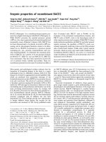
A schematic presentation of the two human pro-BACE2
variants, pro-BACE2-T1 (PB2-T1) and pro-BACE2-T2
(PB2-T2), is shown in Fig. 1, as compared to
pro-BACE1-T1 (PB1-T1) [10,11]. The cDNA of PB2-T1
and PB2-T2 was amplified from a human placenta cDNA
library (Clontech) using oligonucleotide primers: 5¢ primer,
5¢-GGATCCGCCGCCCCGGAGCTGGCCCCCGCGC
3¢;3¢ primer for T1, 5¢-GGATCCTCAGGGCTCGCTCAA
AGACTGAGCGGG-3¢;and3¢ primer for T2, 5¢-GGAT
CCTCAGCTCGCTGCGAAGCCCACCCTC-3¢. These
primers contain a BamHI site at the 5¢ end(shownin
italics). In addition, a stop codon was inserted prior to the
BamHI site in the 3¢ primers (shown in boldface). The PCR
products were cloned into the BamHI site of pET11a
(Novagen), resulting in pET11-PB2-T1 and pET11-PB2-T2.
A schematic presentation of the resulting expressed proteins
is shown in Fig. 1. Expression, inclusion body isolation,
refolding, and purification of BACE2 are described below.
E. coli BL21 (DE3) cells transformed with the expression
vector (pET11-PB2-T1 or pET11-PB2-T2) were grown in
Luria–Bertani broth and induced by the addition of
isopropyl-b-
D
-thiogalactopyranoside (final concentration,
1m
M
) for inclusion body production. The inclusion body
was dissolved in 50 mL of a denaturation buffer (8
M
urea,
1m
M
glycine, 0.1 m
M
EDTA, 10 m
M
b-mercaptoethanol,
10 m
M
dithiothreitol, 1 m
M
reduced glutathione, 0.1 m
M
oxidized glutathione, 20 m
M
Tris/HCl, pH 10.5) to a protein
concentration of 1.2 mgÆmL
)1
. The denatured proteins
were refolded in 10 vols 20 m
M
Tris base using a rapid
dilution method [10,28], followed by adjusting the pH to 8.0.
The refolded protein was concentrated by ultrafiltration,
and further purified by two steps of chromatography on
columns of Sephacryl S-300 (5 · 100 cm, Amersham Phar-
macia Biotech) and Resource-Q (1.6 · 3 cm, prepacked,
Amersham Pharmacia Biotech). The enzyme fractions
obtained from the last column were pooled, concentrated
by ultrafiltration, and used for further experiments.
Activity assay and kinetics measurement of pro-BACE2
To rudimentarily identify the substrate specificity of the
purified PB2-T1 and PB2-T2, each enzyme sample was
incubated separately with different polypeptide substrates
(40 lg) in 40 lL of a reaction mixture containing 50 m
M
sodium phosphate buffer (pH 6.5) at 37 °Cfor2or20h.
Some of the peptide substrates were custom synthesized by a
commercial source (Research Genetics; Huntsville, AL,
USA), and the remainder were purchased (Sigma). The 11
Fig. 1. Schematic diagram of the primary
structures of pro-BACE1-T1 (PB1-T1), pro-
BACE2-T1 (PB2-T1), and pro-BACE2-T2
(PB2-T2). The primary structure of each of
these enzymes consists sequentially of a T7 tag
sequence, a pro, and a mature protease
domain (with or without the C-terminal
extension). Two active-site aspartic acids in
D(T/S)G motifs (D-93/289 for BACE1 and
D-110/303 for BACE2) are marked. The
cysteine residues and possible disulfide bonds
are labeled. Open circles indicate possible free
cysteine residues in PB2-T2.
Ó FEBS 2002 Enzymatic properties of BACE2 (Eur. J. Biochem. 269) 5669
polypeptides are as follows (sequences shown in Table 1):
NCH-c, c-secretase cleavage site of notch [29]; APP-a,
a-secretase cleavage site of APP; APP-b, b-secretase clea-
vage site of APP; swAPP-b, b-secretase cleavage site of
Swedish APP; APP-c, c-secretase cleavage site of APP;
ENK-1, preproenkephalin fragment 129–138 peptide; insu-
lin B chain (Sigma, I6383); kinetensin (Sigma, K1879);
mastoparan (Sigma, M3545); neuropeptide (Sigma,
M0421); and preproenkephalin fragment 128–140 (Sigma,
P7162). The peptide fragments produced from the enzy-
matic reaction were separated by HPLC using a Magic 2002
system (Michrom BioResources, Inc., Aubum, CA, USA)
and a Magic C18 reverse-phase column (1.0 · 150 mm).
Elution was performed with a gradient from 5% acetonitrile
in 0.06% trifluoroacetic acid to 95% acetonitrile in 0.08%
trifluoroacetic acid and monitored at 215 nm. The incuba-
ted samples were also subjected to HPLC/MS (LC/MS,
Molecular Biology Resource Facility, University of Okla-
homa Medical Center) to identify the hydrolytic products
(average error in mass determination was 0.02%). For LC/
MS analysis, the HPLC effluent was fed into the electro-
spray ion source of the mass spectrometer at 40 lLÆmin
)1
.A
Sciex QSTAR hybrid quadruple time-of-flight mass spec-
trometer (Applied Biosystems-Sciex, Inc.) was used to
produce positive ions from a pneumatically assisted elec-
trospray interface. Sample ions were analyzed over the mass
range of 300–3000 amu. The two BACE2 variants were also
incubated with different proteins (40 lg) in 40 lLofa
reaction mixture containing 50 m
M
sodium phosphate
buffer (pH 6.5) at 37 °C for 4 h. The proteins (Sigma) used
were as follows: human serum albumin, cytochrome C,
lysozyme, alcohol dehydrogenase, b-amylase, and carbonic
anhydrase. The reaction mixtures were run in 20% SDS/
PAGE under reducing conditions for identification of the
possible hydrolytic products.
Kinetic parameters (K
m
and V
max
)ofPB2-T1were
routinely determined using the NCH-c peptide as a
substrate. In a typical assay, the reaction was carried out
at 37 °C for 5–30 min in a 40-lL reaction mixture
containing 50 m
M
sodium phosphate buffer (pH 6.5), and
0.8 m
M
substrate with an enzyme concentration of 6.26 l
M
.
The reaction was initiated by the addition of substrate at
concentrations varying in the range of 0.1–2 m
M
,andwas
terminated with 40 lL 2% trifluoroacetic acid. The reaction
5670 Y T. Kim et al. (Eur. J. Biochem. 269) Ó FEBS 2002
mixture was analyzed by HPLC as described above. The
kinetic parameters were obtained from the fitting of the data
using nonlinear regression analysis software GraFit [30].
The protein concentration was estimated colorimetrically
with a protein assay kit (Bio-Rad) using BSA as standard.
Activation of pro-BACE2 by BACE1
To identify the interaction between BACE1 and BACE2,
PB2-T1 was incubated with PB1-T1. The reaction was
carried out at 37 °Cfor60minin50m
M
Tris/BisTris/
sodium acetate/Caps buffer pH 4.5–12 and the aliquots
were applied to a 10% tricine/SDS gel (Novex). The gel
bands produced from the reaction were transferred to a
PVDF membrane and the N-terminal sequence was
analyzed by using automated Edman degradation.
Determination of enzymatic properties
The pH dependencies of PB2-T1 activity toward two
synthetic peptide substrates (NCH-c and ENK-1) were
determined in 50 m
M
sodium acetate (pH 3.0–5.0), 50 m
M
sodium phosphate (pH 5.5–6.5), 50 m
M
Tris/HCl (pH 7.0–
9.0), 50 m
M
Caps/NaOH (pH 9.5–10.5), and 50 m
M
Na
2
HPO
4
/NaOH (pH 11.0–13.0). To investigate the pH
stability, the enzymes were preincubated for 2 h at 25 °Cin
the buffers listed above. The pH of the mixture was adjusted
to 10.0 by the addition of 0.6 vol 0.5
M
Caps/NaOH
(pH 10.0) or 0.1
M
NaOH, and then the enzymatic activity
with NCH-c was determined as described above. To test the
effects of different protease inhibitors, the enzyme solution
containing each inhibitor was preincubated in 50 m
M
sodium
phosphate (pH 6.5) and 50 m
M
Caps/NaOH (pH 10.0) at
37 °C for 10 min, respectively, then assayed using NCH-c as
a substrate. The following inhibitors were tested: 0.1 m
M
antipain, 0.1 m
M
chymostatin, 0.1 m
M
E-64, 0.1 m
M
leu-
peptin, 0.5 m
M
pepstatin, 0.2 m
M
phosphoramidon, 1.0 m
M
pefabloc SC, 10 m
M
EDTA, and 0.01 m
M
aprotinin.
CD spectroscopic study on the thermal stability
of pro-BACE2
CD measurements of PB2-T1 and PB2-T2 at different
temperatures were performed using a JASCO 715 spectro-
polarimeter equipped with a Peltier temperature control
accessory PTC348WI. The temperature scans of the molar
ellipticity were recorded using an optical cell with a 0.1-cm
pathlength for the far-UV region and performed at a rate of
30 °CÆh
)1
. The protein concentrations of PB2-T1 and PB2-
T2 were 23.1 l
M
and 29.7 l
M
, respectively.
RESULTS
Cloning, expression, purification, and activity
of pro-BACE2 variants
Two designed E. coli expression constructs of pro-BACE2,
named pro-BACE2-T1 (PB2-T1) and pro-BACE2-T2
(PB2-T2) are shown in Fig. 1, as compared with pro-
BACE1-T1 (PB1-T1) [10,11]. PB2-T1 was constructed
based on the sequence homology between BACE2 and
BACE1 (PB1-T1) of which a crystal structure has been
recently solved [11]. PB2-T2 was constructed based on the
sequence homology with the pepsin catalytic domain. Both
variant forms of the enzyme were expressed in E. coli BL21
(DE3), then refolded in vitro as described in ÔExperimental
proceduresÕ. The enzymes were purified by consecutive
column chromatographic procedures using Sephacryl S-300
and Resource-Q (data not shown), and gave a single band
on SDS/PAGE (Fig. 2A). Although two free cysteines,
Cys233 and Cys292, exist in PB2-T2 based on sequence
homology (Fig. 1), no intermolecular disulfide bond was
found, as demonstrated by the nonreducing SDS/PAGE
(Fig. 2A). The molecular masses of recombinant PB2-T1
and PB2-T2 were estimated to be 49 183 and 45 747 Da,
respectively, by MALDI-TOF MS (data not shown). These
molecular masses are consistent with the molecular mass
calculated from the deduced amino acid sequences for PB2-
T1 (49 173) and PB2-T2 (45 756), with the standard error of
the MS at 0.02%. The N-terminal sequences of the
recombinant proteins were determined to be Ala-Ser-Met-
Thr-Gly, consistent with the designed sequence. The
enzymatic activities of PB2-T1 and PB2-T2 were determined
using a synthetic peptide substrate, NCH-c (Fig. 2B). The
specific activity of PB2-T1 enzyme was 15 120 (pmolÆ
min
)1
Æmg
)1
). In contrast, the PB2-T2 enzyme exhibited
activity that was only 17% of that of PB2-T1. These results
show that the refolded and purified pro-BACE2 enzymes
(PB2-T1 and PB2-T2) are active in hydrolyzing a synthetic
peptide, NCH-c.
Fig. 2. SDS/PAGE and activities of the puri-
fied PB2-T1 and PB2-T2. (A) SDS/PAGE of
the purified PB2-T1 and PB2-T2. SDS/PAGE
(12.5%) was run under nonreducing condi-
tions followed by Coomassie brilliant blue
staining. Protein standards are shown on the
left. (B) Specific activities of PB2-T1 and PB2-
T2. The enzyme activity was determined in
50 m
M
sodium phosphate buffer (pH 6.5)
with 0.8 m
M
NCH-c at 37 °Cfor30minas
described in ÔExperimental proceduresÕ.
Ó FEBS 2002 Enzymatic properties of BACE2 (Eur. J. Biochem. 269) 5671
Processing of BACE2 propeptide by BACE1
To test whether PB2-T1 can auto-activate either intra or
intermolecularly, the zymogen was incubated under various
conditions, including different pH, buffers, and tempera-
tures. The pH range used was from 4.5 to 12.0, the
incubation time used was 2 or 20 h, and the temperature
was 25 and 37 °C. Auto-activation was not observed under
any of the conditions tested (Fig. 3A). To clarify the
relationship between BACE1 and BACE2 and to study
their possible interactions, PB2-T1 was incubated with PB1-
T1 [10]. Under experimental conditions, pro-BACE2 (PB2-
T1) could be ÔactivatedÕ by BACE1 (PB1-T1), while BACE2
did not activate pro-BACE1 (Fig. 3B and C). The
N-terminal sequence of the lower band in the gel shown
in Fig. 3B (left lane, pH 4.5 and 6.0) contained the sequence
Ala-Leu-Glu-Pro-Ala as the first five amino acid residues,
which is the N-terminal sequence of mature BACE2
observed in vivo [24]. Therefore, these results indicate that
BACE1 is capable of activating pro-BACE2 by removing its
pro-peptide.
pH Dependency and stability
The pH dependence of the PB2-T1 activity toward a
synthetic substrate (NCH-c) is shown in Fig. 4A. PB2-T1
was active over a broad pH range, from 6.0 to 11.0, with
maximum activity at pH 9.5. PB2-T2 was also active in
the same range with maximum activity at pH 9.0–10.0
(data not shown). To confirm whether the pH dependence
of PB2-T1 activity could be changed depending on the
substrate used, the pH dependence of PB2-T1 was also
determined using a different substrate (ENK-1). The
optimum pH of the enzyme using ENK-1 substrate was
6.0 (Fig. 4B), closer to a ÔnormalÕ aspartic protease. These
results show that the pH dependence of PB2-T1 activity
varied depending on the substrate. To investigate the
stability of BACE2 at different pH levels, PB2-T1 and
PB2-T2 were preincubated at various pHs before the
activity was measured. As shown in Fig. 4C, PB2-T1
retained > 80% of the maximum activity after preincu-
bation in the buffers at pH between 4 and 12. The pH
stability of PB2-T2 is similar to that of PB2-T1 (data not
shown). These data show that BACE2 is a new type of
aspartic protease in spite of the conservation of two
active-site aspartic acid residues in D(T/S)G motifs and
the high degree of homology to BACE1 [10].
Thermostability of the secondary structure of BACE2
In PB2-T2, the C-terminal ÔextensionÕ of the protease
domain of BACE2 was deleted, resulting in potential
disruption of two disulfide bonds (Fig. 1). Therefore, the
structure of PB2-T2 may be less stable than that of PB2-T1.
To assay the structural stability, a CD spectropolarimeter
was used to monitor the secondary structure of the proteins
at increasing temperatures. The thermal unfolding of PB2-
T1 and PB2-T2, measured by the changes in ellipticity at
215 nm, is shown in Fig. 5. The figure shows that the major
transition of the secondary structure of PB2-T1 occurs
between 90 and 120 °C, while that of PB2-T2 occurs
between 50 and 80 °C. The secondary structure of PB2-T2
was completely denaturated at temperatures over 80 °C.
However, even at 120 °C, PB2-T1 exhibits 50% of the
far-UV ellipticity of the native enzyme. These results
indicate that the secondary structure of PB2-T1 is unusually
stable, while that of PB2-T2 is considerably less stable.
Possible inhibition of BACE2 by different protease
inhibitors and metal ions
Using NCH-c as a substrate, the possible inhibitory effects
of different protease inhibitors and metal ions were tested
on PB2-T1. The potential inhibitors are listed in Experi-
mental procedures. None of the protease inhibitors tested,
including a high concentration of pepstatin, had any
significant inhibitory effect toward BACE2 (data not
shown). These results are consistent with similar experi-
ments on BACE1 [10]. Two metal ions (Cu
2+
and Zn
2+
),
however, did inhibit PB2-T1 significantly (> 70% inhibi-
tion) at 1 m
M
concentration. It was previously shown that
the inhibition of proteolytic activity by metal ions could be
nonspecific. For example, E. coli leader peptidase is inhib-
ited nonspecifically by Hg
2+
and Cu
2+
ions (60% inhibition
Fig. 3. Processing of pro-BACE2 (PB2-T1) by
BACE1 (PB1-T1). PB2-T1, PB2-T1/PB1-T1,
and PB1-T1 were incubated in 50 m
M
Tris/
BisTris/sodium acetate/Caps buffer (pH 4.5,
6.0, 8.0, 10.0, and 12.0) at 37 °C for 60 min,
respectively. The reaction mixtures were
separated by SDS/PAGE (12.5%) under
reducing conditions. The arrowheads indicate
pro-BACE2-T1 (PB2-T1), pro-BACE1-T1
(PB1-T1), and the mature form of BACE2-T1
(B2-T1). (A) SDS/PAGE of PB2-T1. (B) SDS/
PAGE of PB2-T1/PB1-T1. (C) SDS/PAGE of
PB1-T1.
5672 Y T. Kim et al. (Eur. J. Biochem. 269) Ó FEBS 2002
[31]); an endoprotease from porcine antral mucosal mem-
branes is inhibited by Fe
2+
,Cu
2+
,Zn
2+
,andHg
2+
ions
(100% inhibition [32]), among others [33,34]. Therefore, it is
speculated that the inhibition of BACE2 by the metal ions is
also nonspecific.
Activity and specificity of PB2-T1 and PB2-T2
toward NCH-c
The specificities of PB2-T1 and PB2-T2 towards NCH-c
were measured. The two variants of pro-BACE2 clearly had
different substrate specificities. In this case, PB2-T1 pre-
ferred to cleave between Leu and Ser with a minor cleavage
site between Ser and Arg, while PB2-T2 preferred to cleave
between Ser and Arg with a minor cleavage site between
Leu and Ser (Table 1). These results suggest that the
BACE2 variants have at least two different substrate
specificities. The steady-state enzyme kinetics of PB2-T1
toward substrate NCH-c was also determined (data not
shown). Under the experimental conditions, the processing
site of the substrate was mainly VGSGVLL/SRK, and the
Ser–Arg processing site was insignificant. Therefore, only a
single processing site was measured in the kinetic experi-
ments. The kinetic parameters of PB2-T1 toward the NCH-
c substrates are: K
m
¼ 0.2 m
M
,andV
max
¼ 0.054 l
M
Æs
)1
.
Activity of PB2-T1 and PB2-T2 toward various peptide
and protein substrates
Because BACE2 is highly homologous to BACE1, the
enzymatic activity of PB2-T1 and PB2-T2 toward various
peptide substrates designed according to the a-, b-, and
c-secretase cleavage site of APP was investigated. The
substrate cleavage was assayed and quantified by HPLC
and HPLC/MS. In addition, due to the initial discovery
that PB2-T2 cut at the N-terminal site of the paired basic
residues in NCH-c, some specific peptides derived from
enzyme processing sites of pro-hormones were also tested.
Table 1 summarizes the results of the specificity of PB2-T1
and PB2-T2 toward some of the peptides used. The table
shows that recombinant pro-BACE2 cleaves at b-secretase
recognition site (M/D and L/D, b-secretase recognition
site of APP and Swedish mutation APP, respectively) of
both APP-b and swAPP-b. However, APP-c substrate is
not cleaved by the pro-BACE2 variants under the
experimental conditions used. These results indicate that
recombinant BACE2 exhibits the same activity as that of
b-secretase (BACE1), although the cleavage rate of the b-
secretase recognition site by the enzyme is low. PB2-T1
and PB2-T2 cleaved several positions of kinetensin,
mastoparan, neuropeptide, and preproenkephalin frag-
ment 128–140 at a significant rate. The APP-a,ENK-1
and oxidized insulin B chain were also hydrolyzed at
several sites with poor cleavage rate. These results show
that PB2-T1 demonstrates broad substrate specificities,
preferring bulky residues at the P1 site, and various
residues at the P1¢ site. The substrate specificity of PB2-
T2, in contrast, seems more constrained, apparently
preferring small residues at the P1 site, and basic residues
Fig. 5. Thermostability of the secondary structure of PB2-T1 and PB2-
T2. CD spectropolarimeter was used to measure the thermo-
unfolding of the secondary structures. The ellipticities of PB2-T1 (solid
line) and PB2-T2 (dotted line) were monitored at 215 nm in 20 m
M
Tris/HCl, pH 8.0, 0.4
M
urea.
Fig. 4. pH dependence and pH stability of the activity of PB2-T1. (A) pH dependence of PB2-T1 toward NCH-c. Assay of the enzyme activity was
carried out as described in ÔExperimental proceduresÕ, using a synthetic peptide substrate (NCH-c), except for the use of the following buffers:
50 m
M
sodium acetate (pH 3.0–5.0); 50 m
M
sodium phosphate (pH 5.5–6.5); 50 m
M
Tris/HCl (pH 7.0–9.0); 50 m
M
Caps/NaOH (pH 9.5–10.5);
and Na
2
HPO
4
/NaOH (pH 11.0–13.0). (B) pH dependence of PB2-T1 toward ENK-1. The enzyme assay was carried out as described in
ÔExperimental proceduresÕ with the exception of the above buffers. (C) pH stability of PB2-T1. The enzyme was preincubated for 2 h at 25 °Cinthe
same buffers used for the pH dependence study. Then, the pH of each preincubation mixture was adjusted to 10.0 by the addition of 0.6 vol. 0.5
M
Caps/NaOH (pH 10.0) or 0.1
M
NaOH, and the enzyme activity was determined.
Ó FEBS 2002 Enzymatic properties of BACE2 (Eur. J. Biochem. 269) 5673
at P1¢ and P2¢ sites. These results show that the substrate
specificity of PB2-T1 is different from that of PB2-T2
(Table 1). Thus, the C-terminal extension domain of
BACE2 (Pro432–Pro466) may affect the substrate speci-
ficity of the enzyme.
To explore further the substrate specificity of PB2-T1
and PB2-T2 toward intact protein substrates, some
commercially available proteins, which include human
serum albumin, cytochrome C, lysozyme, alcohol dehy-
drogenase, b-amylase and carbonate anhydrase, were used
in the activity assays. The substrate proteins were incubated
with PB2-T1 in a 1 : 10 enzyme/substrate weight ratio and
various reaction conditions were as follows: the pH range
used was 4.5–12.0, the temperature was 25 and 37 °C, and
the incubation time was 2 or 20 h. None of the above
proteins were processed by PB2-T1 (data not shown). These
results suggest that BACE2 is different from general
purpose aspartic proteases, such as pepsin, but similar to
BACE1, which has also been shown to lack the ability to
process native protein substrates in vitro [10].
DISCUSSION
BACE2 is a newly identified human aspartic protease. To
study its biochemical properties and possible biological
functions, two variants of pro-BACE2, PB2-T1 and PB2-
T2, have been constructed, expressed in E. coli,and
purified. PB2-T1 consists of the pro and protease domains,
similar to a pro-BACE1 variant, PB1-T1, for which a high-
resolution crystal structure has been determined [11]. The
other variant, PB2-T2, is a truncated version of PB2-T1 as
illustrated in Fig. 1. Its protease domain is terminated at the
C-terminal position of homologous pepsin, and is 34-
residues shorter at the C terminus than PB2-T1. Although
the primary structures of the enzymes are in pro-forms, both
PB2-T1 and PB2-T2 have apparent enzymatic activity
consistent with enzymatically active pro-BACE1 (PB1-T1)
[10], indicating that the conformation of the pro-domain of
BACE2 is flexible and that an equilibrium exists under the
reaction conditions between an ÔopenÕ, or active conforma-
tion, and a ÔclosedÕ, or inactive conformation [35].
The activation of most mammalian aspartic proteases is
brought about by removal of the pro-peptide by either auto-
activation or other proteolytic enzymes [17,36]. We showed
here that PB2-T1 does not auto-activate in the wide ranges
of pH, temperature and buffer conditions tested. We started
the experiment with the following intriguing facts in mind.
First, it has been shown that pro-BACE1, which is highly
homologous to pro-BACE2, can be Ôauto-activatedÕ in
acidic conditions [10], although the cleavage site in such
activation is different from that of the in vivo activation site.
In fact, the in vivo pro-BACE1 processing is catalyzed by
furin or related enzymes that recognize basic residues at the
cleavage site [19–21]. Since BACE2 often cleaves at basic or
paired basic residues (Table 1), it was interesting to test
whether BACE2 is able to activate BACE1. Second, cell
culture experiments [24] showed that a mature BACE2
protein starts from residue Ala63, suggesting its in vivo
activation site is the peptide bond between Leu62 and
Ala63. As there is no basic amino acid residue at, or near,
this activation site, it is unlikely that pro-BACE2 is also
activated by furin or related enzymes. Third, we found in
previous experiments that one cleavage site preferred by
BACE1 is between Leu and Ala (data not shown). The
results presented here demonstrate that under the experi-
mental conditions used, BACE2 cannot activate pro-
BACE1 (Fig. 3B), while pro-BACE2 can be activated by
BACE1 at the in vivo maturation position. These results
raise an interesting possibility that BACE1 may be one of
the physiological enzymes activating BACE2. Although we
have shown that both BACE1 [10] and BACE2 (this paper)
cleaves various peptide substrates in vitro, it remains to be
demonstrated that protein substrates can be processed
under similar conditions. To date, the only confirmed
cleavage site of protein substrate for BACE1 is the
b-secretase site of APP or related mutants. Thus PB2-T1
becomes the second protein substrate in this list. It has been
suggested [21] that the pro-peptide of BACE1 is not
evolutionarily developed for the regulation of enzyme
activity, as some other zymogens are [36], but to facilitate
protein folding. Whether the in vivo activity of pro-BACE2
requires preactivation remains the subject of further inves-
tigation. Nevertheless, both BACE1 and BACE2 are
activated in vivo, leaving a defined N terminus of the
mature enzyme [7,8,24]. Thus, the possibility still exists that
zymogen activation of BACE1 and BACE2 may be a means
of regulating their enzymatic activities under an in vivo
condition. Our results apparently contradict recent reports
from other laboratories [37,38], which show that mamma-
lian and insect cell expressed fusion protein BACE2 can self-
activate under acidic conditions. This contradiction may be
due to the different expression systems used. In the case of
BACE1, the rate of substrate turnover (k
cat
/K
m
)ofBACE1
expressed in insect cells is 10-fold higher than that of the
enzyme expressed in E. coli [39]. Furthermore, it has been
shown that glycosylation of BACE1 influences the proteo-
lytic activity and ensures optimum interaction between
BACE1 and a substrate [40]. Therefore, unglycosylated
BACE2 expressed by E. coli may exhibit different activity
from those expressed in mammalian or insect cell lines.
A ÔtypicalÕ aspartic protease is active at acidic pH between
2 and 5 [17]. For example, pepsin has an optimum pH of
near 2.0 [17], gastricsin at pH 3.0 [41], cathepsin D at
pH 3.5–5.0 [42], yapsin at pH 4.5 [43], and BACE1 at
pH 4.0 (recombinant BACE1 from E. coli) [10], or 4.5
(recombinant BACE1 from mammalian or insect cells)
[7,39]. Thus it is surprising to find that the activity of
BACE2 continuously rose with increasing pH up to pH 9.5
when NCH-c was used as a substrate (Fig. 4A). In this
work, a synthetic substrate, NCH-c (Val-Gly-Ser-Gly-Val-
Leu-Leu-Ser-Arg-Lys), was mainly used for the activity
assay. The substrate has a Lys residue (P2¢/P3¢ site)attheC
terminus, which may influence the pH-dependent activity
for this particular substrate. The pK of the e-amino group in
the Lys side chain is close to the pH optimum of the enzyme
activity. Thus it is probable that the enzyme prefers the
deprotonated-Lys form (free base) of the substrate. Com-
pared with the BACE1 substrate binding pockets, S4–S4¢
[11], BACE2 contains the following nonconservative muta-
tions in its substrate binding cleft: Arg307 fi GlninS4,
Gln12 fi Arg in S3, Pro70 fi Lys in both S2¢ and S3¢,and
Glu125 fi Thr in P4¢. The + 2 net charge increase in S2¢–
S4¢ pockets in neutral conditions may provide an explan-
ation for the observation that the optimum enzymatic
activity towards substrate NCH-c shifts to a more basic pH
region relative to other substrates. To demonstrate this
5674 Y T. Kim et al. (Eur. J. Biochem. 269) Ó FEBS 2002
point, a different peptide substrate was used for measuring
the pH dependent activity. The result showed that the
optimum pH of PB2–T1 using ENK-1 (Fig. 4B) was at
pH 6.0. Some results differ from those using purified
BACE2 from different expression systems or using different
substrates [37,38]. It seems that the precise optimum pH of
BACE2 varies depending on substrates, buffers, expression
systems (E. coli, insect cell line, and mammalian cell line),
and expression vector construction (full-length form, trun-
cated form, and full-length/T7 or His tag form). Further-
more, there exist several other examples of aspartic
proteases that are enzymatically active at neutral and
weakly alkaline pH as follows: renin has an optimum pH of
5.5–7.5 [44]; mouse submandibular renin at pH 6.5–8.3 [45];
and signal peptidase II at pH 7 [46].
BACE2 has a high degree of homology with BACE1,
with more than 50% amino acid sequence identity. All six
cysteine residues are conserved between BACE1 and
BACE2. Based on the crystal structure of BACE1 [11],
one can predict, with reasonable confidence, the three-
dimensional positions of most residues of BACE2, including
three disulfide bonds formed by the six cysteine residues
(Figs 1 and 6). Thus in BACE2, the three disulfide bonds
are assumed to be Cys233–Cys433, Cys292–Cys457, and
Cys344–Cys393. Such a disulfide bond pattern of BACE1
and BACE2 is distinctively different from, for example, that
observed in pepsin and cathepsin D [47]. Particularly, the
two disulfide bonds in the C-terminal subdomain, Cys233–
Cys433 and Cys292–Cys457, fasten the C-terminal peptide
to the main body of the catalytic unit (Fig. 6). Both disulfide
bonds as well as the C-terminal peptide are absent in pepsin
and other eukaryotic aspartic proteases. It suggests that the
catalytic domain of BACE2 may be tolerable to a trunca-
tion from the C terminus up to Ser432 without interfering
with the overall folding. The corresponding construct, PB2-
T2, is likely to result in two free cysteine residues, Cys233
and Cys292. Spatial positions of these two cysteine residues
in the homologous model (30 A
˚
for the C
233
a
–C
292
a
distance)
prohibit them from forming an intramolecular disulfide
bond, if the same overall folding of BACE1 is assumed for
BACE2. In addition, the fact that PB2-T2 shows a
monomeric molecular weight in nonreduced SDS/PAGE
(Fig. 2A) indicates that the refolding and purification
protocol used is sufficient to produce protein samples
without introducing intermolecular disulfide bonds, in spite
of the fact that both the free cysteine residues are probably
exposed to solvent.
The high primary sequence homology between BACE2
and BACE1 suggests that their soluble domains share
essentially the same three-dimensional structure. There are
only three deletions in the soluble domain of BACE2
relative to that of BACE1: a three-residue deletion around
residue 240, and two one-residue deletions around residues
390 and 455, respectively. All are located in the corres-
ponding variable loop regions in BACE1 as compared to
pepsin. These deletions in BACE2 change the loop length
only slightly, thus presumably perturbing the overall
structure very little. The C-terminal tail, which is unique
to BACE1 and BACE2, is located on the backside of the
catalytic domain from the active site, connecting the
catalytic domain to the transmembrane domain. The one-
residue deletion in the C-terminal loop region (around
residue 455) in BACE2 relative to BACE1 is unlikely to
affect the formation of the last putative disulfide bond
Fig. 6. Ribbon diagram of the BACE2 cata-
lytic domain. This BACE2 molecular model is
built based on the crystal structure of BACE1
and the primary sequence homology between
them. The view is of the opposite side from the
active site with the substrate binding cleft
roughly horizontal. The C-terminal tail is
shownindarkblue.Catalyticasparticresidues
are shown as yellow stick models. The three
disulfide bonds are shown as red stick models.
Regions containing insertion/deletion as
compared to BACE1 are colored orange. This
figure was produced with the program
MOLSCRIPT
[48].
Ó FEBS 2002 Enzymatic properties of BACE2 (Eur. J. Biochem. 269) 5675
(Cys292–Cys457). In addition to connecting the soluble
domain to the trans-membrane domain, the C-tail also
provides structural enforcement to the soluble domain
through the two disulfide bonds, and an extended b-sheet
and hydrophobic side chain interactions. Together, they are
believed to contribute significantly to the overall stability in
BACE1 [11]. The dramatic thermal stability difference
between PB2-T1 and PB2-T2 observed using CD spectro-
scopic method provides direct evidence supporting the same
notion in BACE2 (Fig. 5). On the other hand, our data
indicate that these structural enforcements are not essential
for the enzymatic activity of BACE2. Deletion of the C-tail
is tolerable for the enzyme activity, although some subtle
structural changes may occur that are associated with the
substrate specificity changes. Such structural integrity of the
soluble domain in the absence of the C-tail is consistent with
the high degree of homology in three-dimensional structures
between BACE1, BACE2 and pepsin, the latter of which
does not contain the C-tail. In addition to the overall
structural stability, the presence/absence of the C-tail
apparently affects the substrate specificity of the enzyme.
Indirectly, the rigidity associated with the C-tail, particularly
the two disulfide bonds, may keep the dynamic structure of
BACE2 in a more open form, thus making it more
accessible to different substrates. In a more direct way, the
loss of the disulfide bond Cys233–Cys433 may affect the
substrate binding at P4 position mediated through a b-turn
around residue 88. Similarly, the free C-terminal end of the
longer version of our BACE2 constructs may wrap around
the soluble domain and reach the putative S4¢ substrate
binding pocket in BACE2. The corresponding terminus in
BACE1 is mobile in the crystal structure [11] and likely to
become more fixed if it attaches to the trans-membrane
domain.
ACKNOWLEDGEMENTS
The authors thank K. Takahashi, School of Life Science, Tokyo
University of Pharmacy and Life Science, for helpful discussion of this
work; K. K. Rodgers, Department of Biochemistry and Molecular
Biology, University of Oklahoma Medical Center, for advice on CD
experiments; and K. Jackson and C. Batson, Molecular Biology
Resource Facility, Warren Medical Research Institute, University of
Oklahoma Medical Center for assistance with MS, amino acid analysis,
and N-terminal sequencing. This work is supported by the National
Institute of Health Grant RO1-AI46298 (to X. Lin).
REFERENCES
1. Selkoe, D.J. (1997) Alzheimer’s disease: genotypes, phenotypes,
and treatments. Science 275, 630–631.
2. Hardy, J. (1997) The Alzheimer family of diseases: Many ethio-
logies, one pathogenesis? Proc. Natl Acad. Sci. USA 94, 2095–2097.
3. Wolfe,M.S.,Xia,W.,Ostaszewski,B.L.,Diehl,T.S.,Kimberly,
W.T. & Selkoe, D.J. (1999) Two transmembrane aspartates in
presenilin-1 required for presenilin endoproteolysis and c-secretase
activity. Nature 398, 513–517.
4. Kimberly,W.T.,Xia,W.,Rahmati,T.,Wolfe,M.S.&Selkoe,
D.J. (2000) The transmembrane aspartates in presenilin 1 and 2
are obligatory for c-secretase activity & amyloid b-protein gen-
eration. J. Biol. Chem. 275, 3173–3178.
5. Sinha, S. & Lieberburg, I. (1999) Cellular mechanisms of b-amy-
loid production and secretion. Proc. Natl Acad. Sci. USA 96,
11049–11053.
6. Sinha, S., Anderson, J.P., Barbour, R., Basi, G.S., Caccavello, R.,
Davis,D.,Doan,M.,Dovey,H.F.,Frigon,N.,Hong,J.,Jacob-
son-Croak,K.,Jewett,N.,Keim,P.,Knops,J.,Lieberburg,I.,
Power,M.,Tan,H.,Tatsuno,G.,Tung,J.,Schenk,D.,Seubert,
P., Suomensaari, S.M., Wang, S., Walker, D., John, V., et al.
(1999) Purification and cloning of amyloid precursor protein
b-secretase from human brain. Nature 402, 537–540.
7. Vassar,R.,Bennett,B.D.,Babu-Khan,S.,Kahn,S.,Mendiaz,
E.A.,Denis,P.,Teplow,D.B.,Ross,S.,Amarante,P.,Loeloff,
R.,Luo,Y.,Fisher,S.,Fuller,J.,Edenson,S.,Lile,J.,
Jarosinski,M.A.,Biere,A.L.,Curran,E.,Burgess,T.,Louis,
J.C., Collins, F., Treanor, J., Rogers, G. & Citron, M. (1999)
b-Secretase cleavage of Alzheimer’s amyloid precursor protein by
the transmembrane aspartic protease BACE. Science 286,
735–741.
8. Yan,R.,Bienkowski,M.J.,Shuck,M.E.,Miao,H.,Tory,M.C.,
Pauley,A.M.,Brashier,J.R.,Stratman,N.C.,Mathews,W.R.,
Buhl, A.E., Carter, D.B., Tomasselli, A.G., Parodi, L.A.,
Heinrikson, R.L. & Gurney, M.E. (1999) Membrane-anchored
aspartyl protease with Alzheimer’s disease b-secretase activity.
Nature 402, 533–537.
9. Hussain, I., Powell, D., Howlett, D.R., Tew, D.G., Meek, T.D.,
Chapman,C.,Gloger,I.S.,Murphy,K.E.,Southan,C.D.,Ryan,
D.M.,Smith,T.S.,Simmons,D.L.,Walsh,F.S.,Dingwall,C.&
Christie, G. (1999) Identification of a novel aspartic protease
(Asp2) as b-secretase. Mol. Cell. Neurosci. 14, 419–427.
10. Lin,X.,Koelsch,G.,Wu,S.,Downs,D.,Dashti,A.&Tang,J.
(2000) Human aspartic protease memapsin 2 cleaves the b-secre-
tase site of b-amyloid precursor protein. Proc.NatlAcad.Sci.
USA 97, 1456–1460.
11. Hong,L.,Koelsch,G.,Lin,X.,Wu,S.,Terzyan,S.,Ghosh,A.K.,
Zhang, X.C. & Tang, J. (2000) Structure of the protease domain of
memapsin 2 (b-secretase) complexed with inhibitior. Science 290,
150–153.
12. Luo, Y., Bolon, B., Kahn, S., Bennett, B.D., Babu-Khan, S.,
Denis,P.,Fan,W.,Kha,H.,Zhang,J.,Gong,Y.,Martin,L.,
Louis, J.C., Yan, Q., Richards, W.G., Citron, M. & Vassar, R.
(2001) Mice deficient in BACE1, the Alzheimer’s beta-secretase,
have normal phenotype and abolished beta-amyloid generation.
Nature Neurosci. 4, 231–232.
13. Cai,H.,Wang,Y.,McCarthy,D.,Wen,H.,Borchelt,D.,Price,
D.L. & Wong, P.C. (2001) BACE1 is the major b-secretase for
generation of Ab peptides by neurons. Nature Neurosci. 4,233–
234.
14.Saunders,A.J.,Kim,T W.,Tanzi,R.E.,Fan,W.,Bennett,
B.D.,Babu-Kahn,S.,Luo,Y.,Louis,J C.,McCaleb,M.,
Citron, M., Vassar, R. & Richards, W.G. (1999) BACE maps to
chromosome 11 and a BACE homolog, BACE2, reside in the
obligate down syndrome region of chromosome 21. Science 286,
1255a–1255a.
15. Acquati,F.,Accarino,M.,Nucci,C.,Fumagalli,P.,Jovine,L.,
Ottolenghi, S. & Taramelli, R. (2000) The gene encoding DRAP
(BACE2), a glycosylated transmembrane protein of the aspartic
protease family, maps to the Down critical region. FEBS Lett. 468,
59–64.
16. Solans, A., Estivill, X. & de La Luna, S. (2000) A new aspartyl
protease on 21q22.3, BACE2, is highly similar to Alzheimer’s
amyloid precursor protein b-secretase. Cytogenet. Cell. Genet. 89,
177–184.
17. Tang, J. & Wong, R.N. (1987) Evolution in the structure and
function of aspartic proteases. J. Cell. Biochem. 33, 53–63.
18. Jutras, I. & Reudelhuber, T.L. (1999) Prorenin processing by
cathepsin B in vitro and in transfected cells. FEBS Lett. 443,48–
52.
19. Creemers, J.W., Dominguez, D.I., Plets, E., Serneels, L., Taylor,
N.A.,Multhaup,G.,Craessaerts,K.,Annaert,W.&De
Strooper, B. (2001) Processing of b-secretase by furin and other
5676 Y T. Kim et al. (Eur. J. Biochem. 269) Ó FEBS 2002
members of the proprotein convertase family. J. Biol. Chem. 276,
4211–4217.
20. Bennett,B.D.,Denis,P.,Haniu,M.,Teplow,D.B.,Kahn,S.,
Louis, J.C., Citron, M. & Vassar, R. (2000) A furin-like convertase
mediates propeptide cleavage of BACE, the Alzheimer’s b-secr-
etase. J. Biol. Chem. 275, 37712–37717.
21. Shi,X.P.,Chen,E.,Yin,K.C.,Na,S.,Garsky,V.M.,Lai,M.T.,
Li,Y.M.,Platchek,M.,Register,R.B.,Sardana,M.K.,Tang,
M.J., Thiebeau, J., Wood, T., Shafer, J.A. & Gardell, S.J. (2001)
The pro domain of b-secretase does not confer strict zymogen-like
properties but does assist proper folding of the protease domain.
J. Biol. Chem. 276, 10366–10373.
22. Tatnell,P.J.,Powell,D.J.,Hill,J.,Smith,T.S.,Tew,D.G.&Kay,
J. (1998) Napsins: new human aspartic proteinases. Distinction
between two closely related genes. FEBS Lett. 441, 43–48.
23. Chuman,Y.,Bergman,A.,Ueno,T.,Saito,S.,Sakaguchi,K.,
Alaiya,A.A.,Franzen,B.,Bergman,T.,Arnott,D.,Auer,G.,
Appella, E., Jornvall, H. & Linder, S. (1999) Napsin A, a member
of the aspartic protease family, is abundantly expressed in normal
lung and kidney tissue and is expressed in lung adenocarcinomas.
FEBS Lett. 462, 129–134.
24. Hussain,I.,Powell,D.J.,Howlett,D.R.,Chapman,G.A.,Gil-
mour, L., Murdock, P.R., Tew, D.G., Meek, T.D., Chapman,
C.,Schneider,K.,Ratcliffe,S.J.,Tattersall,D.,Testa,T.T.,
Southan,C.,Ryan,D.M.,Simmons,D.L.,Walsh,F.S.,Ding-
wall, C. & Christie, G. (2000) ASP1 (BACE2) cleaves the amy-
loid precursor protein at the b-secretase site. Mol. Cell. Neurosci.
16, 609–619.
25. Farzan, M., Schnitzler, C.E., Vasilieva, N., Leung, D. & Choe, H.
(2000) BACE2, a b-secretase homolog, cleaves at the b site and
within the amyloid-b region of the amyloid-b precursor protein.
Proc. Natl Acad. Sci. USA 97, 9712–9717.
26. Marcinkiewicz, M. & Seidah, N.G. (2000) Coordinated expression
of b-amyloid precursor protein and the putative b-secretase BACE
and a-secretase ADAM10 in mouse and human brain. J. Neuro-
chem. 75, 2133–2143.
27. Bennett, B.D., Babu-Khan, S., Loeloff, R., Louis, J.C., Curran,
E., Citron, M. & Vassar, R. (2000) Expression analysis of BACE2
in brain and peripheral tissues. J. Biol. Chem. 275, 20647–20651.
28. Lin, X., Lin, Y. & Tang, J. (1994) Relationships of human
immunodeficiency virus protease with eukaryotic aspartic
proteases. Methods Enzymol. 241, 195–224.
29. Schroeter, E.H., Kisslinger, J.A. & Kopan, R. (1998) Notch-1
signalling requires ligand-induced proteolytic release of
intracellular domain. Nature 393, 382–386.
30. Leatherbarrow, R.J. (1998) Grafit,Version4.0Staines,UK.
31. Kim, Y.T., Muramatsu, T. & Takahashi, K. (1995) Leader pepti-
dase from E. coli: Overexpression, characterization, and
inactivation by modification of tryptophan residues 300 and
310 with N-bromosuccinimide. J. Biochem. (Tokyo) 117, 535–544.
32. Jeohn, G.H. & Takahashi, K. (1995) Purification and character-
ization of a vasoactive intestinal polypeptide-degrading
endoprotease from porcine antral mucosal membranes. J. Biol.
Chem. 270, 7809–7815.
33. Lee,M.J.,Kang,B.S.,Kim,D.S.,Kim,Y.T.,Kim,S.K.,Chung,
K.H., Kim, J.K., Nam, K.S., Lee, Y.C. & Kim, C.H. (1997)
Processing of an intracellular immature pullulanase to the mature
form involves enzymatic activation and stabilization in alkaliphilic
Bacillus sp. S-1. J. Biochem. Mol. Biol. 30, 46–54.
34. Jeohn, G.H., Serizawa, S., Iwamatsu, A. & Takahashi, K. (1995)
Isolation and characterization of gastric trypsin from the micro-
somal fraction of porcine gastric antral mucosa. J. Biol. Chem.
270, 14748–14755.
35. Ermoliff, J., Loy, J.A., Koelsch, G. & Tang, J. (2000) Proteolytic
activation of recombinant pro-memapsin 2 (pro-b-secretase)
studied with new fluorogenic substrates. Biochemistry 39, 12450–
12456.
36. Khan, A.R., Khazanovich-Bernstein, N., Bergmann, E.M. &
James, M.N. (1999) Structural aspects of activation pathways of
aspartic protease zymogens and viral 3C protease precursors.
Proc. Natl Acad. Sci. USA 96, 10968–10975.
37. Hussain, I., Christie, G., Schneider, K., Moore, S. & Dingwall, C.
(2001) Prodomain processing of Asp1 (BACE2) is autocatalytic.
J. Biol. Chem. 276, 23322–23328.
38. Yan, R., Munzner, J.B., Shuck, M.E. & Bienkowski, M.J. (2001)
BACE2 functions as an alternative a-secretase in cells. J. Biol.
Chem. 276, 34019–34027.
39. Mallender,W.D.,Yager,D.,Onstead,L.,Nichols,M.R.,Eck-
man, C., Sambamurti, K., Kopcho, L.M., Marcinkeviciene, J.,
Copeland, R.A. & Rosenberry, T.L. (2001) Characterization of
recombinant, soluble b-secretase from an insect cell expression
system. Mol. Pharmacol. 59, 619–626.
40. Charlwood, J., Dingwall, C., Matico, R., Hussain, I., Johanson,
K., Moore, S., Powell, D.J., Skehel, J.M., Ratcliffe, S., Clarke, B.,
Trill, J., Sweitzer, S. & Camilleri, P. (2001) Characterization of the
glycosylation profiles of Alzheimer’s b-secretase protein Asp-2
expressed in a variety of cell lines. J. Biol. Chem. 276, 16739–
16748.
41. Tang, J., Wolf, S., Caputto, R. & Trucco, R.E. (1959) Isolation
and crystallization of gastricsin from human gastric juice. J. Biol.
Chem. 234, 1174–1178.
42. Barrett, A.J. (1970) Cathepsin D. Purification of isoenzymes from
human and chicken liver. Biochem. J. 117, 601–607.
43. Cawley, N.X., Wong, M., Pu, L.P., Tam, W. & Loh, Y.P. (1995)
Secretion of yeast aspartic protease 3 is regulated by its carboxy-
terminal tail: characterization of secreted YAP3p. Biochemistry 34,
7430–7437.
44. Pickens,P.T.,Bumpus,F.M.,Lloyd,A.M.,Smeby,R.R.&Page,
I.H. (1965) Measurement of renin activity in human plasma. Circ.
Res. 17, 438–448.
45. Figueiredo, A.F.S., Takii, Y., Tsuji, H., Kato, K. & Inagami, T.
(1983) Rat kidney renin and cathepsin D: purification and com-
parison of properties. Biochemistry 22, 5476–5481.
46. Dev, I.K. & Ray, P.H. (1984) Rapid assay and purification of a
unique signal peptidase that processes the prolipoprotein from
Escherichia coli B. J. Biol. Chem. 259, 11114–11120.
47. Haniu,M.,Denis,P.,Young,Y.,Mendiaz,E.A.,Fuller,J.,Hui,
J.O.,Bennett,B.D.,Kahn,S.,Ross,S.,Burgess,T.,Katta,V.,
Rogers, G., Vassar, R. & Citron, M. (2000) Characterization of
Alzheimer’s b-secretase protein BACE. J. Biol. Chem. 275, 21099–
21106.
48. Kraulis, P.J. (1991) Molscript: a program to produce both detailed
and schematic plots of protein structures. J. Appl. Crystallogr. 24,
946–950.
Ó FEBS 2002 Enzymatic properties of BACE2 (Eur. J. Biochem. 269) 5677