Tài liệu Báo cáo Y học: Kinetic properties of bifunctional 6-phosphofructo-2-kinase/ fructose-2,6-bisphosphatase from spinach leaves pdf
Bạn đang xem bản rút gọn của tài liệu. Xem và tải ngay bản đầy đủ của tài liệu tại đây (557.75 KB, 11 trang )
Kinetic properties of bifunctional 6-phosphofructo-2-kinase/
fructose-2,6-bisphosphatase from spinach leaves
Jonathan E. Markham* and Nicholas J. Kruger
Department of Plant Sciences, University of Oxford, South Parks Road, Oxford, OX1 3RB, UK
A cDNA encoding 6-phosphofructo-2-kinase/fructose-2,6-
bisphosphatase was isolated from a Spinacia oleracea leaf
library and used to express a recombinant enzyme in
Escherichia coli and Spodoptera frugiperda cells. The insol-
uble protein expressed in E. coli was purified and used to
raise an tibodies. Western blot analysis of a protein extract
from spinach leaf showed a single band of 90.8 kDa. Soluble
protein was purified to homogeneity from S. frugiperda cells
infected with recombinant baculovirus harboring the isola-
ted cDNA. The soluble protein had a molecular mass of
320 kDa, estimated by gel filtration chromatography,
and a subunit size of 90.8 kDa. The purified protein
had activity of both 6-phosphofructo-2-kinase (specific acti-
vity 10.4–15.9 nmolÆmin
)1
Æmg protein
)1
) and fructose-2,6-
bisphosphatase (specific activity 1.65–1.75 nmolÆmin
)1
Æmg
protein
)1
). The 6-phosphofructo-2-kinase activity was acti-
vated by inorganic phosphate, and inhibited by 3-carbon
phosphorylated metabolites and pyrophosphate. In the
presence of phosphate, 3-phosphoglycerate was a mixed
inhibitor with respect to both fructose 6-phosphate and
ATP. Fructose-2,6-bisphosphatase activity was sensitive to
product inhibition; inhibition by inorganic phosphate was
uncompetitive, w hereas inh ibition by fructose 6-phosphate
was mixed. T hese kinetic p roperties support the view that the
level of fructose 2,6-bisphosphate in leaves is determined b y
the relative concentrations of hexose phosphates, three-car-
bon phosphate esters and inorganic phosphate in the cytosol
through reciprocal modulation of 6-phosphofructo-2-kinase
and fructose-2,6-bisphosphatase activities of the bifunc-
tional e nzyme.
Keywords: fructose 2,6-bisphosphate; 6-phosphofructo-
2-kinase; fructose-2,6-bisphosphatase; spinach leaf; Spinacia
oleracea.
Fructose 2,6-bisphosphate (Fru-2,6-P
2
) is an important
regulator of photosynthetic carbon metabolism in higher
plants. It i s a poten t allosteric inhibitor of cytosolic fructose
1,6-bisphosphatase, which is respon sible f or the c onversion
of fructose 1,6-bisp hosphate to fructose 6-phosphate (Fru-
6-P) during formation of sucrose from triose phosphates [1].
In leaves, Fru-2,6-P
2
contributes both to the coordination of
sucrose synthesis with the rate of CO
2
fixation, and
indirectly to the control of assimilate partitioning between
sucrose and starch [1,2]. Direct evidence for the involvement
of Fru-2,6-P
2
in the regulation of these processes is provided
by studies of transgenic tobacco, kalanchoe
¨
and arabidopsis
in which changes in the rates o f sucrose and s tarch synthesis
correlated with changes in Fru-2,6-P
2
concentration when
the latter was modified by genetic manipulation [3–6].
However, any explanation o f how Fru-2,6- P
2
level serves to
integrate photoassimilatory carbon partitioning must
include a consideration of the factors that influence the
concentration of this signal metabolite.
In common with other eukaryotes, the level of Fru-2,6-P
2
in higher plants is determined by the relative activities of
6-phosphofructo-2-kinase (6PF2K) and fructose-2,6-bis-
phosphatase (Fru-2,6-P
2
ase), which catalyse its synthesis
and degradation, respectively [7]. In leaves, both activities
are subjected to reciprocal fine control by metabolic
intermediates of the pathway of sucrose synthesis; 6PF2K
activity is stimulated by Fru-6-P and P
i
, and inhibited by
three-carbon phosphate esters (including 3-phosphoglycer-
ate and dihydroxyacetone phosphate), whereas Fru-2,6-
P
2
ase activity is inhibited b y Fru-6-P and P
i
[1]. These
properties allow the level of Fru-2,6-P
2
to respond sensi-
tively to the availability of photosynthate and the accumu-
lation of sucrose (the major photosynthetic end product),
and provide the basis for a model describing the regulation
of sucrose synthesis in leaves in the light [2].
Although both 6PF2K and Fru-2,6-P
2
ase activities
have been measured in a range of plant tissues, detailed
kinetic analyses are largely restricted to the a ctivities from
spinach leaves [1]. Consequently it is these activities that
form the basis for our current understanding. However,
the extent to w hich the reported p roperties of spinach
6PF2K and Fru-2,6-P
2
ase activities reflect those of the
Correspondence to N. J. Kruger, Department of Plant Sciences,
University of Oxford, South Parks Road, Oxford, OX1 3RB, UK.
Fax: +44 1865 275074, Tel.: +44 1865 275000,
E-mail:
Abbreviations:Fru-2,6-P
2
, fructose 2,6-bisphosphate; Fru-2,6-P
2
ase,
fructose-2,6-bisphosphatase; Fru-6-P, fructose 6-phosphate; 6PF2K,
6-phosphofructo-2-kinase; PFP, pyrophosphate:fructose 6-phosphate
1-phosphotransferase.
Enzymes: 6-phosphofructo-2-kinase (EC 2.7.1.105); f ructose-2,6-bis-
phosphatase (fructose-2,6-bisphosphate 2-phosphatase, E C 3.1.3.46).
Note: The nu cleotide sequence for s pinach leaf 6PF2K/Fru-2,6-P
2
ase
cDNA described i n this paper is available from the EMBL sequence
database under accession n umber AF041848.
*Present addr ess: D epartment of Molecular B iology of Plants,
Research School GB B, University of Gro ningen, Haren, the
Netherlands.
(Received 26 July 2001, revised 14 December 2001, accepted 8 January
2002)
Eur. J. Biochem. 269, 1267–1277 (2002) Ó FEBS 2002
enzyme(s) in vivo is uncertain. Much of the initial
characterization of the activities was performed on
relatively crude preparations of the enz yme(s) in which
little e ffort was made to protect the sample f rom
proteolysis during isolation [8–10]. There has been only
one study in which a bifunctional enzyme has been
purified to near-homogeneity [11]. That report identified
two forms of the enzyme possessing both 6 PF2K and
Fru-2, 6-P
2
ase activity. The smaller
L
-form of the enzyme
(native M
r
132 000) consisted of a variable group of
catalytically active polypeptides with M
r
of 44 000–
70 000. Despite the presence of protease inhibitors, these
polypeptides are likely to have been generated during
extraction from the proteolytic degradation of a larger
H-form (native M
r
390 000, subunit M
r
90 000) [11,12].
The affinity of the 6PF2K activity of the smaller
L
-form
for its substrates and P
i
, an allosteric activator, was lower
than that of the corresponding activity of the larger
H-form of the enzyme, whereas the corresponding affinity
for i ts inhibitors was 10-fold greater [11]. Furthermore the
ratio of 6PF2K activity to Fru-2,6-P
2
ase activity of the
smaller form of the bifunctional e nzyme was far lower
than that of the larger form of the enzyme [11]. This is
reminiscent of the enzyme from rat liver in which partial
proteolysis destroyed 6PF2K activity while incre asing Fru-
2,6-P
2
ase activity [13]. Differences in the 6PF2K/Fru-2,6-
P
2
ase ratio are a common feature of isoforms of the
bifunctional enzyme from plants [11,12,14], suggesting
that such proteolysis may be a widespread problem. The
sensitivity of the plant bifunctional enzyme to degradation
by endogenous proteases during isolation, and the dem-
onstrable effects of proteolysis on the kinetic characteris-
tics of the component activities of the enzyme compromise
the evidence on which our current understanding of the
regulation of photosynthetic carbon partitioning is based.
Additionally, a monofunctional Fru-2,6-P
2
ase has been
purified from spinach leaves. This activity is specific for
hydrolysis of Fru-2,6-P
2
, and is inhibited by Fru-6-P and
P
i
, although the affinities for these inhibitors differ from
those of the Fru-2,6-P
2
ase activity of the bifunctional
enzyme. The protein has a native M
r
of 50 000–76 000
with a subunit M
r
of 33 000 [15]. The relationship
between this monofunctional Fru-2,6-P
2
ase and the
bifunctional enzyme is uncertain, and the role of the
monofunctional enzyme in Fru-2,6-P
2
ase metabolism has
yet to be resolved [15,16].
Recently cDNA clones encoding homologues of the
mammalian bifunctional enzyme have been isolated from
potato leaf [17] and arabidopsis hypocotyls [18]. The
deduced amino-acid sequence of both clones contain a
region in which about 40–50% of the residues a re identical
to those of the 400-residue Ôcatalytic coreÕ of the mamma-
lian, avian a nd yeast e nzymes [19]. When e xpressed in
E. coli , the proteins encoded by the two plant cDNA display
both 6PF2K and F ru-2,6-P
2
ase activities [17,18]. These
developments provide the opportunity to examine the
kinetic properties of plant 6PF2K/Fru-2,6-P
2
ase purified
from a h eterologous expression system, thus circumventing
problems associated with potential modification of the
enzyme by endogenous plant proteases during extraction.
Here we report o n the kinetic p roperties of a s pinach
bifunctional 6PF2K/Fru-2,6-P
2
ase produced in insect cells
using a baculovirus expression system.
EXPERIMENTAL PROCEDURES
Materials
Superscript Choice System for cDNA synthesis, TC100
medium, SF-900 II serum-free medium, fetal bovine s erum
and FastBac expression system were from Invitrogen Life
Technologies (Paisley, UK). G enescreen Plus membrane
and [a-
32
P]dCTP were from NEN Life Science Products
(Hounslow, Middlesex, UK), and restriction enzymes were
from New England Biolabs (Hitchin, Herts, UK). Chro-
matography media a nd columns were from Amersham
Biosciences (Little Chalfont, Bucks, UK). Pyrophos-
phate:fructose 6-phosphate 1-phosphotransferase (PFP)
was purified from mature tubers of potato (Solanum
tuberosum), as described p reviously [20]. Other coupling
enzymes and Triton X-100 were supplied by Roche
Diagnostics (Lewes, East Sussex, UK). Phenol was from
Qbiogene (Harefield, Middlesex, UK) and all other chem-
icals were from Sigma-Aldrich or Merck (both of Poole,
Dorset, UK).
CDNA library construction
Total RNA was isolated from recently expanded mature
leaves of Spinacia oleracea, as described previously [21].
PolyA
+
RNA was purified using the Oligotex purification
system (Qiagen, Crawley, West Sussex, UK), a nd 3 lgwas
used for cDNA synthesis using oligo dT
(n)
primers. Size
selected cDNA (>1kbp) was cloned into EcoRI-digested
lambda ZAP II (Stratagene, A msterdam, the Netherlands).
The host bacterial strain was XL1-Blue (Stratagene).
Northern analysis
Approximately 20 lg total RNA were separated in 1.4%
agarose gels containing 6.3% formaldehyde and transferred
by capillary action to Hybond-N membrane (Amersham
Biosciences).
Southern analysis
Genomic DNA was isolated f rom mature spinach leaves by
the CTAB extraction procedure [22]. DNA was digested
with restriction enzymes (10 U Ælg
)1
DNA) in buffer sup-
plied by the manufacturer for 24 h . The DNA fragments
were separated on a 0.8% agarose gel and transferred to
Hybond-N membrane by capillary transfer.
Probe labelling and hybridization
DNA probes for both Southern and Northern analysis were
labelled with [a-
32
P]dCTP using Ready-to-Go labeling
reactions and separated from unincorporated nucleotides
through ProbeQuant G-50 Micro-columns (Amersham
Biosciences). The complete cDNA sequence was used as
template for probe synthesis. Membranes were hybridized
in ExpressHyb hybridization solution (Clontech, Basing-
stoke, Hampshire, UK), according to the manufacturer’s
instructions. Following hybridization with the probe,
membran es were rinse d in 2 · NaCl/Cit/0.5% SDS at
room temperature and then washed twice in 0.2 · NaCl/
Cit/0.1% SDS at 42 °C, each time for 30 min.
1268 J. E. Markham and N. J. Kruger (Eur. J. Biochem. 269) Ó FEBS 2002
Sequencing and sequence analysis
DNA sequences were determined by cycle sequencing using
an ABI Prism automated sequencer (Applied Biosystems
Inc, Warrington, Cheshire, UK) at the Durham University
Sequencing Service and D epartment o f Pathology, Univer-
sity of Oxford, UK. Sequence data were processed using
DNASTRIDER
and
GCG
computer programmes.
Preparation of antibodies
The coding region from the 6PF2K/Fru-2,6-P
2
ase cDNA
was amplified from the lambda ZAP II-derived clone by
PCR using the primer 5¢-TTAGGAGAGAGACAT
ATGGG-3¢ and the M13 reverse primer. The amplified
fragment was cloned in-frame into pET 30 expression vector
(Invitrogen Life Technologies) using NdeIandNotI r estric-
tion sites and transformed into E. coli strain BL21(kDE3).
Protein expression was induced in cells growing logarith-
mically in terrific broth [23] at 37 °C by adding isopropyl
thio-b-
D
-galactoside at a fi nal concentration of 1 m
M
.
Bacteria we re harvested, lysed and the i nclusion bodies
were isolated by centrifugation [23].
Approximately 75 mg of insoluble protein derived from
inclusion bodies were fractionated by continuous-elution
SDS/PAGE on a 35 · 100 mm 7% acrylamide gel using a
Model 491 Prep Cell (Bio-Rad, Hemel Hempsted, Herts,
UK), according to the manufacturer’s instructions. Frac-
tions containing the pure recombinant protein (M
r
%
90 800) were identified by analytical SDS/PAGE and the
protein recovered from the pooled fractions by methanol/
chloroform precipitation. The protein was redissolved in
1mL6
M
guanidium/HCl and dialysed exhaustively against
NaCl/P
i
. The resulting protein suspension was used t o raise
polyclonal antibodies in New Z ealand white r abbits at
Harlan Sera Laboratories (Loughborough, Leics, UK).
PAGE and immunoblotting
Analytical SDS/PAGE was p erformed using a P hastgel
system (Amersham Biosciences) run according to the
manufacturer’s recommended conditions. For immuno-
chemical analysis, protein was transferred onto a poly(vinyl-
idene difluoride) membrane (Millipore, Watford, Herts,
UK) and probed with rabbit anti-(6PF2K/Fru-2,6-P
2
ase) Ig
at a 1 : 1000 dilution. Primary antibodies bound to the
membrane were detected using alkaline phosphatase-con-
jugated secondary goat anti-(rabbit IgG) Ig, as described
previously [24].
Expression in
Spodoptera frugiperda
cells
Routine subcultures of S. frugiperda (cell line SF21) were
grown in TC100 medium supplemented with 1 0% fetal
bovine serum and 0.1% Pluronic F-68 in shake flasks at
80 r.p.m. and 27 °C. Recombinant baculovirus was engin-
eered using the FastBac system from Invitrogen Life Tech-
nologies, according to the m anufacturer’s i nstructions. The
primers 5¢-TTAGGATCCAGAAAAATGGGG-3¢ and
5¢-AACAAACAGCGGCCGCGGGCACTTTAATCC-3¢
were used in PCR to amplify the coding region of the cDNA
and introduce appropriate restriction s ites. The plasmid
pFASTBac-1 and the PCR product were ligated after
digestion with BamHI and NotI. The subsequent plasmid
was used to produce r ecombinant baculovirus p articles.
Large-scale cultures of b aculovirus (666 mL) were grown in
a 2-L flask in a mixture comprising 75% SF-900 II and 25%
TC100/10% fetal bovine serum/0.1% F-68. Amplification
of viral stocks was carried out using a multiplicity of
infection of £ 0.1 for at least 4 days. For protein produc-
tion, 666 m L of cells were inoculated with recombinant
baculovirus at a multiplicity of infection of 2–3 and grown
for 60–72 h.
Purification of recombinant 6PF2K/Fru-2,6-
P
2
ase
S. frugiperda cells were harvested from % 700 mL of cell
culture by centrifugation at 1000 g for 10 min. The cells
were resuspended i n 100 mL of buffer A (50 m
M
Tris/
acetate (pH 7.8), 5 m
M
Mg/acetate, 2.5 m
M
dithiothreitol,
1 lg ÆmL
)1
leupeptin) supplemented with 100 m
M
K/acetate
(pH 7.8), 0.1 mgÆmL
)1
4-(2-aminoethyl)benzenesulfonyl
fluoride (AEBSF), 1 lgÆmL
)1
E-64 and 1 lgÆmL
)1
pepstatin
and l ysed by sonication until > 95% of the cells were
broken. Insoluble material was removed by centrifugation
at 10 000 g for 20 min. The supernatant was adjusted to 3%
poly(ethylene glycol) 4000 by adding 0.11 vol. of a 30%
poly(ethylene g lycol) solution in buffer A . After 5 min,
precipitated protein was removed by centrifugation at
10 000 g for 20 min. The supernatant was adjusted to
15% poly(ethylene glycol) by the addition of 0.67 vol. of
30% poly(ethylene glycol) in buffer A, and after 10 min
centrifuged at 10 000 g for 20 min. The resulting pellet was
resuspended in 50 mL of buffer A containing 50 m
M
KCl
and applied to a 50-mL DEAE–Sepharose column equil-
ibrated in the same buffer. Protein was eluted with a 450-mL
linear gradient of 50–500 m
M
KCl in buffer A . Fractions
containing the peak of 6PF2K activity were combined and
applied to a 20-mL Blue Sepharose FF column equilibrated
in buffer A. After loading, the Blue Sepharose column was
washed with 20 mL of buffer A containing 14 m
M
ATP
and 28 m
M
Mg/acetate. Protein was eluted from the column
with 200 mL buffer A containing 9 m
M
ATP, 18 m
M
Mg/
acetate, 2 m
M
Fru-6-P,2.5m
M
glycerol 3-phosphate,
2.5 m
M
phosphoenolpyruvate and 200 m
M
K/acetate
(pH 7.8). The active fractions were combined and concen-
trated by u ltrafiltration (YM10 m embrane, Millipore) to a
final volume of 10 mL. This was diluted to 50 mL with
buffer B [25 m
M
Tris/acetate (pH 7.8), 5 m
M
Mg/acetate,
5m
M
dithiothreitol and concentrated again to 10 mL]. The
concentrated sample was applied to a Mono-Q HR5/5
column equilibrated with buffer B and eluted with a linear
gradient over 20 mL of 0–500 m
M
KCl. The eluate was
collected in 0.5-mL aliquots. Fractions from the Mono-Q
column were purified further by gel filtration chromato-
graphy by applying 200-lL samples to a Superose 12
HR10/30 column equilibrated with buffer B supplemented
with 150 m
M
NaCl. Samples were eluted at a flow rate of
0.3 mLÆmin
)1
and collected in 200-lL fractions.
Enzyme assays
The activities of 6PF2K and Fru-2,6-P
2
ase were determined
by measuring the formation o r disappearance of Fru-2,6-P
2
[25]. Unless otherwise specified, 6PF2K activity was assayed
in 100 m
M
Tris/Cl (pH 7.8), 4 m
M
MgCl
2
,2m
M
ATP,
Ó FEBS 2002 Spinach 6PF2K/Fru-2,6-P
2
ase (Eur. J. Biochem. 269) 1269
2m
M
Fru-6-P,5m
M
KH
2
PO
4
,5m
M
dithiothreitol,
2mgÆmL
)1
BSA and 20 m
M
KF, i n a final volume of
200 lL. The assay for Fru-2,6-P
2
ase activity normally
contained 50 m
M
K/Hepes (pH 7.5), 5 m
M
MgCl
2
,5m
M
dithiothreitol, 2 mgÆmL
)1
BSA and 100 n
M
Fru-2,6-P
2
.
In both assays, activity was calculated by measuring the
amount of Fru-2,6-P
2
present in 10-lL aliquots (usually
four) of the reaction mixture removed at timed intervals
after t he beginning of the assay. Each aliquot was a dded to
40 lL250m
M
KOH immediately after withdrawal from
the reaction mixture to inactivate the enzymes, and the Fru-
2,6-P
2
content of a 10-lL sample of the resulting mixture
was determined by measuring its ability to activate PFP.
For each determination of 6PF2K and Fru-2,6-P
2
ase
activity, the activation of PFP was calibrated against an
internal standard of authentic Fru-2,6-P
2
added to an
aliquot of the assay mixture that had been removed at the
beginning of the assay and acid-treated (to remove endo-
genous Fru-2,6-P
2
) prior to analysis. The activity of PFP
was measured spectrophotometrically using an automated
microplate reader (model EL340; Bio-Tek Instruments,
Winooski, Vermont, USA) in a final v olume o f 200 lL, by
coupling t he production of fructose 1,6-bisphosphate to the
oxidation of NADH as described previously [26]. The
concentration of Fru-2,6-P
2
used as an internal standard
was determined e nzymatically after hydrolysis o f an a liquot
of the concentrated stock solution to F ru-6-P [25]. For
kinetic studies, contaminating P
i
was removed from Fru-6-P
and ATP [27]. One unit of enzyme activity (U) is the amount
of enzyme that synthesizes or degrades 1 lmol o f Fru- 2,6-P
2
per minute at 25 °C.
Determination of kinetic parameters
All kinetic constants and corresponding asymptotic stand-
ard errors we re determined by nonlinear r egression analysis
of the untransformed data using the Marquardt–Levenberg
algorithm [28]. Data were fitted to the appropriate kinetic
equations using
SIGMAPLOT
2000 (SPSS, Chicago, Illinois,
USA). In each analysis the correlation coefficient was
greater than 0.975. Kinetic constants are those defined by
Cornish–Bowden [29].
Protein determination
Protein concentrations were determined by the Bradford
method [30] using bovine c-globulin as a standard.
RESULTS
Isolation of cDNA for spinach leaf 6PF2K/Fru-2,6-P
2
ase
A k phage cDNA library constructed from mature spinach
leaves was screened with a 450-bp EST clone from Pinus
taeda (partial sequence, GenBank accession number
H75207) homologous to the Fru-2,6-P
2
ase domain of the
bifunctional enzyme from mammalian sources. From
% 3 · 10
5
unamplified plaques, two strongly hybridizing
cDNA clones were isolated. The larger clone (GenBank
accession number AF041848) contained 2520 bp (excluding
the polyA
+
tail) and possessed a single ORF beginning at
nucleotide 29 and t erminating with a 242-bp 3¢ noncoding
region. This sequence encodes a polypeptide of 750 amino
acids with a predicte d molecular m ass of 83 374 Da and a
theoretical pI of 5.88. The DNA sequence of the second
clone, which was inserted into the vector in the opposite
orientation, was 16 bp shorter at the 5¢ end but otherwise
identical to that of the larger clone.
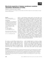
Alignment of the deduced amino-acid sequence against
6PF2K/Fru-2,6-P
2
ase from other sources (Fig. 1) revealed
two distinct regions of similarity. The section of the
polypeptide from about Ile351 to the C-terminus was very
similar to the known sequences of 6PF2K/Fru-2,6-P
2
ase
from other plants (potato tuber, 88%; arabidopsis hypo-
cotyl, 88%; mangrove, 87%; maize leaf, 81%) and similar
to those from other eukaryotes (mammalian liver, s keletal
muscle, brain a nd testis, 45–47%). This r egion c ontains the
domains f or both 6PF2K and Fru-2,6-P
2
ase activities and
forms the catalytic core of the bifunctional enzyme [19].
Within this region all nine residues known to be crucial for
Fru-2,6-P
2
ase activities in the liver isoform of t he mamma-
lian enzyme are conserved in the same relative positions
within the spinach leaf sequence (Fig. 1). Similarly, 17 of the
21 residues identified as being important for 6PF2K activity
in the rat liver or te stes isozymes are conserved in the
alignment of the spinach leaf enzyme (Fig. 1). The
N-terminal region from Met1 to Ala350 is similar to
the N-terminal region of corresponding 6PF2K/Fru-2,6-
P
2
ase cDNA from arabidopsis (56% identity) and man-
grove (59% identity), and to a partial cDNA from
potato (58% i dentity), but is unrelated t o sequences of
6PF2K/Fru-2,6-P
2
ase from nonplant sources.
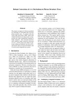
Detection of the gene, transcript and protein
for 6PF2K/Fru-2,6
2
Pase in spinach
A probe generated from the c DNA hybridized to multiple
fragments o n blots of genomic DNA digested with BamHI,
EcoRI or HinDIII, confirming the presence of this sequence
within the spinach genome (data not shown). On blots of
total R NA from spinach leaves, the same probe hybridized
to a single band o f % 2500 bp, corresponding to the length
of the isolated cDNA (Fig. 2A).
Expression of the coding regio n of 6PF2K/Fru-2,6-P
2
ase
in E. coli led to the production of large amounts of insoluble
protein. Antibodies were raised against the recombinant
polypeptide purified from inclu sion bodies. These antibod-
ies detected a single b and with an a pparent molecular mass
of 90.8 kDa on immunoblots of spinach leaf protein
(Fig. 2B). Although both 6PF2K and Fru-2,6-P
2
ase activ-
ities were detectable in extracts of E. coli expressing the
recombinant protein, the kinetic properties of the enzyme
from this source were not studied in detail because the
majority of the soluble activity was asso ciated with several
truncated proteins f rom which the full-length 90.8 kDa
polypeptide could not be separated by conventional non-
denaturing chromatographic techniques (data not shown).
Expression and purification of soluble
6PF2K/Fru-2,6-
P
2
ase
Soluble, recombinant 6PF2K/Fru-2,6-P
2
ase was produced
by expression in S. frugipe rda cell culture using a baculo-
virus expression system. The recombinant enzyme was
purified to app arent homogeneity b y poly(ethylene glycol)
precipitation, followed by chromatography on DEAE–
1270 J. E. Markham and N. J. Kruger (Eur. J. Biochem. 269) Ó FEBS 2002
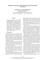
Sepharose, Blue–Sepharose, Mono Q and Superose-12. The
yield of enzyme based on 6PF2K activity was typically 10%.
The purified protein eluted with an a pparent molecular
mass of 320 kDa during gel filtration (Fig. 3) and yielded a
single polypeptide with a molecular mass o f 90.8 kDa when
analysed by SDS/PAGE (Fig. 2C).
Kinetic properties of recombinant 6PF2K/Fru-2,6-
P
2
ase
The purified recombinant protein possessed both 6PF2K
and Fru-2,6-P
2
ase activities. The 6PF2K activity was
markedly stimulated by P
i
. This activity d isplayed standard
Michaelis–Menten kinetics with respect to both ATP and
Fru-6-P in the presence and absence of P
i
(Fig. 4).
Activation by P
i
resulted from both an increase in V
app
max
and a decrease in K
app
m
for each substrate (Table 1). This
activity was also inhibited by a range of three-carbon
phosphate esters and by PP
i
. Each of these compounds
displayed h yperbolic inhibition kinetics at fixed concen-
trations of ATP and Fru-6-P. In the presence of 2 m
M
P
i
, 3-phosphoglycerate, 2-phosphoglycerate and phos-
phoenolpyruvate were all effective inhibitors at m icromolar
concentrations (Table 2). The enzyme activity was less
sensitive to inorganic pyrophosphate, g lycerol 3-phosphate
Fig. 1. Alignment of the amino-acid sequences of 6PF2K/Fru-2,6-P
2
ase from various sources. The origin of the sequences compared are s pinach
(GenBank accession number AF041848), arabidopsis (AF190739) and rat (liver isozyme, Y00702). Grey boxes show identity between the spinach
and other sequences. Residues highligh ted in black are th ose p reviou sly i dentified as essential for 6P F2K or Fru-2,6-P
2
ase. Ile-135, referred to in the
text, is i ndicated (.).
Fig. 2. Detection of 6PF2K/Fru-2,6-P
2
ase transcript and protein in
spinach. (A) Northern blot of total RNA from spinach leaves (B)
Western b lot of t otal protein extract of spinach leaves, and (C) SDS/
PAGE of 1 lg of recombinant protein purified from S. frugiperda
stained with Coomassie blue. Values alongside each track indicate the
size of molecular mass standards presented as (A) nucleotides, and
(B,C) kDa.
Ó FEBS 2002 Spinach 6PF2K/Fru-2,6-P
2
ase (Eur. J. Biochem. 269) 1271
and dihydroxyacetone phosphate under the conditions used
in this investigation (Table 2). We chose to s tudy inhibition
by 3-phosphoglycerate i n more d etail by e xamining the
effect of this compound on the kinetic response of 6PF2K
activity to varying substrate concentrations. The activity
displayed normal hyperbolic kinetics over the range
0–1.0 m
M
3-phosphoglycerate ( Fig. 5). Inhibition was
caused by progressive decreases in V
app
max
and increases in
K
app
m
for both ATP and Fru-6-P as the concentration of
3-phosphoglycerate was increased (Table 3). Inhibition by
3-phosphoglycerate was overcome by increasing concentra-
tions of P
i
, which increased V
app
max
and decreased K
app
m
.Inthe
presence of 2 m
M
Fru-6-P,0.2m
M
3-phosphoglycerate and
2m
M
P
i
,V
app
max
was 7.00 ± 0.38 mUÆmg protein
)1
and K
app
m
for A TP w as 0 .46 ± 0.08 m
M
; t he corresponding values in
the presence of 10 m
M
P
i
were 11.11 ± 0.42 mUÆmg pro -
tein
)1
and 0.34 ± 0.05 m
M
, respectively (Fig. 6). Similar
effects were observed when Fru-6-P was the varied substrate
(data not shown).
As Fru-2,6-P
2
ase from plants is reported to be sensitive to
product inhibition [1], we determined the effect of both Fru-
6-P and P
i
on the Fru-2,6-P
2
ase a ctivity associated w ith the
recombinant bifunctional enzyme. The activity of Fru-2,6-
P
2
ase displayed normal h yperbolic substrate k inetics a t
each of the concentrations of P
i
and Fru-6-P studied
(Fig. 7). Over t he range 0 –5.0 m
M
,P
i
was an uncompetitive
Fig. 3. Native molecular mass of recombinant 6PF2K/Fru-2,6-P
2
ase.
Elution of 6PF2K activity from a Superose-12 gel filtration co lumn
(m). The elution of other proteins used t o calibrate the column are as
indicated (d). Elution volume (V
e
) is expressed relative to the void
volume of the column (V
0
) determined from the elution of blue
dextran.
Fig. 4. Effect of P
i
on the affinity of 6PF2K for Fru-6-P and ATP.
Enzyme activity w as measured over the range 0.01–5.0 m
M
ATP at
2m
M
Fru-6-P (A), and 0 .01–5.0 m
M
Fru-6-P at 2 m
M
ATP (B). The
concentration o f P
i
was 0 m
M
(.), 0.5 m
M
(m), 2.0 m
M
(j), or 5.0 m
M
(d). Each value is a single determination of activity based on a 4-point
timecourse of Fru-2,6-P
2
production. Hill coefficients were between
0.82 ± 0.18 and 0.90 ± 0.09 with respect to ATP (A) and b etween
1.05 ± 0 .09 and 1.15 ± 0 .19 with respect to Fru-6-P (B); none o f
these values w as significantly different from un ity.
Table 1. Effect of P
i
on the kinetic constants o f 6PF2K. Enzyme a ctivity was measured at the concentration of A TP or Fru-6-P showninFig. 4while
the concentration of the cosubstrate was maintained at 2 m
M
. Kinetic constants were obtained by fitting data to the equation for a single-substrate
Michaelis–Menten r eaction and are expressed as the best-fit estimate ± SE from eight m easurements.
P
i
(m
M
)
ATP Fru-6-P
V
app
max
(mUÆmg protein
)1
) K
app
m
(m
M
) V
app
max
(mUÆmg protein
)1
) K
app
m
(m
M
)
0 4.08 ± 0.49 1.32 ± 0.40 1.41 ± 0.18 1.41 ± 0.47
0.5 11.47 ± 0.99 1.29 ± 0.28 9.58 ± 0.33 0.92 ± 0.09
2.0 12.45 ± 0.62 0.90 ± 0.13 10.92 ± 0.61 0.55 ± 0.10
5.0 13.16 ± 0.82 0.53 ± 0.11 11.51 ± 0.60 0.53 ± 0.09
1272 J. E. Markham and N. J. Kruger (Eur. J. Biochem. 269) Ó FEBS 2002
inhibitor. Nonlinear regression analysis of the untrans-
formed data yielded the following values: V
max
, 1.75 ± 0.12
mUÆmg protein
)1
; K
m
, 65.9 ± 4.58 n
M
; K
iu
,1.20±0.11
m
M
, in which the values are the best-fit estimates ± SE from
21 measurements. Attempts to fit the same data to the
kinetic equation describing mixed inhibition produced an
estimate for K
ic
> 100 m
M
, demonstrating that there was a
negligible competitive component to the i nhibition of Fru-
2,6-P
2
ase a ctivity by P
i
. I n c on trast, comparable analysis o f
the effects of 0–1.0 m
M
Fru-6-P yielded the following
constants: V
max
, 1.65 ± 0.22 mUÆmg protein
)1
; K
m
,61.9 ±
3.17 n
M
; K
ic
, 0 .65 ± 0.03 m
M
; K
iu
,1.55±0.14m
M
These
values indicate that Fru-6-P is a mixed inhibitor with
significant competitive and uncompetitive components.
Based on the V
max
values for the two a ctivities obtained
in these analyses, the 6PF2K/Fru-2,6-P
2
ase ratio of the
recombinant bifunctional spinach enzyme was 6.5–9.6.
DISCUSSION
The recombinant protein investigated in the present study is
likely to represent the complete bifunctional 6PF2K/Fru-
2,6-P
2
ase from spinach leaves. The length of the isolated
cDNA corresponds closely to the s ize of the transcript
identified by hybridization against spinach leaf RNA.
Moreover, the protein expressed in insect cells is the same
size as the polypeptide identified in crude extracts of spinach
leaves by antibodies raised against the recombinant protein.
The size of this protein is very similar to t hat of t he H-form
of the bifunctional enzyme previously purified from spinach
leaves [11]. More recently, transcripts and polypeptides of
similar sizes have been identified in arabidopsis seedlings
[18].
The structure of the spinach leaf enzyme studied in this
paper conforms to the pattern of all other bifunctional
6PF2K/Fru-2,6-P
2
ase proteins so far studied [7]. It is
composed of four regions; a central core consisting of the
6PF2K and Fru-2,6- P
2
ase domains t hat are flanked b y
variable N- and C-termini. As might be anticipated, the
central catalytic core shares a high degree of sequenc e
identity with the corresponding region of the bifunctional
enzyme from other eukaryotic sources (Fig. 1). Notably,
only f our of the known catalytic residues a re not conserved
in the same relative positions in the spinach and mammalian
enzyme. However, one of these (Lys479, spinach) is found in
an adjacent position in the strict alignment (Fig. 1).
Furthermore, for each of the other three discrepancies, the
amino-acid substitutions found in the spinach sequence
(Ser441, Gln531, Asn536) are also present in the bifunc-
tional enzymes from arabidopsis [18], potato [17], mangrove
(AB061797) and maize (AF007582).
A striking f eature of the deduced amino-acid sequence of
spinach 6PF2K/Fru-2,6-P
2
ase is t he size of the N-terminal
region preceding the catalytic core. This 350-residue section
contains several m otifs t hat a re found in the c orresponding
region of the bifunctional enzyme from other plants, but
Table 2. Inhibition of 6-phosphofructo-2-kinase activity by phosphate
esters. Enzyme activity was determined using 2 m
M
Fru-6-P,2m
M
ATP. The concentration of phosphate ester producing half-maximum
inhibition (I
0.5
) is presented as the best-fit estimate ± SE from eight
measurements.
Compound I
0.5
(m
M
)
Pyrophosphate 0.106 ± 0.018
Glycerol 3-phosphate 8.07 ± 0.305
Phosphoenolpyruvate 0.045 ± 0.007
2-Phosphoglycerate 0.029 ± 0.004
3-Phosphoglycerate 0.084 ± 0.005
Dihydroxyacetone phosphate 0.737 ± 0.218
Fig. 5. Effect of 3-phosphoglycerate on the affinity of 6PF2K for Fru-6-P
and ATP. Enzyme a ctivity was measured ov er the range 0.0 1–5.0 m
M
ATPat2m
M
Fru-6-P (A), and 0.01–5.0 m
M
Fru-6-P at 2 m
M
ATP
(B) in the presence of 2 m
M
P
i
. The concentration o f 3-phosphogly-
cerate was 0 m
M
(d), 0.2 m
M
(j), or 1.0 m
M
(m). Each value is a
single determination o f activity based on a four-point timecourse of
Fru-2,6-P
2
production. Hill coefficients were between 0.89 ± 0.11 and
1.26 ± 0 .20 with respect to ATP (A) and between 0.87 ± 0.14 and
0.92 ± 0 .08 with respect to Fru-6-P (B); none of these values was
significantly different from unity. 3-PGA, 3-phosphoglycerate.
Ó FEBS 2002 Spinach 6PF2K/Fru-2,6-P
2
ase (Eur. J. Biochem. 269) 1273
otherwise has no significant homology with any known
sequences. In the bifunctional enzyme from other eukary-
otes, regions flanking the catalytic domains have a profound
influence on the kinetic properties of the enzyme. For
example, removal of these regions from the rat liver enzyme
decreases V
max
of 6PF2K and its affinity for Fru-6-P,and
increases V
max
of Fru-2,6- P
2
ase t hus d ecreasing t he activity
of 6PF2K relative to that of Fru-2,6-P
2
ase [19]. Further-
more, structural variation in the N- and C-termini, as well as
the nature and distribution of phosphorylation sites within
these regions, is believed to contribute to the differences
between specific isoforms in the properties of the component
6PF2K and Fru-2,6-P
2
ase activities and their response to
post-translational m odification [7,31,32]. The N-terminal
region is likely to serve a comparable r egulatory function in
plants. Preliminary studies of the recombinant spinach
6PF2K/Fru-2,6-P
2
ase indicate that N-terminal-truncated
forms of the enzyme have a much lower activity of 6PF 2K
relative to Fru-2,6-P
2
ase than the full-length protein studied
in this paper ( J. E. Markham & N. J. Kruger, unpublished
results). Similar differences in the ratio of activities of
6PF2K/Fru-2,6-P
2
ase have been reported for the full-length
and truncated proteins f rom arabidopsis [18]. These obser-
vations show that the N -terminal region can influence t he
component activities of the enzyme and suggest that, by
analogy w ith the mammalian enzyme [7], differences in
the N -terminal region (which is less highly conserved than
the catalytic core ) may be re sponsib le for differences in the
regulatory properties of the enzyme between plant species or
even tissues.
There is circumstantial evidence to suggest that spinach
leaf 6PF2K/Fru-2,6-P
2
ase may be regulated by reversible
phosphorylation [33–35]. Analysis of the N-terminal por-
tion of the deduced amino-acid sequence using
PHOSPHO-
BASE
[36] suggests 1 4 poten tial sites f or phosphorylation b y
calmodulin-dependent protein kinase II and protein kinases
A and C. Six of these sites are identified during compar-
able analyses of the corresponding 6PF2K/Fru-2,6-P
2
ase
sequences from arabidopsis and m angrove. Of the f our
potential phosphorylation sites common to all of these plant
sequences, three (Ser138, Ser155 and Ser224 in spinach)
yield predictive scores greater than 0.90 du ring analysis for
phosphorylation sites using NetPhos, wh ich exploits a
complementary neural network approach [37]. Whether
these, or other, residues are phosphorylated in vivo remains
to be established. Recently, direct evidence has been
obtained for phosphorylation of serine residues in 6 PF2K/
Fru-2,6-P
2
ase in the rosette leaves of arabidopsis [38],
although the identity of the specific sites that are modified
has yet to be determined.
The kinetic properties of the recombinant 6PF2K/Fru-
2,6-P
2
ase are broadly similar to those reported previously
for the bifunctional enzyme from spinach leaves [10,11]. The
6PF2K activity of the recombinant protein is activated by P
i
and inhibited b y a r ange of three-carbon phosphate esters
and PP
i
. The kinetic constants for Fru-6-P and ATP
determined in this paper are consistent with the substrate
affinities of the enzyme r eported i n e arlier stud ies [11].
However, in contrast to previous reports on the partially
purified enzyme [8,10], the activity displays standard
hyperbolic kinetics with both substrates and there is no
evidence for sigmoidal kinetics with respect to Fru-6-P,even
in presence of 3-phosphoglycerate. One possible explanation
for the apparent sigmoidal kinetics observed by others is
contamination of Fru-6-P by P
i
. This would result in a
progressive increase in activation by P
i
as the concentration
of substrate was increased.
Fig. 6 . Influence of P
i
on inhibition of 6PF2K by 3-phosphoglycerate.
Enzyme activity was measured in the presence of 2 m
M
Fru-6-P,
0.2 m
M
3-phosphoglycerate a nd eith er 2 m
M
(d)or10m
M
(s)P
i
.The
concentration of ATP was varied as shown. Each value is a single
determination of activity based on a four-point timecourse of
Fru-2,6-P
2
production. Hill coefficients were 0 . 89 ± 0.11 a t 2 m
M
P
i
and 0.94 ± 0.0 9 at 10 m
M
P
i
; neither of these values was significantly
different from unity.
Table 3. Effect of 3-phosphoglycerate on the kinetic constants of 6PF2K. Enz yme activity was measured in the presence of 2 m
M
P
i
.Thecon-
centration of either ATP or F ru -6-P was varied as sh own in Fig. 5 while the concentration of the cosubstrate was maintained at 2 m
M
.Kinetic
constants were obtained by fitting data to the equation for a single-substrate Michaelis–Menten reaction and are expressed as the best-fit
estimate ± SE from eight measu rements.
3-Phosphoglycerate (m
M
)
ATP Fru-6-P
K
app
m
(m
M
)
V
app
max
(mUÆmg protein
)1
) K
app
m
(m
M
) V
app
max
(mUÆmg protein
)1
)
0 10.40 ± 0.75 0.32 ± 0.09 15.92 ± 0.54 0.96 ± 0.09
0.2 6.25 ± 0.73 0.41 ± 0.16 11.93 ± 0.41 1.02 ± 0.09
1.0 3.89 ± 0.38 0.74 ± 0.13 4.25 ± 0.34 2.36 ± 0.40
1274 J. E. Markham and N. J. Kruger (Eur. J. Biochem. 269) Ó FEBS 2002
The pronounced activation of 6PF2K by P
i
is due to both
an increase in V
app
max
and a de crease in K
app
m
for both of the
substrates. This is similar to the effects of P
i
on rat liver
6PF2K/Fru-2,6-P
2
ase [27] and consistent with the initial
studies on the spinach bifunctional enzyme [10] but
contrasts with the apparent decrease in the affinity for
ATP during activation by P
i
reported for the purified
spinach leaf enzyme [11]. Despite this discrepancy, the
6PF2K activity of the recombinant enzyme is inhibited by
the s ame range of three-carbon phosphorylated intermedi-
ates as that of the enzyme from spinach leaves [8,10,11].
In the present study the effect of 3-phosphoglycerate was
to decrease V
app
max
and increase K
app
m
for both Fru-6-P
and ATP. The changes i n these apparent kinetic parameters
are consistent with 3-phosphoglycerate acting as a mixed
inhibitor [K
ic
¼ 0.182 ± 0.067 m
M
, K
iu
¼ 0.517 ±
0.133 m
M
with respect to ATP; K
ic
¼ 0.283 ± 0.104 m
M
,
K
iu
¼ 0.421 ± 0.099 m
M
with respect to Fru-6-P (best- fit
estimate ± SE, n ¼ 24, calculated f rom d ata presented in
Fig. 5)], although measurements over a greater range of
substrate and effector concentrations would be required to
establish this relationship. As reported for the enzyme
isolated from spinach leaves, the inhibition by 3-phospho-
glycerate i s r everse d b y P
i
. I n c ontrast to the c orresponding
activity of the bifunctional e nzyme from r at liver and other
mammalian sources [39], 6PF2K is not strongly inhibited by
glycerol 3-phosphoglycerate, but is inhibited by PP
i
. The
latter effect is consistent with an earlier observation on the
enzyme purified from spinach leaves [11].
The relatively high affinity of the Fru-2,6-P
2
ase activity of
the recombinant enzyme for Fru-2,6-P
2
(K
m
% 60 n
M
)and
the sensitivity of this activity to inhibition by both P
i
and
Fru-6-P are comparable to the properties of the bifunctional
enzyme isolated from spinach leaves [10,11,15]. Never-
theless, we note that whereas P
i
is a largely uncompetitive
inhibitor of the recombinant enzyme, previous studies
suggest that it acts competitively even though these
reports also claim that P
i
induces sigmoidal kinetics
[10] or increases V
max
[11] neither of which is consistent
with pure competitive inhibition . Insufficient data a re
provided in the previous reports to resolve these apparent
contradictions.
Irrespective of t he minor quantitative differences des-
cribed above, the kinetic properties of the recombinant
6PF2K/Fru-2,6-P
2
ase are in broad agreement w ith those o f
the bifunctional enzyme isolated from spinach leaves, and in
particular the 90-kDa H-form that has been purified to
apparent homogeneity [11]. The affinities of the component
activities for their substrates and effectors are within the
range of concentrations likely to occur in the cytosol of
spinach leaf mesophyll cells (see Table 1 of [26]). This
suggests that the levels of these metabolites, w hich are
known to vary throughout the photoperiod, will affect the
relative activities of 6PF2K and Fru-2,6-P
2
ase t hus altering
the steady-state level of Fru-2,6-P
2
and contribute to the
regulation of flux through cytosolic FBPase in vivo.
However, the relative significance of inhibition of 6PF2K
activity by 3-phosphoglycerate, 2-phosphoglycerate, phos-
phoenolpyruvate and dihydroxyacetone phosphate will
depend upon the in vivo concentration of each of these
metabolites and of P
i
, as discussed previously [1].
In conclusion, the kinetic properties of the recombinant
enzyme are in a greement with t hose of t he enzyme isolated
from spinach leaves. This suggests t hat the properties of the
latter have not been appreciably modified due to proteolysis
during e xtraction. These results corroborate t he current
view of Fru-2,6-P
2
as an internal regulator of sucrose
synthesis, integrating t he m etabolic responses to changes i n
the relative concentrations of three-carbon phosphate esters,
hexose phosphates and P
i
through allosteric modulation of
6PF2K/Fru-2,6-P
2
ase [2].
Fig. 7. Inhibition o f Fru-2,6-P
2
ase by P
i
and Fru-6-P. Enzyme activity
was measured over the range 20–100 n
M
Fru-2,6-P
2
in the presence of
P
i
(A) or Fru-6-P (B). The concentration of P
i
was 0 m
M
(d), 1.0 m
M
(j), or 5.0 m
M
(m). The concentration of Fru-6-P was 0 m
M
(d),
0.25 m
M
(j), o r 1.0 m
M
(m). Each value is a single determination of
activity based on a f our-point timecourse of Fru -2,6- P
2
hydrolysis. Hill
coefficients were between 0.92 ± 0.15 and 1.15 ± 0.2 9 in the presence
of P
i
(A) and between 0.85 ± 0.26 and 1.34 ± 0.2 9 in the presence of
Fru-6-P (B); non e o f these values was significantly different from unity.
Data are displayed as L ineweaver–Burk plots for presentational pur-
poses only. The l ines are the theoretical curves at each concentration o f
product b ased on kinetic constants de rived from nonlinear regression
analysis of the e ntire data s et.
Ó FEBS 2002 Spinach 6PF2K/Fru-2,6-P
2
ase (Eur. J. Biochem. 269) 1275
ACKNOWLEDGEMENTS
We are grateful to Dr Claire Kinlaw (Dendrome Project, USDA
Institute of Forest G enetics, Albany, California, USA) for p roviding
the original loblolly pine EST clone 2541s (dbEST ID 377114). This
research was supported by t he Bio tec hnology a nd Biological Sciences
Research Council, U K (Grant n umber 43/P05839).
REFERENCES
1. Stitt, M. (1990) Fructose 2,6- bisphosphate as a regulatory mole-
cule in plants. Annu. Rev. Plant Physiol. Plant Mol. Biol. 41,
153–185.
2. Stitt, M. (1997) The flux of carbo n between t he chlo roplast a nd
cytoplasm. In Plant Metabolism (Dennis, D.T., Turpin, D.H.,
Lefebvre, D.D. & Layzell, D.B., eds), pp. 382–400. Longman,
Harlow.
3. Scott, P., Lange, A.J., Pilkis, S.J. & Krug er, N .J. ( 1995) Carb on
metabolisminleavesoftransgenictobacco(Nicotiana tabacum L.)
containing elevated fructose 2,6-bisphosphate levels. Plant J. 7,
461–469.
4. Scott, P., Lange, A.J. & Kruger, N.J. (2000) Photosynthetic
carbon metabolism in leaves of transgenic tobacco (Nicotiana
tabacum L.) containing decreased amou nts of fructose
2,6-bisphosphate. Planta 211, 864–873.
5. Truesdale, M.R., Toldi, O. & Scott, P. (1999) The effect of
elevated concentrations of fructose 2,6-bisphosphate on carb on
metabolism during deacidification in the crassulacean acid
metabolism plant Kalanchoe
¨
daigremontiana. Plant Physiol. 12 1 ,
957–964.
6. Draborg, H., Villadsen, D. & Nielsen, T.H. (2001) Transgenic
arabidopsis plants with decreased activity of fructose-6-phos-
phate,2-kinase/fructose-2,6-bisphosphatase have altered carbon
partitioning. Plant P hysiol. 126, 7 50–758.
7. Okar, D.A., Manz ano, A., N a varro-S abate , A., Rier a, L., Ba r-
trons, R. & Lange, A.J. (2001) PFK-2/FBPase-2: maker and
breaker of the essential biofactor fructose 2,6-bisphosphate.
Trends Bi ochem. Sci. 26, 30–35.
8. Cse
´
ke, C. & Buchanan, B.B. (1983) An enzyme synthesizing
fructose 2,6-bisphosphate occurs in leaves and is regulated by
metabolite effectors. FEBS Lett. 155, 139–142.
9. Cse
´
ke, C., Stitt, M., Balogh, A. & Buchanan, B.B . (1983) A pro-
duct-regulated fructose 2,6-bisphosphatase occurs i n green leaves.
FEBS Lett. 162, 103–106.
10. Stitt, M., Cse
´
ke, C. & Buchanan, B.B. (1984) Regulation of
fructose 2,6-bisphosphate concentration in spinach leaves. Eur.
J. Bioc hem. 143, 8 9–93.
11. Larondelle, Y., Mertens, E., Van Schaftingen, E . & Hers, H G.
(1986) Purification and properties of spinach l eaf phospho-
fructokinase 2/fructose 2,6-bisphosphatase. Eur. J. Biochem. 161,
351–357.
12. Macdonald, F.D., Cse
´
ke, C., Chou, Q. & Buchanan, B.B. (1987)
Activities synthesizing and degrading fructose 2,6-bisphosphate in
spinach leaves r eside o n different proteins. Proc. Natl Acad. Sci.
USA 84, 2742–2746.
13. El-Maghrabi, M.R., Pate, T.M., Murray, K.J. & Pilkis, S.J. (1984)
Differential effects o f proteolysis and protein modification on the
activities of 6-phosphofructo-2-kinase/fructose-2,6-bisph ospha-
tase. J. Biol. C hem. 259, 13096–13103.
14. Walker, G.H. & Huber, S.C. (1987) ATP-dependent activation of
a new form of spinach leaf 6-phosphofructo-2-kinase/fruct ose-2,6-
bisphosphatase. Arch. B io chem. Biophys. 258, 5 8–64.
15. Macdonald, F.D., Chou, Q., Buchanan, B.B. & Stitt, M. (1989)
Purification and characterisation of fru ctose-2,6-bisp hosphatase, a
substrate-specific c ytosolic enzym e from leaves. J. Biol. Chem. 264,
5540–5544.
16. Larondelle, Y., Mertens, E., Van Schaftingen, E. & Hers, H G.
(1989) Fructose 2,6-bisphosphate h ydrolysing enzymes in higher
plants. Plant Physiol. 90, 827–834.
17. Draborg, H., Villadsen, D. & Nielsen, T.H. (1999) Cloning,
characterization and expression of a bifunctional fructose-
6-phosphate, 2-kinase/fructose-2,6-bisphosphtase from potato.
Plant Mol. Biol. 39 , 709–720.
18. Villadsen, D., Rung, J.H., Draborg, H. & Nielsen, T.H.
(2000) Structure and heterologous expression of a gene
encoding fructose-6-phosphate,2-kinase/fructose-2,6-bisphospha-
tase from Arabidopsis thanliana. Biochim. Biophys. Acta 1492,
406–413.
19. Pilkis, S.J., Claus, T.H., Kurland, I.J. & Lange, A.J. (1995)
6-Phosphofructo-2-kinase/fructose-2,6-bisphosphatase: a meta-
bolic signalling enzyme. Annu.Rev.Biochem.64 , 799–835.
20. Montavon, P. & Kruger, N.J. (1993) Essential arginyl residue
at the active site of pyro phosphate: fructose 6-phosphate
1-phosphotransferase from potato t uber. Plant Physiol. 101,765–
771.
21. Logemann, J., Schell, J. & Willmitzer, L. (1987) Improved method
for t he isolation o f RNA from plant tissues. Anal. B iochem. 163,
16–20.
22. Dean, C., Sjodin, C., Page, T., Jones, J. & Lister, C. (1992)
Behaviour of the maize transposable element Ac. in Arabidopsis
thaliana. Plant J . 2, 6 9–81.
23. Sambrook, J., Frich, E.F. & M aniatis, T. (1989) Molecular
Cloning: a Laboratory Manual. Cold Spring Harbor Press, New
York.
24. Kruger, N.J. (2002) Detection of polypeptides on immunoblots
using enzyme-conjugated or rad io labelled seco ndary ligands.
In Protein Protocols Handbook, 2nd edn. (Walker, J.M., ed.),
pp. 405–415. Humana Press, Totowa, New J ersey.
25. Stitt, M. (1990) Fructose 2,6-bisphosphate. In Methods in Plant
Biochemistry, Vol. 3 (Dey, P.M. & Harborne, J.B., eds), pp. 87–92.
Academic Press, Lon don.
26. Theodorou, M.E. & Kruger, N.J. (2001) Physiological relevance
of fructose 2,6-bisphosp hate in the regulation of spinach leaf
pyrophosphate:fructose6-phosphate1-phosphotransferase. Planta
213, 147–157.
27. Laloux, M., Van Schaftingen, E., Francois, J. & Hers, H G.
(1985) Phosphate dependency of phosphofructokinase 2. Eur.
J. Bioc hem. 148, 1 55–159.
28. Marquardt, D.W. (1963) An algorithm for least squares estima-
tion of param eters. J. Soc. Ind. Appl. Math. 11, 431–441.
29. Cornish-Bowden, A. (1995) Fundamentals of Enzyme Analysis,
2nd e dn. Portland Press, London.
30. Bradford, M.M. (1976) A rapid and sensitive method for the
quantitation of microgram quantities of protein utilizing the
principle o f protein dye b inging. Anal. Biochem. 72, 248 –254.
31. Kurland, I.J ., Chapman, B. & El-Maghrabi, M.R. (2000) N- and
C-termini modulate the effects of pH and phosphorylation
on hepatic 6-phosphofructo-2-kinase/fructose-2,6-bip hosphatase.
Biochem. J . 347, 4 59–467.
32. Zhu,Z.,Ling,S.,Yang,Q.H.&Li,L.(2000)Thedifferenceinthe
carboxy-terminal sequence is responsible for the difference in the
activity of chicken and rat liver fructose-2,6-bisphosphatase. Biol.
Chem. 381 , 1195–1202.
33. Stitt, M., M ieskes, G., So
¨
ling, H D., Grosse, H . & Heldt, H.W.
(1986) Diurnal changes of fructose-6-phosphate,2-kinase and
fructose-2,6-bisphosphatase activities in spinach leaves. Z. Nat-
urforsch. 41c, 2 91–296.
34. Walker, G.H. & Huber, S.C. (1987) Spinach leaf 6-phosphofructo-
2-kinase. FEBS Lett. 213 , 375–380.
35.Rowntree,E.&Kruger,N.J.(1995)Covalentmodulationof
6-phosphofructo-2-kinase/fructose-2,6-bisphosphate in spinach
leaves . In Photosynthesis: from Ligh t t o B iosphere (Mathis, P., ed.),
1276 J. E. Markham and N. J. Kruger (Eur. J. Biochem. 269) Ó FEBS 2002
Vol. 5, pp. 111–114. Kluwer Academic Publishers, Dordrecht, the
Netherlands.
36. Kreegipuu, A., Blom, N. & B runak, S. (1999) PhosphoBase, a
database of phosphorylation sites: release 2.0. Nu cleic Acids Re s.
27, 237 –239.
37. Blom, N., Gammeltoft, S. & Brunak, S. (1999) Sequence- and
structure-based prediction o f eukaryotic p rotein phosphorylation
sites. J. Mol. Biol. 29 4, 1351–1362.
38. Furumoto, T., Teramoto, M., Inada, N., Ito, M., Nishida, I . &
Watanabe, A. (2001) P hosphorylatio n of a bifunctional enzyme,
6-phosphofructo-2-kinase/fructose-2,6-bisphosphate 2-phospha-
tase, i s regulated physiologically and developmentally in rosette
leaves of Arabidopsis t haliana. Plant C ell Physiol. 42, 1044–
1048.
39. Van Schaftingen, E. (1987) Fructose 2,6-bisphosphate. Adv. Enzy-
mol. R elat. Areas Mol . Biol. 59 , 315–395.
Ó FEBS 2002 Spinach 6PF2K/Fru-2,6-P
2
ase (Eur. J. Biochem. 269) 1277