Báo cáo khoa học: Dematin interacts with the Ras-guanine nucleotide exchange factor Ras-GRF2 and modulates mitogen-activated protein kinase pathways doc
Bạn đang xem bản rút gọn của tài liệu. Xem và tải ngay bản đầy đủ của tài liệu tại đây (394.12 KB, 12 trang )
Dematin interacts with the Ras-guanine nucleotide exchange factor
Ras-GRF2 and modulates mitogen-activated protein kinase pathways
Mohini Lutchman
1
, Anthony C. Kim
1
, Li Cheng
2
, Ian P. Whitehead
2
, S. Steven Oh
1
, Manjit Hanspal
1
,
Andrey A. Boukharov
1
, Toshihiko Hanada
1
and Athar H. Chishti
1
1
Section of Hematology-Oncology Research, Departments of Medicine, Anatomy, and Cellular Biology, St Elizabeth's Medical
Center, Tufts University School of Medicine, Boston, MA, USA;
2
Department of Microbiology and Molecular Genetics,
UMDNJ-New Jersey Medical School, Newark, NJ, USA
Erythroid dematin is a major component of red blood cell
junctional complexes that link the spectrin±actin cytoskel-
eton to the overlying plasma membrane. Transcripts of
dematin are widely distributed including human brain, heart,
lung, skeletal muscle, and kidney. In vitro, dematin binds and
bundles actin ®laments in a phosphorylation-dependent
manner. The primary structure of d ematin consists of a
C-terminal domain homologous to the ÔheadpieceÕ domain
of villin, a n a ctin-binding protein of the brush border c yto-
skeleton. Except ®lamentous actin, no other binding part-
ners of dematin h ave b een i denti®ed. T o investigate the
physiological function of dematin, we employed the y east
two-hybrid assay t o identify dematin-interacting pr oteins in
the adult human brain. Here, we show that dematin interacts
with the guanine nucleotide e xchange f actor R as-GRF2 b y
yeast two-hybrid assay, and this interaction is fur ther
con®rmed by blo t overlay, s urface plasmon r esonance,
co-transfection, and co-immunoprecipitation assays.
Human Ras-GRF2 is expressed in a variety of tissues and,
similar to other guanine nucleotide exchange factors (GEFs),
displays anchorage independent growth in soft agar.
Co-transfection and immunoblotting experiments revealed
that dematin blocks t ranscriptional a ctivation of Jun by
Ras-GRF2 and activates ERK1 via a Ra s-GRF2 indepen-
dent pathway. Because much o f t he present evidence has
centered on the identi®cation of the Rho family of GTPases
as k ey r egulators o f t he actin cytoskeleton, the d irect
association between dematin and Ras-GRF2 may p rovide
an alternate mechanism for regulating the activation of Rac
and R as GTPases via the actin cytoskeleton.
Keywords: d ematin, e rythrocyte, limatin, Ras-GRF2, head-
piece domain.
Dematin is a cytoskeletal protein that binds and bundles
actin ® laments in vitro [1,2]. It was originally identi®ed as a
component of human erythrocyte membrane skeleton, and
migrates in the zone of polypeptides collectively designated
as band 4.9 on polyacrylamide gels [1,2]. Phosphorylation
by the c AMP-dependent protein kinase a bolishes dematin's
actin-bundling activity that is restored by dephosphoryla-
tion [2]. Dematin is part of a junctional c omplex, together
with protein 4 .1, adducin, t ropomyosin, and tropomodulin,
that links spectr in tetramers and a ctin proto®laments to the
erythrocyte plasma membrane [3]. E rythroid dematin exists
as a trimer consisting of one polypeptide of 52-kDa and two
polypeptides of 48-kDa [1,4]. Recently, we have character-
ized the dematin gene and have identi®ed exon 13 as an
alternatively spliced exon present in the 52-kDa polypeptide
but absent in the 48-kDa s ubunit [5,6]. E xon 13 e ncodes a
22-amino-acid insertion t hat i ncludes a motif homologous
to protein 4.2 and a motif that binds to ATP in vitro [7].
Although the functional signi®cance of this insertion is not
known, we have postulated that the 52-kDa subunit
provides a m olecular framework for the f ormation of
disul®de-linked trimeric dematin [4].
Dematin was originally isolated from red b lood cells.
However, dematin t ranscripts have been detected in a wide
variety of t issues including brain, heart, kidney, skeletal
muscle, and lung [5,6,8]. The C-terminal 75-residue
domain of dematin is homologous to the Ôheadpiece Õ
domain of v illin, an actin-bind ing protein of the brush
border cytoskeleton [ 5,9]. Previously, it was believed that
this module played a crucial role in the morphogenesis of
microvilli [10]. H owever, t he rec ent gene ration of villin null
mice strongly suggests t hat villin's role in the micro®lament
assembly of microvilli in absorptive tissues is compensated
for by dematin and/or other ÔheadpieceÕ-containing proteins
[11,12]. The N-terminal core domain of dematin is homo-
logous to only one other known p rotein, a ÔLIMÕ protein
termed limatin (abLIM) [13]. Limatin contains four double
zinc ®nger LIM domains at its N-terminus with the
C-terminus sharing 50% identity to f ull-length dematin
Correspondence to A. Chishti, Biomedical Research, ACH-404,
St Elizabeth's Medical Center, 736 Cambridge Street, Boston, MA
02135, USA. Fax: + 1 617 789 3111, Tel.: + 1 617 789 3118,
E-mail:
Abbreviations: GRF, guanine nucleotide releasing factor; GEF,
guanine-nucleotide exchange factor; DH, Dbl homology domain;
PH, pleckstrin homology dom a in; AbLIM, acti n-binding LIM p rotein;
IQ, Ilimaquinone; NHS, N-hydroxysuccinimide; EDC, N-ethyl-
N¢-[3-(diethylamino)propyl]carbodiimide; Sos, Son of Sevenless;
SAPK, stress-activated protein kinase;
JNK, Jun N-terminal kinase.
Note: M. Lutchman, A. C. Kim, and L. Cheng contributed equally to
this work.
Note: the nucleotide sequences reported in t h is paper have been sub-
mitted to the GenBank with t he accession numbe rs AF181250 and
AF186017.
(Received 2 5 September 200 1, accepted 20 November 2001)
Eur. J. Biochem. 269, 638±649 (2002) Ó FEBS 2002
[13]. The dematin and limatin genes are located o n human
chromosomes 8p21.1 and 10q25, respectively, regions
frequently deleted in prostate and other epithelial cancers
[4,14]. Interestingly, w e have recently demonstrated the loss
of heterozygosity of the dematin gene in a majority of 8p21-
linked prostate tumors [14].
The Ras superfamily of GTPases plays critical roles in the
regulation of signaling pathways from the cell surface to the
nucleus [15]. Approximately 40% of h uman c an cers a re
caused by activated ras alleles [16]. In addition, Ras proteins
are also involved in synaptic t ransmission and long-term
potentiation [17]. These observations generated a great d eal
of interest in proteins that are involved i n the r egulation of
Ras proteins. Ras GTPases cycl e between an active GTP-
bound state a nd an inactive GDP-bound state. GTPase
activating proteins (GAPs) catalyze the intrinsic GTPase
activity of Ras proteins, thereby down-regulating Ras
signaling molecules [17± 19]. In contrast, t he Ras-guanine
nucleotide exchange factor (GEF) proteins are factors that
catalyze the exchange of GDP for GTP, thus activating Ras
GTPases. Two o f the better-known GEFs are Son of
Sevenless (Sos) and the Ras guanine nucleotide release
factor (Ras-GRF) [20±24]. Both p roteins contain a C-ter-
minal domain homologous to the Saccharomyces cerevisiae
Cdc25 protein, a Ras-GEF, and regions homologous to the
Dbl oncogene product (DH domain) in tandem with a
pleckstrin homology (PH) domain [21±23]. The Sos protein
contains C-terminal proline-rich domain not found in the
other related GEFs. It is via this proline-rich domain that
Sos is constitutively associated with the SH3 domain of the
adaptor protein Grb2 [2 0]. Grb2 protein also contains an
SH2 domain that interacts with a phosphorylated tyrosine
residue of activated EGF receptor [20]. The formation of
this complex r ecruits the So s exchange facto r within
proximity of membrane-bound Ras, thus providing a
coupling mechanism between receptor tyrosine kinases
and Ras signaling [20±24].
While the upstream events that lead to Sos activation and
the su bsequent activation of the Ras-MAP kinase cascade
are well known, the signals i nvolved in t he Ras-GRF
activation are not yet fully characterized. Ras -GRFs are of
two types, the neuronally expressed R as-GRF1, and the
more widely expressed Ras-GRF2 [19,21,22,24]. Both Ras-
GRFs are exchange factors for Ras-GTPases via their
Cdc25-like catalytic domains. Recent in vitro evidence
suggests that the Ras-GRFs are activated by G-protein
coupled receptors [23]. Stimulation of muscarinic receptors
or the e xpression of t he G-protein bc subunits is known to
stimulate the exchange activity of Ras-GRF1 (or
CDC25
Mm
) in a phosphorylation-dependent manner [23].
Calcium in¯ux is also shown t o activate Ras-GRF1 [24].
The DH domain of Ras-GRF1 catalyzes nucleotide
exchange of Rac1 in response to a sign al triggered by t he
Gbc25. Moreover, the co-expression of Ras-GRF1 and G
bc
subunits leads to the activation of the MAP kinases JNK1
and ERK2 in heterologous cells [25]. R as-GRF2 stimulates
the ERK1 MAP kinase in a Ras- and ilimaquinone-
dependent manner [22]. More r ecent evidence has shown
that the DH domain of Ras-GRF2 also activates the JNK
pathway in a Rac-dependent manner [26].
To further understand the role of dematin in normal cells,
we proceeded t o identify binding partners that interact with
dematin. The yeast two-hybrid assay was used to screen an
adult human brain library with the C-terminal h alf of
dematin a s t he bait probe. T he identi®cation of Ras-GRF2
as a binding partner for the dematin provides evidence for a
direct association between Ras-GRF2 and dematin and
therefore suggests a novel mechanism f or linking the Ras
signaling complex to the actin cytoskeleton. The functional
signi®cance of t he dematin interaction with Ras-GRF2 was
further explored b y examining t he modulatory e ffects of
dematin on the pathways of ERK a nd JNK activation.
EXPERIMENTAL PROCEDURES
Yeast two-hybrid screen
The v ectors, yeast s trains, and lib rary employed in two-
hybrid s creen werepurchased from C lontech. The C-terminal
half of human 48 kDa dematin (a mino acids 224±383) was
subcloned in-frame into the EcoRI/BamHI site of the GAL4
DNA binding domain plasmid pAS2-1 and used to screen a
human brain Matchmaker cDNA library constructed in the
GAL4 activation domain plasmid pGAD10. The dematin
bait and the library was transformed into CG-1945 and
plated on media lacking the amino acids tryptophan,
leucine, and histidine in the presence of 3-amino-1,2,4-
triazole (5 m
M
). Colonies that grew o n selective m edia were
then scored for b-galactosidase activity by the ®lter assay
according to the manufacturer's instructions (Clontech).
Plasmid DNA from the positive clone, as shown by a blue
color, was recovered from y east and transformed into
bacteria for DNA isolation.
Yeast mating
Yeast mating experiments were utilized to test the speci®city
of interaction b etween dematin and Ras-GRF2. L imatin
and R as-GRF1, the closest kno wn homologues of dematin
and Ras-GRF2, respectively, were included in these exper-
iments. The segment o f limatin (amino acids 597±778)
corresponding to the dematin Ôbait Õ sequence was subcloned
into pAS2-1, while the segment of Ras-GRF1 (amino
acids 172±471), corresponding to the isolated fragment of
Ras-GRF2, was subcloned into pGAD10. The pAS2-1
constructs (including pAS2-1 only) were transformed into
the yeast strain Y187 while pGAD10 constructs (including
pGAD10 only) were subcloned into strain CG1945. Pair-
wise matings between all pAS2-1 transformants and all
pGAD10 transformants were p lated on minimal media a nd
scored for b-galactosidase activity.
Cloning of
Ras-GRF2
cDNA and expression constructs
Primer pair 7/8 (7 : 5¢-ATGCAGAAGAGCGTGCGC
TAC-3¢;8:5¢-TCAAGCAGGGAGTCGAGGTTC-3¢)
was used to a mplify the full-length R as-GRF2 from a
human fetal brain cDNA pool (Invitrogen, CA). These
primers were designed from the murine Ras-GRF2 cDNA
sequence due to the high nucleotide identity. A single b and
of 3.7 kb was ampli®ed and subcloned into the vector
pCR2.1 (Invitrogen, CA, USA) for sequence analysis. The
full-length Ras-GRF2 cDNA was PCR-ampli®ed with
BamHI adaptors and subcloned into the mammalian
expression vector pcDNA3.1/myc-His (Invitrogen).
Immunodetection o f Ras-GRF2 p rotein was carried out
Ó FEBS 2002 Dematin binds to Ras-GRF2 nucleotide exchange factor (Eur. J. Biochem. 269) 639
using a monoclonal a ntibody directed against the
myc-epitope (9E10 clone, U pstate Biotechnology, Lake
Placid, NY, USA). The full-length 48-kDa subunit of
dematin c DNA ( 1.15 kb) was subcloned into th e BamHI
site of pcDNA3.0GFPmyc vector in s ense and antisense
orientations. The following cDNAs w ere PCR-ampli®ed
with Bam HI/EcoRI adaptors for in-frame subcloning into
the bacterial expression vector pGEX-2T (Pharmacia Bio-
tech): Ras-GRF2 (amino acids 176±474), Ras-GRF2 (ami-
no acids 909±1237), Ras-GRF1 (amino acids 172±471),
dematin (amino acids 224±383), and limatin (amino acids
597±778). These constructs will be referred to in this
manuscript as GST±GRF2-DH, GST±GRF2-Cdc25,
GST±GRF1-DH, GST±dematin(224±383) and GST±lima-
tin(597±778), respectively. Recombinant proteins were
expressed and puri®ed accordig to the manufacturer's
instructions (Pharmacia Biotech).
Expression analysis
The primer pair 31/21 (31 : 5¢-AGC GCCTCTT GGAAC
GACTGA-3¢;21:5¢-GCGGCGGCTTTCCTTTCTT-3¢)
wasusedtoamplifya961-bpRas-GRF2fragmentto
probe the Hum an Multiple Tissue Northern Blot ( Clo n-
tech). The probe was
32
P-labeled with t he DECAprime
DNA labeling kit (Ambion) and hybridized to the Northern
blot in Rapid-Hyb buffer a ccording t o the manufacturer's
instructions (Pharmacia Biotech). The primer pair 33/21
(33 : 5¢-CCGCTGCGTCTCCACCACCACAC-3¢)was
used to amplify the Multiple Tissue cDNA Panel #2
(Clontech). These primers amplify a 577-bp product from
the Ras-GRF2 cDNA. Primers speci®c for glyceraldehyde-
3-phosphate dehydro genase ( G3PDH)werealsousedto
ensure equal cDNA loading.
Blot overlay assay
Equal amounts ( 2 lg) of GST and GST±GRF2-DH
fusion proteins were se parated by SDS/PAGE and either
Coomassie-stained or transferred to a nitrocellulose mem-
brane. The nitrocellulose blot was blocked overnight at 4 °C
in 5% (w/v) nonfat dry milk/NaCl/Tris (25 m
M
Tris,
137 m
M
NaCl, 2.5 m
M
KCl, pH 8)/0.1% Tween-20 (block-
ing solution). The blot was t hen incubated i n the blocking
solution containin g 10 lg of puri®ed dematin. Dematin,
which is a trimeric protein of t wo 48-kDa polypeptides and
one 52-kDa polypeptide, was puri®ed from human erythro-
cyte membranes [27]. After an o vernight incubation in the
cold room, the blot was washed twice for 10 min at room
temperature in NaCl/Tris/0.1% Tween-20 and incubated for
1 h in a 1 : 3000 dilution of af®nity-puri®ed polyclonal anti-
dematin Ig. Following two 10-min washes, the blot was then
incubated in a n horseradish peroxidase-conjugated second-
ary antibody (1 : 3000 dilution) for 1 h at room tempera-
ture. After two ®nal washes, bound dematin w as
immunodetected using the ECL system (Pharmacia B iotech).
Surface plasmon resonance analysis
A BIAcore 1000 (Pharmacia Biosensor, NJ, USA) was used
to measure the s peci®c interaction and to d etermine the
binding af® nity between the C-terminal domain of dematin
[dematin(224±383)] and GST±Ras-GRF2. The GST±
dematin(224±383) fusion protein was af®nity-puri®ed
using GSH-Sepharose 4B beads, and treated with
thrombin (Pharmacia Biotech) to proteolytically cleave the
dematin(224±383) domain from the GST fusion p rotein.
A homogeneous sample of the dematin(224±383) (free of the
GST domain) was immobilized ( 1.0 ng of protein per
mm
2
of surface) to the Dextran matrix of a CM5 sensor
chip (Pharm acia Biosensor) using an amine coupling kit
(Pharmacia Biosensor), as previously described [28]. Puri-
®ed G ST±Ras-GRF2-DH fusion protein ( 66 kDa) was
extensively d ialyzed against HBS buffer (10 m
M
Hepes,
pH 7.4, 150 m
M
NaCl, 3.0 m
M
EDTA, 0.005% v/v
Surfactant P20) and diluted to desired con centrations using
the same buffer. Puri®ed recombinant GST was u sed as a
control sample. Association and dissociation rates were
measured at 25 °C at a ¯ow rate of 10 lLámin
)1
.The
binding surface was successfully regenerated with a short
pulse (5.0 lL) of 20 m
M
HCl followed by a short pulse
(5.0 lL) of 0.01% SDS. After the last injection o f a nalyte
samples, the analyte at an initial concentration was
re-injected to check for signi®cant d enaturation of t he
immobilized ligand during the repeated cycles of regener-
ationprocess.Thecontributionofbulksolutioninthe
surface plasmon resonance (SPR) signal were minimal as
determined by injecting the analyte sample onto a blank
CM5 sensor chip surface activated with a 1 : 1 mixture of
N-hydroxysuccinimide (NHS) and N-ethyl-N¢-[3-(diethyla-
mino)propyl]carbodiimide (EDC) and blocked with 1
M
ethanolamine hydrochloride (pH 8.5). The d ata were ana-
lyzed using the
BIAEVALUATION
3.0 (Pharmacia B iosensor)
software .
Transfection of Ras-GRF2 and dematin
into NIH 3T3 cells
The pcDNA3.1-GRF2-myc-His (full length R as-GRF2)
plasmid was transfected into NIH 3 T3 cells using the
pFx-6 lipid reagent following the manufacturer's protocol
(Invitrogen). Cells were plated in duplic ate on plastic and
glass discs in six-well Falcon plates. After 5±8 h in
Opti-Mem (Gibco-BRL) and 24 h in complete media
[Dulbecco's modi®ed Eagle's serum (DMEM) plus 10%
fetal bovine serum; Hyclone, Logan, UT, USA], Ras-GRF2
expressing colonies were selected by growth in medium
containing 400 lgámL
)1
of G418 o ver a period of 2 weeks.
Stable clones were expanded for further analysis. After
2 m onths of selection, Ras-GRF2 stable clones were
cotransfected with pcDNA3-GFPdematin (full length
48-kDa subunit of human dematin) and selected in G418
using the procedures described above.
Immunocytochemistry
Stable NIH 3T3 clones expressing both Ras-GRF2 and
dematin were plated a t 40% con¯uency f or use in i mmuno-
localization studies. Stable clones were washe d in N aCl/P
i
(137 m
M
NaCl, 2.7 m
M
KCl, 10 m
M
Na
2
HPO
4
,1.8m
M
KH
2
PO
4
) and ®xed with formaldehyde (Sigma). After
washing in N aCl/P
i
, cells we re p ermeabilized in NaCl/Tris/
1% Triton X-100 for 5 min. Cells were washed in NaCl/P
i
and incubated in a 1 : 100 d ilution of monoclonal anti-myc
Ig for 1 h. Stable clones were washed in NaCl/P
i
and incu-
bated with a ¯uorescein isothiocyanate (FITC)-conjugated
640 M. Lutchman et al. ( Eur. J. Biochem. 269) Ó FEBS 2002
goat anti-(mouse I gG) Ig (Pierce; 1 : 64 dilution) (Sigma)
for 1 h. After rinsing in NaCl/P
i
, cells were incubated for 1 h
with polyclonal anti-dematin Ig followed by subsequent
washes in NaCl/P
i
and incubation with a rhodamine-
conjugated g oat anti-(rabbit IgG) Ig (Pierce; 1 : 100 dilu-
tion; Sigma) for 1 h. After two ®nal washes, cover slips were
mounted onto slides using an Antifade reagent (Bio-Rad)
and observed under a Zeiss ¯uorescence microscope linked
to a Cooke CCD camera. Photographs were taken using
IMAGE-PRO PLUS
v. 300 (Mediacybernatics, Silver Spring,
MD, USA).
ERK1 activation
A293 cells were transiently transfected with Lipofectamine
2000 (Gibco-BRL). After transfection, the cells were
allowedtorecoverfor48hinDMEM/10%fetalbovine
serum. The cells were then starved for 18 h and treated with
5 l
M
ionomycin (Calbiochem) for 5 min at 37 °C. Cells
were scraped with cell lysis buffer and used for E RK
activation assays. E RK1 a ssays were as described previ-
ously [22]. T he anti-(phospho-ERK) Ig (sc-94, Santa Cruz)
and anti-ERK1 Ig (sc-93, Santa Cruz) were used for the
ERK activation assays. Antibodies were used at dilutions of
1 : 1000 for Western blots. Blots were normalized with the
monoclonal anti-(a-tubulin) Ig (CP06, Oncogene Science,
Cambridge, MA, USA).
Molecular constructs
RacI (WT) and RacI (12 V) encode wild-type and constit-
utively a ctivated derivatives of RacI, respe ctively, that have
been described previously [29]. The reporter construct
utilized in the lu ciferase-coupled tr anscriptional a ssay has
been described previously [30]. The 5XGal4-luc contains the
luciferase gene under the control of a minimal promoter that
contains ®ve Gal4 DNA-binding sites. Gal-Jun(1±223)
contains the Gal4 DNA-binding domain fused to the
transactivation domain o f Jun. The pCMVnlac encodes the
sequences for the b-galactosidase gene under the control o f
the cytomegalovirus promoter.
Transient-expression reporter gene assays
For transient expression reporter assays, C OS-7 cells were
transfected by DEAE-dextran, a s d escribed p reviously [31].
COS-7 cells were maintained in high glucose DMEM
supplemented with 10% fetal bovine serum. Cells were
allowedtorecoverfor30 h,andwerethenstarvedinDMEM
supplemented with 0.5% f etal bovine serum for 1 4 h before
lysate preparation. Analysis of luciferase expression was as
described previously [30] with enhanced chemiluminescent
reagents and a Monolight 3010 luminometer (Analytical
Luminescence, San Diego, CA, USA). b-Galactosidase
activity was determined using Lumi-Gal substrate (Lumigen,
South®eld, M I, USA) according to the manufacturer's
instructions. All assays were performed i n triplicate.
Rac1 activation assay
The p21-binding domain of Pak3 was expressed as a GST
fusion in Escherichia coli and immobilized by binding to
glutathione-coupled Sepharose 4B beads (Amersham Phar-
macia, Piscataway, NJ, USA). The immobilized RacI
binding domain was then u sed to precipitate activated
GTP-bound Rac1 from COS-7 cell lysates. Cells were
washed in cold NaCl/P
i
and t hen lysed in 50 m
M
Tris/HCl,
pH 8.0, 2 m
M
MgCl
2
,0.2m
M
Na
2
S
2
O
5
, 10% glycerol,
20% sucrose, 2 m
M
dithiothreitol, 1 lgámL
)1
leupeptin,
1 lgámL
)1
pepstatin, and 1 lgámL
)1
aprotinin. Cell lysates
were then cleared by centrifugation at 10 000 g for 10 min
at 4 °C. The e xpression of proteins was c on®rmed by
Western blotting prior to af®nity puri®cation. Lysates u sed
for af®nity puri®cation were normalized for endogenous
RacI levels. Af®nity puri®cations were carried out at 4 °C
for 1 h, washed three times in an excess of lysis buffer, and
then analyzed by Western blot. GTP-Rac1 was detected
with the monoclonal anti-(C-14) Ig (Santa Cruz Biotech-
nology, Santa Cruz, CA, USA).
RESULTS
Isolation of human Ras-GRF2 by yeast two-hybrid
screening
To investigate the function of erythroid dematin in none-
rythroid tissues, we employed the yeas t two-hybrid a ssay to
identify the dematin-interacting proteins. As the dematin
transcript is most abundantly expressed in brain [5,6], we
screened a brain cDNA library prepared from adult human
brain tissue to isolate cDNAs encoding for the dematin-
interacting proteins. In the initial screen, the full-length
coding s equence o f human erythroid dematin (48-kDa
polypeptide) was used as the bait. However, control tests
with the b ait alone indicated that the full-length dematin
cDNA strongly autoactivated transcription thereby pre-
cluding its u se as a bait in the yeast two-hybrid assay (data
not shown). To overcome this limitation, several c DNA
constructs were designed that encoded de®ned segments of
dematin and tested for the autoactivation of transcription.
The bait construct c ontaining the C-terminal half of
dematin was used to screen a human brain cDNA library.
This construct, designated as dematin(224±383), includes
complete headpiece domain (75 amino a cids) and a portion
of the d ematin core domain (85 amino a cids) that p recedes
the headpiece domain (Fig. 1). The dematin(224±383)
construct does not include the PEST s equence or the
poly(glutamic acid) motif that have been previously iden-
ti®ed in the dematin core domain [5,8]. A total of
6.0 ´ 10
5
clones of the brain cDNA library were screened
using d ematin(224±383) a s t he bait. Five colonies that grew
on media lacking histidine were assayed for b-galactosidase
activity as described in the Experimental procedures.
Sequence analysis of the plasmid inserts identi®ed the clones
as Ras-GRF2 encoding for the IQ motif, the DH domain,
and a small portion of the s econd PH domain (Fig. 1). The
interaction between dematin and Ras-GRF2 was con®rmed
using controls as speci®ed by the m anufacture's protocol.
This indicated that t he two p roteins interacted in vitro using
the yeast two-hybrid assay.
Cloning and complete primary structure
of human Ras-GRF2
Our initial identi®cation of the human Ras-G RF2 cDNA
was based on its sequence alignment with the mouse
Ó FEBS 2002 Dematin binds to Ras-GRF2 nucleotide exchange factor (Eur. J. Biochem. 269) 641
Ras-GRF2 cDNA that was isolated from t he mouse brain
cDNA library [22]. To isolate full-length human Ras-GRF2
cDNA, a PCR-based strategy was used to amplify the
required cDNA from human fetal brain cDNA pool. The
details of the ampli®cation strategy are described in
Experimental p rocedures. Both s trands of cD NA were
sequenced to con® rm the identity o f the human Ras-GRF2
and e nsure t he ®delity of P CR. The predicted s equence of
human Ras-GRF2 consists of 1237 amino acids and
encodes a protein of 140 763 Da with an isoelectric point
of 7.44 ( GeneBank accession no. AF181250, data reviewed
but not shown ). Sequence alignment analysis between
human and mouse brain Ras-GRF2 sequences indicated
that human Ras-GRF2 protein contains several well-
de®ned motifs including: a n N-terminal PH ( pleckstrin
homology) domain, an a helical coiled coil (cc) motif, an IQ
motif that is known to bind calmodulin, a DH (Dbl
homology) domain, a second PH domain, a Ras exchanger
motif (REM) that is conserved among the Ras-speci®c
exchange factors, a CDB motif similar to the cyclin destruc-
tion box, and a Cdc25-like catalytic exchange domain at the
C-terminus (Fig. 1A) [21]. The pri mary structure of human
Ras-GRF2 is 90.5% identical to the mouse Ras-GRF2 [22],
65.2% identical to human Ras-GRF1 (22), and 64.1%
identical to the mouse R as-GRF1 [ 22]. Th e e xtent o f
sequence identity is even greater when individual protein
domains are compared, as shown by the 97.7% identity
between DH domains of human and mouse Ras-GRF2
proteins. One notable difference is the presence o f an
additional 50 amino-acid sequence found in the human
Ras-GRF2. The I
1
insertion sequence is located between the
CDB and Cdc25-like domains of human Ras-GRF2 protein
(Fig. 1 A,C). These results indicate that the o verall domain
organization of Ras-GRF2 is highly conserved across
species thus permitting functional analysis o f human and
murine Ras-GRF2 proteins by switching their cDNAs in
mutagenesis and immunohistochemistry experiments.
Human Ras-GRF2 is widely distributed
but most abundantly expressed in brain
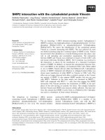
Northern blot analysis showed an abundant expression of
Ras-GRF2 transcript ( 8.0 kb) in human brain tissue
(Fig. 2 A). The enrichment of Ras-GRF2 in human brain i s
consistent with the highly abundant expression of dematin
in human brain [5,6]. In a ddition, low levels of the Ras-
GRF2 transcript were also detected in human heart,
placenta, kidney, and pancreas (Fig. 2A) . A highly sensitive
PCR-based assay w as then used to detect R as-GRF2 in the
cDNA pool of human tissues. As shown in Fig. 2B, a
relatively signi®cant amount of Ras-GRF2 was detected in
human ovary and spleen tissues. In the testis, an additional
band was detected that migrated just above the expected size
of the PCR product (Fig. 2B). The extra band was
subcloned and its cDNA was sequenced. The additional
PCR band encoded a 50-amino acid insert (I
1
for insertion 1)
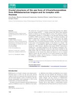
Fig. 1. Yeast two-hybrid a nalysis. (A) Sche-
matic representation o f dematin and R as±
GRF2 interaction. The carboxyl-terminal half
of dematin (amino acids 224±383) was used as
the bait for the yeast two -hybrid screening.
Yeast transformed with bo th dematin and
Ras-GRF2 grew on me dia lacking histidine
(+) and turned blue (marked with a B) in the
presence of X-gal indicative of a binding
interaction. Absence of growth was designated
by (±) while failure to activate the L acZ
reporter gene was designated as (W). (B) Yeast
mating between dematin an d Ras-GRF1 and
between limatin and Ras-GRF 1 and Ras -
GRF2. (C) Amino-acid sequence of insertion-
1 sequence. The ÔextraÕ ex on is located between
the amino aci ds KHAQ-Insertion1-DFEL of
the human Ra s-GRF2 sequence. The under-
lined sequence of insertion-1 shows homology
with an isofo rm of T rio nucleotide exchanger
as discussed in t he Results section.
642 M. Lutchman et al. ( Eur. J. Biochem. 269) Ó FEBS 2002
and is located between the candidate-destruction box and
Cdc25-like catalytic domain s of Ras-GRF2 (Fig. 1 C).
Genebank database analysis revealed that a 16-amino-acid
segment of insertion 1 is 75% identical to a sequence found
in an isoform of the Trio protein (Fig. 1C).
Speci®city of the binding interaction between dematin
and human Ras-GRF2
Several independent techniques w ere employed to e stablish
the speci®city of binding interaction between dematin and
Ras-GRF2. First, the yeast two-hybrid assay was used to
demonstrate the speci®city of binding between members
of the dematin and Ras-GRF families. As shown in
Fig. 1B, the C-terminal half of dematin [dematin (224±
383)] binds to the DH domain of human Ras-GRF2. The
dematin(224±383) construct was inte ntionally engineered to
delete the poly(glutamic) acid motif found in the N-terminal
half of the dematin core domain [5,6]. In preliminary control
tests, the poly(glutamic) acid motif appeared to contribute
in the autoactivation o f the full-length dematin c onstruct.
The design of the dematin(224±383) construct was also
in¯uenced by our previous studies showing a stable
expression of the headpiece domain in solution whereas
the bacterially expressed core domain o f d ematin was
relatively susceptible to proteolysis [4,5]. For this reason, the
dematin(224±383) construct was selected for the y east two-
hybrid and other biochemical assays.
A secon d bait construct for the yeast two-hybrid screen
contained only the headpiece do main of dematin. The
dematin(309)383) headpiece construct failed to bind the
DH domain of Ras-GRF2 in the yeast two-hybrid assay
(data not shown) suggesting that the Ras-GRF2 binding site
is likely to be located within the 84-residue [dematin(224±
308)] segment o f the core domain of dematin. Similarly, the
dematin(224±383) construct failed to bind to the DH
domain of human Ras-GRF1 that is 88% identical to
the DH domain of human Ras-GRF2. T his r esult suggests
that the human dematin binds speci®cally to the DH
domain of human Ras-GRF2 but not human Ras-GRF1
(Fig. 1B). We have recently identi®ed human limatin
(abLIM) as the closest homologue of dematin in mamma-
lian tissues [13]. A construct of human limatin(597±778)
corresponding to dematin(224±383) ( 40% identity) also did
not bind to the DH domain o f either Ras-GRF2 or R as-
GRF1 (Fig. 1B). Based on the results of the yeast two-
hybrid assay, we conclude that the interaction between
dematin and Ras-GRF2 is highly speci®c and is mediated by
a novel sequence located within the core domain of dematin.
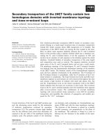
An in vitro overlay assay was used to demonstrate direct
biochemical interaction between dematin and Ras-GRF2.
Native dematin was puri®ed from human erythrocyte
membranes and tested for b inding to the recombinant
Ras-GRF2-DH protein immobilized on the nitrocellulose
membrane. As shown on Fig. 3A, native dematin speci®-
cally bound to the GST fusion protein of Ras-GRF2-DH
domain but not GST alone. Again, no binding was observed
between native dematin an d the GST fusion protein of
human Ras-GRF1-DH domain (data not shown). Speci®c
binding of the G ST fu sion protein of Ras-GRF2-DH
domain to the dematin(224±383) was quanti®ed by surface
plasmon resonance technique using a BIAcore biosensor
instrument. A homogeneous preparation of dematin(224±
383) domain (18 kDa) (free of GST) was immobilized to a
CM5 s ensor chip by a standard amine coupling protocol
[28]. The binding interaction of GST±Ras-GRF2-DH
domain (66 kDa) to the immobilized demat in(224±383)
was concentration dependent (Fig. 3 B). No such binding
was observed when GST samples were injected at increasing
concentrations (up to 6 .6 l
M
) onto the same dematin(224±
383)-immobilized ligand surface under the same experimen-
tal c onditions. T he binding was r eproducible after repeated
cycles of the regeneration process. These results demonstrate
that the DH domain of Ras-GRF2 protein speci®cally binds
to a segment of dematin encoded by dematin(224±383).
Apparent on/off rate constants for the observed binding
interaction between dematin and Ras-GRF2 protein was
determined from the association a nd dissociation phases of
the sensorgram using a nonlinear regression algorithm in the
BIAEVALUATION
3.0 software package. Estimated kinetic
constants for the immobilized dematin(224±383) and GST±
Ras-GRF2±DH interaction were k
a
7.64 ´ 10
3
M
)1
ás
)1
and k
d
3.53 ´ 10
)3
s
)1
. An apparent dissociation con-
stant K
d
462 n
M
was obtained from the ratio of k
d
/k
a
.Itis
noteworthy here that the GST domain of ligand-bound and
free GST±Ras-GRF2-DH domain could in principal,
undergo dimerization causing an avidity effect in both
association and dissociation phases of the interaction.
Dematin and Ras-GRF2 associate in mouse brain lysate
and in transfected epithelial cells
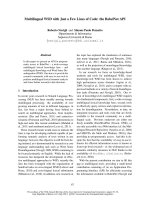
To test whether Ras-GRF2 and dematin associate in vivo,
we examined their association in mouse brain lysate and
mammalian cells. Dematin was immunoprecipitated from
mouse brain lysate using an af®nity-puri®ed polyclonal anti-
dematin I g. The dematin immunoprecipitate was analyzed
Fig. 2. Tissue expression of human Ras-GRF2. (A) No rthern blot
analysis of Ras-GRF2. Ras-GRF2 e xpression is most abundant in
the brain. A single band of 7.5kbisdetectedinmosttissues.
(B) A mu lt iple tissue cDNA panel was s cree ned b y P CR using Ras-
GRF2 speci®c p rimers. The bottom p anel shows equal amount of
starting cDNA pool in each tissue as d etected by the glyceraldehyde
3-phosphate dehydrogenase-speci®c primers.
Ó FEBS 2002 Dematin binds to Ras-GRF2 nucleotide exchange factor (Eur. J. Biochem. 269) 643
by SDS/PAGE and Western blotted with the Ras-GRF2
monoclonal antibody generated against the PH domain o f
Ras-GRF2 (Transduction Laboratories, Lexington, KY,
USA). A control without the addition of anti-dematin Ig did
not show any Ras-GRF2 band (Fig. 4A, lane 1). A speci®c
140-kDa b and consistent with the mobility of mouse Ras-
GRF2 was detected in total lysate ( Fig. 4A, lane 2) and in
lysate immunoprecipitated with the polyclonal anti-dematin
Ig (Fig. 4A, lan e 3). These results demonstrate that endog-
enous dematin and Ras-GRF2 associate within the same
protein complex in mouse brain lysate. To examine this
interaction further, we transfected human embryonic kidney
epithelial cells (A293) with either dematin or Ras-GRF2 or
both. The expression of Ras-GRF2 and dematin in the
transfected cells was con®rmed using an anti-myc Ig (data
not shown). Dematin, Ras-GRF2, and dematin/Ras-GRF2
lysates were i mmunoprecipitated with the anti-dematin Ig
and immunoprecipitates were blotted w ith the monoclonal
anti-(Ras-GRF2) Ig (Fig. 4B). Tot al Ras-GRF2 ly sate was
used as the control indicating the position o f 140-kDa band
(Fig. 4 B). T he Ras-GRF2 band was d etected only in the
cotransfected A293 cells (Fig. 4 B). Together, these results
indicate that dematin and Ras-GRF2 ass ociate with e ach
other in vivo under th e conditions described above.
Ras-GRF2 and dematin colocalize in the transfected
®broblasts
Direct binding of dematin to Ras-GRF2 suggested that the
two p roteins might colocalize whe n o ver-expressed i n t he
Fig. 3. Interaction of dematin with the DH domain of human Ras-GRF2. (A) Blot overlay assay. Approximately 2 lg of GST and GST-Ras-
GRF2-DH fusion protein was immobilized on the nitrocellulose. The immunoblot was incubated with puri®ed native dematin, and the binding of
dematin was detected by immunoblot analysis. The details of the blot overlay are described in the Experimental procedures. A similar analysis was
carried out using GS T-Ras-GRF1-DH fusion p rote in. No b indin g was ob served b etween dem atin and R as-GRF1 ( data not sh own). (B ) An overlay
plot of sensorgra ms s howing the binding interaction of GST±Ras-GRF2 a nd the C-terminal domain of de matin [dematin(224±383)]. A homo-
geneous sample of the dematin(224±383) protein was i mmobilized to the dextran matrix of a CM 5 sensor chip by a s tandard a mine coupling
procedure (1.0 ng p roteinámm
)2
). The sensorgrams were generated by i njecting dierent concentrations of GST±Ras-GRF2 (2.3 l
M
,1.2l
M
,
0.46 l
M
) at a ¯ow rate of 10 lLámin
)1
at 25 °C. Puri®ed recombinant GST (6.6 l
M
) did not bind under the same conditions. Apparent association
and dissociation rate constants were estimated from the sensorgrams using
BIAEVALUATION
3.0 s oftware: k
a
7.64 ´ 10
3
M
)1
ás
)1
and
k
d
3.53 ´ 10
)3
s
)1
. An a pparent dissociation c onstant (K
D
)of462n
M
was obtained from t he ratio of k
d
/k
a
. The avidity eect c aused by the
dimerization of the GST d om ain has not been discounted from the data i n the determination of kinetic constants.
Fig. 4. In vivo interaction of dematin with Ras-GRF2. (A) Co-immunoprecipitation of d ematin and R as-GRF2 from m ouse brain l ysate. Mouse
brain w as homogenized in NP-40 lysis b uer and t he homogenate was centrifuged at 14 000 g. The supernatant was precleared with protein G
beads and incubated w ith anti-dematin Ig. The i mmune co mplexes w ere recovered by pro tein G bead s that were exte nsively washed. Lane 1, p rotein
G beads were added in samples that were not incubated with anti-dematin Ig (negative control). Lane 2, total brain lysate (positive control). Lane 3,
dematin immune complexes t hat were immunoblotted with Ras-GRF2 antibody. The140 kDa band corresponds to Ras-GRF2.
(B) Co-transfection and coimmunoprecpitation of dematin and Ras-GRF2 com plex from A293 epithelial cells. A 293 cells were tran siently
transfected with either dematin or Ras-G RF2 or both for immunopre cipitation experiments. Lane 1, total lysate of the dematin/Ras-GRF2
cotransfected cells. Lane 2, a nti-dematin i mmunoprecipitate of dematin t ransfected cells. Lane 3, anti-dematin imm unoprecipate o f Ras-GRF2
transfected cells. Lane 4 shows anti-dematin immunoprecipitate of dematin/Ras-GRF2 cotransfected cells. Note that the 140 kDa Ras-GRF2 was
detected only in the c otransfe cted cells.
644 M. Lutchman et al. ( Eur. J. Biochem. 269) Ó FEBS 2002
mammalian cells. Full-length cDNA co nstructs of de matin
and Ras-GRF2 were transfected into NIH 3T3 ®broblasts to
generate stable cell lines. The expression of Ras-GRF2
protein in the stable clones w as con®rmed by the detection
of a 140-kDa polypeptide by Western blot analysis using an
anti-myc Ig (data not shown). The overexpression of
dematin was detected using a speci®c anti-dematin Ig. By
indirect immuno¯uorescence analysis, dematin and Ras-
GRF2 were colocalized in the perinuclear and c ytoplasmic
compartments of the transfected ®broblasts (Fig. 5).
Nuclear staining of neither dematin nor Ras-GRF2 was
not d etectable under these conditions. These results suggest
that the t wo proteins may interact with each o ther in the
cytoplasmic compartment, and directly or indirectly mod-
ulate the in vivo function of small GTPases in mammalian
cells.
Effect of dematin expression on ERK1
and JNK activation
Recent studies have shown that t he Cdc25-like domain of
Ras-GRF2 stimulates the activation of the MAP kinase
ERK1 and Ras upon in¯ux of intracellular calcium in A293
cells [22,26]. First, we wanted to test whether the binding of
dematin to the DH domain o f human Ras-GRF2 had a ny
downstream regulatory effects on the activation of ERK1
via its Cdc25 domain. The recombinant Cdc25-like domain
of human Ras-GRF2 stimulated guanine nucleotide
exchange on Ha-Ras protein (data reviewed but not shown).
We then transfected the A 293 cells with various constructs
and measured the extracellular-signal-regulated kinase
(ERK) activity as described in the Experimental procedures.
Interestingly, the transfection of dematin alone in A293 cells
caused a s igni®cant enhancement of ionomycin-induced
activation of ERK1 (Fig. 6A). However, dematin over-
expression did not result in any measurable modulatory
Fig. 6. Eect of dematin on ERK1 activation. (A) A 293 cells were
transfected with either vector, or constitutively active Ras, or dematin,
or Ra s-GRF2. C ells were stimulated wit h ionomycin, as described i n
the Experimental p rocedures, and lysates were immunoblotted with
respective antibodies. A nti-tubulin Ig w as used to normalize the pro-
tein content of each lysate. ERK1 activation was detected with an
antibody against phospho -ERK1. This a ntibody detects a doublet of
activated ERK1. N ote t hat d ematin overexpression alone induce d
signi®cant increase in the activation of ERK1. (B) Dematin does not
modulate the Ras-GRF2 induced activation of ERK1. Anti-tubulin Ig
normalized lysates were then tested for the presence of total ERK
protein using an anti-ERK2 Ig. Activated E RK1 was detected as
described in ( A).
Fig. 5. Immuno¯uorescent colocalization of
dematin and Ras-GRF2. (A) Phase contrast
picture of stably c otransfecte d dematin/Ras-
GRF2 NIH 3T3 cells. (B) Rhodamine-labeled
dematin antibody s howing localization of
dematin in the p erinucle ar and cytoplasmic
compartments of the transfected cells.
(C) FITC-labeled anti-myc i n the stably
transfected cells showing perinuclear and
cytoplasmic l ocalization of human
Ras-GRF2. (D) An ov e rlay of B/C panels
indicating that dematin a nd Ras-GRF2
localize to the s ame compartments of these
overexpressing cells. Magni®cation 100´.
Ó FEBS 2002 Dematin binds to Ras-GRF2 nucleotide exchange factor (Eur. J. Biochem. 269) 645
effect o n the ionomycin-induced activation of ERK1
through Ras-GRF2 (Fig. 6B). These results suggest t hat
dematin does not directly modulate the Ras s ignaling
pathway mediated by t he Cdc25 domain of human Ras-
GRF2.
The DH domain of several exchange proteins has been
shown to exhibit guanine nucleotide exchange activity
[22,23,25,26]. To investigate the nucleotide exchange activity
of the DH domain o f human Ras-GRF2, w e ®rst tested
whether the recombinant DH domain could catalyze the
nucleotide exchange of RhoA GTPase. In vitro exchange
assays did not sh ow any stimulation of the nucleotide
exchange on RhoA irrespective of whether dematin was
bound to the DH domain of Ras-GRF2 (data reviewed but
not shown). Recently, the DH domain of mouse Ras-GRF2
has been reported to enhance t he nucleotide e xchange
activity of Rac1 and stimulates stress-activated protein
kinase (SAPK), also known as Jun N-terminal kinase
(JNK), in transfected 293 cells [26]. Indeed, the human
Ras-GRF2 activated Rac1 in transfected COS-7 cells as
demonstrated by a GST-pulldown assay (Fig. 7). More-
over, the coexpression of dematin did not modulate the Rac
activation (Fig. 7 ). Although it appears that the dematin
overexperssion may slightly inhibit the Rac exchange
activity (Fig. 7), it is probably accounted for by the slightly
lower expression of R as-GRF2 in that particular condition.
We then proceeded to examine the effect of dematin
overexpression on JNK activation via Ras-GRF2 in the
transfected COS-7 cells. The JNK activation w as quanti®ed
by measuring the transcriptional activation of Jun by human
Ras-GRF2. As expected, the expression o f R as-GRF2 a nd
constitutively active Rac(12V) resulted in the transcriptional
activation o f Jun (Fig. 8). Interestingly, the coexpression of
dematin c aused a signi®cant inhibition of Jun activation by
Ras-GRF2 as well as Rac(12V) (Fig. 8). Similarly, cot rans-
fection of d ematin and Ras-GRF2 in A293 cells suppressed
JNK activation by ®vefold (data reviewed but not
shown). Together, these results indicate that dematin
functions downstream of the signaling cascade mediated
by Rac1 an d Ras-GRF2 in t he mammalian epithelial cells.
DISCUSSION
The identi®cation of dematin as a component of erythrocyte
cytoskeleton revealed many aspects of its actin binding/
bundling properties [1,2,27]. However, the function of
dematin in nonerythroid cells remains t o be elucidated.
The primary structure of dematin suggested that its modular
sequence might encode distinct cellular functions [4,5]. The
C-terminal head piece domain of d ematin is specialized for
its actin binding function, and is likely to modulate
dematin's actin bundling activity [2,27]. In contrast, the
core domain of d ematin may serve as a docking site for t he
binding of unknown proteins. With this modular s tructure,
dematin could be ideally suited as a molecular adaptor
linking the cytoplasmic or membrane-associated proteins to
the actin cytoskeleton. Due t o t he abundant expression of
dematin in the brain, we searched for dematin-interacting
proteins by screening a human brain cDNA library using
the yeast two-hybrid system. Guided by our previous studies
Fig. 7. Dematin does not regulate Ras-GRF2 encoded R ac-GRF activ-
ity. COS-7 cells were transien tly transfected with pAX142-RacI (WT)
and with pCDNA3 that contained the indicated cDNAs. Lysates were
collected at 48 h and examined by Western blot for expression of RacI
(B), Ras-GRF2 (C), and Dematin (D). L ysates were then normalized
forRacIexpressionandsubjectedtoanityprecipitationusing
immobilized GST-Pak. GTP-bound RacI that was precipitated with
GST-Pak w as visualized by Western blot (A) using an anti-RacI Ig
(C14, Santa Cruz Biotechnology). Dematin was immunoblotted using
a monoclonal antibody from T ransduction Laboratories.
Fig. 8. Dematin blocks transcriptional activation of Jun by Ras-GRF2.
COS-7 cells were transfected with plasmids encoding the indicated
proteins (3 lg each), along with an expre ssion vector for th e Gal4
DNA binding domain fused to transactivation domain of Jun [0.25 lg
Gal-Jun (1±223)] and a Gal4 luciferase r eporter (2.5 lg 5XGal4-luc).
For each c ondition , pCMVnlac (0.25 lg) was a lso included in the
transfection as an internal con trol for transfection eciency and/ or
growth inhibition. All val ues were normalized against b-galactosidase
activity. Fold a ctivation was determined by the number o f l uciferase
units relative to the number of units seen with the vector control. Data
shown are representative of at least three independent assays p er-
formed on duplicate p lates. The error b ars indicate standard d evi-
ations.
646 M. Lutchman et al. ( Eur. J. Biochem. 269) Ó FEBS 2002
showing poor expression of the core domain, most likely
due to the presence of a PEST sequence that marks proteins
for proteolysis, we designed a dematin bait construct
expressing only 84 amino acids of the core domain fused
to the h eadpiece domain. The headpiece domain is a
protease-resistant module that expresses as a stable recom-
binant protein in vitro [4]. This bait construct of dematin
containing 84 amino acids of the core domain and complete
headpiece domain mediated binding with the DH domain of
human Ras-GRF2 (Fig. 1). In contrast, a bait construct
containing only the headpiece domain of dematin failed to
bind to the DH domain of human Ras-GRF2 (data not
shown). This observation suggests that a novel 84-amino-
acid sequence originating from the core domain mediates
dematin b inding to the DH domain of human Ras-GRF2
protein. Clearly, a d etailed evaluation by in vitro mutagen-
esis will be required t o p recisely map the Ras±GRF2
binding interface and its s tability within t he core domain of
dematin.
The inability of dematin to bind to the DH domain of
human Ras-GRF1, as well as l ack of binding between
limatin (abLIM) and Ras-GRF2/Ras-GRF1 underscores
the s peci®city of the binding interaction between dematin
and Ras-GRF2. The primary structure of human brain
Ras-GRF2 encodes a highly conserved multidomain p ro-
tein consisting of an N-ter minal PH domain, followed by
the coiled coil (cc) and IQ motifs, a single DH domain that is
closely linked to an other PH domain, REM and CDB
motifs, and a C-terminal Cdc25 exchanger domain (Fig. 1).
The overall domain organization of human Ras-GRF2 is
similar to its mouse homologue except for the presence of an
additional sequence of 50 amino acids located just upstream
of the Cdc25 exchanger domain (Fig. 1) [22]. The I
1
insertion s equence w as identi®ed during PCR ampli®cation
of human testis cDNA pool, and likely to represent an
alternatively s pliced exon. Interestingly, a s egment of the I
1
insertion sequence shows signi®cant homology with another
nucleotide exchanger termed Trio [32]. Trio is a multi-
domain protein consisting of Rac- and Rho-speci®c guanine
nucleotide exchanger domains, and binds to the leukocyte
antigen-related transmembrane tyrosine phosphatase [32].
Whether the Ras-GRF2 isoform bearing the I
1
insertion
sequence binds to a similar transmembrane protein remains
to be determined. W hile our manuscript w as under r eview,
the primary structure of human Ras-GRF2 was published
[33]. Our results are consistent with the r eported primary
structure of human Ras-GRF2 [33]. The presence of I
1
insertion upstream of the Cdc25-like domain of Ras-GRF2
remains unique in our sequence (Fig. 1).
The widespread tissue distribution of Ras-GRF2 (Fig. 2),
in contrast to restricted neuronal expression of Ras-GRF1,
is consistent with the tissue expression of dematin [5,6]. Both
dematin a nd Ras -GRF2 are enriched in human brain
suggesting a functional interdependence of their interaction
in vivo. The co-immunoprecipitation of dematin and Ras-
GRF2 from brain lysate (Fig. 4A) and transfected A293
epithelial cells (Fig. 4 B) suggest that the two proteins are
found in the same protein complex in vivo. Biochemical
analysis of cellular fractionation assays revealed that the two
proteins are p redominantly associated with the particulate
fraction of transfected cells (data not shown). This result,
together with the cytosolic and p erinuclear localization of
dematin and Ras-GRF2 in transfected ®bro blasts (Fig. 5),
suggests that t he protein c omplex may r egulate cytoskeletal
reorganization in mammalian cells.
Direct binding of dematin t o the DH d omain of Ras-
GRF2 raises important issues regarding the function of
these domains in Ras signaling and actin reorganization.
Nucleotide exchange factor proteins carrying deletions and
targeted mutations within the DH domains lose their
transformation potential and catalytic exchange activity
[34]. A physical link between the DH domains, cellular
transformation, and cytoskeletal association is likely to be
afforded by the activation of Rho and Rac family GTPases
[34]. T hese observations imply that a n alternate mechanism
must exist that can couple Ras-GRF exchangers to
micro®lament reorganization. It has recently been demon-
strated that R as-GRF1 and Ras-GR F2 can form homo-
and hetero-oligomers via their DH d omains [33]. T his
observation s uggests t hat D H domains, in a ddition to their
nucleotide exchange function, may be involved in protein±
protein interactions. While our results indicate that dematin
does not directly interact with Ras-GRF1, dematin may
indirectly recruit GRF1 to the actin cytoskeleton via its
association with Ras-GRF2. It is therefore plausible that the
direct binding of dematin to the DH domain of R as-GRF2
may provide a functional link between Ras signaling and the
actin cytoskeleton.
Elucidation of the crystal structure of tandem DH a nd
PH domains of human Sos1 protein highlights the dramatic
complexity of the DH domain±mediated interactions [35].
The c rystal structure revealed that the DH domain i s
composed of three h elical segments, two of which provide a
highly conserved surface bearing functionally critical r esi-
dues [35]. The adjacent P H domain s tructure is so oriented
that its interaction with inositol(1,4,5)-triphosphate is likely
to in¯uence t he binding of DH domain with potential
GTPases. This pivotal insight into the structure of the DH±
PH domains opens a case for precise mapping of dematin
binding to a speci®c helical segment(s) of Ras-GRF2
protein. The reported interaction of dematin with the DH
domain of Ras-GRF2 may therefore provide a rationale for
the modulation of cytoskeletal integrity by phosphorylation,
phospholipid binding, and GTPase activation.
Much of the c urrent evidence implicates the Rho family
of GTP ase s a s key regulators of the actin cytoskeleton [36].
For instance, the activation of the Rho GTPase leads to
stress ®ber and focal adhesion formation while the activa-
tion of Rac and cdc42 leads to the formation of lamello-
podia and ®lopodia, respectively [36]. The i nduction of
membrane ruf¯es by microinjection of activated mutant Ras
into ®broblasts strongly suggested a role of Ras in t he
remodeling of actin cytoskeleton [37]. The association of
Ras-GRF2 with dematin, an actin binding and bundling
protein, provides a potential coupling mechan ism between
Ras signaling a nd the a ctin cytoskeleton without Rho
protein intermediaries. Although our data indicate that the
direct binding of dematin to the DH domain does not affect
the activation of E RK1 via th e C dc25-like domain o f Ras-
GRF2 (Fig. 6), the activation of E RK1 by d ematin alone
suggests a potential modulatory role of t he actin cytoskel-
eton in the Ras signaling pathways. More importan tly, the
data shown in Figs 7 and 8 provide the ®rst evidence for a
functional role of d ematin in the regulation of Rac1-JNK
signaling pathway. Suppression of JNK activation by t he
overexpression of dematin, irrespective of whether the signal
Ó FEBS 2002 Dematin binds to Ras-GRF2 nucleotide exchange factor (Eur. J. Biochem. 269) 647
is transmitted v ia Ras-GR F2 or Rac 1, h ightlights t he
functional importance of the dematin-mediated reorganiza-
tion of the actin cytoskeleton in intracellular signaling
pathways. It is noteworthy here that Vav, a proto-oncogene
that plays a major role i n cell proliferation and cytoskeletal
organization, activates Rac1 and JNK pathway only upon
phosphorylation of i ts tyrosine residues [38]. As dematin's
actin bundling activity is completely dependent upon its
state of phosphorylation, a possibility remains that a
physical link b etween dematin and Ras-GRF2 may man-
ifest functionally upon post-translational modi®cation of
either protein in vivo under sp eci®c stimulatory conditions.
DH domain-containing proteins, of w hich there a re
greater than 20 members, constitute the largest family of
oncogenes [34]. In fact, many DH domain p roteins were
discovered by virtue of their transforming ability when
expressed in ®broblasts. For instance, Tiam-1 is an exchange
factor for Rac and was ide nti®ed by virtue of its contribu-
tion in tumor invasion and metastasis pathways [39,40].
Similarly, the APC colon tumor suppressor b inds to a Rac-
speci®c guanine nucleotide exchange factor (Asef) a nd
regulates membrane ruf¯ing and l amellipodia formation i n
epithelial cells [41]. The mechanism by which these nucle-
otide exchangers modulate cell signaling and cytoskeletal
reorganization is poorly understood. It is of interest to note
that w e h ave recently reported loss of heterozygozity of the
dematin gene in a majority of 8p21-linked prostate tumors
[14]. Based on these observations, we postulate that dematin
may play a role in the regulation o f cell s hape with
implications in understanding the mechanism of cellular
transformation and tumor progression in malignant cells.
This proposed function of dematin would be analogous to
the recently discovered role of the neuro®bromatosis type II
(NF2) tumor suppressor p rotein in the i nhibition of Rac-
induced signaling as a possible mechanism of tumor
initiation and progression [42].
ACKNOWLEDGEMENTS
The National Institutes of Health Grants HL51445 (AHC) and
CA77493 (IPW) supported this work. We are grateful to Dr Larry Feig
of Tufts University Biochemistry Department for sharing t he cDNA
constructs and giving us v aluable advice during the course of these
studies. We thank Dr J. Samulski for providing the pCMVnlac
construct. We are a lso thankful to Donna Marie-Mironchu k f or help
with the artwork and D r Richie K hanna of St. Elizabeth's Medical
Center for critically reading the m anuscript.
REFERENCES
1. Siegel,D.L.&Branton,D.(1985)Partialpuri®cationandchar-
acterization o f an actin-bundling protein, band 4.9, from human
erythrocytes. J. Cell Biol. 100, 775±785.
2. Chishti, A., Levin, A. & Branton, D. (1988) Abolition of actin-
bundling by phosphorylation of human erythrocyte protein 4 .9.
Nature 334, 718±721.
3. Gilligan, D.M. & Bennett, V. (1993) The junctional complex of the
membrane skeleton. Seminars Hematol. 30, 74±83.
4. Azim, A.C., Knoll, J.H., Beggs, A.H. & Chishti, A.H. (1995)
Isoform cloning, actin binding, and chromosomal localization of
human erythroid dematin, a member of the villin superfamily.
J. Biol. Chem. 270, 17407±17413.
5. Rana, A.P., Ru, P., Maalouf, G.J., Speicher, D.W. & Chishti,
A.H. (1993) Cloning of human erythroid dematin reveals another
member of the villin family. Proc. Natl Acad. Sci. USA 90, 6651±
6655.
6. Kim, A.C., Azim, A.C. & Chishti, A.H. (1998) Alternative splicing
and structure of the human erythroid dematin gene. Biochim.
Biophys. Acta 1398, 3 82±386.
7. Azim, A.C., Marfatia, S.M., Korsgren, C., Dotimas, E., Cohen,
C.M. & Chishti, A.H. (1996) Human erythrocyte dematin and
protein 4 .2 (pallidin) are ATP binding proteins. Biochemistry 35,
3001±3006.
8. Azim,A.C.,Kim,A.C.,Lutchman,M.,Andrabi,S.,Peters,L.L.
& Chishti, A.H. (1999) cDNA sequence, genomic structure, and
expression of the mouse d ematin gene. Mamm. G en. 10 , 1026±
1029.
9. Arpin, M., Pringault, E., Finidori, J., Garcia, A., Jeltsch, J.M.,
Vandekerckhove, J. & Louvard, D. (1988) Sequence of human
villin: a large duplicated domain homologous with o ther actin-
severing proteins and a unique small carboxy-terminal domain
related to villin speci®city. J. Ce ll Biol. 107, 1759±1766.
10. Friederich, E., Vancompernolle, K., Huet, C., Goethals, M.,
Finidori, J., Vandekerckhove, J. & L ouvard, D. (1992) An actin-
binding s ite c ontaining a conserved motif of c harged amino acid
residues is essential for the morphogenic eect of villin. Cell 70,
81±92.
11. Pinson, K.I., Dunbar, L., Samuelson, L. & Gumucio, D.L. (1998)
Targeted disruption of the m ouse villin ge ne does not impair t he
morphogenesis of m icrovilli. Dev. Dyna m. 211, 109±121.
12. Ferrary, E., Cohen-Tannoudji, M., Pehau-Arnaudet, G.,
Lapillonne, A., Athman, R., Ruiz, T., B oulouh a, L., El Marjou,
F.,Doye,A.,Fontaine,J.J.,Antony,C.,Babinet,C.,Louvard,D.,
Jaisser, F. & Robine, S. (1999) In vivo, villin is required for
Ca(2+)-dependent F-actin disruption in intestinal brush borders.
J. Cell Biol. 146, 819±830.
13. Roof, D.J., Hayes, A., Adamian, M., Chishti, A.H. & Li, T. (1997)
Molecular c haracterization of abLIM, a novel actin-binding and
double zinc ®nger p ro tein. J. Cell Biol . 138, 575±588.
14. Lutchman,M.,Pack,S.,Kim,A.C.,Azim,A.,Emmert-Buck,M.,
Huel, C.V., Zhuang, Z . & Chishti, A.H. (1999) Loss of he te ro-
zygosity on 8p in prostate cancer implicates a r ole for de matin in
tumor progression. Cancer Genet. Cytogen. 115, 65±69.
15. Boguski, M. S. & McCormick, F. (1993) Proteins reg ulating Ras
and its relatives. Na ture 366, 643 ±654.
16. Bos, J.L. (1989) Ras oncogenes in human cancer. Cancer Res. 49,
4682±4689.
17. Brambilla, R., Gnesutta, N., Minichiello, L., White, G., Roylance,
A.J., Herron, C.E., Ramsey, M., Wolfer, D.P., Cestari, V., Rossi-
Arnaud, C., Gra nt, S.G., Chapman, P.F., Lipp, H.P., Sturani, E.
& K lein, R. ( 1997) A role for the Ras signalling pathway in sy n-
aptic transmission and long-term memory. Na tu re 390, 281±286.
18. Rozakis-Adcock, M., Fernley, R., Wade, J., Pawson, T. &
Bowtell, D. (1993) The S H2 and SH3 domains of mammalian
Grb2 couple the EGF receptor to the Ras activator mSos1. Nature
363, 83±85.
19. Shou, C., Farnsworth, C.L., Neel, B.G. & Feig, L.A. (1992)
Molecular cloning of cDNAs encoding a guanine-nucleotide-
releasing factor for Ras p21. Nature 358, 351±354.
20. Lowenstein, E.J., Daly, R .J., Batzer, A.G., Li, W., Margolis, B.,
Lammers, R., U llrich, A., S kolnik, E .Y., Bar-Sagi, D. & Schl es-
singer, J. (1992) The SH2 and SH3 domain-containing protein
GRB2 links receptor tyrosine kinases to ras signaling. Cell 70,
431±442.
21. Wei, W., Mosteller, R.D., Sanyal, P., Gonzales, E., McKinney,
D.,Dasgupta,C.,Li,P.,Liu,B.X.&Broek,D.(1992)Identi®-
cation of a m ammalian gene structurally and f unctionally related
to the CDC25 gene of Saccharomyces cerevisiae. Proc. Natl Acad.
Sci. USA 89, 7100±7104.
22. Fam, N.P., Fan, W.T., Wang, Z., Zhang, L.J., Chen, H. & Moran,
M.F. (1997) Cloning a nd characterization of Ras-GRF2, a novel
648 M. Lutchman et al. ( Eur. J. Biochem. 269) Ó FEBS 2002
guanine nucleotide exchange factor f or Ras. Mol. Cell. Biol. 17,
1396±1406.
23. Mattingly, R.R. & Macara, I.G. (1996) Phosphorylation-de pen-
dent activation o f the Ras-GRF/CDC25Mm e xchange f actor b y
muscarinic receptors and G-protein beta gamma subunits. Nature
382, 268±272.
24. F arnswort h, C.L., Fre shney, N.W., Rosen, L.B., Ghosh, A.,
Greenberg, M.E. & Feig, L.A. (1995) Calcium activation of R as
mediated by neuronal exchange factor Ras-GR F. Na ture 376,
524±527.
25. Kiyono, M., Satoh, T. & Kaziro, Y. (1999) G protein beta gamma
subunit-dependent Rac-guanine nu cleotide exchange activity of
Ras-GRF1/CDC25 (Mm). Proc. Natl Acad. Sci. USA 96 , 4 826±
4831.
26. F an, W.T., Koch, C.A., de Hoog, C.L., Fam, N.P. & Moran,
M.F. (1998) The exchange factor Ras-GRF2 activates Ras-
dependent and Rac-dependent mitogen-activated protein kinase
pathways. Cur r. Biol. 8, 935±938.
27. Ch ishti, A ., F aquin, W., Wu, C.C. & B ranton, D . ( 1989) Puri®-
cation of erythrocyte dematin (protein 4.9) reveals an endogenous
protein kinase that modulates actin-bundling activity. J. Biol.
Chem. 264, 8985±8991.
28. Johnsson, B., Lofas, S. & Lindquist, G. (1991) Immobilization of
proteins to a carboxymethyldextran-modi®ed gold surface for
biospeci®c interaction analysis i n surface plasmon resonance sen-
sors. Anal. Biochem. 198, 268± 277.
29. Whitehead, I.P., Khosravi-Far, R., Kirk, H., Trigo-Gonzalez, G.,
Der, C.J. & Kay, R. (1996) Expression cloning of lsc,anovel
oncogene with structural similarities to the Dbl family of guanine
nucleotide exchange factors. J. Biol. Chem. 271, 18643±18650.
30. Whitehead,I.P.,Lambert,Q.T.,Glaven,J.A.,Abe,K.,Rossman,
K.L., M ahon, G.M., Trzaskos, J.M., Kay, R., Campbell, S.L. &
Der, C.J. (1999) Dependence of Dbl and Dbs transformation
on MEK and N F-kappaB a ctivation . Mol. Cell. Biol. 19, 7759±
7770.
31. Whitehead, I., Kirk, H., Tognon, C., Trigo-Gonzalez, G. & Kay,
R. (1995) Expression cloning of lfc, a novel oncogene with struc-
tural similarities to guanine nucleotide exchange factors and to the
regulatory region of protein kinase C . J. Biol. Chem. 270, 18388±
18395.
32. D ebant, A., Serra-Pages, C., Seipel, K., O'Brien, S., Tang, M.,
Park, S.H. & St reuli, M. (1996) The multidomain protein Trio
binds the LAR transmembrane tyrosine phosphatase, contains a
protein kinase domain, and h as separate rac-speci®c and rho-
speci®c guanine nucleotide exchange factor domain s. Proc. N atl
Acad.Sci.USA93, 5466±5471.
33. Anborgh, P.H., Qian, X., Papageorge, A.G., Vass, W.C.,
DeClue, J.E. & Lowy, D.R. (1999) Ras-speci®c exchange factor
GRF: oligomerization through its Dbl homology domain
and calcium-dependent activation of Raf. Mol. Cell. B io l. 19,
4611±4622.
34. Whitehead, I.P., Campbell, S., Rossman, K.L. & Der, C.J. (1997)
Dbl family proteins. Bi ochim. Biophys. Acta 1332, 1±23.
35. Soisson, S.M., Nimnual, A.S., Uy, M., Bar-Sagi, D. &
Kuriyan, J. (1998) Crystal structure of the Dbl and pleckstrin
homology domains from the human Son of sevenless protein. Cell
95, 259±268.
36. Hall, A. (1998) Rho G TPases and the actin c ytoskeleton. Science
279, 509±514.
37. Bar-Sagi, D. & Feramisco, J.R. (1986) Induction o f m embrane
ruing and ¯uid-phase pinocytosis in quiescent ®broblasts by ras
proteins. Science 233, 1061±1068.
38. Crespo, P., Schuebel, K.E., Ostrom, A.A., Gutkind, J.S. &
Bustelo, X.R. (1997) Phosphotyrosine-dependent activation of
Rac-1 GDP/GTP exchange by the vav proto-oncogene product .
Nature 385, 169± 172.
39. Habets,G.G.,Scholtes,E.H.,Zuydgeest,D.,vanderKammen,
R.A., Stam, J.C., Berns, A. & Collard, J.G. (1994) Identi®cation of
an invasion-in ducing gene, Tiam-1, that encodes a protein with
homology to GDP-GTP exchangers for Rho-like proteins. Cell 77,
537±549.
40. H ordijk, P.L., ten Klooster, J.P., van der Kammen, R.A.,
Michiels, F ., Oomen, L.C. & Collard, J.G. (1997) Inhibition of
invasion of epithelial cells by Tiam1-Rac signaling. Science 278,
1464±1466.
41. Kawasaki, Y., Senda, T., Ishidate, T., Koyama, R., Morishita, T.,
Iwayama, Y., Higuchi, O. & A kiyama, T. (2000) A sef, a link
between the tumor suppressor APC and G-protein signaling.
Science 289, 1194±1197.
42. Shaw,R.J.,Paez,J.G.,Curto,M.,Yaktine,A.,Pruitt,W.M.,
Saotome,I.,O'Bryan,J.P.,Gupta,V.,DerRatner,N.C.J.,
Jacks, T. & M cClatchey, A .I. (2001) The Nf2 tumor suppres-
sor, Merlin, functions in Rac-dependent signaling. Dev. Cell 1,
63±72.
Ó FEBS 2002 Dematin binds to Ras-GRF2 nucleotide exchange factor (Eur. J. Biochem. 269) 649