Báo cáo khoa học: A chloroplast RNA binding protein from stromal thylakoid membranes specifically binds to the 5¢ untranslated region of the psbA mRNA potx
Bạn đang xem bản rút gọn của tài liệu. Xem và tải ngay bản đầy đủ của tài liệu tại đây (235.32 KB, 8 trang )
A chloroplast RNA binding protein from stromal thylakoid membranes
specifically binds to the 5¢ untranslated region of the
psbA
mRNA
Friedrich Ossenbu¨hl*, Kristina Hartmann and Jo¨ rg Nickelsen
Lehrstuhl fu
¨
r Allgemeine und Molekulare Botanik, Ruhr-Universita
¨
t Bochum, Bochum, Germany
The intrachloroplastic localization of post-transcriptional
gene expression steps represents one key determinant for the
regulation of chloroplast development. We have character-
ized an RNA binding protein of 63 kDa (RBP63) from
Chlamydomonas reinhardtii chloroplasts, which cofraction-
ates with stromal thylakoid membranes. Solubility proper-
ties suggest that RBP63 is a peripheral membrane protein.
Among RNA probes from different 5¢ untranslated regions
of chloroplast transcripts, RBP63 preferentially binds to the
psbA leader. This binding is dependent on a region com-
prising seven consecutive A residues, which is required for
D1 protein synthesis. A possible role for this newly discov-
ered RNA binding protein in membrane targeting of psbA
gene expression is discussed.
Keywords: chloroplast gene expression; D1 synthesis; mem-
brane targeting; RNA binding; thylakoid.
Chloroplast gene expression within plant or algal cells has
been shown to be dependent upon nuclear gene products,
which are translated in the cytoplasm and, subsequently, are
imported by the organelle. Herein, they fulfil their function
by interacting with distinct elements and/or factors associ-
ated with the chloroplast gene expression machinery [1,2].
While the molecular mechanisms of regulatory interaction
during these processes are being pieced together, relatively
little is known about the intrachloroplast localization of
different steps of gene expression.
For instance, the chloroplast DNA is organized in
nucleoids. In developing higher plants, these are associated
with the inner plastid envelope through the PEND protein.
Upon full chloroplast maturation, the cpDNA is localized
to thylakoid membranes by an undetermined mechanism
[3]. This suggests that the plastid transcription machinery is
distributed in a similar way. Further evidence for subcom-
partmentalization of chloroplast gene expression has been
obtained by the recent cloning of genetically defined loci,
which are required for distinct post-transcriptional steps of
chloroplast gene expression. These factors could be detected
in the stromal compartment like Crp1 and Crs2 in maize
[4,5], or Maa3, Mbb1 and Nac2 in Chlamydomonas
reinhardtii [6,7,8], which are involved in processing/splicing
or stabilization of specific chloroplast transcripts, respec-
tively. Conversely, association with the inner plastid enve-
lope and/or the so-called low density membranes (LDM),
which resemble the inner envelope membrane with regard to
their lipid composition [9], has been observed for the
translation termination factor RF4 from spinach and the
RNA splicing factor Maa2 from C. reinhardtii [10,11].
By application of in vitro run-on translation systems, a
cotranslational insertion of thylakoid membrane proteins
has been reported [12]. This is consistent with the finding
that chloroplast psbA and psbD transcripts are associated
with thylakoids [13,14]. Further evidence for an essential
role of the thylakoid membrane for chloroplast gene
expression was deduced from the analysis of a maize
mutant lacking the chloroplast SecY homologue of the
thylakoid protein translocation machinery. In this mutant,
chloroplast translation is defective [15].
Moreover, in vitro assays revealed a still growing number
of various RNA binding proteins (RBPs), which have been
implicated in the control of post-transcriptional gene
expression steps. Some of these RBPs appear to mediate
their function via distinct cis-acting elements within the 5¢
untranslated regions (UTRs) of plastid mRNAs, which are
essential for mRNA maturation/stabilization and/or trans-
lation initiation [16–20]. Whereas some of these RNA
binding activities are localized to the chloroplast stroma
[18,21], recent work suggests that many other RBPs are
associated with the abovementioned LDM system [9].
In C. reinhardtii,the5¢ UTR of the psbA mRNA
encoding the D1 protein of the photosystem (PS) II reaction
centre was shown to interact with RB47, a member of the
polyA-binding protein family, which forms a complex with
major proteins of 38, 55 and 60 kDa [22]. RB60 represents a
protein disulfide isomerase that exhibits no RNA binding
activity, but is involved in the light and/or redox regulation
of D1 synthesis. RB47 was localized to the LDM system [9]
and, in addition, RB60 was shown to be partitioned
between the stroma and the membrane phase following
chloroplast fractionation experiments [23].
We have previously reported on a set of chloroplast
RBPs, which interact with the 5¢ UTR of the psbD mRNA
encoding the D2 protein of PS II of C. reinhardtii. Amongst
those, a protein of 63 kDa (RBP63) cofractionated with
thylakoid membranes during separation of chloroplast
lysates by sucrose step gradient centrifugation [18]. In this
Correspondence to J. Nickelsen, Lehrstuhl fu
¨
r Allgemeine und Mole-
kulare Botanik, Ruhr-Universita
¨
t Bochum, 44780 Bochum, Germany.
Fax: + 49 2343214184, Tel.: + 49 2343225539,
E-mail:
Abbreviations: LDM, low density membranes; RBP, RNA binding
protein; UTR, untranslated region; PS, photosystem.
*Present address: Department fu
¨
r Biologie I, Ludwig-Maximilians
Universita
¨
t, Menzinger Str. 67, 80638 Mu
¨
nchen, Germany.
(Received 13 March 2002, revised 7 June 2002, accepted 19 June 2002)
Eur. J. Biochem. 269, 3912–3919 (2002) Ó FEBS 2002 doi:10.1046/j.1432-1033.2002.03057.x
report, this particular protein is characterized further. We
were able to show that RBP63 is a stromal thylakoid
membrane protein. It preferentially binds to the 5¢ UTR of
the psbA message determined by an A-rich region eight
nucleotides upstream of the AUG start codon. To the best
of our knowledge, this is the first RNA binding activity
found exclusively within thylakoid membranes.
MATERIALS AND METHODS
Algal strains, preparation of protein fractions
and western analysis
The C. reinhardtii strain used harboured the cw15 mutation,
which facilitates chloroplast isolation. It was maintained on
Tris/acetate/phosphate medium [24] at 25 °C and cultures
weregrowntoadensityof2· 10
6
cells Æ mL
)1
in this
medium containing 1% sorbitol. Cells were harvested by
centrifugation and chloroplasts and chloroplast subfrac-
tions were prepared exactly as described previously [18].
For the separation of stroma and grana thylakoids,
isolated unstacked thylakoid membranes were resuspended
at 0.4 mgÆmL
)1
chlorophyll in buffer B (15 m
M
tricine/
KOH pH 7.9, 0.1
M
sorbitol, 10 m
M
NaCl, 5 m
M
MgCl
2
,
10 m
M
NaF) and incubated for 30 min at 4 °C to allow for
restacking [25]. Subsequent fractionation was carried out as
described previously [26]. In brief, restacked thylakoid
membranes were incubated with 0.4% digitonin in buffer B
for 2 min at room temperature. The incubation was stopped
by adding 10 vol. buffer B. The suspension was centrifuged
four times at 4 °C. Each supernatant was used for the next
centrifugation step. The relative acceleration rates were
1000 g for10min,10000g for30min,40000g for 30 min
and 150 000 g for 1 h. The different pellets corresponding
to thylakoid membranes (P
1000g
), grana thylakoids (P
10000g
),
intermediate membranes (P
40000g
) and stroma thylakoids
(P
150000g
) were resuspended in 2 · lysis buffer, diluted with
75% glycerol and stored at )20 °C [18].
Western analyses were carried out as described previously
[18]. Protein and chlorophyll concentrations were deter-
mined as described previously [27,28].
Membrane solubilization analysis
For membrane solubilization analysis, isolated thylakoid
membranes corresponding to 1 mg chlorophyll were incu-
bated as indicated in Fig. 2 at 4 °C for 30 min and
centrifuged at 100 000 g for 1 h. The pellets were resus-
pended in 2 · lysis buffer. NaCl in the supernatants of the
salt washes was removed by centrifugation through ultrafree
centrifugal filter (Millipore Corporation). All fractions were
diluted with 75% glycerol and stored at )20 °C. Equal
amounts of the soluble and the membrane fractions
corresponding to 1 lg chlorophyll of untreated thylakoid
membranes were analysed by UV cross-linking assay.
Sedimentation analysis
For sedimentation analysis of RBP63 activity, isolated
chloroplasts were hypotonically lysed in buffer containing
5m
M
6-amino hexanoic acid, 25 lgÆmL
)1
pepstatin A,
10 lgÆmL
)1
leupeptin, 1 m
M
benzamidine HCl and 1 m
M
phenylmethanesulfonyl fluoride. After centrifugation for
1 h at 100 000 g the sedimented membranes were solubi-
lized in the same buffer containing 0.5% Triton X-100,
loaded on a 15–80% linear glycerol gradient and centrifuged
for 18 h at 180 000 g. The gradient was fractionated into 22
fractions of 500 lL; 10 lL of these fractions were used for
UV cross-linking experiments.
In vitro
synthesis of RNA and UV cross-linking of RNA
with proteins
Templates for the in vitro synthesis of psbD leader RNA
probes, KS-RNA, and psbC-RNA were generated as
described [9,18]. A PCR fragment comprising positions
+1041 to +1157 relative to the AUG of the psbA mRNA
(corresponding to the coding region of the C-terminal
amino acids of D1 and the 3¢ UTR of the psbA mRNA) was
amplified with the oligonucleotides psbA3/1 (5¢-CTCTAGC
TCAAACAACT-3¢)andpsbA3/2 (5¢-GCCTATGGTAGC
TATTA)3¢) and cloned into the pBluescriptII KS
+
vector.
The resulting clone, p41.a9, was sequenced (MWG-Biotech
AG, Ebersberg, Germany). For in vitro synthesis of the
psbA 3¢ UTR RNA (psbA3¢ RNA), p41.a9 was digested
with EcoRI. Other templates for in vitro synthesis of the
different 5¢ UTR RNAs were generated by PCR with the
following oligonucleotides: psbA RNA (wild-type sequence
of the psbA mRNA corresponding to positions )91 to +13
relative to the AUG); T7-psbA5¢ (5¢-GTAATACGACTCA
CTATAGGGTACCATGCTTTTAATAGAAG-3¢)and
2054 (5¢-GATCCATGGTCATATGTTAATTTTTTTAA
AG-3¢); )36-RNA (wild-type sequence of the psbA mRNA
corresponding to positions )36 to +13 relative to the
AUG); T7–36ntA5¢ (5¢-GTAATACGACTCACTATAGG
GTTTACGGAGAAATTAAAAC-3¢) and 2054; M1-
RNA (sequence of the psbA mRNA corresponding to
positions )36 to +13 relative to the AUG with an exchange
at positions )27 to )19 to C residues); psbA-T7mut1 (5¢-GT
AATACGACTCACTATAGGGTTTACGGAGCCCCC
CCCCC-3¢)andpsbA3¢mut1 (5¢-GATCCATGGTCATAT
GTTAATTTTTTTAAAGGGGGGGGGGC-3¢); M2-
RNA (sequence of the psbA mRNA corresponding to
positions )36 to +13 relative to the AUG with an exchange
at positions )14 to )4 to C residues): T7-36ntA5¢ and
psbA3¢mut2 (5¢-GATCCATGGTCATATGGGGGGGG
GGGGAAAGTTTTAATTTC-3¢); M2a-RNA (sequence
of the psbA mRNA corresponding to positions )36 to +13
relative to the AUG with an exchange at positions )17 to
)12 to C residues); T7-36ntA5¢ and psbA3¢mut2a
(5¢-GATCCATGGTCATATGTTAATTTTGGGGGGG
TTTTAATTTC-3¢); M2b-RNA (sequence of the psbA
mRNA corresponding to positions )36 to +13 relative to
the AUG with an exchange at positions )11 to )8toC
residues); T7-36ntA5¢ and psbA3¢mut2b (5¢-GAT
CCATGGTCATATGTTAAGGGGTTTAAAGTTTTA
ATTTC-3¢); M2c-RNA (sequence of the psbA mRNA
corresponding to positions )36 to +13 relative to the AUG
with an exchange at positions )7to)4 to C residues);
T7-36ntA5¢ and psbA3¢mut2c (5¢-GATCCATGGTCATA
TGGGGGTTTT TTTAAAGTTTTAATTTC-3¢); M2d-
RNA (sequence of the psbA mRNA corresponding to
positions )36 to +13 relative to the AUG with an exchange
at positions )17 to )15 to C residues); T7-36ntA5¢ and
psbA3¢mut3 (5¢-GA TCCATGGTCATATGTTAATTTT
TTTGGGGTTTTAATTTC-3¢); psbB-RNA (wild-type
Ó FEBS 2002 Protein binding to the psbA 5¢ UTR (Eur. J. Biochem. 269) 3913
sequence of the psbB mRNA corresponding to positions
)147 to +24 relative to the AUG): T7-psbB5¢
(5¢-GTAATACGACTCACTATAGGGTAAATTAATT
TAATTTAAAATC-3¢)andpsbB3¢ (5¢-TACACGATA
CCAAGGTAAACC-3¢). Each template contained the pro-
motor of the T7 RNA polymerase fused to the 5¢ end of the
described fragments. In vitro transcription of RNA, UV
cross-linking of RNAs with proteins and quantification of
binding signals was carried out as described [18]. For
competition experiments radiolabelled RNA and non-
labelled competitors were mixed prior to the addition of
proteins. Quantification of competitor RNA amounts was
performed by measuring the incorporation of low levels of
radioactivity into transcripts [18]. Signal intensities in com-
petition binding experiments were quantified densitometri-
cally by using an ICU-1 unit and the
IMAGE DOC
/
EASY WIN
2
software from Herolab.
RESULTS
RBP63 cofractionates with stromal thylakoids
Previously, we have analysed interactions of chloroplast
proteins with the 5¢ UTR of the psbD mRNA in C. rein-
hardtii [18]. Chloroplast lysates were fractionated by
centrifugation through a 1.0
M
sucrose cushion in the
absence of MgCl
2
. Under these experimental conditions,
neither stroma nor LDMs entered the cushion [9,18], and
could be collected together in one fraction, which contained
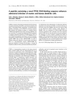
the majority of RBPs (Fig. 1A, lane 3).
By using the UV cross-linking technique, RNA binding
activities of 40, 63 and 90 kDa were detected in cT fractions
representing crude thylakoid membranes, which sedimented
through the sucrose cushion (Fig. 1A, lane 4). After further
purification of these membranes by flotation in a second
sucrose step gradient (1.3
M
/1.8
M
sucrose) and subsequent
washing by sedimentation [18], only the 63 kDa (RBP63)
and trace amounts of the 90 kDa species were detectable
(Fig. 1A, lane 5). This suggests that RBP63 is associated
with thylakoid membranes, while RBP40 and RBP90
represent stromal contamination of the cT fraction, which
still contained substantial amounts of the stromal Rubisco
enzyme (Fig. 1C).
However, a signal in the range of 63 kDa was also
detected in the stromal fraction (Fig. 1A, lane 3). To test,
whether this activity represents stroma-localized RBP63,
high resolution SDS/PAGE was performed. As shown in
Fig. 1B, the stromal component is approximately 61 kDa
(RBP61) in size and, thus, distinctly different from RBP63.
Further evidence supporting this finding was obtained from
sedimentation analyses, which demonstrated that RBP61
and RBP63 are part of two different high molecular weight
complexes of 450 kDa and 700 kDa, respectively
(Fig. 1E, fractions 5 and 9–15, respectively). From these
data, it can be concluded that active RBP63 is associated
exclusively with the thylakoid membrane and is not
partitioned between the stroma and the thylakoids.
Thylakoid membranes can be divided into stroma
lamellae and stacked grana regions. To test whether
RBP63 shows any selective accumulation within these
thylakoid membrane subfractions, appressed grana and
unappressed intermediate and stroma thylakoids were
separated by differential centrifugation following digitonin
treatment as described earlier [26]. As shown in Fig. 1D,
RBP63 activity was found to be significantly enriched in the
stromal thylakoid membrane fraction together with the CF1
subunit of the chloroplast ATPase and the PsaD subunit of
PS I, which serve as marker molecules for stromal thylak-
oids [29]. Low amounts of RBP63 were detectable in the
intermediate fraction, whereas PS I and ATPase already
showed a significant enrichment. In view of actual models of
the domain structure of the photosynthetic membrane it
Fig. 1. Fractionation pattern of RBP63. Chloroplast lysate (A) and -subfractions (C) were analysed by UV cross-linking to the psbD 5¢ UTR-RNA.
(B) The stromal (S) and the crude thylakoid membrane fraction (cT) were analysed as in (A), but proteins were separated on an 8% instead of a
10% acrylamide/SDS gel. The sizes of marker proteins are indicated in kDa. P, Protein-free control; T, thylakoid membrane fraction. (C)
Chloroplast fractionation was controlled by Western analysis with antibodies against Rubisco (RbcL), PS I (PsaD) and ATP-synthase (CF1). (D)
Floated thylakoid membranes (T) were isolated and separated into grana (GT), intermediate (IT), and stroma (ST) thylakoids. Identical amounts of
chlorophyll were either analysed by UV cross-linking using the psbD-RNA (2 lg chlorophyll) or by Western analysis (20 lg chlorophyll) with
antibodies against PS I (PsaD) and ATP-synthase (CF1). (E) Chloroplast lysates were centrifuged on a linear 15–80% glycerol gradient. Given
fractions were analysed by UV cross-linking with the psbD RNA. Sedimentation of marker proteins (in kDa) is indicated at the top. The arrows
point to RBP63, and RBP61 is marked by asterisks.
3914 F. Ossenbu
¨
hl et al. (Eur. J. Biochem. 269) Ó FEBS 2002
appears likely that the intermediate fraction might be
enriched in thylakoid margin regions which constitute a
distinct subdomain and contain amounts of PS I and
ATPase similar to stromal thylakoids [29]. However, the
data clearly indicate that RBP63 does not cofractionate with
the grana thylakoid membranes.
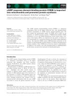
To test the nature of the association between RBP63 and
thylakoids, membranes were washed with high salt (0.1–
2.0
M
NaCl) or with buffer containing 0.1–2.0% of either
detergent Brij 35 or Triton X-100. Salt treatment did not
induce any release of RBP63 into the soluble phase,
although some activity was lost in the membrane phase,
probably due to degradative processes during extensive
washing of membranes which was required to completely
remove NaCl (Fig. 2A). In contrast, complete release was
observed with high concentrations (2.0%) of Brij 35. This
nonionic detergent preferentially dissolves peripheral/ex-
trinsic membrane proteins because of its high hydrophilic-
lipophile balance number [30,31]. Under these conditions,
chlorophyll could not be measured in the supernatants
(Fig. 2B) demonstrating that intrinsic membrane proteins
are not dissolved [32]. By using Triton X-100, both
chlorophyll and RBP63 were readily detected in the
supernatant fraction. Complete release of chlorophyll was
achieved with high concentrations (1.0%) of Triton X-100,
whereas only low concentrations (0.1%) were required to
release all RBP63 activity (Fig. 2C). Taken together, these
results indicate that RBP63 is a membrane protein of
thylakoids. It might be peripherally associated with the
membrane via a hydrophobic polypeptide anchor, as has
been suggested for thylakoid phosphatases from
C. reinhardtii [33].
RBP63 preferentially binds to the
psbA
5¢ UTR
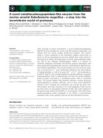
Initially, RBP63 was detected by using a radiolabelled
RNA probe containing the psbD 5¢ UTR. However, other
tested radiolabelled 5¢ UTR-probes from the psbA mRNA
(Fig. 3A) or psbB, psbC, rbcL and rps4 mRNAs (data not
shown) were also able to generate a similar RBP63 signal
under noncompetitive and, hence, unphysiological condi-
tions. To distinguish between different affinities of RBP63
to the various RNAs, comparative competition experi-
ments were performed. These used radiolabelled psbD-
RNA and an excess of unlabeled 5¢ UTR RNA probes
from other chloroplast mRNAs encoding subunits of the
PS II core, including psbA, psbB,andpsbC.ThepsbB,
psbC and even the psbD RNA exhibited only moderate
and nearly the same competition effects. However,
surprisingly, a very strong reduction of the RBP63 signal
was obtained when the psbA RNA was used as a
competitor (Fig. 3B), thus suggesting a high affinity of
RBP63 for the psbA 5¢ UTR RNA. Consequently,
competition experiments similar to those described in
Fig. 3B were performed, except that psbA 5¢ UTR was
used in place of psbD as the radiolabelled probe. Again,
the homologous psbA RNA led to the most significant
competition effect, whereas the addition of an excess of
psbB, psbC,andpsbD RNAs resulted in only minor
competition (Fig. 3C). Additional RNAs were tested and
included the 5¢ UTRs of rbcL and rps4 mRNAs as well as
an unrelated in vitro transcript comprising the polylinker
region of the pBluescript KS
+
vector, which competed at
low levels in the same range (data not shown). Similar to
several other chloroplast RNA binding proteins, RBP63
exhibited a high affinity for the ribohomopolymers polyA
and polyU, whereas polyG and polyC were not recog-
nized (data not shown). Also the addition of a 500-fold
excess of dsDNA from the psbA 5¢ region had no effect
on RBP63-binding (data not shown). In conclusion, these
data confirm the high affinity of RBP63 for the psbA
leader.
Analysis of the
cis
-acting determinants
for RBP63-binding
Within chloroplasts of C. reinhardtii, two forms of the psbA
mRNA exist: a larger form with a 5¢ UTR of 91 nucleotides
(which had been used in the binding assays shown in
Fig. 3C) and the predominant, shorter form with a leader of
36 nucleotides (Fig. 5A) [34,35]. It has been hypothesized
that the larger form represents the precursor to the shorter
mRNA which is generated by a 5¢ processing event [36].
Similar to the situation found for psbD and psbB gene
expression [37,38], a tight molecular connection between
processes of 5¢ RNA maturation and translation initiation
had been postulated for the psbA gene [36]. In order to
distinguish whether RBP63 also binds to the shorter psbA
message, further comparative competition experiments were
carried out by using the two different psbA 5¢ UTR forms as
unlabeled competitors. The two RNAs reduced the RBP63
signal with almost the same efficiency indicating that RBP63
recognizes the psbA mRNA via an element located between
position )36 and +1 of its leader (Fig. 4). In contrast, an
RNA probe covering the psbA 3¢ UTR competed only to a
low level (Fig. 4).
Fig. 2. Association of RBP63 with thylakoid membranes. Thylakoid
membranes were incubated with NaCl (A), Brij35 (B) and Triton
X-100 (C) as indicated. The soluble (S) and the membrane phases (M)
were separated and analysed by UV cross-linking assays with the psbD
RNA. The relative amounts of chlorophyll released into the soluble
phases during the treatments are indicated (Chl).
Ó FEBS 2002 Protein binding to the psbA 5¢ UTR (Eur. J. Biochem. 269) 3915
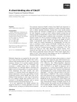
For further examination of the cis-acting determinants,
which are essential for RBP63 binding, we generated several
mutant versions of the shorter psbA 5¢ UTR.Basedonthe
observation that RBP63 exhibits high affinity to stretches of
A and U residues, the two A-rich regions at position )27 to
)19 (A-tract 1) and position )14 to )4(A-tract2)relative
to the AUG start codon of the psbA leader were changed
into C-tracts, resulting in the mutant RNAs M1 and M2,
respectively (Fig. 5A). When these mutant RNAs were used
as competitor RNAs in competition experiments, M1-RNA
still reduced the RBP63 signal as efficiently as did )36-RNA
(Fig. 5B) suggesting that A-tract 1 is not essential for
RBP63 binding. In contrast, a significantly weaker compe-
tition effect was observed with M2 RNA, indicating that
crucial cis-acting determinants for RBP63-binding activity
are located within A-tract 2.
To analyse these in more detail, four additional leader
mutants of A-tract 2 were generated, which contained
C-tracts localized at positions )17 to )12 (M2a RNA), )11
to )8(M2bRNA),)7to)4(M2cRNA)and)17 to )15
(M2d RNA) (Fig. 5A). These new mutant RNAs were used
as competitor RNAs as described above. As shown in
Fig. 5C, both M2c RNA and M2d RNA were able to
reducetheRBP63signaltothesamedegreeaswild-type
)36-RNA. Addition of either M2a RNA or M2b RNA,
however, caused only weak competition effects in the range
of those obtained with M2 RNA. This indicated that
RBP63 binding to the psbA leader is determined mainly by
the tract of seven A residues located between position )14
and )8 relative to the AUG start codon.
DISCUSSION
In this study, we report on the identification and character-
ization of the chloroplast RNA binding protein RBP63,
which is part of a high molecular weight complex of
700 kDa. RBP63 exhibits a high affinity for the 5¢ UTR of
the psbA message when compared to various different 5¢
UTRs of chloroplast genes. Moreover, its binding activity in
vitro is dependent on a tract of seven consecutive A-residues
located 14–8 nucleotides upstream of the psbA AUG start
codon. Based on the fact that chloroplast mRNAs often
contain long stretches of A residues, it appears unlikely that
Fig. 3. RBP63 binds with high affinity to the psbA 5¢ UTR. (A) Floated thylakoid membranes were analysed by UV cross-linking assays with
radiolabelled 5¢ UTR probes of either the psbD or the psbA mRNA (psbD-andpsbA-RNA, respectively). (B) cT-fractions were incubated with
radiolabelled psbD-RNA and a 5-, 50-, or 500-fold molar excess of the indicated competitor RNAs representing the 5¢ UTR RNAs of the psbA,
psbB, psbC and psbD mRNA.(C)Asin(B)exceptthatpsbA RNA instead of psbD RNA was radiolabelled. Each diagram displays the intensities of
RBP63 signals in relation to the RBP63 signal without competitor from one representative experiment, in which the exposure time was the same for
all lanes.
Fig. 4. RBP63 binds to the short psbA leader form. Competition
experiments similar to those described in Fig. 3C were performed by
using the larger (psbA) and the shorter ()36) psbA 5¢ UTR (Fig. 5A) as
well as the psbA 3¢ UTR.
3916 F. Ossenbu
¨
hl et al. (Eur. J. Biochem. 269) Ó FEBS 2002
thisA-stretchonitsownisalreadysufficienttomediatehigh
affinity binding of RBP63. Rather, additional cis-acting
determinants, such as the secondary structure of the leader,
for example, might facilitate site-specific RNA recognition
by RBP63, similar to RNA–protein complex formation in
other systems [20,39]. Nevertheless, the A-rich region had
previously been shown to be required for D1 synthesis in
C. reinhardtii. In the chloroplast transformant RBS11,
deletion of the sequence between position )26 and )11 led
to the elimination of psbA mRNA translation in vivo,
whereas the stability and 5¢ maturation of the message were
unaffected (Fig. 5A) [36]. As the deletion in the translational
RBS11 mutant covers four of the seven A residues of the
psbA leader that are essential for RBP63-binding, a corre-
lation between RNA binding activity in vitro and transla-
tional activity in vivo becomes obvious. Thus, we speculate
that RBP63 might be involved in the translational control of
psbA gene expression in C. reinhardtii. This resembles the
situation found for the regulation of translation initiation of
the psbD gene in chloroplasts of C. reinhardtii. In the case of
the psbD 5¢ UTR, a U-rich element located 25 to 14
nucleotides upstream of its AUG start codon was shown to
be required for translation but not for RNA stabilization or
5¢ maturation [38]. This region is recognized by a stromally
localized 40 kDa protein (RBP40), which has been postu-
latedtobeinvolvedinD2synthesis[18].
In spinach, the ribosomal protein S1 has been shown to
interact with A- or U-rich sequence elements within the
psbA 5¢ UTR [40]. In contrast, detailed competition binding
experiments revealed that several other chloroplast RNA
probes are also bound by S1 with the same affinity [41]. In
C. reinhardtii, similar to Escherichia coli, the chloroplast S1
protein has a size in the range of 60 kDa [18], thus
resembling RBP63. However, when thylakoid membrane
proteins were analysed with a polyclonal antiserum against
the E. coli S1 protein, no immunoreactive material was
detected (data not shown). These data, as well as the fact
that RBP63 binds with high affinity solely to the psbA leader
strongly support the idea that these two factors are distinct,
though, formally, we cannot exclude that RBP63 represents
a membrane-bound version of the S1 protein.
However, one of the most intriguing features of RBP63 is
its association with stromal thylakoid membranes. To our
knowledge, this represents the first example of a chloroplast
RNA binding protein within thylakoids. While analyses of
such proteins were initiated mainly from the soluble stromal
phase, more recently a partitioning of various RBPs
between the soluble and the membrane fraction was
reported following the use of a radiolabelled RNA probe
of the psbC 5¢ UTR in UV cross-linking experiments [9].
Further fractionation of the membrane phase revealed that
the bulk of RBPs is specifically enriched (over 100-fold)
within the above mentioned LDM, which can easily be
separated from thylakoids by sucrose density centrifugation
in the absence of MgCl
2
[9]. During the course of this work,
stroma and LDM phase were not separated and, except for
RBP63, all RBPs were detected in the stroma/LDM fraction
by using either a psbD or a psbC 5¢ UTR probe (Fig. 1A;
data not shown), indicating that the floated thylakoid
membranes were not contaminated by LDM membranes
(Fig. 1A, lane 5).
It has been hypothesized that LDMs, which resemble the
inner envelope with regard to their lipid composition,
represent the sites of thylakoid membrane protein synthesis
and that de novo formed photosynthetic complexes are
transported via vesicles from the inner envelope to the
thylakoids [42,43]. This appears to be consistent with the
finding that many RBPs, which are likely to be involved in
post-trancriptional gene expression steps in the chloroplast,
cofractionate with LDMs. On the other hand, the repair
mechanism of PS II in mature chloroplasts mainly involves
the exchange of photo-damaged D1 protein by a newly
Fig. 5. cis-acting determinants of RBP63
binding to the short psbA leader. (A) Sequence
alignment of the larger (psbA) and the shorter
()36) psbA 5¢ UTRs from the wild-type and
different mutated 5¢ UTR versions. Asterisks
represent conserved residues. Positions rela-
tive to the initiation codon and the 5¢ pro-
cessing site (vertical arrow) are marked above
the sequence. The region that has previously
been deleted in the chloroplast mutant RBS11
[34] is boxed. (B and C) Competition experi-
ments were carried out similar to those
described in Fig. 3C by using the indicated
RNAs. Each diagram displays the intensities
ofRBP63signalsinrelationtotheRBP63
signal without competitor from one represen-
tative experiment, in which the exposure time
was the same for all lanes. Competition with
M2d-RNA was performed independently and,
thus, RBP63 signals were quantified in rela-
tion to the 0x value given in the respective
M2d-RNA lane.
Ó FEBS 2002 Protein binding to the psbA 5¢ UTR (Eur. J. Biochem. 269) 3917
synthesized one and has been localized to the stroma
lamellae of thylakoid membranes. Subsequently, intact
PS II is moving to its functional localization in the grana
regions [44]. Assuming a cotranslational insertion of D1
[45], it appears likely that gene expression for this repair
mechanism is restricted to the subfraction of the stromal
thylakoid membranes, especially, as the tightly stacked
grana regions would not allow access of the RNA
polymerase, ribosomes or other soluble complexes involved
in chloroplast gene expression to the membrane. In
conjunction with data obtained from yeast mitochondria
[46], this leads to the model of a molecular tether, which
localizes chloroplast transcripts encoding integral mem-
brane proteins to stromal thylakoids [47,48]. RBP63
appears to be a good candidate for a molecular tether of
chloroplast mRNAs. It combines properties, which have to
be postulated for such a factor, namely, it is associated with
stromal thylakoids and binds to RNA. In particular, the
fact that RBP63 binds with high affinity to the psbA 5¢ UTR
and might thereby target the mRNA at stromal thylakoid
membranes is striking, as most translational activity in
mature chloroplasts is restricted to D1 synthesis due to
constraintsofPSIIrepair[44].
In conclusion, two different processes of PS II generation
have to be considered, which might overlap in time: the
de novo assembly in premature developing chloroplasts and
the mechanisms, which are involved in PS II maintenance in
mature chloroplasts [49]. Based on available data and actual
hypotheses, we hence propose the following scenario for the
control of psbA gene expression in C. reinhardtii. In devel-
oping chloroplasts, psbA mRNA translation is regulated via
its 5¢ UTR in a light and/or redox-controlled manner by the
previously described complex of RB47, RB60, RB38 and
RB55. This process may take place at the LDM system, since
it has been shown immunologically that RB47 is localized to
this chloroplast subfraction [9]. Furthermore, RB60 has also
been demonstrated to be partitioned between the soluble and
membrane phase during chloroplast fractionation experi-
ments, although no distinction between LDMs and thylak-
oid membranes has been made during this analysis [23]. As
RB60 exhibits no RNA binding activity in UV cross-linking
experiments, it is clearly distinct from RBP63 which is
described here [16]. If new D1 protein is required for the
repair of PS II in mature chloroplasts, then psbA translation
is targeted at the stromal thylakoid region by RBP63, which
might interact with or replace the RB47/RB60 complex and
promote the first assembly of ribosomes on the 5¢ UTR. In
contrast with RB47 [22], the RNA binding activity of RBP63
is not significantly altered by the energy and/or redox status
of the chloroplast (data not shown). However, the originat-
ing nascent polypeptide chain compounded with ribosomes
(the so-called ribosome nascent chain complexes) has then to
be targeted at the D1 insertion point within the stromal
thylakoids, a process which appears to be mediated by the
chloroplast homologue of the 54 kDa signal recognition
particle protein [50].
Although this model has still to be considered speculative,
it is based on data that show for the first time that putative
trans-acting regulatory factors of psbA mRNA translation
initiation are localized in different membrane subcompart-
ments of the chloroplast. Furthermore, it takes into account
that two different pathways for D1 synthesis might exist
strongly depending on the developmental stage of the
chloroplast.
ACKNOWLEDGEMENTS
We thank T. Stratmann and T. Arndt for excellent technical assistance
and U. Ku
¨
ck for providing laboratory space. Antisera against the
Rubisco holoenzyme, the CF1 subunit of the chloroplast ATPase and
PsaD were kindly provided by G. Wildner, R. Berzborn and
J D. Rochaix, respectively. Plasmid pDH245 was a generous gift of
W. Zerges. This work was supported by a grant from the Deutsche
Forschungsgemeinschaft (SFB 480-TP B8).
REFERENCES
1. Leon, P., Arroyo, A. & Mackenzie, S. (1998) Nuclear control of
plastid and mitochondrial development in higher plants. Ann. Rev.
Plant Physiol. Plant Mol. Biol. 49, 453–480.
2. Goldschmidt-Clermont, M. (1998) Coordination of nuclear and
chloroplast gene expression in plant cells. Internat. Rev. Cytol. 177,
115–180.
3. Sato, N., Rolland, N., Block, M.A. & Joyard, J. (1999) Do plastid
envelope membranes play a role in the expression of the plastid
genome? Biochimie 81, 619–629.
4. Fisk,D.G.,Walker,M.B.&Barkan,A.(1999)Molecularcloning
of the maize gene crp1 reveals similarity between regulators of
mitochondrial and chloroplast gene expression. EMBO J. 18,
2621–2630.
5. Jenkins, B.D. & Barkan, A. (2001) Recruitment of a peptidyl-
tRNA hydrolase as a facilitator of group II intron splicing in
chloroplasts. EMBO J. 20, 872–879.
6. Rivier, C., Goldschmidt-Clermont, M. & Rochaix, J D. (2001)
Identification of an RNA-protein complex involved in chloroplast
group II intron trans-splicing in Chlamydomonas reinhardtii.
EMBO J. 20, 1765–1773.
7. Vaistij, F.E., Boudreau, E., Lemaire, S.D., Goldschmidt-Cler-
mont, M. & Rochaix, J.D. (2000) Characterization of Mbb1, a
nucleus-encoded tetratricopeptide repeat-like protein required for
expression of the chloroplast psbB/psbT/psbH gene cluster in
Chlamydomonas reinhardtii. Proc. Natl Acad. Sci. USA 97, 14813–
14818.
8. Boudreau, E., Nickelsen, J., Lemaire, S.D., Ossenbu
¨
hl,F.&
Rochaix, J.D. (2000) The Nac2 gene of Chlamydomonas reinhardtii
encodes a chloroplast TPR-like protein involved in psbD mRNA
stability. EMBO J. 19, 3366–3376.
9. Zerges, W. & Rochaix, J.D. (1998) Low density membranes are
associated with RNA-binding proteins and thylakoids in the
chloroplast of Chlamydomonas reinhardtii. J. Cell Biol. 140,
101–110.
10. Rolland, N., Janosi, L., Block, M.A., Shuda, M., Teyssier, E.,
Miege, C., Cheniclet, C., Carde, J.P., Kaji, A. & Joyard, J. (1999)
Plant ribosome recycling factor homologue is a chloroplastic
protein and is bactericidal in Escherichia coli carrying temperature-
sensitive ribosome recycling factor. Proc. Natl Acad. Sci. USA 96,
5464–5469.
11. Perron, K., Goldschmidt-Clermont, M. & Rochaix, J.D. (1999) A
factor related to pseudouridine synthases is required for chlor-
oplast group II intron trans-splicing in Chlamydomonas reinhardtii.
EMBO J. 18, 6481–6490.
12. Wollman, F.A., Minai, L. & Nechushtai, R. (1999) The biogenesis
and assembly of photosynthetic proteins in thylakoid membranes.
Biochim. Biophys. Acta 1411, 21–85.
13. Breidenbach, E., Jenni, E. & Boschetti, A. (1988) Synthesis of two
proteins in chloroplasts and mRNA distribution between thyla-
koids and stroma during the cell cycle of Chlamydomonas
reinhardtii. Eur. J. Biochem. 177, 225–232.
3918 F. Ossenbu
¨
hl et al. (Eur. J. Biochem. 269) Ó FEBS 2002
14. Herrin, D. & Michaels, A. (1985) The chloroplast 32 kDa protein
is synthesized on thylakoid-bound ribosomes in Chlamydomonas
reinhardtii. FEBS Lett. 184, 90–95.
15. Roy, L.M. & Barkan, A. (1998) A SecY homologue is required
for the elaboration of the chloroplast thylakoid membrane and
for normal chloroplast gene expression. J. Cell Biol. 141,
385–395.
16. Danon, A. & Mayfield, S.P. (1991) Light-regulated translational
activators: identification of chloroplast gene specific mRNA-
binding proteins. EMBO J. 10, 3993–4001.
17. Zerges, W. & Rochaix, J D. (1994) The 5¢ leader of a chloroplast
mRNA mediates the translational requirements for two nucleus-
encoded functions in Chlamydomonas reinhardtii. Mol. Cell. Biol.
14, 5268–5277.
18. Ossenbu
¨
hl, F. & Nickelsen, J. (2000) Cis-andtrans-acting
determinants for translation of psbD mRNA in Chlamydomonas
reinhardtii. Mol. Cell. Biol. 20, 8134–8142.
19. McCormac, D.J., Litz, H., Wang, J., Gollnick, P.D. & Berry, J.O.
(2001) Light-associated and processing-dependent protein binding
to 5¢ regions of rbcL mRNA in the chloroplasts of a C4 plant.
J. Biol. Chem. 276, 3476–3483.
20. Fargo, D.C., Boynton, J.E. & Gillham, N.W. (2001) Chloroplast
ribosomal protein S7 of Chlamydomonas binds to chloroplast
mRNA leader sequences and may be involved in translation
initiation. Plant Cell 13, 207–218.
21. Nakamura, T., Ohta, M., Sugiura, M. & Sugita, M. (2001)
Chloroplast ribonucleoproteins function as a stabilizing factor of
ribosome-free mRNAs in the stroma. J. Biol. Chem. 276, 147–152.
22. Bruick, R.K. & Mayfield, S.P. (1999) Light-activated translation
of chloroplast mRNAs. Trends Plant Sci. 4, 190–195.
23. Trebitsh,T.,Meiri,E.,Ostersetzer,O.,Adam,Z.&Danon,A.
(2001) The protein disulfide isomerase-like RB60 is partitioned
between stroma and thylakoids in Chlamydomonas reinhardtii
chloroplasts. J. Biol. Chem. 276, 4564–4569.
24. Gorman, D.S. & Levine, R.P. (1965) Cytochrome f and plasto-
cyanin: their sequence in the photosynthetic electron transport
chain of Chlamydomonas reinhardtii. Proc. Natl Acad. Sci. USA
54, 1665–1669.
25. Ojakian, G.K. & Satir, P. (1974) Particle movements in chlor-
oplast membranes: quantitative measurements of membrane
fluidity by the freeze-fracture technique. Proc. Natl Acad. Sci.
USA 71, 2052–2056.
26.Adir,N.,Shochat,S.&Ohad,I.(1990)Light-dependentD1
protein synthesis and translocation is regulated by reaction center
II: reaction center II serves as an acceptor for the D1 precursor.
J. Biol. Chem. 265, 12563–12568.
27. Smith, P.K., Krohn, R.I., Hermanson, G.T., Mallia, A.K.,
Gartner, F.H., Provenzano, M.D., Fujimoto, E.K., Goeke, N.M.,
Olson, B.J. & Klenk, D.C. (1985) Measurement of protein using
bicinchoninic acid. Anal. Biochem. 150, 76–85.
28. Arnon, D.J. (1949) Copper enzymes in isolated chloroplast:
polyphenoloxidase in Beta vulgaris. Plant Physiol. 24, 1–15.
29. Albertson, P.A. (2001) A quantitative model of the domain
structure of the photosynthetic membrane. Trends Plant Sci. 6,
349–354.
30. Bhairi, S.M. (2001) A Guide to the Properties and Uses of Deter-
gents in Biology and Biochemistry. Calbiochem-Novabiochem.
Corporation, San Diego, CA, USA.
31. Jagow, G. & Scha
¨
gger, H. (1994) Membrane Protein Purification.
Academic Press Inc, London.
32.Thornber,J.P.,Morishige,D.T.,Anandan,S.&Peter,G.F.
(1991) Chlorophyll-carotenoid proteins of higher plant thylakoids.
In Chlorophylls (Scheer, H., ed.), pp. 549–585. CRC Press, Boca
Raton.
33. Matika, A. (1999) Die Regulation der Photosynthese Durch Pro-
teinphosphatasen in Chlamydomonas Reinhardtii. Dissertation,
Ruhr University, Bochum.
34. Erickson, J.M., Rahire, M. & Rochaix, J D. (1984) Chlamydo-
monas reinhardii gene for the 32,000 mol. wt. protein of photo-
system II contains four large introns and is located entirely within
the chloroplast inverted repeat. EMBO J. 3, 2753–2762.
35. Nickelsen, J., van Dillewijn, J., Rahire, M. & Rochaix, J D.
(1994) Determinants for stability of the chloroplast psbD RNA
are located within ist short leader region in Chlamydomonas
reinhardtii. EMBO J. 13, 3182–3191.
36. Bruick, R.K. & Mayfield, S.P. (1998) Processing of the psbA 5¢
untranslated region in Chlamydomonas reinhardtii depends upon
factors mediating ribosome association. J. Cell Biol. 143, 1145–
1153.
37. Vaistij, F.E., Goldschmidt-Clermont, M., Wostrikoff, K. &
Rochaix, J D. (2000) Stability determinants in the chloroplast
psbB/T/H mRNAs of Chlamydomonas reinhardtii. Plant J. 21,
469–482.
38. Nickelsen, J., Fleischmann, M., Boudreau, E., Rahire, M. &
Rochaix, J.D. (1999) Identification of cis-acting RNA leader ele-
ments required for chloroplast psbD gene expression in Chlamy-
domonas. Plant Cell 11, 957–970.
39. Klaff, P., Mundt, S.M. & Steger, G. (1997) Complex formation of
the spinach chloroplast psbA mRNA 5¢ untranslated region with
proteins is dependent on the RNA structure. RNA 3, 1468–1479.
40. Alexander, C., Faber, N. & Klaff, P. (1998) Characterization of
protein-binding to the spinach chloroplast psbA mRNA 5¢
untranslated region. Nucleic Acids Res. 26, 2265–2272.
41. Shteiman-Kotler, A. & Schuster, G. (2000) RNA-binding char-
acteristics of the chloroplast S1-like ribosomal protein CS1.
Nucleic Acids Res. 28, 3310–3315.
42. Zerges, W. (2000) Translation in chloroplasts. Biochimie 82,
583–601.
43. Kroll, D., Meierhoff, K., Bechtold, N., Kinoshita, M., Westphal,
S., Vothknecht, U.C., Soll, J. & Westhoff, P. (2001) VIPP1, a
nuclear gene of Arabidopsis thaliana essential for thylakoid
membrane formation. Proc. Natl Acad. Sci. USA 98, 4243–4248.
44. Erickson, J.M. (1998) Assembly of photosystem II. In The
Molecular Biology of Chloroplasts and Mitochondria in
Chlamydomonas (Rochaix, J.D., Goldschmidt-Clermont, M. &
Merchant, S., eds), pp. 255–285. Kluwer Academic Publishers,
Dordrecht.
45. Zhang, L., Paakkarinen, V., van Wijk, K.J. & Aro, E.M. (1999)
Co-translational assembly of the D1 protein into photosystem II.
J. Biol. Chem. 274, 16062–16067.
46. Fox, T.D. (1996) Genetics of Mitochondrial Translation: Transla-
tional Control, Monograph Series 30. pp. 733–758. Cold Spring
Harbor Laboratory Press, Cold Spring Harbor, New York.
47. Rochaix, J.D. (1996) Post-transcriptional regulation of chloro-
plast gene expression in Chlamydomonas reinhardtii. Plant Mol.
Biol. 32, 327–341.
48. Hauser, C.R., Gillham, N.W. & Boynton, J.E. (1998) Regulation
of chloroplast translation. In The Molecular Biology of Chloro-
plasts and Mitochondria in Chlamydomonas (Rochaix, J.D.,
Goldschmidt-Clermont, M. & Merchant, S., eds), pp. 197–217.
Kluwer Academic Publishers, Dordrecht.
49. Choquet, Y. & Vallon, O. (2000) Synthesis, assembly and
degradation of thylakoid membrane proteins. Biochimie 82,
615–634.
50. Nilsson, R., Brunner, J., Hoffman, N.E. & van Wijk, K.J. (1999)
Interactions of ribosome nascent chain complexes of the chloro-
plast-encoded D1 thylakoid membrane protein with cpSRP54.
EMBO J. 18, 733–742.
Ó FEBS 2002 Protein binding to the psbA 5¢ UTR (Eur. J. Biochem. 269) 3919