Diabetic foot ulcers in a Kenyan referral and teaching hospital: Risk factors, patient characteristics and clinical outcomes
Bạn đang xem bản rút gọn của tài liệu. Xem và tải ngay bản đầy đủ của tài liệu tại đây (481.97 KB, 11 trang )
Series of Endocrinology, Diabetes and Metabolism
Research Article
Vol 1 Iss 2
Diabetic Foot Ulcers in a Kenyan Referral and Teaching Hospital:
Risk Factors, Patient Characteristics and Clinical Outcomes
Mutonga DM1*, Mureithi MW2,3, Ngugi NN4 and Otieno FC5
1
Institute of Tropical and Infectious Diseases, College of Health Sciences, University of Nairobi, Kenya
KAVI-Institute of Clinical Research, Kenya
3
Department of Medical Microbiology, College of Health Sciences, University of Nairobi, Kenya
4
Department of Medicine, Kenyatta National Hospital, Kenya
5
Department of Clinical Medicine and Therapeutics, College of Health Sciences, University of Nairobi, Kenya
2
*
Correspondence: Daniel Munyambu Mutonga, Institute of Tropical and Infectious Diseases, College of Health
Sciences, University of Nairobi, Kenya
Received on 18 July 2019; Accepted on 29 August 2019; Published on 03 September 2019
Copyright © 2019 Mutonga DM, et al. This is an open access article and is distributed under the Creative Commons
Attribution License, which permits unrestricted use, distribution, and reproduction in any medium, provided the
original work is properly cited.
Abstract
Introduction: The burden of diabetes mellitus (DM) is increasing in resource-poor settings
leading to a rise in diabetic complications. Foot complications result in almost half of all
hospital admissions among diabetic patients and may result in amputations or death.
Objective: To investigate the sociodemographic, clinic-laboratory characteristics and clinical
outcomes of patients with diabetic foot ulcers (DFU) in a clinical setting.
Materials and Methods: A cross-sectional study of 84 adult consecutive inpatients and
outpatients at Kenyatta National Hospital (KNH) with any type of DM and having active DFU
was conducted over 12 months. History and physical examinations findings were recorded
through a structured questionnaire. Relevant data on the most recent blood tests and clinical
outcomes for patients with foot ulcers were retrieved from the patients’ medical notes and
analysed.
Results: Majority (68%) were inpatients. The mean age was 60.30 years with 68% living in
urban areas and 60% having minimal or no formal education. 8% were newly diagnosed with
DM. The median duration of DM was 6.5 years. A majority (96%) had type 2 diabetes mellitus
(T2DM). 45% were on insulin only, 18% on oral drugs only and 32% on a combination of both.
The median random blood sugar was 9.60 mmol/L and glycated haemoglobin was 8.80%.
Although 61% of patients had co-morbid hypertension, only about 40% had elevated systolic
blood pressure (BP) while 23% had elevated diastolic pressures. A majority of the patients had
good lipid profile, 85% with desirable total cholesterol and 70% having ideal low-density
lipoproteins. The mortality rate among patients with DFU was 11%.
Conclusion: There are poor outcomes for patients with DFU in this setting such as poor wound
healing, high recurrence rates, increased amputations and mortality compared to previous
studies. However, the prevalence of uncontrolled hypertension, dyslipidaemia and neuropathy
was much lower than earlier local reports.
Citation: Mutonga DM, Mureithi MW, Ngugi NN, et al. Diabetic foot ulcers in a Kenyan referral and teaching hospital:
risk factors, patient characteristics and clinical outcomes. Series Endo Diab Met. 2019;1(2):41-51.
Series of Endocrinology, Diabetes and Metabolism
2019 | Vol 1 | Iss 2
Keywords: diabetic foot ulcers, Kenya, risk factors, clinical outcomes
Abbreviations: BP: blood pressure; DFU: diabetic foot ulcers; DM: diabetes mellitus; IQR: interquartile range; KNH:
Kenyatta National Hospital; T2DM: type 2 diabetes mellitus; UoN: University of Nairobi
Introduction
The global prevalence of DM is 8.8% [1]. By 2030, diabetes will have affected 188 million adults in their fourth and
fifth decades and 80% of these patients will come from resource-poor countries [2]. Globally, foot complications result
in 25–50% of all hospital admissions in DM patients [3].
In Africa, the prevalence of DFU ranges from 3.2–19.1% [4–10]. A DFU is any foot with ulceration and linked to
neuropathy and/or peripheral vascular disease in a patient with DM [11]. Previous reports reveal that the prevalence
of diabetic polyneuropathy in Uganda, Kenya and Tanzania vary from 29.4–44% [6, 7, 12, 13]. In addition, 40% of
diabetic patients in Nigeria present with the peripheral arterial disease [7]. The occurrence of diabetic amputations, a
common complication of DM, varies in Sub-Saharan Africa but is generally thought to be low especially in the rural
areas [14, 15]. However, from recent accounts, the prevalence of lower limb amputations among DM patients ranges
widely from 11.2–78% [4, 5, 16–19].
Global healthcare expenditure to treat and prevent DM was between USD 376 billion (2010) and 673 billion (2015)
[1, 2]. The estimated cost of treating a DFU in resource-rich countries is 6 to 10 times that in resource-poor countries
revealing inequalities in access to health resources [20–22]. Moreover, DFU have mortality rates ranging from 2–
14.3% in the African setting [4, 23–25).
There is limited data on DFU in Kenya. Previous local publications have looked at the prevalence, associated risk
factors, types, and duration of DFU among the study participants [26–29]. There is no mention of outcomes on patients
with DFU in the country. The present study aimed to investigate the sociodemographic, clinical characteristics and
clinical outcomes of DFU among adult diabetic patients at KNH.
Materials and Methods
These study methods were adapted from studies by Nyamu et al. and Chalya et al. [5, 27].
Study design
A hospital-based cross-sectional study was conducted over 12 months from September 2017 to August 2018. Adult
diabetic patients with active DFU were recruited by consecutive sampling from medical inpatient departments and the
diabetes outpatient clinic at KNH, Nairobi, Kenya, this is the largest hospital in East and Central Africa.
Data collection
Basic patient and clinical characteristics were collected as part of the routine history taking and usual physical
examinations. Patients’ basic characteristics included age, sex, area of residence, and level of education. Clinical
characteristics including presence of comorbidities, smoking history, alcohol use, duration of diabetes, type of
diabetes, duration of DFU, current diabetes medications, previous history of healed foot ulcers, type of DFU, Wagner’s
classification, and location of the lesion were recorded for each patient. Peripheral neuropathy and peripheral vascular
disease were also assessed. Height, weight and BP with the adult cuff standard technique were also measured.
The outcomes of the DFU were collected for patients at the end of the study by reviewing their medical records.
Relevant information from the latest case notes, diabetic clinic or medical wards was extracted, classified [complete
healing, non-healing, surgical intervention (minor or amputation) or mortality] and analysed.
42
Series of Endocrinology, Diabetes and Metabolism
2019 | Vol 1 | Iss 2
Statistical analyses
Microsoft® Excel was used for data entry. Descriptive analyses was performed for all variables and data presented in
charts and tables. Continuous variables such as age, time and, blood tests were converted into categorical variables
using cut-off points. Independent two-tailed t-test was used to compare means, Chi-square test was used to verify the
association of categorical variables and one-way ANOVA for comparison across multiple groups. Results were
considered significant at p < 0.05. All analyses were performed using IBM® SPSS® Statistical Package Version 23.0.
Ethical considerations
Approval was sought from the KNH-UoN Ethics and Research Committee (Approval Number P769/10/2016; website:
to conduct the study and patients enrolled only after informed consent was taken. Patient
confidentiality and data security was guaranteed.
Results
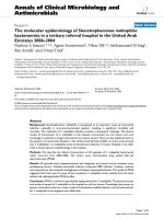
Eighty-four patients were enrolled in the study (Figure 1). Most of the study patients were inpatients (68.4%). The
mean age was 60.30 + 12.88 years. The youngest patient was 25 years while the oldest was 108 years, with most
patients being in their fifth and sixth decades. The females were 54.1%. Most of the patients (68.3%) lived in urban
areas while 31.7% lived in rural areas. A majority of 44.2% had primary education and 15.6% did not have any formal
education. Males were more educated than females (p = 0.001) (Table 1).
The median interquartile range (IQR) duration of DM was 6.5 (1.25, 12.5). A small proportion, (8.5%) of patients
were newly-diagnosed with diabetes, 17.3% had ever smoked cigarettes while 33.3% had ever taken alcohol. Males
were more likely to be smokers (p = 0.004). Fourteen patients (16.5%) had no comorbidities, while the rest of the
patients (83.5%) had one or more comorbidities. The most common comorbidities were hypertension, kidney disease,
heart disease and anemia. Most of the study patients had T2DM and were currently on medication (Table 2).
The mean BP was within normal ranges but 39.7% had elevated systolic BP and 23.3% had high diastolic BP. A large
number of patients did not have recent laboratory results; 63.53% lacked HbA1c levels, 70% lacked lipid profile tests,
47% lacked liver function tests and 30% lacked kidney function tests. 39.3% of patients had high urea levels and
58.9% had elevated creatinine levels. Females had significantly better creatinine levels. A majority of the patients
had desirable lipid profile tests (Table 3).
The median IQR duration of the DFU was 8 (4, 16) weeks. The longest duration of DFU was 312 weeks. Half of the
patients had a previous history of DFU and a third had a prior history of amputation. At the time of the study, 77.6%
had recently used antibiotics to treat the ulcer. A majority (88.10%) of the patients had ulcers on only one foot. Fortynine ulcers (53.26%) were located on the right foot and 43 ulcers (46.74%) on the left. Most ulcers were located on
the forefoot and hindfoot. The DFU were mainly neuropathic or ischaemic and Wagner Stage 1 or 2. Of all the DFU,
54.3% had signs of diabetic neuropathy (Table 4).
The interval between the first and second interview was calculated and its median IQR was 98 (147) days. A majority
of the patients were reviewed within 1–3 months after the first interview (28.9%), 3–6 months (28.9%) and 6–12
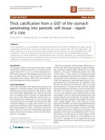
months (21.1%). We reviewed 36 patients (42.9%) while 48 patients (57.1%) were lost to follow-up. A majority of
the patients had good progress while 25% of the patients on follow-up had non-healing DFU. There was a mortality
rate of 11.1% (Figure 2).
43
Series of Endocrinology, Diabetes and Metabolism
2019 | Vol 1 | Iss 2
Figure 1: Flow chart of the study. The figure depicts the recruitment of patients into the study and study directed
activities. Microbiological procedures and results are described in an earlier publication. *VITEK 2 machine was
utilised for automated culture and sensitivity tests; **RT-PCR: real-time polymerase chain reaction.
Sex
Age group
Age (years) mean (SD)
n (%)
39 (45.9)
46 (54.1)
1 (1.2)
15 (18.1)
42 (50.6)
20 (24.1)
5 (6.0)
Male
Female
≤30
31–50
51–65
66–80
≥81
Total population
Male
Female
p value
60.30 (12.88)
58.62 (11.38)
61.65 (13.95)
0.29
None
12 (15.6)
2 (5.9)
10 (23.3)
Primary
34 (44.2)
12 (35.3)
22(51.2)
Education level n (%)
0.001
Secondary 24 (31.2)
13 (38.2)
11(25.6)
Tertiary
7 (9.1)
7 (20.6)
0 (0.0)
Urban
56 (68.3)
29 (78.4)
27 (60.0)
Residence n (%)
0.097
Rural
26(31.7)
8 (21.6)
18 (40.0)
Table 1: Patients’ sociodemographic characteristics. This table shows the sociodemographic characteristics of the
study patients. The lower part of the table shows differences in variables across gender and were considered
statistically significant at p < 0.05.
44
Series of Endocrinology, Diabetes and Metabolism
Type of diabetes mellitus n (%)
History of diabetes mellitus n (%)
Duration of DM (years) median (IQR)
Duration of DM (years) n (%)
Type of treatment n (%)
Risk factors
Smoking habits n (%)
Alcohol intake n (%)
2019 | Vol 1 | Iss 2
Type 1 DM
Type 2 DM
Gestational DM
Yes (previously diagnosed)
No (newly diagnosed)
6.5 (11.25*)
<5
6–10
10–20
> 21
None
Diet
OHA
Insulin
Both diet and OHA
Both OHA and insulin
Total population Male
n (%)
2 (2.4)
81 (96.4)
1 (1.2)
75 (91.5)
7 (8.5)
36 (46.2)
18 (23.1)
19 (24.4)
5 (6.4)
2 (2.4)
1 (1.2)
15 (17.6)
38 (44.7)
2 (2.4)
27 (31.8)
Female
Yes
13 (17.3)
11 (31.4)
2 (5.0)
No
62 (82.7)
24 (68.6)
38 (95.0)
Yes
25 (33.3)
18 (51.4)
7 (17.5)
p value
0.004
0.003
No
50 (66.7)
17 (48.6)
33 (82.5)
None
14 (16.5)
7 (17.9)
7 (15.2)
1
33 (38.8)
15 (38.5)
18 (39.1)
2
19 (22.4)
7 (17.9)
12 (26.1)
Number of comorbidities n (%)
0.509
3
13 (15.3)
5 (12.8)
8 (17.4)
4
5 (5.9)
4 (10.3)
1 (2.2)
5
1 (1.2)
1 (2.6)
0 (0.0)
Table 2: Clinical characteristics from patients’ history. This table represents descriptive statistics of the clinical
findings from patients’ medical history, where both mean and median were reported, the data was not normally
distributed. Differences across gender were statistically significant when p < 0.05.
Parameter
Systolic blood pressure
(mmHg)
Diastolic blood pressure
(mmHg)
RBS1 (mmol/L)
HbA1c2 (%)
Urea (mmol/L)
Total mean (SD)
14.12 (11.03)
8.40 (2.29)
12.05 (10.58)
Creat3 (mmol/L)
147.53 (74.06)
LDL-C4 (mmol/L)
1.40 (0.34)
Total median (IQR)
Male
Female
p value
136.05 (34.51)
138.25 (34.72)
138.39 (24.77)
0.985
80.08 (18.74)
80.56 (19.47)
80.61 (12.41)
0.991
9.60 (8.95)
8.80 (2.80)
7.1 (11.85)
10.08 (4.96)
9.59 (3.75)
19.36 (20.90)
0.373
0.358
114.55 (111.2)
228.19 (156.38)
11.57 (7.28)
8.51(1.74)
9.67 (16.68)
133.12
(100.72)
1.82 (0.79)
0.627
1.51 (0.81)
1.65 (0.90)
0.049
0.015
Albumin (g/L)
29.97 (6.81)
28.5 (7.53)
30.66 (6.46)
0.333
Table 3: Clinical and laboratory parameters. This table represents descriptive statistics of the clinical and laboratory
parameters from physical examination and reviewing of patients’ clinical records, where both mean and median were
reported, the data was not normally distributed. Statistically significant p < 0.05. 1Random blood sugar 2Glycated
hemoglobin 3Creatinine 4Low-density lipoprotein-cholesterol.
45
Series of Endocrinology, Diabetes and Metabolism
2019 | Vol 1 | Iss 2
n (%)
Duration of DFU (weeks) 8 (12*)
>6
27 (37.5)
7–26
38 (52.8)
27–52
7 (9.7)
> 52
0 (0.0)
Yes
40 (50)
No
40 (50)
Yes
26 (33.3)
No
52 (66.7)
Yes
45 (77.6)
No
13 (22.4)
Right
Left
Total
Forefoot
25 (51.0)
24 (55.8)
49 (53.3)
Midfoot
7 (14.3)
2 (4.7)
9 (9.8)
Hindfoot
11 (22.4)
16 (37.2)
27 (29.4)
Forefoot & midfoot
4 (8.2)
0 (0.0)
4 (4.4)
Midfoot & hindfoot
1 (2.0)
0 (0.0)
1 (1.1)
Forefoot, midfoot & hindfoot
1 (2.0)
1 (2.3)
2 (2.2)
Ischaemic
20 (40.8)
22 (51.2)
42 (45.6)
Neuropathic
25 (51.0)
18 (41.9)
43 (46.7)
Neuroischaemic
4 (8.2)
3 (7.0)
7 (7.6)
0
1 (2.0)
0 (0.0)
1 (1.1)
1
11 (22.4)
16 (37.2)
27 (29.4)
2
16 (32.7)
12 (27.9)
28 (30.4)
3
10 (20.4)
9 (20.9)
19 (20.7)
4
10 (20.4)
4 (9.3)
14 (15.2)
Duration of DFU (weeks) n (%)
History of previous DFU n (%)
History of previous amputation n (%)
History of any recent antibiotic use n (%)
Anatomic site of foot ulcer
n (%)
Type of ulcer n (%)
Wagner stage n (%)
5
1 (2.0)
2 (4.7)
3 (3.3)
Table 4: Clinical characteristics of the DFU. This table represents duration, types, location and other clinical
characteristics of DFU among the study patients. *The interquartile range of duration of DFU was from 4 to 16.
46
Series of Endocrinology, Diabetes and Metabolism
2019 | Vol 1 | Iss 2
Figure 2: Distribution of clinical outcomes of DFU for the study patients. The chart depicts the proportion of study
patients reviewed with a particular outcome of interest. Complete and ongoing healing were considered to be good
outcomes while non-healing wounds and mortality as poor outcomes.
Discussion
Africa is the second most common region to be affected by DFU according to a recent global meta-analysis [30]. In
the present study, there was a high recurrence rate of DFU (50%), comparable to facilities that follow best practices
(70%) [31]. This warrants further investigation to see if patients who have had prior DFU adhere to proper foot care.
The mean age of patients in this study was slightly higher than in earlier studies [5, 27, 29]. Most of the study patients
were however in their fifth and sixth decades just like in other studies in East Africa [5, 8, 9, 28, 29]. There were more
females than males enrolled in this present study similar to an earlier study in Kenya but in contrast to other studies
in Sudan, Tanzania, and Ethiopia where males were more [5, 8, 27, 28, 32].
In the present study, about 60% had minimal or no formal education similar to studies in Sudan and Tanzania [5, 32].
Moreover, a majority of patients in this study were from urban areas similar to publications from Sudan and Ethiopia
[9, 10, 32]. A study in Tanzania also enrolled patients mainly from rural areas [5]. In this current study, more patients
were from urban areas mainly because of KNH’s location in Nairobi.
Mariam et al. reported T2DM to be a significant risk factor for developing a DFU [9]. In this present study, almost all
of the patients had T2DM. A small proportion of the study patients were newly-diagnosed with DM comparable to an
earlier study [27]. The median duration of DM was 6.5 years, revealing that DFU are developing much earlier than
before. This could be an indicator of poor foot care or inadequate foot care.
In the present study, only a small proportion of patients were not on medication. A majority were on insulin therapy,
whether alone or in combination with oral drugs. The high number of patients on insulin could result from the fact
that inpatients often have deranged glucose levels and require insulin for strict glucose control. However, less than
half of the current study patients were on insulin therapy only which is comparable to earlier studies in Kenya and
Ethiopia [9, 27]. Although poor glycaemic control often leads to DFU, insulin treatment is in itself a risk factor for
developing DFU in Tanzania [6, 27, 29]. The glycemic control based on HbA1c level for patients in this present study
was similar to an earlier study in KNH but much worse than in a recent study in rural Kenya [27, 29].
The prevalence of uncontrolled hypertension in this study was much lower than an earlier study in Kenya [29]. In the
current study, the rates of kidney disease were similar to findings in Ethiopia, while heart disease was higher than in
47
Series of Endocrinology, Diabetes and Metabolism
2019 | Vol 1 | Iss 2
Sudan [8, 32]. In this present study, the lipid profile was much better than that reported by Nyamu et al. and could be
a result of improved patient awareness or better management of dyslipidaemia by the clinicians [27].
The median duration of DFU was 8 weeks in this current study. This was shorter than in earlier studies in Kenya and
Tanzania and could perhaps indicate increased patient awareness, increased foot care, and increased foot care
assessment by the clinicians [5, 27]. DFU is often undiagnosed because of both patient or clinician related factors
[33]. It is important that patients examine their feet regularly and clinicians not to omit to ask or examine for the
diabetic foot.
Neuropathy is a significant risk factor for developing DFU [6, 9]. Peripheral neuropathy usually begins with autonomic
dysfunction, then sensory nerves are affected and finally motor nerves deteriorate [31]. Although the prevalence of
neuropathy in this present study is much lower than in earlier Kenyan studies [27, 28], diabetic polyneuropathy among
the study patients was much higher than in Uganda and Nigeria [7, 12]. This confirms a higher rate of neuropathy
among Kenyan patients. A majority of the DFU in this present study was in Wagner Stage 1 and 2 in contrast to studies
in Tanzania and Libya [5, 24]. This could also indicate an early presentation of the patients to this hospital.
In the present study, a third of the study patients had a prior amputation, which was thrice the rate of previous
amputation in Tanzania [5]. However, after follow-up, only 14% of patients had a surgical procedure compared to
90% in Tanzania, mainly because the current study was conducted in medical and not surgical departments [5]. Half
of the patients followed-up in this present study had good clinical outcomes. The mortality rate in this present study
was high and similar to the study in Tanzania and a literature review from 19 African countries [4, 5]. Mortality in
Tanzania was associated with diabetes complications and advanced DFU [5]. In this present study, there was a nonhealing rate of 25% among the study patients who were followed-up. Healing of wounds is a complex immunological
response to injury [20, 31, 33].
Limitations
One of the limitations in this study is that funding for this study was allocated towards microbiological tests to isolate
the bacteria from DFU as presented in an earlier publication [34]. As such, we relied on extraction of clinical data
from patients medical records for retrieval of laboratory tests. Moreover, outcome of DFU in some instances depended
on the review of medical notes and not repeat physical examinations.
Conclusion
There are poor outcomes for patients with DFU in this setting such as poor wound healing, high recurrence rates,
increased lower-limb amputations and mortality compared to previous studies. The study patients were slightly older,
female less educated but urbanised. The study patients had better control of hypertension and dyslipidaemia, lower
levels of neuropathy and earlier presentation of DFU reflecting increased patient awareness and better management
by clinicians. Majority of the patients in this study were on insulin and antibiotics.
Recommendations
We recommend the following:
The high prevalence of poor outcomes for patients with DFU warrants the need to investigate biopsychosocial risk factors.
Screening and foot assessment should be encouraged during each clinical visit. Examination focuses on
peripheral neuropathy, peripheral arterial disease and plantar pressure [31]. Certain professional
organisations recommend different prevention measures based on the patients’ risk levels [20]. Patients
with DM should be educated on smoking cessation and foot-care.
Patients with DM should have laboratory tests such as UECs, LFTs, Lipid profile, and HbA1c every 6
months to rule out and properly manage diabetic complications.
Conflicts of Interest
The authors declare that they have no competing interests.
48
Series of Endocrinology, Diabetes and Metabolism
2019 | Vol 1 | Iss 2
Funding Statement
DMM received partial funding from KNH Research and Programs Department (KNH Study Registration Number:
Med/101/2017) to conduct this study (website: Most of the funding sought was for microbiology
tests and PCR tests presented in an earlier publication. The funders had no role in study design, data collection and
analysis, decision to publish or preparation of the manuscript.
Acknowledgements
We are grateful to all patients who participated in this study. Our sincere thanks goes towards the nursing staff in the
Medicine Department and the Diabetes and Endocrinology Outpatient Clinic at Kenyatta National Hospital for their
support. We also acknowledge Mr. Wycliffe Ayieko for his assistance in data analysis and Dr. Jackline Ngigi for her
assistance in data collection.
References
1.
IDF Diabetes Atlas 7th Edition (2015).
2.
IDF Diabetes Atlas 4th Edition (2009).
3.
Ezera Agwu, Ephraim O. Dafiewhare and Peter E. Ekanem (December 9th 2011). Possible Diabetic-Foot
Complications in Sub-Saharan Africa, Global Perspective on Diabetic Foot Ulcerations, Thanh Dinh,
IntechOpen.
4.
Rigato M, Pizzol D, Tiago A, et al. Characteristics, prevalence, and outcomes of diabetic foot ulcers in Africa.
A systemic review and meta-analysis. Diabetes Res Clin Pract. 2018;142(1):63-73.
5.
Chalya PL, Mabula JB, Dass RM, et al. Surgical management of diabetic foot ulcers: A Tanzanian university
teaching hospital experience. BMC Res Notes. 2011;4(365):1-7.
6.
Chiwanga FS, Njelekela MA. Diabetic foot: prevalence, knowledge, and foot self-care practices among
diabetic patients in Dar es Salaam, Tanzania - a cross-sectional study. J Foot Ankle Res. 2015;8(20):1-7.
7.
Ogbera AO, Adeleye O, Solagberu B, et al. Screening for peripheral neuropathy and peripheral arterial
disease in persons with diabetes mellitus in a Nigerian University Teaching Hospital. BMC Res Notes.
2015;8(533):1-6.
8.
Deribe B, Woldemichael K, Nemera G. Prevalence and factors Influencing diabetic foot ulcer among diabetic
patients attending Arbaminch Hospital, South Ethiopia. J Diabetes Metab. 2014;5(1):1-7.
9.
Mariam TG, Alemayehu A, Tesfaye E, et al. Prevalence of diabetic foot ulcer and associated factors among
adult diabetic patients who attend the diabetic follow-up clinic at the University of Gondar Referral Hospital,
North West Ethiopia, 2016: Institutional-Based Cross-Sectional study. J Diabetes Res.
2017;2017(2879249):1-8.
10. Awadalla H, Noor SK, Elmadhoun WM, et al. Diabetes complications in Sudanese individuals with type 2
diabetes: Overlooked problems in sub-Saharan Africa? Diabetes Metab Syndr. 2017;11(S2):S1047-S51.
11. Alexiadou K, Doupis J. Management of diabetic foot ulcers. Diabetes Ther. 2012;3(1):1-15.
12. Kisozi T, Mutebi E, Kisekka M, et al. Prevalence, severity and factors associated with peripheral neuropathy
among newly diagnosed diabetic patients attending Mulago hospital: a cross-sectional study. Afr Health Sci.
2017;17(2):463-73.
13. Mugambi-Nturibi E, Otieno CF, Kwasa T, et al. Stratification of persons with diabetes into risk categories
for foot ulceration. East Afr Med J. 2009;86(5):233-39.
49
Series of Endocrinology, Diabetes and Metabolism
2019 | Vol 1 | Iss 2
14. Abbas ZG, Archibald LK. Epidemiology of the diabetic foot in Africa. Med Sci Monit. 2005;11(8):262-70.
15. Kengne AP, Choukem SP, Dehayem YM, et al. Diabetic foot ulcers in Cameroon: can microflora prevalence
inform probabilistic antibiotic treatment? J Wound Car. 2006;15(8):363-66.
16. Verla TG. Terence. Diabetes related amputations in the north-west region of Cameroon. 2011.
17. Essoh JBS, Kodo M, Djè VDB, et al. Limb amputations in adults in an Ivorian Teaching Hospital. Niger J
Clin Pract. 2009;12(3):245-47.
18. Nwosu C, Babalola MO, Ibrahim MH, et al. Major limb amputations in a tertiary hospital in North Western
Nigeria. Afr Health Sci. 2017;17(2):508-12.
19. Udeabor SE, Adisa AO, Orlowska A, et al. Tumor-associated macrophages, angiogenesis, and tumor cell
migration in oral squamous cell carcinoma. Ann Afr Med. 2017;16(4):181-85.
20. Singh N, Armstrong DG, Lipsky BA. Preventing foot ulcers in patients with diabetes. JAMA.
2005;293(2):217-28.
21. Kasiya MM, Mang’anda GD, Heyes S, et al. The challenge of diabetic foot care: Review of the literature and
experience at Queen Elizabeth Central Hospital in Blantyre, Malawi. Malawi Med J. 2017;29(2):218-23.
22. Danmusa UM, Terhile I, Nasir IA, et al. Prevalence and healthcare costs associated with the management of
diabetic foot ulcer in patients attending Ahmadu Bello University Teaching Hospital, Nigeria. Int J Health
Sci (Qassim). 2016;10(2)219-28.
23. Edo AE, Edo GO, Ezeani IU. Risk factors, ulcer grade and management outcome of diabetic foot ulcers in a
Tropical Tertiary Care Hospital. Niger Med J. 2013;54(1):59-63.
24. Benkhadoura M, Alswehly M, Elbarsha A. Clinical profile and surgical management of diabetic foot in
Benghazi, Libya. Foot Ankle Surg. 2016;22(1):55-58.
25. Foryoung JB, Ditah C, Fon PN, et al. Long-term mortality in outpatients with type 2 diabetes in a reference
hospital in Cameroon: A retrospective cohort study. BMJ Open. 2018;8(2):1-7.
26. McLigeyo, Otieno LS. Diabetic ulcers--a clinical and bacteriological study. East Afr Med J. 1991;68(3):20410.
27. Nyamu PN, Otieno CF, Amayo EO, et al. Risk factors and prevalence of diabetic foot ulcers at Kenyatta
National Hospital , Nairobi. East Afr Med J. 2003;80(1):36-43.
28. Obimbo MM, Bundi PK, Collis F, et aL. Foot complications among diabetics attending a district hospital in
Kenya : Predisposing factors and possible intervention. Ann Afr Surg. 2008;2(6):3-8.
29. Kibachio J, Omolo J, Muriuki Z, et al. Risk factors for diabetic foot ulcers in type 2 diabetes: a case control
study, Nyeri, Kenya. Afr J Diab Med. 2013;21(1):1-4.
30. Zhang P, Lu J, Jing Y, et al. Global epidemiology of diabetic foot ulceration: a systematic review and metaanalysis. Ann Med. 2017;49(2):106-16.
31. Mendes JJ, Neves J. Diabetic foot infections: current diagnosis and treatment. J Diab Foot Complicat.
2012;4(2):26-45.
32. Shigidi M, Abdelgafar H, Taha ES. Awareness regarding diabetes control and diabetic nephropathy among
Sudanese adults admitted with diabetic foot: a cross-sectional study. Pan Afr Med J. 2013;16(157):1-6.
50
Series of Endocrinology, Diabetes and Metabolism
2019 | Vol 1 | Iss 2
33. Bruhn-Olszewska B, Korzon-Burakowska A, Gabig-Cimińska M, et al. Molecular factors involved in the
development of diabetic foot syndrome. Acta Biochim Pol. 2012;59(4):507-13.
34. Mutonga DM, Mureithi MW, Ngugi NN, et al. Bacterial isolation and antibiotic susceptibility from diabetic
foot ulcers in Kenya using microbiological tests and comparison with RT‑ PCR in detection of S. aureus and
MRSA. BMC Res Notes. 2019;12(1)1-6.
51