Báo cáo khoa học: C-terminal truncated cannabinoid receptor 1 coexpressed with G protein trimer in Sf9 cells exists in a precoupled state and shows constitutive activity ppt
Bạn đang xem bản rút gọn của tài liệu. Xem và tải ngay bản đầy đủ của tài liệu tại đây (365.19 KB, 10 trang )
C-terminal truncated cannabinoid receptor 1 coexpressed
with G protein trimer in Sf9 cells exists in a precoupled
state and shows constitutive activity
Chandramouli Reddy Chillakuri, Christoph Reinhart and Hartmut Michel
Department of Molecular Membrane Biology, Max-Planck-Institute for Biophysics, Frankfurt ⁄ Main, Germany
Cannabinoid receptors belong to the seven transmem-
brane G protein coupled receptor (GPCR) family.
Two different subtypes have been reported in humans,
namely cannabinoid receptor 1 (CB1) [1] and cannabi-
noid receptor 2 (CB2) [2]. A splice variant of CB1,
called CB1a, has also been identified to be expressed in
low levels in rodent brain [3]. Cannabinoid receptors
form the site of action for the active ingredients of
marijuana (D
9
-tetrahydrocannabinol, D
9
THC), ananda-
mide being the important endocannabinoid. CB1 is
present predominantly in the nerve axons of the cen-
tral nervous system, is known to be neuroprotective in
its function and thus forms an important target in the
pharmaceutical industry [4]. CB1 is coupled to the
G
i ⁄ o
family of heterotrimeric G proteins. Activation
of the CB1-mediated G
i ⁄ o
proteins inhibits adenylyl
cyclases to reduce cAMP production, inhibit calcium
channels and increase inwardly rectifying potassium
currents. The modulation of ion channels has been
shown to be independent of cAMP production,
indicating that G proteins, especially Gbc, may inter-
act directly with the effector molecules [5,6].
An earlier concept states that agonist-bound GPCR
alone can couple to G proteins to transduce the
signal. However, this rather historical hypothesis is
opposed today because of several reports confirming
Keywords
cannabinoid receptor; G protein coupled
receptor; G proteins; membrane proteins;
signal transduction
Correspondence
H. Michel, Department of Molecular
Membrane Biology, Max-Planck-Institute for
Biophysics, Max-von-Laue Str.3,
D-60438 Frankfurt ⁄ Main, Germany
Fax: +49 69 6303 1002
Tel: +49 69 6303 1001
E-mail: Hartmut.Michel@mpibp-frankfurt.
mpg.de
(Received 23 July 2007, revised 15 Septem-
ber 2007, accepted 8 October 2007)
doi:10.1111/j.1742-4658.2007.06132.x
We have investigated the existence of a precoupled form of the distal C-ter-
minal truncated cannabinoid receptor 1 (CB1-417) and heterotrimeric
G proteins in a heterologous insect cell expression system. CB1-417 showed
higher production levels than the full-length receptor. The production lev-
els obtained in our expression system were double the values reported in
the literature. We also observed that at least the distal C-terminus of the
receptor was not involved in receptor dimerization, as was predicted in the
literature. Using fluorescence resonance energy transfer, we found that
CB1-417 and Ga
i1
b
1
c
2
proteins were colocalized in the cells. GTPcS bind-
ing assays with the Sf9 cell membranes containing CB1-417 and the G pro-
tein trimer showed that the receptor could constitutively activate the Ga
i1
protein in the absence of agonists. A CB1-specific antagonist (SR 141716A)
inhibited this constitutive activity of the truncated receptor. We found that
the CB1-417 ⁄ Ga
i1
b
1
c
2
complex could be solubilized from Sf9 cell mem-
branes and coimmunoprecipitated. In this study, we have proven that the
receptor and G proteins can be coexpressed in higher yields using Sf9 cells,
and that the protein complex is stable in detergent solution. Thus, our sys-
tem can be used to produce sufficient quantities of the protein complex to
start structural studies.
Abbreviations
B
max
, maximum binding capacity; CB1, cannabinoid receptor 1; CB2, cannabinoid receptor 2; CFP, cyan fluorescent protein; CHS, cholesterol
hemisuccinate; DM, decylmaltoside; FRET, fluorescence resonance energy transfer; GPCR, G protein coupled receptor; K
d
, equilibrium
dissociation constant; m.o.i., multiplicity of infection; D
9
THC, D
9
-tetrahydrocannabinol; YFP, yellow fluorescent protein.
6106 FEBS Journal 274 (2007) 6106–6115 ª 2007 The Authors Journal compilation ª 2007 FEBS
the precoupled form of GPCR and G proteins. In a
recent study, using fluorescence resonance energy trans-
fer (FRET), several receptors were identified, such as
the muscarinic receptor M4, the adrenergic receptor
a2A, the adenosine receptor A1 and the dopamine
receptor D2, in a precoupled form to the G protein tri-
mer (Ga
o
b
1
c
2
) [7]. It was suggested that GPCR dimers
and the G protein heterotrimer are present in cell mem-
branes in a resting state as a pentameric complex in the
absence of agonists. However, it may not be true that
this whole complex is resting and inactive, and can only
be activated by agonists. Reports constantly support
the constitutive activity of GPCRs, indicating that
GPCRs have some basal activity in the cell even in the
absence of agonists. Inverse agonists (antagonists) to
several GPCRs have shown that these ligands can
reverse this basal activation of GPCRs, demonstrating
the existence of constitutively active receptors [8].
Solubilization of the cannabinoid receptor from rat
brain and GTP binding studies initiated the concept of
the presence of a stable CB1 ⁄ G protein complex [9].
The development of the CB1-specific antagonist
SR 141716A led to the identification of constitutively
active receptors [10]. The existence of such an active
form was tested, and it was found that CB1 can
sequester G proteins from a common pool, making
them unavailable to other GPCRs present in the cell
[11]. Meanwhile, it was found that a CB1 C-terminal
peptide (CB1-401–417) was able to activate the specific
G proteins in brain [12]. Further, a C-terminal trun-
cated CB1 (CB1-417) was found to have enhanced
ability to sequester G proteins and exhibited increased
constitutive activity [13]. Recently, it has been reported
that different subtypes of Ga
i
coimmunoprecipitate
with CB1 from N18TG2 cell membranes in the
absence of exogenously added agonists [14]. Most of
the results discussed above were either performed on
native tissue or mammalian cells. In this work, for the
first time, we investigated the existence of a precoupled
form of truncated CB1 in a heterologous Sf9 insect cell
expression system. Insect cell expression systems are
cheaper than mammalian expression systems, and are
therefore more feasible for the large-scale production
of receptor required for structural determination.
Results
Functional production of CB1
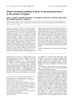
The C-terminal truncated CB1 (CB1-417) was pro-
duced in a functional form in Sf9 insect cells (the gene
constructs used in this study are shown in Fig. 1).
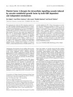
Analysis of the protein produced, by immunoblotting
with antiflag M2 IgG, showed (Fig. 2) a monomeric
band at 47 kDa and an oligomeric band specific to the
cannabinoid receptor at a size of approximately
Strep-tagIICB1 HisFlag
P
PH
Melittin
Tev
1 to 472 aa
Strep-tagIICB1 HisFlag
P
PH
Melittin
Tev
1 to 417 aa
YFPCB1 HisFlag
P
PH
Melittin
Tev
1 to 417 aa
G?i1CFPG?i1
P
PH
Strep-tagIICB1 HisFlag
P
PH
Melittin
Tev
1 to 472 aa
Strep-tagIICB1 HisFlag
P
PH
Melittin
Tev
1 to 417 aa
YFPCB1 HisFlag
P
PH
Melittin
Tev
1 to 417 aa
CFPG
α
i1 G
α
i1
P
PH
Fig. 1. Gene constructs used for the expression in insect cells. The four gene constructs were prepared using the basic pVL baculovirus
transfer vector. CFP, cyan fluorescent protein; Flag, flag epitope used for immunoblotting; His, decahistidine tag; P
PH
, polyhedrin promoter;
Strep-tagII, used for the purification of the receptor; YFP, yellow fluorescent protein. The names of each construct mentioned in this work
are given before each graphic representation.
C. R. Chillakuri et al. Precoupled form of human CB1 and G protein trimer
FEBS Journal 274 (2007) 6106–6115 ª 2007 The Authors Journal compilation ª 2007 FEBS 6107
160 kDa. A similar higher oligomeric band was
reported for full-length CB1 between 160 and 200 kDa.
Wager–Miller et al. [15] took this oligomeric form of
the cannabinoid receptor as an example to explain the
dimerization of GPCRs. Radioligand binding assay
showed saturation binding on the cell membranes
(Fig. 3). It was found that the use of 10 multiplicity of
infection (m.o.i.) virus and incubation for 72 h resulted
in the best production levels (data not shown). The
production levels of both constructs of this receptor
were much higher than the values reported so far in
the literature. The full-length receptor (FHTCB1StII)
gave a maximum binding capacity (B
max
) of 39.7 ±
1.3 pmolÆmg
)1
, and the C-terminal truncated version
[FHTCB1(417)StII] gave a B
max
value of 52 ±
3.09 pmolÆmg
)1
. The equilibrium dissociation constants
(K
d
) observed were 2.5 and 3.6 nm, which fall within
the range of the values reported earlier. The best value
reported so far is 24.5 pmolÆmg
)1
for N-terminal histi-
dine-tagged CB1 in the Sf21 cell line [16]. Therefore,
the gene constructs used in this report with a StrepII
tag on the C-terminus gave a two-fold increase in pro-
duction levels compared with the constructs used in the
literature. The truncated receptor showed a 30% higher
production than the full-length receptor.
Determining the colocalization of the CB1
⁄
G
protein complex by FRET
The cannabinoid receptor exists in a precoupled form
to G proteins. The receptor R can exist in an RG
GDP
175
83
62
47.5
32.5
KDa M 1 2
Fig. 2. Immunoblotting of full-length and truncated CB1. The full-
length and distal C-terminal truncated CB1 were produced in Sf9
cells. Thirty micrograms of cell membrane were used to run
SDS ⁄ PAGE, and were immunoblotted using antiflag M2 IgG. The
lower band is the monomeric band and the upper band is the
oligomeric band (presumably tetrameric). Lane 1, marker in kDa;
lane 2, full-length CB1; lane 3, truncated construct of CB1 (CB1-
417).
0 0.5x10
4
1x10
4
1.5x10
4
2.0x10
4
2.5x10
4
CB1
[SR 141716A]
pmol/mg
40
30
20
10
0
0 0.2x10
4
0.6x10
4
1.0x10
4
1.4x10
4
CB1(417)
[SR 141716A]
pmol/mg
0 0.5x10
4
1x10
4
1.5x10
4
2.0x10
4
2.5x10
4
40
35
30
25
20
15
10
5
0
CB1
[SR 141716A]
pmol/mg
50
0 0.2x10
4
0.6x10
4
1.0x10
4
1.4x10
4
CB1(417)
[SR 141716A]
pmol/mg
Fig. 3. Saturation binding curves of full-length and truncated CB1. One microgram of cell membrane containing either full-length or truncated
CB1 was used to determine the production level of the protein. Eight concentrations (200 p
M to 25 nM) of the radioactive cannabinoid ligand
SR 141716A were used. Each point is the specific binding calculated from the mean of triplicates of the positive reaction and duplicates of
the negative reaction. The x-axis represents the concentration of the radioactive ligand in picomoles. The y-axis represents the specific bind-
ing in pmolÆmg
)1
of total cell membranes. The full-length receptor gave a B
max
value of 39.7 ± 1.3 pmolÆmg
)1
and K
d
value of 2.5 nM. The
truncated receptor gave a B
max
value of 52 ± 3.09 pmolÆmg
)1
and K
d
value of 3.6 nM.
Precoupled form of human CB1 and G protein trimer C. R. Chillakuri et al.
6108 FEBS Journal 274 (2007) 6106–6115 ª 2007 The Authors Journal compilation ª 2007 FEBS
(GDP-bound form) or RG
–
(nucleotide-lacking form)
in addition to the free R form [17]. These G protein-
bound forms of the receptor are believed to be respon-
sible for the sequestration and constitutive activity
mechanisms of the receptor. In this study, we investi-
gated the presence of such a precoupled complex in
heterologous Sf9 cells using the FRET technique.
FRET occurs when a donor (cyan fluorescent protein,
CFP) transfers its energy obtained on excitation to an
acceptor (yellow fluorescent protein, YFP) by dipole–
dipole interactions. This phenomenon occurs when
FRET partners specifically interact and are present
within a close proximity of less than 100 A
˚
.
In this study, we used FHTCB1(417)-YFP (accep-
tor) and G
i1
-CFP (donor) fusion proteins. Sf9 cells
coexpressing FHTCB1(417)-YFP, G
i1
-CFP and b
1
c
2
were imaged using a laser scanning confocal micro-
scope. No cannabinoid ligands were included in the
cell cultures or buffers. Nevertheless, both proteins
were found to be colocalized in the cell membrane
(Fig. 4). The fluorescence energy transfer between the
proteins was investigated by acceptor bleaching and
donor dequenching experiments. The conditions used
for bleaching, with a 515 nm laser, were optimal for
> 90% acceptor bleaching and minimal donor
bleaching. When the acceptor protein was bleached,
there was an increase in donor fluorescence. The
increase in donor fluorescence calculated for 10 cells
was 7% ± 2%. As a negative control CB1-CFP and
histamine1 receptor-YFP were coexpressed to almost
equal levels and the experiment was repeated in the
same way as above. These two GPCRs have not been
reported to form a dimer complex. An increase of
less than 2% was detected, which could be a result of
the random collision of fluorescent molecules in the
membrane. This was considered as the background
signal.
Constitutive activity of the truncated cannabinoid
receptor
The cannabinoid receptor has been shown to exhibit
constitutive activity, thereby transducing the biological
signal, even in the absence of ligand [18]. Truncation
of the distal C-terminal tail has been shown to enhance
the constitutive activity and sequestration ability of the
cannabinoid receptor [13]. Using the patch clamp tech-
nique on neurones, it has been shown that the C-termi-
nal distal tail constrains the receptor from interacting
with G proteins. In this study, we investigated the con-
stitutive activity of the truncated cannabinoid receptor
in heterologous Sf9 cell membranes. We used fluores-
cent and radioactive GTPcS binding assays to observe
the constitutive activity. In the fluorescence experi-
ment, the cell membranes containing CB1-417 were
incubated with purified Ga
i1
or Ga
sL
proteins. CB1-
417 enhanced the fluorescent GTPcS binding to the
Ga
i1
protein, whereas no effect was seen when Ga
sL
protein was used, which is not a physiological partner
to the cannabinoid receptor (Fig. 5A). This increase in
GTPcS binding was observed even in the absence of
agonist. Wild-type Sf9 cell membranes were included
in the reactions to monitor the basal activity of the
Ga proteins used. Purified Ga
i1
protein showed an
increased activity in the presence of cell membranes,
although the reason was unclear. However, this
increase in GTPcS binding was not additive with
increasing wild-type membrane concentration, unlike
the CB1-417 membrane, which showed an additive
effect on GTPcS binding (Fig. 5B). GDP (10 lm) was
CFP YFP Overlap
Post-bleach Pre-bleach
Fig. 4. Confocal images showing the colo-
calization of the receptor and G protein. Sf9
cells producing the CB1-417-YFP fusion pro-
tein and G
i1
-CFP fusion protein were imaged
using a laser scanning confocal microscope.
CFP was excited with a 458 nm laser and
YFP with a 515 nm laser. Images were col-
lected using the filters 475–525 nm (CFP)
and > 530 nm (YFP). Overlap images show
the colocalized receptor and the G protein.
YFP was bleached using a 515 nm laser in a
donor dequenching experiment. Donor de-
quenching gave a 7% increase in acceptor
fluorescence. The white rectangle in the
images shows the area bleached using the
laser.
C. R. Chillakuri et al. Precoupled form of human CB1 and G protein trimer
FEBS Journal 274 (2007) 6106–6115 ª 2007 The Authors Journal compilation ª 2007 FEBS 6109
used in the reactions in order to reduce the basal activ-
ity of the purified G proteins.
Sf9 cell membranes containing heterotrimeric G pro-
teins alone or together with the cannabinoid receptor
were used for the radioactive GTPcS binding assay.
The production of all subunits was confirmed by
immunoblotting against each subunit. There was a sig-
nificant increase in GTP binding when the receptor
was coexpressed together with the G proteins (Fig. 6).
This increase in the absence of ligand shows the
constitutive activity of the receptor. The antagonist
AM251 inhibited this GTP binding to G proteins. The
agonist WIN 55,212–2 increased GTP binding to a
lesser extent. These ligand-dependent effects were not
seen in the membranes lacking the receptor. Similar
results have been reported in [19], where cannabinoid
receptors produced in Sf9 cell membranes and G pro-
teins purified from brain were reconstituted. In the
present study, a defined receptor and G protein
complex was used rather than a whole pool of Ga
subunits.
Coimmunoprecipitation of CB1-417 and the
G protein complex
Sf9 cell membranes containing the Ga
i1
b
1
c
2
protein
complex together with FHTCB1(417)StII were used
for coimmunoprecipitation (Fig. 7). The membranes
Time (min)
0312 45
Time (min)
0312 45
Intensity (AU)Intensity (AU)
24
A
B
23
21
19
17
15
13
11
34
32
28
24
20
16
12
Fig. 5. Fluorescent Bodipy GTPcS binding assay. (A) Specific
increase in GTPcS binding to the G
i1
(r) protein and not the G
sL
(j) protein in the presence of membranes containing the truncated
cannabinoid receptor. Symbols s and d represent GTPcS binding
to pure G
i1
and G
sL
in the absence of cell membranes. Symbols e
and h represent GTPcS binding to G
i1
and G
sL
in the presence of
Sf9 cell membranes. (B) Increase in GTPcS binding to G
i1
is addi-
tive because of CB1 and not just because of the cell membranes.
Doubling the concentration of cell membranes with CB1 (r) adds
to the GTPcS binding, whereas Sf9 cell membranes (e) do not
show a similar effect. Symbols n and m represent the binding of
GTPcStoG
i1
protein in the presence of a 1· concentration of Sf9
membranes or CB1 membranes, respectively. (The bullets used in
this figure are not data points, but are used to distinguish between
the different spectra.)
0
10000
20000
30000
40000
50000
60000
70000
80000
90000
G
CB1 + G
CB1 + G + A
CB1 + G + IA
Dpm (GTP
[S
35
])
Fig. 6. Radioactive GTPcS binding assay. Thirty micrograms of cell
membrane containing G protein trimer only (G) or G protein trimer
coexpressed with truncated CB1 (R) were used to estimate GTPcS
binding. Coexpression of the receptor together with G proteins
increased GTPcS binding. The cannabinoid receptor agonist
WIN 55,212–2 (A) further increased GTPcS binding. This binding
was inhibited by the cannabinoid selective antagonist AM251 (IA).
Precoupled form of human CB1 and G protein trimer C. R. Chillakuri et al.
6110 FEBS Journal 274 (2007) 6106–6115 ª 2007 The Authors Journal compilation ª 2007 FEBS
containing only G proteins or receptor + G proteins
were solubilized using a 1% decylmaltoside
(DM) + 0.2% cholesterol hemisuccinate (CHS) mix-
ture for 1 h. The presence of CHS during the solubili-
zation and purification of GPCRs has been
demonstrated to be crucial in retaining the functional-
ity of the receptor [20]. We have had a similar experi-
ence with other GPCRs in our laboratory (C. R.
Chillakuri et al., unpublished data). In a recent report,
CHS was used in the purification of CB2 [21]. Mukho-
padhyay & Howlett [14] used Chaps detergent to solu-
bilize the CB1 ⁄ G protein complex from N18TG2
neuroblastoma cell membranes. Immunoprecipitation
of the receptor ⁄ G protein complex was performed as
described in Experimental procedures. The eluted pro-
tein from the antiflag M2 agarose matrix was analysed
using different antibodies. The immunoblot with
antiflag M2 IgG showed the CB1 band at 47 kDa.
Anti-histidine tag immunoblot showed the receptor
band as well as the bc dimer (c subunit has the
histidine tag on the N-terminus) at 34 kDa. The
immunoprecipitated sample from the membranes
containing only G proteins did not show any specific
band in either immunoblot. Anti-G
i1
immunoblot
showed a faint band in the negative control, indicating
a nonspecific interaction of Ga
i1
with the matrix. How-
ever, the signal in the positive control was higher, and
therefore it was concluded that the Ga
i1
protein was
specifically bound to the receptor. The presence of bc
subunits only in the positive reaction supports this
conclusion.
Discussion
One of the prime limiting factors for structural studies
of GPCRs is the availability of material. Obtaining
sufficient quantities of pure and active receptor is in
itself a challenge for many GPCRs, including CB1. In
this study, we focused on the overproduction of CB1
and the investigation of the precoupled form of this
receptor with G proteins. Structural studies of the
receptor ⁄ G protein complex are needed to understand
the mechanism of interaction between the partners.
Instead of producing the subunits separately and using
them for cocrystallization, it may be worthwhile to
isolate the ternary complex for structural studies.
Another proposal behind the choice of the recep-
tor ⁄ G protein complex for three-dimensional crystalli-
zation is that the G protein trimer increases the
hydrophilic portion of the complex, and thus enhances
the chances of crystallization of GPCR, an integral
membrane protein, which has been a challenge for
crystallographers.
We used a heterologous insect cell expression system
for the overproduction of CB1. We obtained two-fold
higher production levels for this receptor than those
reported in the literature [16]. Truncation of the distal
C-terminal tail of CB1 has been reported to increase
the constitutive activity and sequestration tendency of
the receptor [13]. In this study, we observed that this
truncation also increases the production levels of the
receptor in Sf9 insect cells. The truncated receptor was
produced in insect cell culture at up to 500 lgÆL
)1
( 52 pmolÆmg
)1
of 47 kDa protein). These moder-
ately higher production levels provide better scope for
producing more protein required for structural studies.
Truncation of the receptor did not hinder ligand bind-
ing to the receptor, indicating that the receptor was
functional. An important observation from the
immunoblot of the truncated receptor was that this
truncated receptor also exists as an oligomer, as does
full-length CB1. Wager-Miller et al. [15] reported that
the C-terminal tail may be important in the assembly
of the oligomer. Our results show that at least the
distal C-terminal tail (418–472) is not involved in
CB1(417)
G
+G
Anti-Flag M2 IgG
Anti-His tag IgG
Anti-His tag IgG
Anti-G
i1
/G
i2
IgG
Receptor
Receptor
G
G
i1
Fig. 7. Coimmunoprecipitation. Sf9 cell membranes containing
CB1-417 and the Ga
i1
b
1
c
2
trimer complex were solubilized using a
mixture of DM and CHS. The complex was immunoprecipitated
using antiflag M2 IgG agarose matrix. The matrix was washed
thrice and the bound protein was eluted by denaturation with SDS
gel loading buffer. Immunoprecipitated samples: lane 1, cell
membrane containing G protein trimer only; lane 2, cell membrane
containing both receptor and G protein trimer. The antibodies used
to identify the different subunits of the complex are denoted on
the left side of the image. The subunit that was identified is men-
tioned on the right side of the immunoblot image. The anti-G
i1
⁄ G
i2
IgG immunoblot showed that the Ga subunit exhibits nonspecific
binding to the matrix. However, the intensity of the Ga
subunit was much higher when the receptor was present in the
solubilizate.
C. R. Chillakuri et al. Precoupled form of human CB1 and G protein trimer
FEBS Journal 274 (2007) 6106–6115 ª 2007 The Authors Journal compilation ª 2007 FEBS 6111
oligomerization, as the truncated protein was observed
to oligomerize.
According to the classical hypothesis, the presence
of agonist is necessary for receptor ⁄ G protein complex
formation and activation. In contradiction to this
hypothesis, some receptors, such as the j-opioid recep-
tor [22], dopamine receptor D2, adrenergic receptor
a2A, muscarinic receptor M4 and adenosine receptor
A1, have been found to exist as complexes with their
corresponding G proteins prior to ligand activation [7].
CB1 was found to exist in a precoupled form in
N18TG2 cells, even in the absence of ligands [14]. In
this study, we investigated the existence of a precou-
pled form of truncated CB1 in Sf9 insect cells. A
FRET experiment on Sf9 cells producing the truncated
cannabinoid receptor and the G protein heterotrimer
showed that these proteins were colocalized. The fluo-
rescent and radioactive GTPcS binding experiments
showed that the truncated receptor produced in Sf9
cells retained the ability to activate G proteins in the
absence of ligands. The fluorescent GTPcS binding
experiment showed the specificity of the cannabinoid
receptor to Ga
i1
protein and not Ga
sL
protein. The
radioactive GTPcS binding experiment showed that
the receptor was constitutively active and the antago-
nist AM251 inhibited the basal activity of the receptor.
Similar results have been reported previously by Glass
& Northup [19] using the G proteins purified from
bovine brain. The relatively small increase in GTPcS
binding to G protein on addition of the agonist WIN
55,212–2 in our experiments can be explained by the
presence of low levels of the receptor conformational
state recognized by the agonist. We observed that the
maximum binding of antagonist (AM251) to the recep-
tor was five times higher than the maximum binding
of agonist (CP-55 940) (data not shown). This shows
that most of the receptor produced in insect cells is in
a conformation not recognized by the full agonist. A
similar result was reported by Xu et al. [16]. Another
reason may be that the agonist-activated conforma-
tional state found in Sf9 cell membranes prefers other
subtypes of Ga
i ⁄ o
protein than the Ga
i1
used. In this
study, we also investigated the possibility of the solubi-
lization and isolation of the whole receptor ⁄ G protein
complex produced in insect cells. The coimmunopre-
cipitation experiment showed that the complex could
be solubilized using the mild nonionic detergent DM
in combination with CHS. These mild conditions are
necessary to retain the function of the receptor and the
G protein complex.
Taken together, our results confirm that the distal
C-terminal truncated CB1 can be produced in a func-
tional form in Sf9 insect cells in higher yields than
those obtained previously. This receptor exhibits con-
stitutive activity, and it is also possible to coimmuno-
precipitate the whole receptor ⁄ G protein complex in
the absence of any ligands. This paves the way for fur-
ther investigations to determine the possibility of stabi-
lizing and purifying this complex to homogeneity, so
that it can be used for crystallization, in order to
obtain a better understanding of the interaction
between the partners, and the mechanism of signal
transduction.
Experimental procedures
Chemicals and reagents
The general laboratory chemicals used were of analytical
grade and were purchased from Roth (Carl Roth & Co.
KG, Karlsruhe, Germany), Merck (Merck KGaA, Darms-
tadt, Germany) and Fluka ⁄ Sigma (Sigma-Aldrich Chemie
GmbH, Diesenhofen, Germany). Bodipy FL-GTPcS was
purchased from Molecular Probes (Eugene, OR, USA).
Radioactive cannabinoid agonist [
3
H]CP-55 940 was
obtained from Perkin Elmer LAS, (Deutschland) GmbH ⁄
Rodgan-Ju
¨
gesheim, Germany, and antagonist [
3
H]SR
141716A was purchased from Amersham Biosciences.
GTPc[S
35
] was obtained from Perkin Elmer Life Sciences.
Unlabelled cannabinoid ligands WIN 55,212–2 mesylate
and AM251 were purchased from Tocris (Bristol, UK).
DM was purchased from Glycon Biochemicals, Luckenwal-
de, Germany. The protease inhibitors used were obtained
from Biomol Feinchemikalien GmbH, Hamburg, Germany.
Antiflag M2 IgG conjugated to alkaline phosphatase and
anti-polyhistidine antibody conjugated to alkaline phospha-
tase were obtained from Sigma-Aldrich Chemie GmbH
(Munich, Germany). Anti-Ga
i1
⁄ Ga
i2
IgG was purchased
from Calbiochem (Merck KGaA, Darmstadt, Germany).
Anti-flag M2 IgG agarose was obtained from Sigma-
Aldrich Chemie GmbH.
Cloning
The gene encoding CB1 was cloned into modified pVL1393
baculovirus transfer vector with the mellitin signal
sequence. The forward primer for the CB1 gene with the
BamHI restriction site was 5¢-GC G GAT CC G ACC ATG
GCG AAG TCG ATC CTA GAT GGC-3¢. The reverse
primer for the full-length CB1 gene, with EcoRI and NotI
restriction sites, was 5¢-GAA T GC GGC CGC TCA CTT
TTC GAA TTG AGG GTG CGA CCA GAA TTC AGC
CTC GGC AGA CGT GTC TGT GGA-3¢, which contains
the StrepII tag between the EcoRI and NotI sites. The
reverse primer for the truncated receptor with the EcoRI
site was 5¢-CCA GAA TTC GCC TTC ACA AGA GGG
AAA CAT-3¢. The full-length receptor gene (1–472 amino
Precoupled form of human CB1 and G protein trimer C. R. Chillakuri et al.
6112 FEBS Journal 274 (2007) 6106–6115 ª 2007 The Authors Journal compilation ª 2007 FEBS
acids) was cloned between the BamHI and NotI sites of the
vector to prepare pVLMelFHTevCB1StrepII. The truncated
receptor (1–417 amino acids) was cloned into the BamHI
and EcoRI sites of the above vector to prepare
pVLMelFHTevCB1(417)StrepII. In the CB1-417-YFP con-
struct, the YFP gene was cloned between the EcoRI and
NotI sites of the truncated construct. G
i1
-CFP was prepared
by introducing CFP between the NcoI and XbaI sites.
These restriction sites (ACC ATG GTG TCT AGA) were
generated in the G
i1
protein between amino acids Ser62
(TCA) and Glu63 (GAA) by overlap PCR. The G
i1
-CFP
gene was cloned into the pVL vector (no mellitin sequence)
using BamHI and EcoRI. Digestion of DNA and ligation
were performed according to the protocols in the New
England Biolabs GmbH, Frankfurt am Main, Germany.
The DH5a strain of Escherichia coli was used to amplify
and clone the DNA constructs. Ga
i1
and Ga
sL
genes were
cloned into the pDEST14 vector using Gateway cloning
technology (Invitrogen), for protein production in E. coli.
Recombinant virus production and selection
Recombinant virus for the DNA constructs was prepared
according to the protocols given in Invitrogen’s baculovirus
expression system catalogue. Three to five positive plaques
(according to X-gal selection) for each gene were selected,
and virus was produced by infecting Sf9 cells. The virus
from these clones was used to infect the 2 · 10
6
cells in a
tissue culture plate. After 4 days of incubation, the cells
were harvested by spinning in a centrifuge; 2 · 10
5
cells
from each clone were lysed using 1% SDS in a buffer con-
taining protease inhibitors and DNase. This lysate was used
for analysis on SDS ⁄ PAGE. The gel was immunoblotted
using anti-flag M2 IgG to confirm recombinant protein
production. The clone showing the best expression profile
was selected, and the corresponding virus was amplified
and stored at 4 °C. The titre for the virus was calculated
using the 96-well plate end point dilution assay [23]. Virus
for human Ga
i1
and bovine Gb
1
c
2
was obtained from for-
mer laboratory members [24].
Cell culture and cell membrane preparation
Sf9 cells were grown in TNMFH medium (C.C. pro
GmbH, Germany) containing 5% fetal bovine serum (PAA
Laboratories GmbH, Germany). The cells were maintained
in a tissue culture flask. For suspension culture, cells from
the tissue culture flask were added to the medium contain-
ing 0.1% Pluronic F68 at a density of 1 · 10
5
cellsÆmL
)1
.
The conical flask was incubated in a shaker at 27 °C and
125 r.p.m. Cells were grown to a density of 2 · 10
6
cell-
sÆmL
)1
, and harvested using sterile centrifuge tubes by cen-
trifugation at 1000 g. Old medium was removed and an
equal volume of fresh medium was added to the cells and
resuspended gently using a pipette. Virus was used at 10
m.o.i. for protein production. Cells were incubated for
3 days before harvesting. Sf9 cells were harvested by centri-
fugation. The pellet was resuspended in ice-cold lysis buffer
(20 mm Tris ⁄ HCl pH 8.0, 100 mm NaCl, 5 mm MgCl
2
,
250 mm sucrose) containing protease inhibitors (1 lm E64,
5 lgÆmL
)1
leupeptin, 2 lgÆ mL
)1
pepstatin A, 10 l g ÆmL
)1
aprotonin). The cells were broken in a Parr Bomb for 1 h
at 35 kg ⁄ cm
2
. The suspension was collected and centrifuged
at 2000 g to remove the unbroken cells. The turbid super-
natant was ultracentrifuged at 100 000 g to sediment the
cell membranes. The membrane pellet was resuspended in
resuspension buffer (20 mm Tris ⁄ HCl pH 8.0, 100 mm
NaCl, 5 mm MgCl
2
, 10% glycerol) and homogenized using
a potter. The homogenate was aliquoted and stored at
) 80 °C in a freezer.
Radioactive cannabinoid ligand binding
The total protein concentration was estimated by a bicinch-
oninic acid protein assay kit (Pierce, Rockford, IL, USA);
1 lg of Sf9 cell membranes was used for each reaction in a
saturation binding assay. Membranes were added to bind-
ing assay buffer A (20 mm Tris ⁄ HCl, 5 mm MgCl
2
,1mm
EDTA, 1% BSA). Eight concentrations of [
3
H]SR 141716A
were used in the saturation binding assay. Triplicates of
positive reactions (radioactive ligand only) and duplicates
of negative reactions (radioactive ligand + 10 lm cold
ligand AM251) were set up in 1.5 mL Eppendorf tubes for
each radioactive ligand concentration. The reactions
(250 lL) were incubated for 1 h at 30 °C and filtered over
glass fibre filters (GF-B) from Whatmann GmbH (Dassel,
Germany). The filters were washed three times with warm
(30 °C) binding assay buffer. The filters were collected in
5 mL radioactivity counting tubes and 4.5 mL of scintillant
(Roth) was added. The radioactivity was measured in terms
of disintegrations per minute (d.p.m.). The specific d.p.m.
(mean positive reactions ) mean negative reactions) was
used to calculate the receptor concentration in pmolÆmg
)1
.
A Kaleida graph (Synergy software) was used to plot the
receptor binding sites versus radioactive ligand concentra-
tion in a nonlinear regression curve using the following for-
mula: specific binding Y ¼ (M
1
· M
0
) ⁄ (M
2
+ M
0
); M
1
¼
1; M
2
¼ 1. M
1
is B
max
and M
2
is K
d
.
Determination of the colocalization of receptor
and G protein by FRET
Cells were allowed to attach to the glass surface before the
images were taken. An LSM-510 Meta confocal microscope
(Carl-Zeiss AG, Oberkochen, Germany), fitted with a · 60
oil objective, was used to collect the images. CFP was
excited with a 458 nm laser and images were obtained using
a bandpass filter from 475 to 525 nm. YFP was excited
with a 515 nm laser and images were obtained using a
longpass filter above 530 nm. The acceptor bleaching
C. R. Chillakuri et al. Precoupled form of human CB1 and G protein trimer
FEBS Journal 274 (2007) 6106–6115 ª 2007 The Authors Journal compilation ª 2007 FEBS 6113
experiment was performed by sequential scanning. A region
of the cell along the surface was selected manually using
the software provided with the microscope, and was
bleached using a 515 nm laser at 80% laser power. Pre-
bleach and postbleach images were taken for both proteins
using their respective filters. The difference in the donor flu-
orescence was calculated from the background subtracted
images. The percentage FRET was calculated using the fol-
lowing formula: (intensity of postbleach image ) intensity
of prebleach image) ⁄ intensity of prebleach image. Ten cells
were observed and the mean value was calculated. Cross-
talk between the channels was corrected using the cells pro-
ducing only CFP or only YFP. The laser power, pinhole
size and detector gain were chosen optimally to avoid satu-
ration of the images.
Fluorescent Bodipy FL-GTPcS binding assay
To measure the basal G protein activation by the cannabi-
noid receptor, a fluorescent Bodipy FL-GTPcS binding
assay was performed. Sf9 cell membranes coexpressing CB1-
417 and Gb
1
c
2
were used. Ga
i1
and Ga
sL
were produced in
E. coli and purified. The cell membrane concentration was
chosen to contain 20 nm receptor (as estimated by radioli-
gand binding) in the final reaction. Purified G protein in
buffer B (20 mm Tris ⁄ HCl pH 8.0, 100 mm NaCl, 5 mm
MgCl
2
,10lm GDP, 0.25 mm dithiothreitol) was used at a
five-fold molar excess (100 nm) to the receptor. A 20 lL
membrane ⁄ G protein reaction mixture (10·) was made and
incubated at 25 °C for 45 min. This reaction mixture was
diluted in assay buffer (buffer B + 500 nm Bodipy FL-
GTPcS), and the fluorescence was monitored immediately
using a time drive program for 5 min in an LSM-50 lumi-
nescence spectrophotometer (Perkin Elmer Life Sciences).
The fluorescent ligand was excited at 485 nm and the emis-
sion was monitored at 520 nn. In a negative control experi-
ment, wild-type Sf9 cell membranes were used.
Radioactive GTPc[S
35
] binding assay
Radioactive binding assay was used as a complementary
experiment to observe the constitutive activity of the recep-
tor. Sf9 cell membranes containing CB1-417 and Ga
i1
b
1
c
2
were used. Cell membranes containing only G protein sub-
units were used as a negative control. Thirty micrograms of
total cell membrane were used for each reaction. Cell mem-
branes were diluted in buffer C (buffer B containing 0.5%
BSA) to prepare a reaction volume of 200 lL. Agonist or
antagonist (4 lm) dissolved in dimethylsulfoxide was used
as required in the reactions containing ligands. Radioactive
GTPcS was diluted in buffer C and added to each reaction
to obtain a final concentration of 4 nm. The reactions were
incubated at 30 °C for 1 h. The reactions were filtered
under vacuum, over glass fibre filters wetted with buffer C.
The filters were washed thrice with buffer C. The radio-
activity on these filters was counted using a b-counter
calibrated with the
14
C isotope.
Coimmunoprecipitation
Cell membranes containing the receptor and G proteins
were solubilized using a mixture of 1% DM and 0.2%
CHS at 4 °C for 1 h. The supernatant was clarified by ul-
tracentrifugation and incubated with 50 lL of anti-flag M2
IgG agarose (rinsed with buffer B) at 4 °C for 1 h. The
supernatant was removed and the antibody matrix was
washed three times with buffer B containing 0.2% DM and
0.04% CHS. The matrix was resuspended in SDS gel load-
ing buffer to elute the protein bound to the antibody by
denaturation. This eluate was used to run an SDS gel and
analysed by immunoblotting. Anti-M2 IgG was used to
identify the receptor. Anti-polyhistidine tag IgG was used
to detect both the receptor and the Gbc dimer (histidine
tag on the C-terminus of the c subunit). Ga
i1 ⁄ i2
antibody
was used to detect Ga
i1
protein.
Acknowledgements
We would like to thank the UMR cDNA resource
centre for the kind donation of the cDNA for CB1.
We thank Heinz Schewe (Bio-zentrum, University of
Frankfurt, Germany) for his help in using the laser
scanning confocal microscope. This work was funded
by the Max-Planck-Gesselschaft and Sanofi-Aventis.
References
1 Matsuda LA, Lolait SJ, Brownstein MJ, Young AC &
Bonner TI (1990) Structure of a cannabinoid receptor
and functional expression of the cloned cDNA. Nature
346, 561–564.
2 Munro S, Thomas KL & Abu-Shaar M (1993) Molecu-
lar characterization of a peripheral receptor for cannabi-
noids. Nature 365, 61–65.
3 Rinaldi-Carmona M, Calandra B, Shire D, Bouaboula
M, Oustric D, Barth F, Casellas P, Ferrara P & Le Fur
G (1996) Characterization of two cloned human CB1
cannabinoid receptor isoforms. J Pharmacol Exp Ther
278, 871–878.
4 Szabo B & Schlicker E (2005) Effects of cannabinoids
on neurotransmission. Handbook Exp Pharmacol 168,
327–365.
5 Mackie K, Lai Y, Westernbroek R & Mitchell R (1995)
Cannabinoids activate an inwardly rectifying potassium
conductance and inhibit Q type calcium currents in
AtT20 cells transfected with rat brain cannabinoid
receptor. J Neurosci 15, 6552–6561.
6 Guo J & Ikeda SR (2004) Endocannabinoids modulate
N-type calcium channels and G protein coupled
Precoupled form of human CB1 and G protein trimer C. R. Chillakuri et al.
6114 FEBS Journal 274 (2007) 6106–6115 ª 2007 The Authors Journal compilation ª 2007 FEBS
inwardly rectifying potassium channels via CB1 cannab-
inoid receptors heterologously expressed in mammalian
neurons. Mol Pharmacol 65, 665–674.
7 Nobles M, Benians A & Tinker A (2005) Heterotrimeric
G proteins precouple with G protein-coupled receptors
in living cells. Proc Natl Acad Sci USA 102, 18706–
18711.
8 de Light RA, Kourounakis AP & Ijzerman AP (2000)
Inverse agonism at G protein-coupled receptors:
(patho)physiological relevance and implications for drug
discovery. Br J Pharmacol 130, 1–12.
9 Houston DB & Howlett AC (1992) Solubilization of the
cannabinoid receptor from rat brain and its functional
interaction with guanine nucleotide-binding protein.
Mol Pharmacol 43, 17–22.
10 Rinaldi-Carmona M, Barth F, Heaulme M, Shire D,
Calandra B, Congy C, Martinez S, Maruani J, Neliat G
& Caput D (1994) SR141716A, a potent and selective
antagonist of the brain cannabinoid receptor. FEBS
Lett 350, 240–244.
11 Vasquez C & Lewis DL (1999) The CB1 cannabinoid
receptor can sequester G proteins, making them
unavailable to couple to other receptors. J Neurosci 19,
9271–9280.
12 Mukhopadhyay S, Mcintosh HH, Houston DB &
Howlett AC (1999) The CB1 cannabinoid receptor juxta-
membrane C-terminal peptide confers activation to spe-
cific G proteins in brain. Mol Pharmacol 57, 162–170.
13 Nie J & Lewis DL (2001) Structural domains of the
CB1 cannabinoid receptor that contribute to constitu-
tive activity and G-protein sequestration. J Neurosci 21,
8758–8764.
14 Mukhopadhyay S & Howlett AC (2005) Chemically dis-
tinct ligands promote differential CB1 cannabinoid
receptor–Gi protein interactions. Mol Pharmacol 67,
2016–2024.
15 Wager-Miller J, Westenbroek R & Mackie K (2002)
Dimerization of G protein coupled receptors: CB1 can-
nabinoid receptors as an example. Chem Phys Lipids
121, 83–89.
16 Xu W, Filppula SA, Mercier R, Yaddanapudi S,
Pavlopoulos S, Cai J, Pierce WM & Makriyannis A
(2005) Purification and mass spectroscopic analysis
of human CB1 cannabinoid receptor functionally
expressed using the baculovirus system. J Pept Res 66,
138–150.
17 Howlett AC (2004) Efficacy in CB1 receptor-mediated
signal transduction. Nature 142, 1209–1218.
18 Bouaboula M, Perrachon S, Milligan L, Canat X,
Rinaldi-Carmona M, Portier M, Barth F, Calandra B,
Pecceu F, Lupker J, et al. (1997) A selective inverse
agonist for central cannabinoid receptor inhibits
mitogen-activated protein kinase activation stimulation
by insulin or insulin-like growth factor 1. Evidence for a
new model of receptor ⁄ ligand interactions. J Biol Chem
272, 22330–22339.
19 Glass M & Northup JK (1999) Agonist selective regula-
tion of G proteins by cannabinoid CB1 and CB2 recep-
tors. Mol Pharmacol 56, 1362–1369.
20 Weiss HM & Grisshammer R (2002) Purification and
characterization of the human adenosine A(2a) receptor
functionally expressed in Escherichia coli. Eur J Biochem
269, 82–92.
21 Yeliseev AA, Wong KK, Soubias O & Gawrisch K
(2005) Expression of human peripheral cannabinoid
receptor for structural studies. Prot Sci 14, 2638–
2653.
22 Frances B, Puget A, Moisand C & Meunier JC (1990)
Apparent precoupling of j- but not l-opioid receptors
with a G protein in the absence of agonist. Eur J Phar-
macol 189, 1–9.
23 Summers MD & Smith GE (1987) A manual of
methods for baculovirus vectors and insect cell
culture procedures. Texas Agric Exp Station Bull
1555.
24 Gru
¨
newald S, Reila
¨
nder H & Michel H (1996) In vivo
reconstitution of dopamine D2S receptor-mediated G
protein activation in baculovirus infected cells: preferred
coupling to Gil versus Gi2. Biochemistry 35, 15162–
15173.
C. R. Chillakuri et al. Precoupled form of human CB1 and G protein trimer
FEBS Journal 274 (2007) 6106–6115 ª 2007 The Authors Journal compilation ª 2007 FEBS 6115