Báo cáo Y học: Ubiquitination of soluble and membrane-bound tyrosine hydroxylase and degradation of the soluble form ppt
Bạn đang xem bản rút gọn của tài liệu. Xem và tải ngay bản đầy đủ của tài liệu tại đây (254.38 KB, 9 trang )
Ubiquitination of soluble and membrane-bound tyrosine hydroxylase
and degradation of the soluble form
Anne P. Døskeland and Torgeir Flatmark
Department of Biochemistry and Molecular Biology, University of Bergen, Norway
Tyrosine hydroxylase (TH) demonstrates by two-dimen-
sional electrophoresis a microheterogeneity both as a soluble
recombinant human TH (hTH1) and as a membrane-bound
bovine TH (bTH
mem
). Part of the h eterogeneity is likely due
to deamidation of l abile asparagine residues. Wild-type
(wt)-hTH1 was found to be a substrate for the ubiquitin ( Ub)
conjugating enzyme system in a reconstituted in vitro system.
When wt-hTH1 was expressed in a coupled transcription-
translation TnT
R
-T7 reticulolysate system
35
S-labelled
polypeptides of the expected molecular mass of native
enzyme as well as both higher and lower molecular mass
forms were observed. The amount of high-molecular-mass
forms increased by time and was enhanced in the presence of
Ub and clasto-lactacystin b-lactone. In pulse-chase experi-
ments the amount of full-length hTH1 decreased by first-
order kinetics with a half-time of 7.4 h and 2.1 h in the
absence and presence of an ATP-regenerating system,
respectively. The ATP-dependent degradation was inhibited
by clasto-lacta cystin b-lactone. Our findings support t he
conclusion that hTH1 is ubiquitinated and at least p artially
degraded by the proteasomes in the reticulocyte lysate
system. F inally, it is shown that the integral TH of the
bovine adrenal chromaffin granule membrane (bTH
mem
)is
ubiquitinated, most likely m onoubiquitinated. Additional
Ub-conjugates of this membrane, detected by Western blot
analysis, h ave not yet been identified.
Keywords: tyrosine hydroxylase; ubiquitin; proteasome;
chromaffin granule membrane; neuroendocrine cells.
Tyrosine hydroxylase (TH, EC 1.14.16.2) catalyzes the
conversion of
L
-tyrosine to
L
-dihydroxyphenylalanine
(
L
-DOPA), the rate-limiting step in the biosynthesis of
dopamine and noradrenaline/adrenaline [ 1]. The cellular
activity of TH is regulated by several alternative mech-
anisms in response t o, e.g. neuronal and hormonal
stimuli of neuroendocrine target cells. Both long-term
transcriptional and short-term post-transcriptional m ech-
anisms (notably phosphorylation) are involved in its
regulation [2,3]. Beside its l ocalization in the brain [2,4],
TH is present in high amount in the adrenal chromaffin
cells mainly as a soluble cytosolic form (TH
sol
) [5,6] and
partly as a membrane-bound form (TH
mem
), associated
with the catecholamine secretory granules [7–9]. The
molecular and cellular mechanisms involved in the
degradation of t his k ey enzyme of neurotransmitter
biosynthesis is, however, not yet known. The half-life
of rat TH in PC-12 cells, in a subclone of PC-12 cells
and in chromaffin cells has been reported to be 17 h [10],
30 h [11] and 29 ± 3 h [12], respectively, and the
possibility that PEST motifs could be involved in its
turnover has been suggested [13]. The possibility that the
ubiquitin-proteasome pathway could play a role in its
degradation is considered in the present study as the
structurally closely related recombinant h uman phenylal-
anine h ydroxylase (PAH, EC 1.14.16.1) [ 14,15] h as been
shown to be a substrate for the ubiquitin (Ub)-conju-
gating enzyme s ystem of r at liver [16].
MATERIALS AND METHODS
Materials
Mouse monoclonal anti-Ub Ig which recognizes free and
conjugated Ub was obtained from Z ymed laboratories, Inc.
(San Francisco, CA, USA). Polyclonal antibodies directed
against recombinant hTH1 e xpressed in E. coli were
prepared in rabbit and partially purified by ammonium
sulfate precipitation. Peroxidase-conjugated antibodies
[goat anti-(mouse IgG) Ig and goat anti-(rabbit IgG) Ig]
were from Biorad. Rabbit anti-(mouse IgG) Ig was from
Trichem Aps, Denmark. Mouse monoclonal anti-(26S
proteasome) IgG (directed against p27 subunit of 20S
cylinder particles) was from American Research Products
(Belmont, MA, USA). Protein A–Sepharose CL-4B was
from Amersham Pharmacia Biotech (Oslo, Norway). Ub
C-terminal hydrolase, isopeptidase T was from Affiniti
Research Products Ltd (UK), Ub aldehyde (Ubal) and
clasto-lactacystin b-lactone were from Boston Biochem I nc.
(Cambridge, MA, USA). Yeast hexokinase was from
Roche Molecular Biochemicals (Mannheim, Germany).
[
125
I]Protein A and [
35
S]methionine (code AG 1094) were
from Amersham (Buckinghamshire, UK). The TnT
R
-T7
reticulocyte lysate system was from Promega (Madison,
USA).
Correspondence to T. Flatmark, Department of Biochemistry and
Molecular Biology, University of Bergen, A
˚
rstadveien 19, N- 5 009
Bergen, Norway. Fax: + 47 55 586400, Tel.: + 47 55 586428,
E-mail: torgeir.fl
Abbreviations: TH, tyrosine hydroxylase; h TH, human TH; bTH,
bovine TH; PAH, phenylalanine hydroxylase; Ub, u b iquitin;
L
-DOPA,
L
-dihydroxyphenylalanine.
(Received 1 November 200 1, revised 2 January 2 002, accepted
23 January 2002)
Eur. J. Biochem. 269, 1561–1569 (2002) Ó FEBS 2002
Purification of recombinant hTH1 expressed in
E. coli
Isoform 1 of recombinant human TH (hTH 1) expressed i n
Escherich ia coli was p urified by affinity chromatography on
heparin–Sepharose as described previously [17]. The con-
centration of the hydroxylase was expressed in terms of
enzyme subunits of 62 kDa [18].
Ubiquitination of wt-hTH1 in a reconstituted
in vitro
system
Ubiquitination of wt-hTH1 was assayed at 3 7 °Cina
reconstituted in vitro system with [
125
I]ubiquitin and the
isolated Ub-conjugating enzymes [i.e. a fraction containing
the Ub-activating (E1), Ub-carrier (E2s) and Ub-protein
ligase (E3)] as described f or ubiquitination of phenylalanine
hydroxylase [16]. Following preincubation of the
Ub-conjugating enzymes (7.6 lgproteinper55lL assay),
with 1.5 l
M
Ubal, ubiquitination was performed with
18 l
M
[
125
I]Ub by the standard assay procedure in the
absence and presence of 8 l
M
hydroxylase. After 90 min,
the reaction was quenched by t he addition of acetone, and
the p rec ipitated proteins w ere analysed o n two-dimensional
electrophoresis (for details, see below). After electrophor-
esis, the gels were stained with Coomassie Blue R250 and
dried in vacuo at 70 °C between two sheets of cellophane.
To determine the distribution of
125
I-radioactivity, gels
were then exposed to Hyperfilm TM-b-max for autoradi-
ography. T he apparent molecular mass of the
125
I-
containing bands in each lane, representing [
125
I]Ub, free
poly Ub chains and [
125
I]Ub-conjugates, respectively, was
estimated by comparison with the position of the standard
proteins.
Expression and degradation of hTH1 in a coupled
transcription-translation reticulocyte lysate system
The hTH1 w as expressed in a coupled in vitro transcription-
translation system using the pET3a-hTH1 vector [18] and
the TnT-T7 reticulocyte lysate system in the presence of
[
35
S]methionine essentially as described by t he supplier.
1–4 lL[
35
S]methionine and approximately 1 lg of plasmid
DNA were routinely used in the 50 lL assay. Reactions
were incubated at 30 °C for the time periods indicated in the
figure legen ds. From the reaction mixture 5 lL a liquots
were quenched at given time points and subjected to SDS/
PAGE after heating to 56 °C for 15 min in the classical
Laemmli s ample buffer as treatment of proteins at high
temperature (95 °C)hasbeenshowntoresultinthe
formation of aggregates especially for samples containing
membrane proteins [19] and observed in the present study.
The stability of hTH1 was studied in a reaction mixture
containing in a final volume of 50 lL: 15 m
M
Hepes
(pH 7 .5), 5 m
M
MgCl
2
,0.25m
M
dithiothreitol, 1 m
M
methionine and 25 lL of f reshly thawed rabbit reticulocyte
lysate. T he reaction was performed at 37 °C i n the pr esence
of added 0.5 m
M
ATP, 10 m
M
phosphocreatine and
0.2 m gÆmL
)1
creatine phosphokinase (Sigma), or in an
ATP-depleted lysate obtained by a dding 2-deoxy-
D
-glucose
(20 m
M
) and hexokinase (230 UÆmL
)1
). The mixture was
preincubated for 10 min and incu bation started by the
addition of the l ast component, i.e. [
35
S]methionine-labelled
hTH1 (6.5% of the final volume) freshly obtained by the
coupled in vitro transcription-translation system. To mon-
itor hTH1 degradation, aliquots (6 lL) were, a t selected
time points, added to 10 lL of reducing SDS/PAGE sample
buffer containing 2 mercaptoethanol (5%), incubated
15 min at 56 °C,andappliedto10%SDS/PAGEgels.
The distribution of r adioactivity in each sample lane of one-
dimensional gel or in two-dimensional gel was first deter-
mined in unstained gels by a b-scanner (Packard Instant
Imager, Packard Inc., Canberra, Australia) and then
exposed to Biomax MR (Kodak) or H yperfilm TM-b-max
for autoradiography. The app arent molecular mass of t he
35
S-containing bands in each lane, representing [
35
S]hTH1
and its derivatives, was estimated r elative t o t he position of
the standard proteins.
Preparation of chromaffin granule membranes
Chromaffin granules from the bovine adrenal medulla
were isolated by a discontinuous sucrose density-gradient,
lysed (hypotonic) and centrifuged in a final discontinuous
density-gradient to yield chromaffin granule ghosts essen-
tially free from mitochondrial and microsomal contamin-
ation [20].
Polyacrylamide gel electrophoresis
Protein samples for e lectrophoresis, either from ubiquiti-
nation assay or from isolated chromaffin granule ghosts,
were precipitated with ice-cold acetone (sample/acet-
one ¼ 1 : 3 by vol.) and kept on ice for 30 min After
centrifugation (12 000 g for 15 min), the pe llets were
dissolved in sample buffer and subjected to one-dimen-
sional or two-dimensional gel electrophoresis. SDS/PAGE
was performed according to the Laemmli p roce dure [21] in
10% (w/v) gel. One volume of the samples was routinely
mixed with 1 vol. of Laemmli sample buffer a nd incubated
for 15 min at 56 °C. Two-dimensional electrophoresis was
performed as described previously [16]. Acetone precipita-
ted proteins were dissolved in a medium containing 9.5
M
urea, 2% (w/v) Chaps, 1.6% (w/v) Bio-Lyte p H 5–7, 0.4%
Bio-Lyte pH 3–10 and 100 m
M
dithiothreitol and kept at
)20 °C until used. After 1 h pre-electrophoresis at 200 V,
the proteins were loaded at the basic end of the isoelec-
trofocusing gel, and electrophoresis was performed at
400 v for 16 h and at 1000 v for an additional hour. The
second dimension was run according to Laemmli u sing
10% (w/v) acrylamide slab gels (1 mm). The prestained
protein standards (Biorad) used were phosphorylase b
(101 kDa), BSA (79 kDa), ovalbumin (50.1 kDa), car-
bonic anhydrase (34.7 kDa), soybean trypsin inhibitor
(28.4 kDa) and lysozyme (20.8 kDa). The gels w ere stained
with Coomassie Brilliant Blue, dried in vacuo at 70 °C
between two sheets o f c ellophane and analysed f or
radioactive proteins.
Western blot analysis
Proteins from chromaffin granule membranes separated by
SDS/PAGE [10% (w/v) gel] were blotted electrophoretically
for 3 h at 300 mA on a nitrocellulose membrane (0.45 lm
pore diameter, BA 85 from Schleicher & Schuell, Dassel,
Germany) in a buffer containing 48 m
M
Trizma base,
39 m
M
glycine (pH 9.2) and 20% (v/v) methanol.
1562 A. P. Døskeland and T. Flatmark (Eur. J. Biochem. 269) Ó FEBS 2002
Western blot analysis of chromaffin granule membrane
proteins was performed using t he enhanced chemilumines-
cence detection method with polyclonal rabbit anti-hTH1 Ig
or anti-Ub Ig as primary antibody and anti-rabbit or
anti-mouse horseradish peroxidase-labelled s econdary anti-
body.
Isotopic detection and quantitation using [
125
I]protein A
was preferentially used to ensure specificity of the TH and
Ub immunoreactivity. Thus, the transferred proteins were
probed with rabbit anti-TH Ig at dilution 1 : 1000 or in
paralell with anti-Ub serum at the recommanded working
concentration o f 2 lgÆmL
)1
and w ith rabbit anti-(mouse
IgG) Ig as the secondary antibody. Nitrocellulose mem-
branes were then incubated with [
125
I]protein A at the
concentration o f 0.2 lCiÆmL
)1
in phosphate buffered s aline
containing 2.5% (w/v) dried non fat milk and 0.1% (v/v)
Tween-20, and in order to vizualize
125
I-labeled proteins,
they were counted in a b scanner ( Packard Instant Imager,
Packard Inc., Canberra, Australia) or exposed to X-ray film
for autoradiography.
For Western blot analysis of chromaffin granule mem-
brane proteins on one-dimensional gel, 280 lgofproteins
were applied in a large well. After electrophoresis and
blotting, the membrane was divided in two identical parts
and probed against anti-TH Ig or anti-Ub Ig, respectively,
as illustrated below. For analysis of proteins by Western
blot on two-dimensional gel, the membrane blot was, after
immunodetection with one antibody, for example anti-TH
IgG, st ripped a nd probed with a nti-Ub IgG and vice versa.
Stripping of bound antibodies was performed by incuba-
ting the membrane in a buffer containing 63 m
M
Tris/HCl
pH 6.7, 100 m
M
2-mercaptoethanol and 2% (w/v) SDS at
50 °C for 30 min with occasional agitation and finally
extensive washing in a large volume of Tris/NaCl/P
i
/
Tween.
Immunoisolation
Chromaffin granule membrane proteins (200 lg) were
solubilized in 1% (w/v) SDS and incubated at room
temperature for 5 min 10 vol (920 lL) of buffer (50 m
M
potassium phosphate pH 7.0 containing 190 m
M
NaCl,
6m
M
EDTA, 2.5% Triton X-100 and a cocktail of protease
inhibitors including 0.2 m
M
phenylmethanesulfonyl fluor-
ide, 20 lgÆmL
)1
leupeptin, 0.5 mg ÆmL
)1
soybean trypsin
inhibitor, 14 lgÆmL
)1
pepstatin, 1 m
M
benzamidine) were
added, followed by addition of 120 lL immunoadsorbent
Protein A–Sepharose with bound IgG. The immunoad-
sorbent was Protein A–Sepharose ( 10 mg of dry beads
suspended and washed twice in 50 m
M
potassium phos-
phate, pH 8.0) to which were coupled 5 lLanti-THIgGby
incubating for 1 h on a rotating wheel at 4 °C. For
immunoisolation the beads were mixed with samples of
the membrane proteins and rocked in Eppendorf tubes f or
2hat4°C. The p rotein A–Sepharose with bound IgG–TH
was pelleted b y centrifugation a t 12 000 g for 15 s, washed
nine times with phosphate buffer containing 0.2% (w/v)
Triton X-100 a nd finally twice with the same buffer without
Triton X-100. The pellet was kept at )20 °C until used, then
heated (56 °C, 10 min) in sample buffer (40 lL added).
Immunoreactive material resolved by SDS/PAGE was
thereafter immunoblotted with either anti-Ub Ig or anti-
TH Ig, and the immunoreactivities compared.
RESULTS
Ubiquitination of recombinant wt-hTH1
by a reconstituted ubiquitin conjugating
enzyme system
As expected from our previous studies [16] on the ubiqui-
tination of recombinant w ild-type human phenylalanine
hydroxylase (wt-hPAH), it is seen from F ig. 1 that recom-
binant wt-hTH1 i s also a substrate for the reconstituted
Ub-conjugating enzyme system of rat liver. After incubation
of the hydroxylase with
125
I-labelled Ub and a mixture of
the purified preparations of the E 1, E2 and E 3 enzymes, the
2D-electrophoresis revealed the formation of
125
I-labelled
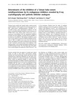
Fig. 1. Mono- and multi/poly ubiquitination of recombinant hTH1 by a
reconstituted ubiquitin conjugating enzyme system. Ubiquitination of
recombinant hTH1 was performed in a reconstituted in vitro system
with the U b c onju gating e nzymes E1, E2 and E3 isolated by affinity
chromatography from rat liver and [
125
I]Ub [1 6]. hPAH wit h subunit
molecularmassof51kDawasusedasapositivereferenceproteinfor
ubiquitination. The reaction mixture (55 lL) co ntained 7.6 lgof
proteins (E1, E2 and E3 proteins), 1.5 l
M
Ubal, 18 l
M
[
125
I]Ub in the
absence of hydroxylase (A), the presence of 8 l
M
hPAH (B) and
(CandD)of8l
M
hTH1. After 90 m i n, the reaction was quenched by
the addition of acetone and precipitated proteins analysed on two-
dimensional electrophoresis [12.5% (w/v) ( gel)] in ( A–C); 10% (w /v)
gel in (D). Inset in B and C: Coomassie Brilliant Blue stained proteins
from the reaction mixture containing PAH (B) and TH (C). The
multiple molecular forms of hTH1 have a molecular mass for the
subunit of 62 kDa; the doublet with a more acidic p I a nd a
molecular mass of 100 kDa r epresents presumably the E1 enzyme
[16,49]. The main forms of hTH1 are also indicated by arrows in Panel
D. The [
125
I]Ub-labelled conjugates of hTH1 are v isualized as diagonal
spots of radioactivity in the autoradiogram (Panel C and D). The
polyUb chains derived from
125
I-labelled Ub with 8.5 kDa a nd neutral
pI are observed as vertical spots (A–C). (D) An expanded view of the
area of interest (i.e. a bove 60 kDa) and co rrespon ds to the pattern of
superimposed profils of stained g el and t he respective autoradio gram
obtained after short exposure time. The main Coomassie Blue stained
spots indicated by arrows correspond to hTH1 and E1. The auto-
radiographic pattern of [
125
I]Ub-labelled conjugates corresponds
mainly to the poly/multi U b-TH conjugates vizu alized as a ladder o f
at least eight distinct Ub-TH conjgates with a microheterogeneity
corresponding to the enzyme as isolated.
Ó FEBS 2002 Ubiquitination of tyrosine hydroxylase (Eur. J. Biochem. 269) 1563
Ub-protein conjugates derived from hTH1 in addition to
poly Ub-chains. Mono-ubiquitinated and poly/multi-ub iq-
uitinated species were visualized as a diagonal pattern of
high M
r
ÔladderÕ of radioactive spots (Fig. 1C) which reflects
the h eterogeneity of the Ub-protein conjugates of wt-hTH1
in terms of size and pI. Multiple molecular forms of
62 kDa and different p I values around 5.5 (Fig. 1C inset)
were observed for the nonubiquitinated enzyme. A ladder of
at least eight Ub-TH conjugates could be identified in the
molecular mass range higher than 66 kDa (Fig. 1, panel
D), which also revealed a microheterogeneity corresponding
to the enzyme as i solated (arrow i n Fig. 1D). The
background in the control was negligible in this relevant
area as earlier reported for this reconstituted in vitro assay
[16]. Thus, the diagonal spots observed on the autoradio-
gram (Fig. 1D) correspond to mono- and multi/poly Ub
adducts, respectively, for the wt-hTH1 with 70 kDa,
78 kDa, etc. (molecular mass increasing by multiple of
8 kDa) and increasing pI. In a ddition, a series of
predominant s pots with the same neutral pI as f ree Ub
and increasing M
r
, observed in the absence (Fig. 1A) or
presence of hydroxylase (Fig. 1B,C), was distributed in a
periodic pattern co rresponding to poly Ub chains [16].
Finally, some insignificant amounts of poly/multi-ubiquiti-
nated proteins, representin g ubiquitination o f not yet
identified liver proteins, present in the E3 preparation [16],
were also o bserved ( Fig. 1A).
Microheterogeneity of wt-hTH1 and bTH as observed
by two-dimensional electrophoresis
The recombinant wt-hTH1 expressed in E. coli revealed a
microheterogeneity on two-dimensional electrophoresis
(Fig. 2A) with 5–6 components of 62 kDa, differing
in pI by 0.1 pH unit. A similar type o f microhetero-
geneity was observed (Fig. 2 B) when the enzyme was
expressed (1 h at 37 °C) in an in vitro transcription-
translation system as a protein of either 62 kDa or
60 kDa subunits, where the difference in molecular
mass is explained by a second initiation site in this
expression system [22].
When the membrane form of bovine TH (bTH
mem
),
extracted from i solated adrenal chromaffin g ranule ghosts,
was subjected to two-dimensional electrophoresis, the
Western blot a nalysis revealed a broad distribution pattern
in terms of pI (Fig. 2C) with the apparent molecular mass of
60 kDa, a value typical of the subunit of bTH in
chromaffin cells [7,9]. The streaky pattern of bTH
mem
(Fig. 2C) is characteristic of proteins with a tendency to
aggregate/precipitate around the pI [23]. In addition, two
post-translational modifications of bTH may also contri-
bute to this pronounced microheterogeneity. Thus, the
enzyme has four possible phosphorylation sites at Ser
residues in the regulatory domain, and each phosphoryla-
tion lowers the pI b y 0.1 U [24,25]. Deamidation o f labile
amide groups has a similar effect on pI as shown for the
structurally related phenylalanine hydroxylase [26] and
most likely e xp lains the microheterogeneity of the nonphos-
phorylated recombinant wt-hTH1 (Fig. 2A). Thus, hTH1
contains three aspargine residues of which Asn414 (posi-
tioned in a short loop between two b strands) [14] is
predicted to be the most labile one on the basis of its nearest
neighbour amino acids (QNG), with a half-life of 1.5 days
days [27] in Tris buffer, pH 7.0. Thus, similarly to hPAH
[26,28], hTH1 also occurs in multiple molecular forms which
could be explained by a progressive deamidation of labile
Asn residue(s).
Ubiquitination and degradation of hTH1
in the reticulocyte lysate system
wt-hTH1 was expressed in the coupled in vitro transcrip-
tion-translation ( TnT
R
) syste m and the net accumulation
of [
35
S]hTH1 was followed as a function of time
(Fig. 3A,B). The typical profile of [
35
S]hTH1 on
SDS
_
PAGE revealed two major bands, corresponding to
subunits of 62 kDa and 60 kDa, respectively. In
Fig. 2. Microheterogeneity of recombinant human tyrosine hydroxylase
(hTH1) an d the me mbrane-bound form o f the bov ine enzyme (bTH
mem
)
as revealed by 2D-electrophoresis. (A) R ecombinant hTH1 (40 lg)
expressed in E. coli and visualized by Coomassie B rilliant Blue stain-
ing. ( B) [
35
S]Methionine-labelled hTH expressed in the in vitro
transcription-translation system (10 lL assay) and dete cted b y a uto-
radiography. (C) bTH
mem
of the bovine adrenal chromaffin granula
membrane. (part a) two-dimensional profil of Coomassie Brilliant
Blue stained membrane proteins (500 lg). ChgA (chromogranin A)
and DBH (dopamine b-hydroxylase) represent the major spots as
described previously [50,51]. The position of the multiple molecular
forms of bTH are indicated by bracket as confirmed by immuno-
blotting using ECL detection with 20 s (part b) a nd 5 min (part c)
exposures.
1564 A. P. Døskeland and T. Flatmark (Eur. J. Biochem. 269) Ó FEBS 2002
addition, on prolonged exposure (> 60 min),
35
S-labelled
proteins of molecular mass around 80 kDa were observed,
concomittantly to the formation of the main product. Less
defined
35
S-labelled proteins were also observed in the
high-molecular-mass region (Fig. 3C), in amount enh anced
by the presence of U b (20 l
M
) and an inhibitor o f
proteasome proteolytic activity [29,30], clasto-lactacystin
b-lactone (2
m
M
) dissolved in dimethylsulfoxide (1%) (data
not shown), and were identified as post-transcriptionally
modified TH such as Ub-conjugates. Furthermore, in the
hTH1-expression sytem,
35
S-labelled peptides of 34 and
28–30 kDa were observed in increasing amount concom-
itantly to the formation a nd subsequent decrease of
[
35
S]TH at longer incubation time points. Thus, degrada-
tion of wt-hTH1 by components in the rabbit reticulocyte
lysate influencing its proteolysis were further studied
(Fig. 3C,D).
MgATP-dependent degradation of wt-hTH1
[
35
S]Methionine-labelled wt-TH1 was incubated with a
reticulocyte lysate as the degradation mac hinery (Fig. 4).
Reticulocytes do not contain any lysosomes [31] and any
MgATP-dependent degradation correlates with proteo-
somal activity [ 32]. In the presence of M gATP a s ignificant
decrease in the a mount of wt-hTH1 was obs erved (Fig. 4A,
lower i nset) while the hTH1 degradation was relatively
moderate on depletion of MgATP (Fig. 4A, upper inset).
The h alf-life of hTH1 disappearance was estimated to be of
7.4 h when the lysate was depleted for MgATP vs. 2.1 h in
the presence of an ATP regenerating sys tem. Based on three
independent experiments the MgATP-dependent proteoly-
sis gave a half-life of 4.3 h (Fig. 4B). Furthermore, when
clasto-lactacystin b-lactone (2 l
M
) a nd anti-(26S protea-
some) IgG (2 lLper50lL assay) were added to the
degradation assay in the presence of an excess of MgATP,
the MgATP-dependent degradation was reduced by
60%. This finding further supports the conclusion that
proteasomes in the reticulocyte lysate are involved in t he
degradation of TH.
Fig. 4. MgATP-dependent degradation of hTH1. Semilogarithmic plot
of the degradation of [
35
S]methionine labelled full-length ( 62 kDa )
and truncated form ( 60 kDa) of hTH1 p rotei n synthesize d by the
coupled in vitro transcription-translation (reticulocyte lysate) system.
After synthesis for 1.5 h at 30 °C, 1 m
M
cold me thionine was added
and incubated at 37 °C in the presence of ex cess (j, h)ordepletionof
MgATP (m) (fo r details, see Materials a nd method s). Aliquots w ere
removed at t imed inter vals, and labelled hTH1 ( 62 plus 60 kDa
forms) was quantitated by
INSTANT IMAGER
or subjected to
autoradiography a fter SDS
_
PAGE. (A) The autoradiograms shown as
inset represent experiments with excess of MgATP (lower inset) vs.
depletion of MgATP (upper i nset). The half-lives for TH were esti-
matedto7.4hintheassaydepletedinMgATP(m),andto2.1hwith
excess of MgATP (j, h). (B) The MgATP-dependent degradation
(total minu s MgATP- inde pendent) gave a h alf life of 4.3 h (mean of
three experiments shown by separated symbols). The curves were
drawn b y linea r regression analysis [A , r ¼ 0.809 f or curve (m)and
r ¼ 0.961 f or curve ( j, h); B, r ¼ 0.872 for the curve].
Fig. 3. In vitro ubiquitination and degradation of [
35
S]methionine
labelled hTH1 in the reticulocyte lysate system. hTH1 was expressed in
a coupled in vitro transcription-translation (reticulocyte lysate) system
as described in Materials and metho ds. F rom the 50 lLreaction
mixture 5 lL aliquots were quenched at given time points subjected to
SDS
_
PAGE and image analysis. ( A) Ze ro control; (B) the pattern of
[
35
S]methionine-labelled proteins after 30 min incubation; (C) repre-
sents the image analysis profile of [
35
S]methionine-labelled h TH1 at
150minincubationtime,and(D)theprofileobtainedafteranaddi-
tional 3 h incubation in the presence of a regenerating system for ATP
(degradation assay). The main products in (B) t o (D) co rrespond to
proteins 62 and 60 kDa (i.e. full-length and truncated form of
hTH1) and minor proteolytic products of 34 and 28–30 kDa.
(C) T he area containing [
35
S]methionine-labelled proteins which are
considered to represent U b-conjugates of hTH1 are indicated by
bracket. O, origin and F, dye fr ont. Th e value 1 o n the ordinate c or-
responds to about 214 cpm in (A) and (B), to 76 c.p.m. in (C) and (D).
Ó FEBS 2002 Ubiquitination of tyrosine hydroxylase (Eur. J. Biochem. 269) 1565
Immunodetection of Ub-TH conjugates in bovine
adrenal chromaffin granule ghosts
TH is also a t arget protein for ubiquitination in vivo,as
shown by SDS/PAGE and Western blot analysis of proteins
extracted from highly purified bovine chromaffin granule
ghosts. Immunoblots with anti-Ub IgG revealed several
Ub-conjugates of molecular mass ‡ 55 kDa, with the
highest intensity near the top of the gel (Fig. 5A, lane 2).
One o f t he bands revealed a mobility c orresponding to that
of TH immunoreactivity ( 60 kDa), with a trace amount
of reactivity a t the top of the gel (Fig. 5A, la ne 1 ). O n t wo-
dimensional electrophoresis, t he U b-conjugates were found
to be distributed over a l arge pI interval, m ostly d etected as
a smear, especially dense in t he high-molecular-mass region.
Most interestingly, a strong immunoreactivity was observed
as a s eries of distinct s pots of 5 .0 < pI < 5.8 in t he 63 kDa
region (Fig. 5B, panel 2) w hich revealed cross-r eactivity
with anti-TH Ig (Fig. 5B, panel 1). The same pattern was
obtained whether the membranes were fir st probed w ith
anti-Ub Ig or anti-TH Ig, a nd then r eprobed with anti-TH
Ig and a nti-Ub Ig, r espectively. Due to th e similarity in
molecular mass b etween the proteins cross-reacting wi th
anti-TH Ig and those reacting with anti-Ub Ig, it is a ssumed
that the Ub-conjugates correspond to monoubiquitinated
forms of TH.
The c omigration of TH- and Ub-im munoreactivities was
further studied in chromaffin granule membranes solubi-
lized by the detergents SDS (1%, w/v) and Triton X-100
(2.5%, w/v) [33]. bTH
mem
was immunoisolated, in the
presence of protease inhibitors, on anti-TH IgG coupled t o
protein A–Sepharose beads (see Materials and methods).
After resolution on one dimensional SDS/PAGE and
Western blot analysis, TH of 60 kDa and some higher
molecular mass forms w ere detected with anti-TH IgG. The
immunoisolates were also probed with the anti-Ub Ig, and
positive immunoreactivity w as then observed at 60 kDa
comigrating with TH immunoreactivity (data not shown),
and thus further support the results shown in Fig. 5B.
DISCUSSION
The ubiquitination of many vital p roteins plays an import-
ant role in regulating their functions and turnover in
eukaryotic cells including neurons and n euroendocrine cells
[34,35]. In the present study it is shown that a key regulatory
enzyme in catecholaminergic neuroendocrine cells, i.e.
tyrosine hydroxylase, is a substrate for the Ub-conjugating
enzyme system, both in vitro as a soluble recombinant
human enzyme and in vivo as a membrane-bound form of
the enz yme i n t he c atecholamine storage/secretory gra nules
of the a drenal medulla, w hich may h ave important
functional implications in the central nervous system.
Ubiquitination of the soluble recombinant hTH1
The finding that recombinant hTH1 is a substrate for the
in vitro reconstituted Ub conjugating enzyme system of rat
liver (Fig. 2) was indeed expected as the structurally
homologous enzyme phenylalanine hydroxylase and its
catalytic core enzyme [14] have already been found to be
ubiquitinated [16]. Similarly, the in vivo turnover of the
structurally homologou s enzyme tryptophan h ydroxylase
[14,15] is reported to be mediated by the Ub-proteasome
pathway [36]. Furthermore, ou r previous studies on two
mutant forms of hTH1, associated with the clinical and
metabolic phenotype of
L
-DOPA responsive dystonia and
infantile p arkinsonism, h ave revealed a reduced cellular
stability c ompared t o the wild-type form when expressed in
human embryonic kidney (A293) cells [37] supporting the
in vivo relevance of the observed Ub-conjugates of hTH1
formed in vitro. Thus, elimination of proteins by the
Ub-proteasome pathway is co nsidered to be most active
towards misfolded/misassembled and abnormal mutant
proteins [38].
Energy-dependent degradation of recombinant hTH1
in the
in vitro
reticulolysate system
Further e xp erimental e vidence in s upport of d egradation of
hTH1 by the Ub-proteasome pathway was obtained in
stability studies of recombinant hTH1 in the in vitro
reticulolysate system (Fig. 3). Reticulocyte lysates have
been used as the degradation machinery and are especially
well suitable to study ubiquitin-dependent proteasomal
degradation of specific proteins [32]. Indeed, reticulocytes
Fig. 5. Immunodetection of bTH
mem
and ubiquitin-conjugates in
chromaffin granule membranes. (A) C hromaffin granule ghost proteins
(130 lg) were su bjected to SDS/PAGE and immunoblotted w ith
antibodies directed against TH (lane 1) or Ub (lane 2) and
125
I-Protei n
A as described in the Materials and methods section. O, origin; F,
front. (B) C hromaffin g ranule ghost p roteins ( 800 lg) were subjected
to two-dimensional electrophoresis and imm unob lotted with anti-
bodies directed against hTH1 (panel 1) or Ub (panel 2). Ub-conjugates
were first precisely localized on two-dimensional gel electrophoresis by
immunoblotting with anti-U b Ig. The membrane was th ereafter
stripped an d then reprobed with anti-TH Ig. A s shown in panel 2,
Ub-conjugates were detected at the same position as bTH (panel 1).
The immunoblotting procedure has be en repeated using first anti-TH
and then anti-Ub Ig which also revealed colocalization of the two
immunoreactivities. The time of expo sure (memb rane to film) w hich
was required to detect Ub-conjugates with anti-Ub Ig was usually
twofold to t hreefold longer than that required to d etect TH immuno-
reactivity.
1566 A. P. Døskeland and T. Flatmark (Eur. J. Biochem. 269) Ó FEBS 2002
contain m ultiple proteolytic systems including the MgATP-
ubiquitin-proteasome-dependent pathway, calpains and
MgATP-independent proteases, but they contain no lyso-
somes [31]. Due to the lack of lysosomal activities, the
system has one limitation as compared to regular eukaryotic
cells.
In order to clearly distinguish proteasomal activity from
other proteolytic activities, an established effective concen-
tration (2 l
M
) in vitro of the selective and potent protea-
somal inhibitor clasto-lactacystin b-lactone was used in the
present study. The inhibitor is a lactacystin analog with a
potency of 10 times that of lactacystin and which beside
epoxomycin [39] is the most potent and specific proteasome
inhibitor [29,30,40]. In this coupled transcription-translation
system a time-dependent formation of [
35
S]methionine-
labelled hTH1 w as observed, followed by i ts degradation to
molecular species of 34 and 28–30 kDa, which is related
to the 34-kDa core fragment of hTH1 observed on limited
tryptic proteolysis [4 1]. Its degradation was found to be
partly MgATP-dependent which was inhibited to about
60% by anti-(26S proteasome) IgG plus clasto-lactacystin-
b-lactone.
The o verall half-life of [
35
S]methionine-labelled h TH1
was estimated to 2.1 h in the p resence of an ATP-
regenerating system and to 7.4 h when the lysate was
depleted of MgATP (Fig. 4). By comparison, the half-life of
rat TH estimated in PC-12 cells, in a subclone of PC-12 cells
and in chromaffin cells has been reported to be 17 h [10],
30 h [11] and 29 ± 3 h [12], respectively. The shorter half-
lifes observed in the present reconstituted in vitro system
may be explained in several ways, including a stabilization
of TH in the cells as a result of i ts binding to membranes [9]
and to cytosolic proteins, e.g. the 14.3.3 proteins [42], as
discussed below.
That TH is a substrate for the Ub-conjugating enzyme
system also in vivo is further supported by our finding of
mono-ubiquitinated bTH
mem
in highly purified bovine
adrenal chromaffin granule ghosts (Fig. 5) and for the first
time underlines a physio logical role of this post-translation-
sal modification. Although the TH- and Ub- immunoreac-
tivities revealed a comigration in the t wo selected (one- and
two-dimensional) electrophoretic systems we do not know
if all the TH subunits in the oligomer are ubiquitinated.
A similar observation has been made for the ubiquitinated
form of the m embrane-bound form o f nitric oxide s ynthase
on SDS/PAGE [43]. bTH
mem
behaves as an i ntegral
membrane protein [7,9], but the mechanism b y which it is
bound is not yet resolved. Interestingly, it has been
suggested that ubiquitination might promote a structural
change (unfolding) of linked proteins [44], but it is not
possible at this point to answer the question of whether
bTH
mem
is inserted into the membrane before or after its
ubiquitination. The finding that bTH
mem
is ph osphorylated
by cAMP-dependent protein kinase on Ser40 in the
regulatory domain [9] may support an ubiquitination of
the e nzyme by the cytosolic Ub-conjugating enzyme system
after its membrane insertion.
In contrast to the multi/poly Ub conjugates observed for
the soluble recombinant hTH1 the membrane-bound form
of bTH is mono-ubiquitinated, which m ay be related to t he
function of the u biquitin C-terminal h ydrolase (UCH-L1 o r
PGP9.5) which is widely and often highly expressed in
neuroendocrine cells [34,35], including the rat chromaffin
cells [45]. From a functional point of view, the membrane
localization may protect the catalytically active enzyme
from degradation by t he cytosolic proteases. Thus, ubiqui-
tination may play a role in the degradation of both
membrane-bound and soluble TH. However, the accurate
role of the ubiquitination remains t he subject of further
investigation and the reason why TH is detected mainly as
mono-ubiquitinated form is still unclear.
Ubiquitination of proteins in the chromaffin granule
membrane
Previous studies on subcellular fractions of rat brain
homogenates have revealed that the synaptic membrane
fraction contains multiple Ub-immunoreactive bands, i.e.
Ub-conjugates of 105 , 72, 60, 41 and 38 kDa, and the
majority of the conjugates were found to be integral
membrane proteins including some high-molecular-mass
glycoproteins [46]. As the synaptic membrane fraction
represents a mixture of different types of membranes, the
functional significance of this finding is not clear. The
specific localization of Ub-conjugates to secretory granules
may suggest a function either in membrane fusion events or
in the turnover of the o rganelles. Thus, it has been reported
that an Ub-like conjugating enzyme system is involved in
homotypic membrane fusion in Pichia pastoris [47]. Alter-
natively, Ub appended to membrane proteins may represent
a signal which promotes the selective sorting/sequestration
and turnover of the s ecretory granules by autophagosomes
as already shown for the regulated turnover of other
cytoplasmic organelles (reviewed in [48]).
ACKNOWLEDGEMENTS
The study was supported by grants from the Research Council of
Norway, from t he Novo Nordisk Foudation, The Nansen Fund, T he
Blix Family Fund f or t he Advancement of Medical R esearch and the
Norwegian Council on Cardiovascular Diseases. We greatly appreciate
the expert technical assistance of Sidsel Riise for the preparation of
hTH1 an d that o f S is sel V ik Berg e f or the p rep aration o f c hromaffin
granule ghosts.
REFERENCES
1. Nagatsu, T., Levitt, M. & Ud enfrie nd, S. ( 1964) Tyrosine hydro-
xylase. The initial step in norepinephrine biosynthesis. J. Biol.
Chem. 23 9, 2910–2917.
2. Kaufman, S. (1995) Tyrosine hydroxylase. Adv. Enzymol. Relat.
Areas. Mol . Biol. 70 , 103–220.
3. Flatmark, T. (2000) Catecholamine biosynthesis and physiological
regulation in neuroendocrine cells. Acta Physiol. Scand. 168, 1–17.
4. Kuc zenski, R.T. & Mandell, A.J. (1972) Regulatory properties of
soluble and particulate rat brain tyrosine hydroxylase. J. Biol.
Chem. 24 7, 3114–3122.
5. H aycock, J.W., George, R.J. & Wa ymire, J.C. (1985) In situ
phosphorylation of tyrosine hydroxylase in chromaffin cells:
localization to soluble compartments. Neurochem. Int. 7, 301–308.
6. Haavik, J., Andersson, K.K., Petersson, L. & Flatmark, T. (1988)
Soluble tyrosine hydroxylase (tyrosine 3-monooxygenase) from
bovine adrenal m edulla: large-scale purification and physico-
chemical p roperties. Biochim. Bio phys. Acta 953, 142–156.
7.Treiman,M.,Weber,W.&Gratzl,M.J.(1983)3¢,5¢-Cyclic
adenosine m onophosp hate- and Ca
2+
-calmodulin-depe ndent
endogenous protein phosphorylation activity in membranes of
the bovine chromaffin secretory vesicles: identification of t wo
Ó FEBS 2002 Ubiquitination of tyrosine hydroxylase (Eur. J. Biochem. 269) 1567
phosphorylated components a s tyrosine hydroxylase and protein
kinase regulatory subunit t ype II. J. Neurochem. 40, 6 61–664.
8. Morita, K ., Teraoka, K. & Oka, M. (1987) I nteraction of
cytoplasmic tyrosine hyd roxyla se with chromaffin granule.
In vitro studies on association of soluble enzyme with granule
membranes and alteration in enzyme activity. J. Biol. Chem. 262,
5654–5658.
9. Kuhn, D.M., Arthr, R. Jr, Yoon, H. & Sankaran, K. (1990)
Tyrosine hydroxylase in secretory granules from adrenal medulla.
Evidence for an integral m embrane form. J. Biol. Chem. 26 5,
5780–5786.
10. Wu, D.K. & Cepko, C.L. (1994) The stability of endogenous
tyrosine h ydroxylase protein in PC-12 cells differs from that
expressed in mouse fibroblasts by gene transfer. J. Neurochem. 62,
863–872.
11. Tank, A.W., H am, L. & Curella, P. (1986) In duction of tyrosine
hydroxylase by cyclic AMP and glucocorticoids in a rat pheo-
chromocytoma cell line. effect o f t h e inducing ag ents alone or in
combination on the enzyme levels and rate of synthesis of tyrosine
hydroxylase. Mol. Pharmacol. 30 , 486–496.
12. Fernandez, E. & Craviso, G .L. (1999) Protein synthesis blockade
differentially a ffects the degradation of constitu tive a nd nicotinic
receptor-induced tyrosine hydroxylase protein level in isolated
bovine c hromaffin c ells. J. Neurochem. 73, 1 69–178.
13. Pasinetti, G.M., Osterburg, H.H., Kelly, A.B., Kohama, S.,
Morgan, D.G., Reinhard, J.F. Jr, Stellwagen, R.H. & Finch, C.E.
(1992) Slow changes of tyrosine hydroxylase g ene expression in
dopaminergic brain neurons after neurotoxin lesio ning: a model
for neuron aging. Mol. Brain . Res. 13, 63–73.
14. Flatmark, T. & Ste vens, R.C. (1999) Structural insights into the
aromatic amino acid hydroxylase and their disease related mutant
forms. Chem. Rev. 99 , 2137–2160.
15. Grenett, H.E., Ledley, F.D., Reed, L.L. & Woo, S.L. (1987) Full-
lenght cDN A f or r abbit tryptophan hydroxylase: f unctional
domains and evolution of aromatic amino acid hydroxylases.
Proc. N atl Acad. Sci. USA 84 , 5530–5534.
16. Døskeland, A.P. & Flatmark, T. (2001) Conjugation of phenyl-
alanine hydroxylase with polyubiquitin chains catalysed by rat
liver e nzymes. Biochim. Biophys. Acta 1 547, 379–386.
17. Haavik, J., Le Bourdelles, B., Martı
´
nez, A., Flatmark, T. &
Mallet, J. (1991) Recombinant human tyrosine hydroxylase iso-
zymes. Reconstitution with iron and inhibitory effect of other
metal ions. Eur. J. Biochem. 19 9, 371–378.
18. Le Bourdelle
`
s, B., Horellou, P., Le Caer, J P., Dene
`
fle, P., Latta,
M., Haavik, J., G uibert, B., Mayaux, J F. & Mallet, J. (1991)
Phosphorylation of human recombinant tyrosine hydroxylase
isoforms 1 and 2: an additional phosphorylated residue in isoform
2, generated through alternative splicing. J. Biol. Chem. 266,
17124–17130.
19. Sagne, C., Isambert, M F. & Henry, J . (19 96) -P. & Ga snier, B.
SDS-resistant aggregation of membrane proteins in application to
the purification of vesicular monoamine transporter. Biochem.
J. 316, 8 25–831.
20. Terland, O. & Flatmark, T. (1980) Oxidoreductase activities o f
chromaffin granule ghosts isolated f rom the bovine adrenal
medulla. Bio chim. Biophys. Acta 597, 318–330.
21. Laemmli, U.K. (197 0) Cleavage of structural proteins during t he
assembly of the head of b acteriophage T4. Nature 227, 680–685.
22. Lu
¨
decke, B., Knappskog, P.M., Clayton, P.T., Surtees, R.A.H.,
Clelland, J .D., Heales, S.J., Brand, M.P., Bartholome
´
,K.&
Flatmark, T. (1996) Recessively inherited
L
-DOPA-responsive
parkinsonism in infancy caused by a point mutation (L205P) i n
the tyrosine hydroxylase g ene. Hum. Mol. Genet. 5, 102 3–1028.
23. Gørg,A.,Obermaier,C.,Boguth,G.,Harder,A.,Scheibe,B.,
Wildgruber, R. & Weiss, W. (2000) The current state of two-
dimensional electrophoresis with immobilized pH gradients.
Electrophoresis 21, 1037–1053.
24. Coˆte
´
, A., Doucet, J P. & Trifaro, J M. (1986) Phosphorylation
and dephosphorylation of c hr omaffin cell p ro teins in r espo nse to
stimulation. Ne uroscience 19, 629–645.
25. Pocotte, S.L., H olz, R.W. & Ue da, T. (1986) Cholinergic receptor-
mediated phosphorylation a nd activation of tyrosine hydroxylase
in cultured bovine adrenal chromaffin cells. J. Neurochem. 46,
610–622.
26. Solstad, T. & Flatmark, T. (2000) Microheterogeneity of
recombinant human phenylalanine hydroxylase as a result o f
non-enzymatic deamidations of labile amide containing amino
acids. Effects on catalytic and stability properties. Eur. J. Biochem.
267, 6302–6310.
27. Robinson, N.E. & Robinson, A.B. (2001) Prediction of protein
deamidation rates from p rim ary and three-dimensional structure.
Proc. N atl Acad. Sci. USA 98 , 4367–4372.
28. Carvalho, R .M.N., Solstad, T ., Robinson, N .E., Robinson, A.B.
& Flatmark, T. (2002) Pos sible contributions of labile a sparagine
residues to differences in regulatory properties of human and rat
phenylalanine hydroxylase. In Chemistry and Biology of Pteridines
and Folates 2001 (Parrak, V. & Reibnegger, G., eds), Kluwer,
USA. In press.
1
29. Fenteany,G.,Standaert,R.F.,Lane,W.S.,Choi,S.,Corey,E.J.&
Schreiber, S.L. (1 995) Inhibition of proteasome activities and
subunit-specific amino-terminal threonine modification by lacta-
cystin. Science 268, 726–731.
30. Craiu, A., Gaczynska, M., Akopian, T., Gramm, C.F., Fenteany,
G., G oldberg, A.L. & R ock. K.L. (1997) Lac tacystin and cl asto-
lactac yst in b-lactone modify mu ltiple proteasome b-subunits and
inhibit intracellular protein degradation and m ajor histocompa-
tibility complex class I antigen presentation. J. Biol. Chem. 272,
13437–13445.
31. Watson, A.L., Laszlo, L. & Doherty, F.J. (1991 ) The de gradative
fate of ubiquitin-protein conjugates in nucleated and enucleated
cells. Acta Biol. Hung. 42, 4 9–56.
32. Hershko, A. (1988) Ub iquitin-mediated protein degradation.
J. Biol. Chem. 263, 15237–15240.
33. Stoller, T.J. & Shields, D. ( 1988) Retrovirus-mediated e xpression
of preprosomatostatin: posttranslational p rocessing, intracellular
storage, and sec retion in GH
3
pituitary cells. J. Cell Biol. 107,
2087–2095.
34. Doran, J.F., Jackson, P., Kynoch, P.A. & Thompson, R.J. (1983)
Isolation of PGP 9.5, a new human neuron-specific protein
detected by high-resolution two-dimensional electrophoresis.
J. Neurochem. 40, 1 542–1547.
35. Wilkinson, K.D., L ee, K ., Deshpande, S., D uerksen-Hughes, P.,
Boss, J.M. & Pohl, J. (1989) The neuron-specific protein PGP 9.5
is a ubiq uitin carboxyl-terminal hydrolase. Sci ence 246, 670 –673.
36. Kojima, M., Oguro, K., Sawabe, K., Iida, Y., Ikeda, R.,
Yamashita, A., Nakanishi, N. & Hagesawa, H. (2000) Rapid
turnover of trypto phan hydro xylase i s d riven b y prote asomes in
RBL2H3 cells, a serotonin p roducing mast cell line. J. Biochem.
127, 121–127.
37. Flatmark, T., Knappskog, P.M., B jørgo, E. & Martı
´
nez, A. (1997)
Molecular characterization of disease related mutant forms of
human phenylalanine hydroxylase and tyrosine hydroxylase. In
Chemistry and Biology of Pteridines and Folates (Pfleiderer,W.&
Rokos, H. , eds), pp. 503–508. Blac kwell Science, Oxford, UK.
38. Wickner, S., Maurizi, M. & Gottesman, S. (1999) Posttransla-
tional quality control: fo ld ing, refolding, and degrading prote ins.
Science 28 6, 1888–1893.
39. Meng,L.,Mohan,R.,Kwok,B.H.B.,Elofsson,M.,Sin,N.&
Crews, C.M. (1999) Epoxomicin, a potent and selective protea-
some inhibitor, exhibits in vivo anti-inflammatory activity. Proc.
Natl Acad. S ci. USA 96, 1040 3–10408.
40. Dick, L.R., Cruikshank, A.A., Destree, A.T., Grenier, L.,
McCormack, T.A., Melandri, F.D., Nunes, S .L., Palombella, V .J.,
Parent, L.A., Plamondo n, L. & Stein, R.L. (1997) Mechanistic
1568 A. P. Døskeland and T. Flatmark (Eur. J. Biochem. 269) Ó FEBS 2002
studies on the inactivation of the proteasome by lactacystin in
cultured cells . J. Bio l. Chem. 27 2, 182–188.
41. M artı
´
nez, A., Haavik, J., Flatmark, T., Arrondo, J.L.R. & Muga,
A. (1996) Conformational properties and stability of tyrosine
hydroxylase studied by infrared spectroscopy. Effect of iron/
catecholamine b inding and phosphorylatio n. J. Bi ol. C hem. 271,
19737–19742.
42. Itagaki, C., Isobe, T., Taoka, M., Natsume, T., Nomura, N.,
Horigome,T.,Omata,S.,Ichinose,H.,Nagatsu,T.,Greene,
L.A. & Ichimura, T. (199 9) Stimulus–coupled interaction of
tyrosine hydroxylase with 14–3)3 proteins. B i oche mistr y 38,
15673–15680.
43. Bender, A.T., Demady, D.R. & Osawa, Y. ( 2000) Ubiquitination
of neuro nal nitric-o xide synt hase in vitro and in vivo. J. Biol. C hem.
275, 1 7407–17411.
44. Pickart, C.M. (1998) Polyubiquitin chains. In Ubiquitin and the
Biology of the Cell (Peters, J. -M., H arris, J.B. & Finley, D., eds),
pp. 19–63. P lenum Press, N ew York.
45. Kent, C. & Coupland, R.E. (1989) Localisation of Chromo-
granin A and B, met-enkephalin-arg
6
-gyl
7
-leu
8
and PGP9.5-like
immunoreactivity in the developing and adult rat adrenal medulla
and extra-adrenal c hromaffin tissue. J . Anat. 166 , 213–225.
46. Beesley, P.M., Mummery, R., Tibaldi, J., Chapman, A.P., Smith,
S.J. & Rider, C.C. (1995) Th e post-synap tic density: putative
involvement in synapse stabilization via cadherins and covalent
modification by ubiquitination. Biochem. So c. Tra ns. 23, 5 9–64.
47. Yuan, W., Stromhaug, P.E. & Dunn, W.A. (1999) G lucose-
induced autophagy of peroxisomes in Pichia pastoris requires a
unique E1-like protein. Mol . Biol. C ell 199, 1353 –1366.
48. Klionsky, D.J. & Emr, S.D. (2000) Autophagy as a regulated
pathway of cellu lar degradation. Sc ience 29 0, 1717–1721.
49. Cook, J.C. & Chock, P.B. (1992) Isoforms of mammalian
ubiquitin-activating enzyme. J. Biol. C hem. 267, 2 4315–24321.
50. Bad er, M.F., Hikita, T . & Trifaro, J .M. (1985) Calcium-dependent
calmodulin binding to chromaffin granule membranes: presence of
a 65-kilodalton calmodulin binding protein. J. Neurochem. 44,
526–539.
51. Winkler, H., Apps, D.K. & Fi scher-Colbrie, R. (1986) The
molecular function of adrenal chromaffin granules: established
facts and unresolved t o pics. Neuroscience 18 , 261–290.
Ó FEBS 2002 Ubiquitination of tyrosine hydroxylase (Eur. J. Biochem. 269) 1569