Báo cáo Y học: A comparative biochemical and structural analysis of the intracellular chorismate mutase (Rv0948c) from Mycobacterium tuberculosis H37Rv and the secreted chorismate mutase (y2828) from Yersinia pestis pptx
Bạn đang xem bản rút gọn của tài liệu. Xem và tải ngay bản đầy đủ của tài liệu tại đây (828.06 KB, 12 trang )
A comparative biochemical and structural analysis of
the intracellular chorismate mutase (Rv0948c) from
Mycobacterium tuberculosis H
37
R
v
and the secreted
chorismate mutase (y2828) from Yersinia pestis
Sook-Kyung Kim*, Sathyavelu K. Reddy, Bryant C. Nelson, Howard Robinsonà, Prasad T. Reddy
and Jane E. Ladner
Biochemical Science Division, Chemical Science and Technology Laboratory, National Institute of Standards and Technology, Gaithersburg,
MD, USA
Keywords
chorismate mutase; Mycobacterium
tuberculosis; pathogenesis; shikimate
pathway; Yersinia pestis
Correspondence
P. T. Reddy, Biochemical Science Division,
Bldg 227, Rm B244, Chemical Science and
Technology Laboratory, National Institute of
Standards and Technology, Gaithersburg,
MD 20899, USA
Fax: +1 301 975 8505
Tel: +1 301 975 4871
E-mail:
J. E. Ladner, Biochemical Science Division,
Bldg 227, Rm B244, Chemical Science and
Technology Laboratory, National Institute of
Standards and Technology, Gaithersburg,
MD 20899, USA
Fax: +1 240 314 6225
Tel: +1 240 314 6206
E-mail:
Present addresses
*Division of Metrology for Quality Life,
Korea Research Institute of Standards and
Science, Daejeon, Republic of Korea
Division of Molecular Biology, Department
of Zoology, Sri Venkateswara University,
Tirupati, Andhra Pradesh, India
àBiology Department, Brookhaven National
Laboratory, Upton, NY, USA
(Received 9 May 2008, revised 25 July
2008, accepted 30 July 2008)
doi:10.1111/j.1742-4658.2008.06621.x
The Rv0948c gene from Mycobacterium tuberculosis H
37
R
v
encodes a 90
amino acid protein as the natural gene product with chorismate mutase
(CM) activity. The protein, 90-MtCM, exhibits Michaelis–Menten kinetics
with a k
cat
of 5.5 ± 0.2 s
)1
and a K
m
of 1500 ± 100 lm at 37 °C and
pH 7.5. The 2.0 A
˚
X-ray structure shows that 90-MtCM is an all a-helical
homodimer (Protein Data Bank ID: 2QBV) with the topology of Escheri-
chia coli CM (EcCM), and that both protomers contribute to each catalytic
site. Superimposition onto the structure of EcCM and the sequence align-
ment shows that the C-terminus helix 3 is shortened. The absence of two
residues in the active site of 90-MtCM corresponding to Ser84 and Gln88
of EcCM appears to be one reason for the low k
cat
. Hence, 90-MtCM
belongs to a subfamily of a-helical AroQ CMs termed AroQ
d.
The CM
gene (y2828) from Yersinia pestis encodes a 186 amino acid protein with an
N-terminal signal peptide that directs the protein to the periplasm. The
mature protein, *YpCM, exhibits Michaelis–Menten kinetics with a k
cat
of
70±5s
)1
and K
m
of 500 ± 50 lm at 37 °C and pH 7.5. The 2.1 A
˚
X-ray
structure shows that *YpCM is an all a-helical protein, and functions as a
homodimer, and that each protomer has an independent catalytic unit
(Protein Data Bank ID: 2GBB). *YpCM belongs to the AroQ
c
class of
CMs, and is similar to the secreted CM (Rv1885c, *MtCM) from M. tuber-
culosis.
Abbreviations
*MtCM, secreted chorismate mutase from Mycobacterium tuberculosis; *YpCM, secreted chorismate mutase from Yersinia pestis; CM,
chorismate mutase; EcCM, chorismate mutase domain of chorismate mutase–prephenate dehydratase from Escherichia coli; IPTG,
isopropyl-thio-b-
D-galactoside; MtCM, intracellular chorismate mutase from Mycobacterium tuberculosis; PfCM, chorismate mutase from
Pyrococcus furiosus; ScCM, allosteric chorismate mutase from Saccharomyces cerevisiae; TSA, transition state analog; TtCM, chorismate
mutase from Thermus thermophilus.
4824 FEBS Journal 275 (2008) 4824–4835 Journal compilation ª 2008 FEBS. No claim to original US government works
Chorismate mutase (CM) (EC 5.4.99.5), a shikimate
pathway enzyme [1], catalyzes the pericyclic rearrange-
ment of chorismate to prephenate [2]. Subsequent to
this conversion, prephenate dehydrogenase and
prephenate dehydratase catalyze the biosynthesis of
tyrosine and phenylalanine, respectively. As this bio-
synthetic pathway is absent in mammals but is essen-
tial for the survival of bacteria and fungi, CM is often
targeted for the development of inhibitors for micro-
bial pathogens. This work was aimed at the character-
ization of CM from Mycobacterium tuberculosis
H
37
R
v
, a dreaded pathogen that claims two million
human lives annually [3].
Annotation of the genome of M. tuberculosis H
37
R
v
revealed two genes for CM [4]. The Rv1885c gene
encodes a secreted CM (*MtCM) and the Rv0948c
gene encodes an intracellular CM (MtCM). Sasso et al.
[5], Prakash et al. [6] and Kim et al. [7] have character-
ized *MtCM. Kim et al. [7] have shown that *MtCM
has in fact an extracellular destination in M. tuber-
culosis. Prakash et al. [6] conducted a brief study of
the recombinant MtCM. Our work is aimed at the fur-
ther characterization of MtCM. The true primary
sequence of MtCM is complicated by virtue of a
number of presumptive in-frame initiator methionines
preceded by a reasonable ribosome-binding sequence.
The annotation Rv0948c for MtCM in a laboratory
strain of M. tuberculosis H
37
R
v
would encode a 105
amino acid protein (105-MtCM) [4], whereas the anno-
tation MT0975 for MtCM in the CDC1551 strain
would encode a 217 amino acid protein (217-MtCM)
[8]. Furthermore, alignment of 105-MtCM with the
genetically engineered Escherichia coli CM (EcCM) [9]
shows that the 105 amino acid protein has extra amino
acids beyond the N-terminus of EcCM. Hence, we
cloned 90-MtCM beginning with Met16 in Rv0948c.
We determined the 3D structure of the 90-MtCM and
kinetic parameters of all three proteins. The 90-MtCM
is an AroQ class CM and the protein functions as a
dimer. In this article, we also report on the cloning of
the gene encoding the secreted CM from Yersinia
pestis (*YpCM, y2828), purification of the protein,
investigation of the properties of the enzyme, and the
crystal structure analysis of the protein.
Results and Discussion
Annotation of CMs in M. tuberculosis H
37
R
v
The difference in annotation of MtCM arose from an
in-frame initiator ATG codon in MT0975 (217-MtCM)
and in Rv0948c (105-MtCM). Furthermore, the N-ter-
minus of 105-MtCM has 22 more residues than the
CM domain of E. coli prephenate dehydratase (Fig. 1).
There is an in-frame methionine at position 16 of
105-MtCM and a purine-rich sequence analogous to
the Shine–Dalgarno sequence about 10 nucleotides
upstream of Met16. We reasoned that this Met16
could be the real initiator and consequently would pro-
duce a 90 amino acid protein (90-MtCM). We charac-
terized all three versions of the putative intracellular
CM, i.e. 217-MtCM, 105-MtCM, and 90-MtCM. In a
recent publication, Schneider et al. [10] observed simi-
lar ambiguity about the translation start site(s) in the
gene MSMEG5513 for an intracellular CM, a homolog
of Rv0948c, from the annotation of the Mycobacte-
rium smegmatis genome. They determined the transla-
tion start site by translation fusion with the
b-galactosidase gene, and showed that the first methio-
nine in MSMEG5513 is the ‘real initiator’. Schneider
et al. [10] did not determine the translation start site
for Rv0948c.
Production and purification of MtCM
The 105-MtCM was overproduced as a fusion protein
with the subtilisin prodomain (Fig. 2, lane 2). The
fusion protein was completely soluble (Fig. 2, lane 3).
Cleavage of 105-MtCM from the prodomain was trig-
gered by fluoride-induced subtilisin activity (Fig. 2,
lane 5). We observed three major protein products at
this stage of purification: intact fusion protein (per-
haps not very tightly bound), 105-MtCM, and an
unidentified lower molecular mass protein. Hence,
105-MtCM was further purified by molecular sieve
chromatography to near homogeneity (Fig. 2, lane 6).
The molecular mass of 105-MtCM determined
by MALDI-TOF MS was 11 771 Da (theoretical
mass = 11 771 Da), and established that the protein is
intact. Similarly, 90-MtCM was overproduced as a
fusion protein with the subtilisin prodomain, and the
protein was purified to homogeneity (Fig. 3, lane 5).
As can be seen in lane 5 of Fig. 3, 90-MtCM migrated
as a 6 kDa protein. Hence, we determined the
molecular mass of 90-MtCM by MALDI-TOF MS as
10 090 ± 1 Da, which is identical to the theoretical
mass of 10 090 Da. The yield of 105-MtCM and
90-MtCM was 1 mg per 1 L of culture. Activity mea-
surements for CM using these two proteins showed
that both proteins catalyze the conversion of choris-
mate to prephenate (see kinetic measurements for k
cat
).
The 217-MtCM, purified from the subtilisin column,
had 1 ⁄ 50th of the CM activity of 90-MtCM and 105-
MtCM at the same stage of purification. Hence, we
did not further purify or characterize 217-MtCM. We
conclude that the annotation of the MT0975 gene was
S. -K. Kim et al. AroQ chorismate mutases
FEBS Journal 275 (2008) 4824–4835 Journal compilation ª 2008 FEBS. No claim to original US government works 4825
misled by an upstream in-frame initiator methionine
preceded by the Shine–Dalgarno sequence.
Purification of *YpCM from the periplasmic fluid
of E. coli
*YpCM production was induced with 10 lm isopro-
pyl-thio-b-d-galactoside (IPTG). Periplasmic proteins
were isolated as described for *MtCM [7]. The peri-
plasmic fluid was concentrated to 500 lL in a Milli-
pore centrifugal tube with a 5000 Da molecular mass
cutoff. *YpCM was purified on a 210 mL Biosep
SEC-3000 HPLC column (Phenomenex, Torrance, CA,
USA), equilibrated and eluted with 50 mm Tris ⁄ HCl
(pH 7.5), 1 mm EDTA, and 100 mm NaCl.
Fig. 1. Alignment of 90-MtCM with AroQ
a
CMs. The alignment begins with amino acid 13 in 90-MtCM (the numbering begins with amino
acid 1 in the 90 amino acid protein). Amino acids 1–12 were not seen in the electron density map; their sequence is shown above the align-
ment. In the structural alignment of MtCM, EcCM, PfCM, and TtCM by
MATRAS [12], the top line indicates the average secondary structure
(AVE_SECSTR); H, helical; T hydrogen-bonded turn; C, coil; S, bend. Capital letters indicate agreement for all structures. The active site resi-
dues in EcCM are highlighted, and are shadowed when similar in the other sequences. C-terminal residues that were not visible in the struc-
tures are shown as lower-case letters for MtCM, PfCM, and TtCM. At the top of the figure, the 15 N-terminal residues of the 105-MtCM
construct are shown.
1 2 3 4 5 6 7 kDa
32.5
Subtilisin prodomain:105 aa
MtCM fusion protein
25.0
16.5
105 aa MtCM monomer
6.5
Fig. 2. SDS ⁄ PAGE (16%) of the production and purification of
105-MtCM. Lane 1: uninduced cell-free extract of E. coli BL21(DE3)
harboring the pG58–105-MtCM clone (25 lg of protein). Lane 2:
induced cell-free extract of E. coli BL21(DE3) harboring the pG58–
105-MtCM clone (25 lg of protein). Lane 3: 48 000 g supernatant
of induced cells (same volume as used in lane 2). Lane 4: flow
through from subtilisin column (same volume as used in lane 2).
Lane 5: 10 lg of protein(s) eluted after equilibration with 100 m
M
sodium fluoride. Lane 6: 5 lg of purified 105-MtCM from a Sepha-
dex G-75 column. Lane 7: molecular mass markers.
1 2 3 4 5 6 kDa
32.5
25.0
16.5
Subtilisin prodomain: 90 aa
MtCM fusion protein
6.5
90 aa MtCM monomer
Fig. 3. SDS ⁄ PAGE (16%) of the production and purification of
90-MtCM. Lane 1: induced cell-free extract of E. coli BL21(DE3)
harboring the pG58–90-MtCM clone (25 lg of protein). Lane 2:
48 000 g supernatant of induced cells (same volume as used in
lane 1). Lane 3: flow through from subtilisin column (same volume
as used in lane 1). Lane 4: 10 lg of protein(s) eluted after equilibra-
tion with 100 m
M sodium fluoride. Lane 5: 13 lg of purified
90-MtCM from a Sephadex G-75 column. Lane 6: molecular mass
markers.
AroQ chorismate mutases S. -K. Kim et al.
4826 FEBS Journal 275 (2008) 4824–4835 Journal compilation ª 2008 FEBS. No claim to original US government works
Kinetic measurements
Assays for CM were performed with 90-MtCM and
105-MtCM at chorismate concentrations of 100 lm to
4mm. Both MtCMs catalyzed the conversion of
chorismate to prephenate with a k
cat
of 5.5 ± 0.2 s
)1
and a K
m
of 1500 ± 100 lm at 37 °C and pH 7.5
(Table 1). The k
cat
for MtCM is about 14-fold lower
than that reported for EcCM (72 s
)1
) [11]. The K
m
of
1500 lm chorismate for MtCMs is five times higher
than that observed for EcCM. One obvious reason for
the low k
cat
and high K
m
exhibited by MtCM is the
absence of two of the substrate-binding residues found
in the C-terminus of EcCM (Fig. 1). In contrast,
*YpCM, in which all the catalytic site residues are pre-
served, exhibits a high k
cat
of 70 ± 5 s
)1
, similar to
that for EcCM.
Crystal structure of 90-MtCM
The crystal structure of 90-MtCM shows clearly that the
molecule is an all a-helical homodimer (Protein Data
Table 1. Comparison of the catalytic properties of MtCM, EcCM,
and *YpCM. MtCM proteins and *YpCM were purified as
described in Experimental procedures. One microgram of MtCM or
200 ng of *YpCM in 10 lL was used in each assay of 0.3 mL of
buffer. The buffer consisted of 50 m
M Tris ⁄ HCl (pH 7.5), 0.5 mM
EDTA, 0.1 mg BSAÆmL
)1
, and 10 mM b-mercaptoethanol. Choris-
mate concentrations were varied from 0.25 to 4 m
M. Assays were
performed at 37 °C for 5 min [34], and the reaction was stopped
with 0.3 mL of 1
M HCl. After further incubation for 10 min at
37 °C to convert prephenate to phenylpyruvate, 0.6 mL of 2.5
M
NaOH was added. The absorbance of the phenylpyruvate chromo-
phore was read at 320 nm. Blanks without the enzyme were main-
tained for each of the chorismate concentrations to account for the
nonenzymatic conversion of chorismate to prephenate. Results are
the average of three experiments. The kinetic data for EcCM were
from the literature [11], measured at 30 °C.
Enzyme k
cat
(s
)1
) K
m
(lM)
90-MtCM 5.5 ± 0.2 1500 ± 100
105-MtCM 5.5 ± 0.2 1500 ± 100
217-MtCM 0.1 Not determined
EcCM 72 296 ± 19
*YpCM 70 ± 5 500 ± 50
A
B
C
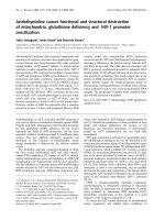
Fig. 4. Crystal structure of 90-MtCM. (A) The homodimer is shown
as a stereo cartoon on the top with one polypeptide chain in blue
and the other in green. (B) The superimposition of a single chain of
TtCM, PfCM and EcCM onto 90-MtCM is shown, with the TSA
from EcCM marking the active site. The approximate positions of
the N-termini and C-termini are labeled in the same color as the
polypeptide chain. The three helices are labeled H1, H2, and H3.
(C) Helix 3 from each of the four structures is shown. The helices
were taken from the superimposed structures and then separated
by translating each horizontally in the plane of the page. The figures
were drawn with
PYMOL ().
S. -K. Kim et al. AroQ chorismate mutases
FEBS Journal 275 (2008) 4824–4835 Journal compilation ª 2008 FEBS. No claim to original US government works 4827
Bank ID: 2QBV; Fig. 4). The polypeptide chain has one
long 36 residue helix (helix 1), an eight residue loop, an
11 residue helix (helix 2), a two residue loop, and a 15
residue helix (helix 3). The buried surface area of the
dimer is 3810 A
˚
2
. This crystal form has one protomer in
the asymmetric unit; the complete molecule is generated
by a crystallographic two-fold. The data and refinement
statistics are shown in Table 2. The Ramachandran plot
has 96.9% of the residues in the most favored region
and 3.1% in the additional allowed region. Five residues
were modeled with alternative conformations. No inter-
pretable density was observed for the first 12 residues or
for the last five residues. When the model is numbered
according to the 90 residue protein, residues 13–85 are
seen in the electron density map. Using matras [12] to
compare the structure with a representative library of
structures, three structures stood out as very similar;
these are Protein Data Bank IDs 2D8D, 1YBZ and
1ECM. 2D8D and 1YBZ are annotated as CMs from
Thermus thermophilus (TtCM) and Pyrococcus furiosus
(PfCM), respectively, from Structural Genomics pro-
jects on these organisms. 1ECM [13] is a genetically
engineered 109 amino acid CM domain from E. coli.
The dimer of 90-MtCM is shown in Fig. 4A, and the
superimposition of the four structures for a single poly-
peptide chain is shown in Fig. 4B. It is apparent from
the structural alignment that helix 3 of 90-MtCM is a
shorter version of helix 3 of EcCM, TtCM and PfCM
lacking two of the binding site residues (Fig. 4C) corre-
sponding to Ser84 and Gln88 of EcCM as discussed
below.
During the preparation of this article, we were made
aware of another deposited Protein Data Bank file for
90-MtCM, 2VKL (M. Okvist, K. Roderer, S. Sasso,
P. Kast, and U. Krengel, unpublished data). In the
structure of 90-MtCM in 2VKL, there is a malate ion
from the buffer in the active site of the enzyme. Malate
mimics endo-oxabicyclic dicarboxylic acid, the transition
state analog (TSA), in much the same fashion as citrate
that we observed in our *YpCM structure (Protein Data
Bank ID: 2GBB). We crystallized 90-MtCM in the pres-
ence of citrate but did not see citrate in the active site.
However, we studied the effect of citrate on 90-MtCM
activity, and found citrate to have some kind of inhibi-
tory effect from preliminary results (data not shown).
The inhibition is not a salt effect, because sodium chlo-
ride and sodium acetate had no effect on the activity.
The rmsd on C-alphas between the two 90-MtCM
structures is 0.69 A
˚
. One difference between the struc-
tures is that five residues at the C-terminal end are dis-
ordered in our structure (2QBV) and only one residue
is disordered in 2VKL. In fact, although the space
group is the same for both structures, the c-axis is
10 A
˚
shorter in 2VKL. This difference is due to crystal
packing, which allows the C-terminal residues of
neighboring molecules (not in the same dimer) to inter-
act and the tail of one protomer to almost reach the
active site of the neighbor.
Active site of 90-MtCM
The structure of EcCM includes the TSA, which
clearly identifies the active site. From structural and
sequence homology with EcCM, the active site residues
of 90-MtCM can be identified, and are shown in
Fig. 5. The striking difference between EcCM and 90-
MtCM is that the EcCM residues Ser84 and Gln88 are
absent in 90-MtCM (Fig. 1). Structurally, Ser84 of
EcCM lines up with Gly84 of 90-MtCM. The final five
residues, GRLGH, of 90-MtCM are not seen in the
electron density map. However, none of these residues
is a candidate for performing the role of Gln88 in
EcCM. Of the other two structures, PfCM has the
conserved Ser70 and Gln74, and TtCM has Ser81 and
Glu85. There are two Protein Data Bank files for
TtCM; in the file 2D8D, Glu85 is only seen in one of
the two chains in the structure, which has a dimer in
the asymmetric unit, and in the file 2D8E, which has
one chain in the asymmetric unit, all of the C-terminal
residues are seen. Another difference is the loop orien-
Table 2. Diffraction data and refinement statistics showing over-
all ⁄ high-resolution shell (2.18–2.10 A
˚
) values where appropriate.
90-MtCM *YpCM SeMet
Diffraction data
Space group P4
3
2
1
2 C222
1
Cell dimensions (a, b, c)(A
˚
) 59.9, 59.9,
47.5
89.0, 144.1,
116.6
Resolution limit (A
˚
) 2.0 2.1
No. of measured intensities 91 040 566 550
No. of unique reflections 6192 ⁄ 815 43 510 ⁄ 4106
Mean redundancy 14.7 ⁄ 14.8 13.0 ⁄ 8.0
R
merge
0.056 ⁄ 0.323 0.113 ⁄ 0.469
0% Completeness 100.0 ⁄ 100.0 99.4 ⁄ 95.3
Average I ⁄ r 25.3 ⁄ 7.0 12.0 ⁄ 2.0
Mosaicity 1.37 0.77
Radiation wavelength 1.54 0.979
Refinement
Resolution limits (A
˚
) 20.0–2.0 20.0–2.1
R-factor (95% of the data) 0.219 0.207
R
free
(5% of the data) 0.298 0.258
No. of water molecules 47 193
Bond length rmsd (A
˚
) 0.021 0.019
Bond angle rmsd (°) 1.98 1.86
Average B (main
chain ⁄ side chain) (A
˚
2
)
42.2 ⁄ 44.0 34.3 ⁄ 36.5
Average B for water (A
˚
2
) 41.5 34.5
AroQ chorismate mutases S. -K. Kim et al.
4828 FEBS Journal 275 (2008) 4824–4835 Journal compilation ª 2008 FEBS. No claim to original US government works
tation between the first long helix and the second helix.
Figure 4B shows that for the EcCM, TtCM and PfCM
structures, this loop aligns very well, but that the
90-MtCM structure is significantly altered; however,
examination of the surface (not shown) demonstrates
that even with this change, the active site remains bur-
ied. We superimposed the EcCM structure with the
TSA onto 90-MtCM to see the TSA in the active site
of 90-MtCM (Figs 5 and 6). As the residues corre-
sponding to Ser84 and Gln88 of EcCM are absent in
90-MtCM, chorismate is unlikely to be as well stabi-
lized in its active site. This at least partly explains the
low k
cat
for 90-MtCM (Table 1).
In an attempt to substantiate the notion that the
lower k
cat
and higher K
m
are due to the missing sub-
strate-binding residues, we engineered a modified
version of helix 3 in 90-MtCM. We replaced the C-ter-
minal seven amino acids GRGRLGH in 90-MtCM
with amino acids
SVLTQQALL or SVLTEQALL, cor-
responding to the C-terminus of EcCM, thus providing
Ser84 and Gln88 ⁄ Glu88 in corresponding positions in
90-MtCM. Production of glutamine and glutamic acid
H
2
N
H
2
N
NH
2
NH
Arg18′
(Arg127)
(Lys54)
(Asp63)
(Gln66)
(Arg43)
Arg58
Arg35
(Ala99)
Ser 84
(Gln 103)
(Glu67)
Gln 88
Residues in EcCM
missing in 90-MtCM
Arg46
Val55
Glu59
NH
O
HN
OH
HN
N
H
H
N
H
H
H
O
O
O
+
-
-
-
+
+
+
O
O
HO
O
O
O
O
NH
2
NH
2
H
2
N
H
2
N
H
2
N
Fig. 6. The active site of 90-MtCM: a diagrammatic view of the
active site of 90-MtCM is shown, with the superimposition of the
TSA from the EcCM structure. The corresponding residue numbers
for *YpCM are shown in parentheses.
A
B
C
Fig. 5. Stereo view of the active sites of EcCM, 90-MtCM, and
*YpCM: a stereo view of the active site of EcCM is shown in (A),
and the corresponding view of 90-MtCM is shown in (B). The
active site residues are shown in stick form, and the rest of the
structure is in cartoon form. In EcCM, one polypeptide chain is gray
and the other is rose. In 90-MtCM, one polypeptide chain is blue
and the other is green. The TSA from the EcCM structure is shown
with yellow carbon atoms in the 90-MtCM structure for orientation.
The active site residues are labeled, and the N-terminus of the
chain that contributes one residue (R11¢ in EcCM and R18¢ in 90-
MtCM) to the active site is labeled. In 90-MtCM, the observed
C-terminus of the structure visible in the electron density is indi-
cated for the second chain. The citrate from the crystal structure of
*YpCM is shown in the active site with yellow carbon atoms in (C).
All of the active site residues belong to the same chain. The figure
was drawn with
PYMOL ().
S. -K. Kim et al. AroQ chorismate mutases
FEBS Journal 275 (2008) 4824–4835 Journal compilation ª 2008 FEBS. No claim to original US government works 4829
variants of the 90-MtCM clones resulted in inclusion
bodies of the overproduced protein(s) under various
conditions of growth and induction. Thus, we could
not experimentally verify our interpretation of the
lower k
cat
and higher K
m.
In an analysis of active site
residues in EcCM by site-directed mutagenesis, Liu
et al. [11] observed lower activity for the Q88A mutant.
They proposed that the side chain of Gln88 in EcCM
hydrogen bonds with O7 of the transition state analog,
endo-oxabicyclic dicarboxylic acid (Fig. 6). This experi-
mental evidence reinforces the low k
cat
that we
observed for 90-MtCM, which has leucine instead of
glutamine in the corresponding position.
Crystal structure of *YpCM
The crystal structure of the secreted, mature, dimeric
*YpCM with a citrate ion in the active site has been
determined to 2.1 A
˚
resolution, using data collected at
a single wavelength for the selenomethionine derivative
of the protein. The protein crystallized in the space
group C222
1
with two homodimers (A ⁄ B and C ⁄ D) in
the asymmetric unit. The protomers superimpose with
average rmsd values in the Ca coordinates of less than
0.8 A
˚
. The final model for *YpCM includes all 155
residues for chains A and C and 154 residues for
chains B and D, where the initial residue, Gln31, is
not ordered. The model also includes four citrate ions,
one in each active site, 13 sulfate ions with 11
modeled at 0.5 occupancy, and 193 water molecules.
[Correction added on 28 August 2008 after first online
publication: in the preceding sentence, ‘13 sulfate ion,
with 11’ was corrected to ‘13 sulfate ions with 11’]. In
the Ramachandran plots, 95.1% of the residues are in
the most favored regions, 4.5% in the additional
allowed regions, and 0.4% in the generously allowed
regions. The structure is all a-helical, and the protomer
has the fold of the EcCM dimer with an inserted loop
connecting the two chains. Each protomer of *YpCM
has one active site, and the molecule forms a homo-
dimer. In this crystal form, citrate ions from the crys-
tallization solution are present in all the active sites.
This is the same fold as for *MtCM [7,14,15]. The
superimposition of *MtCM on *YpCM aligns 132 resi-
dues and yields an rmsd for Ca atoms of 1.8 A
˚
for both
Protein Data Bank files 2F6L and 2FP2. The dimer is
also formed in the same manner as that of *MtCM.
There is only 23% sequence identity over the aligned
residues. As in *MtCM, the active site has residues
mainly from the N-terminal half of the chain, and the
region that would correspond to a second active site by
analogy to the EcCM dimer is closed off by a disulfide
bond. In *MtCM, the disulfide bond between Cys160
and Cys193 links helices that correspond to helix 2
and helix 3 of the second EcCM protomer; in *YpCM,
the disulfide bond is between the third residue of
the mature protomer and the bottom of helix 1 of the
second EcCM protomer, Cys33 and Cys148.
Classification of MtCM
A diverse array of CMs occur in nature: AroQ class
CMs such as EcCM [13], CM from Methanococcus jann-
aschii [16], and allosteric CM from Saccharomyces cere-
visiae (ScCM) [17]; *AroQ class CMs such as Erwinia
herbicola CM (*EhCM) [16], *MtCM [5–7], and
*YpCM; and AroH class CMs such as Bacillus subtilis
CM (BsCM) [18]. [Correction added on 28 August 2008
after first online publication: in the preceding sentence,
‘*Erwinia herbicola (*EhCM) [16], *MtCM [5-7], and
*YpCM; and AroH class CMs such as Bacillus subtilis
(BsCM) [18]’ was corrected to ‘Erwinia herbicola CM
(*EhCM) [16], *MtCM [5-7], and *YpCM; and AroH
class CMs such as Bacillus subtilis CM (BsCM) [18]’].
AroQ class and *AroQ class CMs function as dimers,
whereas AroH class CMs function as trimers. In addi-
tion, ScCM has a domain for regulation of the activity
by tryptophan and tyrosine [19], whereas *MtCM [7,14]
and *YpCM do not have such a regulatory domain.
Furthermore, structural motifs differ among the AroQ
and AroH classes of CMs. AroQ and *AroQ CMs exhi-
bit all a-helical bundles, whereas AroH CMs contain
both a-helices and b-sheets. The active site in EcCM is
formed by residues from all three helices of one pro-
tomer and by a residue from the N-terminal long helix
of the second protomer. In contrast, the active site in
ScCM [17], *MtCM [7,14,15] and *YpCM is formed
within a single protomer.
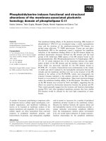
Further subclassification of AroQ CMs on the basis
of their distinct structural prototypes was proposed by
Okvist et al. [14] (Fig. 7). EcCM-like proteins whose
catalytic site is formed with residues from both
protomers are denoted as AroQ
a
. ScCM-like proteins
in which the catalytic site is formed within a single
protomer with a domain for regulation of activity by
tryptophan and tyrosine are denoted as AroQ
b
.
Secreted CMs such as *MtCM and *YpCM, in which
the catalytic site is formed within a single protomer
but without an apparent regulatory domain, are
denoted as AroQ
c
. A fourth subclass of CMs denoted
AroQ
d
was proposed by Okvist et al. [14], on the basis
of the primary sequence alone. The structural motif of
AroQ
d
CMs resembles that of AroQ
a
, with the notable
difference of a shortened third helix that lacks two
substrate-binding site residues. Here we describe the
first 3D structure of such a protein, MtCM.
AroQ chorismate mutases S. -K. Kim et al.
4830 FEBS Journal 275 (2008) 4824–4835 Journal compilation ª 2008 FEBS. No claim to original US government works
Experimental procedures
Materials
All the reagents used in this work were obtained from the
specified sources [7]. A selenomethionine auxotroph of
E. coli strain B834(DE3) was obtained from EMD
Biosciences Inc. (Madison, WI, USA). The M9 salts growth
medium (Cat. No. MD045003) for the incorporation of
selenomethionine into *YpCM was purchased from Medici-
lon Inc. (Chicago, IL, USA).
E. coli strains and plasmids
E. coli strains NovaBlue and BL21(DE3) were used for
cloning the target gene and expression of the cloned gene,
respectively. The plasmid vector pG58 and subtilisin
column were kindly provided by P. N. Bryan (Center for
Advanced Research in Biotechnology, University of Mary-
land Biotechnology Institute). Engineering of the fusion
protein production vector pG58 was described by Ruan
et al. [20]. Briefly, pG58 was designed to produce a target
gene product as a fusion protein with the subtilisin prodo-
main. The fusion protein would be bound to a resin cou-
pled with a stable variant of subtilisin protease. Next,
equilibration with fluoride anion will trigger the cleavage
by subtilisin between the prodomain and the target protein,
thus releasing the target protein in its native form, begin-
ning with the initiator methionine.
Cloning of Rv0948c and MT0975 genes
The Rv0948c ORF for the 105 amino acid protein was ampli-
fied by PCR from M. tuberculosis H
37
R
v
genomic DNA.
Oligonucleotide pair 1 with specific restriction recognition
sequences for cloning into pG58 was: 5 ¢-GCTACG
TTTAAAGCGATGATGAGACCAGAACCCCCACATCA
CG-3¢ (forward primer with DraI site underlined) and
5¢-CG
GAATTCTTAGTGACCGAGGCGGCCCCTGCC-3¢
(reverse primer with EcoRI site underlined). Similarly, the
Rv0948c ORF for the 90 amino acid protein beginning
with Met16 was amplified with oligonucleotide pair 2:
5¢-GCTACG
TTTAAAGCGATGATGAACCTGGAAATG
CTCGAGTCC-3¢ (forward primer with DraI site underlined)
and the same reverse primer. Oligonucleotide pair 3 for
amplification of MT0975 (217 amino acid protein – another
annotation for MtCM) for cloning into pG58 was:
5¢-GCTACG
TTTAAAGCGATGATGGACCGGGAGGCT
TGGCG-3¢ (forward primer with DraI site underlined) and
the same reverse primer as above. Amplification conditions
with all sets of primers were: 95 ° C for 5 min for initial
melting of DNA, followed by 30 cycles of amplification, with
each cycle consisting of melting at 95 °C for 60 s, annealing
at 50 ° C for 60 s, and polymerization at 72 °C for 60 s.
Polymerization was continued at the end for 10 min at
A
B
C
D
Fig. 7. Four subclasses of AroQ CMs: the four subclasses of AroQ
CMs are shown with cartoon drawings of representative structures.
All AroQ CMs are homodimers. One chain is blue and the other
chain is green. The distinguishing features are emphasized by indi-
cating the active sites with red circles. The regulatory sites of the
AroQ
b
class are highlighted with red squares. The shortened third
helices of AroQ
d
are pointed to with red arrows. (A) AroQ
a
is
EcCM. (B) AroQ
b
is ScCM. (C) AroQ
c
is *YpCM. (D) AroQ
d
is
90-MtCM with the TSA from the superimposition of EcCM.
S. -K. Kim et al. AroQ chorismate mutases
FEBS Journal 275 (2008) 4824–4835 Journal compilation ª 2008 FEBS. No claim to original US government works 4831
72 °C. One hundred nanograms of M. tuberculosis H
37
R
v
genomic DNA (generously provided by J. Belisle and
P. Brennan, Colorado State University) and 100 ng of prim-
ers were used in the amplification. The amplified DNA
obtained with oligonucleotide pairs 1, 2 and 3 was digested
with Dra I and EcoRI for cloning into the respective sites of
the pG58 plasmid. A recombinant was isolated from E. coli
Novablue and introduced into E. coli BL21(DE3) for protein
production.
Overproduction of the proteins
E. coli BL21(DE3) harboring either pG58–Rv0948c
(105 amino acids), pG58–Rv0948c (90 amino acids) or
pG58–MT0975 (217 amino acids) recombinant plasmid was
grown in 25 mL of LB medium containing ampicillin
(100 lgÆmL
)1
)at37°CtoanA
600 nm
0.5. Protein pro-
duction was induced with 30 lm IPTG overnight at 24 °C,
except for the pG58–MT0975, clone which was induced
overnight at 15 °C to eliminate the formation of inclusion
bodies of the protein. All three fusion proteins were pro-
duced in fully soluble form under these conditions.
Purification of native MtCM
Cells from 1 L of induced culture of BL21(DE3) harboring
pG58–Rv0948c (encoding either the 105 amino acid protein
or the 90 amino acid protein) were suspended in 40 mL of
lysis buffer (10 mm potassium phosphate, pH 7.4, 15 mm
NaCl), to which a tablet of protease inhibitor cocktail was
added. The cell suspension was passed through a French
press twice at 10 000 lbÆin
)2
, and the extract was centrifuged
at 48 000 g for 1 h. Supernatant containing the subtilisin
prodomain–MtCM fusion protein was loaded onto a 5 mL
subtilisin column at a flow rate of 0.5 mLÆmin
)1
. The resin
was washed with 60 mL of the lysis buffer at a flow rate of
1mLÆmin
)1
. The resin was further washed with 50 mL of
1 m sodium acetate in the lysis buffer. Next, the cleavage of
MtCM from the prodomain was triggered by flushing the
resin at a flow rate of 1 mLÆmin
)1
with 20 mL of 100 mm
sodium fluoride in the lysis buffer and equilibration for
30 min. The resin was washed with 25 mL aliquots of
100 mm sodium fluoride in the lysis buffer. Effluent fractions
containing CM, as judged by SDS ⁄ PAGE and by activity,
were pooled and concentrated to 5 mL in an Amicon cell
using a 5000 Da molecular mass cut-off membrane. MtCM
(105 amino acids ⁄ 90 amino acids) was further purified by
molecular sieve chromatography on a 480 mL Sephadex G-75
superfine column, which was equilibrated and eluted with
50 mm Tris ⁄ HCl (pH 7.5), 1 mm EDTA, and 100 mm NaCl.
Effluent fractions containing pure MtCM (105 amino
acids ⁄ 90 amino acids) were concentrated for protein determi-
nation and biochemical analysis. The 217-MtCM was simi-
larly purified with the subtilisin column. Further purification
was not pursued, as it exhibited extremely low CM activity.
Cloning and expression of the *YpCM gene
(y2828) in E. coli
The gene y2828 from the genome sequence of Y. pestis strain
Kim10+ [21] was annotated as CM. [Correction added on
28 August 2008 after first online publication: in the preceding
sentence, ‘The gene y2828 from the genome sequence of
Y. pestis strain Kim10+ (21) as CM’ was corrected to ‘The
gene y2828 from the genome sequence of Y. pestis strain
Kim10+ (21) was annotated as CM’]. The full-length
*YpCM gene coding sequence, including the signal peptide,
was amplified by PCR using the forward primer 5¢-GG
AATTC
CATATGCAACCCACTCATACGCTAACAAG-3¢
(with the NdeI restriction recognition sequence underlined)
and the reverse primer 5¢-CG
GGATCCTTATTTTAATT
TTACCTGATTGAAGGTTGAG-3¢ (with the BamHI
restriction recognition sequence underlined). Amplification
conditions were: 95 °C for 60 s for initial melting of DNA,
followed by 30 cycles of amplification, with each cycle con-
sisting of melting at 95 °C for 60 s, annealing at 60 °C for
60 s, and polymerization at 72 °C for 60 s. Polymerization
was continued at the end for 10 min at 72 °C. Two hundred
nanograms of Y. pestis strain KIM10+ chromosomal DNA
(kindly provided by R. D. Perry, University of Kentucky)
and 100 ng of primers were used in the amplification. The
amplified DNA was digested with NdeI and BamHI and
cloned into the respective sites of the pET15b plasmid.
A recombinant was isolated from E. coli Novablue and intro-
duced into BL21(DE3) for protein production. E. coli
BL21(DE3) harboring the pET15b–y2828 recombinant
plasmid was grown in 100 mL of LB medium containing
ampicillin (100 lgÆmL
)1
)at37°CtoanA
600 nm
0.6.
Protein production was induced with 10 lm IPTG overnight
at 15 °C. *YpCM was purified by molecular sieve chroma-
tography from the periplasmic fluid of E. coli as described
for *MtCM [7]. The production and purification of *YpCM
was scaled up for crystallization.
Production and purification of selenomethionine
*YpCM
E. coli B834(DE3), a methionine auxotroph, was trans-
formed with the pET15b–y2828 recombinant plasmid. Incor-
poration of selenomethionine into *YpCM was performed
using the M9 salts ⁄ selenomethionine growth medium,
according to the manufacturer’s recommendation. Briefly,
cells were grown in 1 L of LB medium containing ampicillin
(100 lgÆmL
)1
) overnight at 37 °C. Cells were harvested,
washed twice with sterile water, and suspended in 100 mL of
M9 salts medium. Four 1 L volumes of M9 salts media con-
taining ampicillin were inoculated with 25 mL of the culture
per 1 L. Cells were grown at 37 °CtoA
600 nm
= 0.4. At this
stage, selenomethionine was added and induced with 10 lm
IPTG at 15 °C overnight. *YpCM was purified by molecular
sieve chromatography from the periplasmic fluid.
AroQ chorismate mutases S. -K. Kim et al.
4832 FEBS Journal 275 (2008) 4824–4835 Journal compilation ª 2008 FEBS. No claim to original US government works
Crystallization of 90-MtCM
The 90-MtCM was concentrated to 8.3 mgÆmL
)1
in 50 mm
Tris ⁄ HCl (pH 7.5), 1 mm EDTA, and 100 m m sodium chlo-
ride. Crystallization conditions were surveyed by the sitting
drop vapor diffusion method using Emerald BioSystems
Wizard Screens I and II. There were several hits. The crystal
used for data collection was grown with a well solution of
0.1 m Tris ⁄ HCl (pH 8.6), 0.2 m magnesium chloride, and
20% poly(ethylene glycol) 400. The crystallization drops
were made with equal volumes of protein and well solution.
Crystallization of *YpCM
Crystallization conditions were surveyed by the hanging
drop vapor diffusion method using the Wizard II kit from
Emerald BioSystems ().
The protein concentration was 10 mgÆmL
)1
in 50 mm
Tris ⁄ HCl (pH 7.5), 1 mm EDTA, 1 mm dithiothreitol, and
200 mm sodium chloride. The original hit was with solu-
tion 9 (2 m ammonium sulfate, 0.1 m citrate ⁄ phosphate
buffer, pH 4.2). For the refined conditions, a well solution
of 1.5–1.6 m ammonium sulfate and 0.1 m citrate ⁄ phos-
phate buffer (pH 4.2) was used, and the protein concentra-
tion was reduced to 5 mgÆmL
)1
. For the selenomethionine
protein, the well solution was 1.8–2.0 m ammonium sulfate
and 0.1 m citrate ⁄ phosphate buffer (pH 4.2), with a protein
concentration of 2.5–5 mgÆmL
)1
.
Data collection for 90-MtCM
Diffraction data were collected using a home source Riga-
ku 007 generator and a RAXIS IV
++
image plate detector
(Rigaku ⁄ MSC, The Woodlands, TX, USA). The crystal
was cooled to 105 K with a Rigaku Xtream 2000 cryocool-
er. For cryo-data collection, the crystals were mounted
through a layer of paraffin oil placed on top of the crystal-
lization drop. The data were collected and processed with
crystalclear [22], and the statistics are shown in Table 2.
Structure determination for 90-MtCM
The structure of 90-MtCM was solved by molecular
replacement using phaser [23], with the structure of PfCM
(Protein Data Bank ID: 1YBZ). The asymmetric unit of the
P4
3
2
1
2 crystal includes a single chain of 90-MtCM. Molecu-
lar replacement trials using a single protomer failed. How-
ever, when the symmetry was lowered to P4
3
and the dimer
was used as the search model, a solution was found. The
remainder of the structure determination was carried out in
the space group P4
3
2
1
2. refmac5 [24,25] was used to refine
the model, and resolve [26] was used to iteratively rebuild
the model to remove bias. The final refinement statistics are
shown in Table 2. coot [27] was used to view the model
graphically and to build portions not built by resolve. The
stereochemistry was checked with procheck [28] and with
routines inside coot.
Data collection for *YpCM
Preliminary data were collected on the home source
described above, and cryoprotection was accomplished in
the same manner as for 90-MtCM. The selenomethionine
data for *YpCM were collected on beamline X29A of the
National Synchrotron Light Source at wavelength 0.9790 A
˚
with the crystal cooled to 100 K. The statistics are shown
in Table 2.
*YpCM structure determination
The structure of *YpCM was solved using the phasing
information from the anomalous data. The positions of
the selenium atoms were located with shelxd [29], and
the initial phases were calculated with solve [30]. Two
dimers in the asymmetric unit cell gives a Matthews coeffi-
cient of 2.6 and a solvent content of 52.5%. The initial
model was built with resolve [26,31], using iterative
rounds of pattern-matching, fragment identification, den-
sity modification, and refinement. This model included
78% of the residues and placed 71% of the side chains.
The noncrystallographic symmetry was used to combine
the four partial chains to produce a more complete model.
Then, further cycles of model building and refinement
were performed using xtalview [32] and refmac5 [24].
The final refinement statistics are shown in Table 2. The
stereochemistry was checked with procheck [28] and with
molprobity [33]. Four residues in the A chain and one in
the D chain were modeled with alternative side-chain con-
formations. No interpretable electron density was observed
for the first residues (residue 31) of chains B and D. The
residues between Cys148 and Asp155 are somewhat disor-
dered, particularly in the C and D chains, and conse-
quently have increased B-values.
Other methods
CM was assayed by the method of Davidson & Hudson
[34], essentially as described in our previous study [7]. One
microgram of MtCM protein or 200 ng of *YpCM protein
were used in the assay. Protein concentration was deter-
mined by the Micro BCA method with BSA as the stan-
dard (Pierce, Rockford, IL, USA). The monomeric
molecular mass of the native MtCM was determined by
MALDI-TOF MS. Mass spectra were collected and
analyzed using an Applied Biosystems Voyager-DE STR
Biospectrometry Workstation (Foster City, CA, USA). The
DNA sequence of the cloned genes was confirmed by the
dideoxy sequencing method [35], as adopted for the Applied
Biosystems model 3130 Genetic Analyzer.
S. -K. Kim et al. AroQ chorismate mutases
FEBS Journal 275 (2008) 4824–4835 Journal compilation ª 2008 FEBS. No claim to original US government works 4833
Disclaimer
Certain commercial equipment or materials are identified in
this article in order to specify adequately the experimental
procedure. Such identification does not imply recommenda-
tion or endorsement by the National Institute of Standards
and Technology, and nor does it imply that the materials
or equipment identified are necessarily the best available
for the purpose.
Acknowledgements
We thank John Belisle and Patrick Brennan, Colorado
State University, for generously providing Mycobacterium
tuberculosis H37Rv DNA. We also thank Robert Perry,
University of Kentucky, for generously providing Yersinia
pestis Kim10+ DNA. We are grateful to the anonymous
reviewers of this paper for their constructive critique and
valuable suggestions.
References
1 Haslam E (1993) Shikimic Acid: Metabolism and Meta-
bolites. Wiley, New York, NY.
2 Andrews PR, Cain EN, Rizzardo E & Smith GD (1977)
Rearrangement of chorismate to prephenate. Use of
chorismate mutase inhibitors to define the transition
state structure. Biochemistry 22, 4848–4852.
3 Stewart GR, Robertson BD & Young DB (2003)
Tuberculosis: a problem with persistence. Nat Rev
Microbiol 1, 97–105.
4 Cole ST, Brosch R, Parkhill J, Garnier T, Churcher C,
Harris D, Gordon SV, Eiglmeier K, Gas S, Barry CE
III et al. (1998) Deciphering the biology of Mycobacte-
rium tuberculosis from the complete genome sequence.
Nature 393, 537–544.
5 Sasso S, Ramakrishnan C, Gamper M, Hilvert D &
Kast P (2005) Characterization of the secreted choris-
mate mutase from the pathogen Mycobacterium tubercu-
losis. FEBS J 272, 375–389.
6 Prakash P, Aruna B, Sardesai AA & Hasnain SE (2005)
Purified recombinant hypothetical protein coded by
open reading frame Rv1885c of Mycobacterium tubercu-
losis exhibits a monofunctional AroQ class of periplas-
mic chorismate mutase activity. J Biol Chem 280 ,
19641–19648.
7 Kim S-K, Reddy SK, Nelson BC, Vasquez GB, Davis
A, Howard AJ, Patterson S, Gilliland GL, Ladner JE
& Reddy PT (2006) Biochemical and structural charac-
terization of the secreted chorismate mutase (Rv1885c)
from Mycobacterium tuberculosis H
37
R
v
: an *AroQ
enzyme not regulated by the aromatic amino acids.
J Bacteriol 188, 8638–8648.
8 Fleischmann RD, Alland D, Eisen JA, Carpenter L,
White O, Peterson J, DeBoy R, Dodson R, Gwinn M,
Haft D et al. (2002) Whole-genome comparison of
Mycobacterium tuberculosis clinical and laboratory
strains. J Bacteriol 184, 5479–5490.
9 Stewart J, Wilson DB & Ganem B (1990) A genetically
engineered monofunctional chorismate mutase. JAm
Chem Soc 112, 4582–4584.
10 Schneider CZ, Parish T, Basso LA & Santos DS (2008)
The two chorismate mutases from both Mycobacterium
tuberculosis and Mycobacterium smegmatis: biochemical
analysis and limited regulation of promoter activity by
aromatic amino acids. J Bacteriol 190 , 122–134.
11 Liu DR, Cload ST, Pastor RM & Schultz PG (1996)
Analysis of active site residues in Escherichia coli choris-
mate mutase by site-directed mutagenesis. J Am Chem
Soc 118, 1789–1790.
12 Kawabata T (2003) MATRAS: a program for protein
3D structure comparison. Nucleic Acids Res 31, 3367–
3369.
13 Lee AY, Karplus PA, Ganem B & Clardy J (1995)
Atomic structure of the buried catalytic pocket of
Escherichia coli chorismate mutase. J Am Chem Soc
117, 3627–3628.
14 Okvist M, Dey R, Sasso S, Grahn S, Kast P &
Krengel U (2006) 1.6 A
˚
Crystal structure of the secreted
chorismate mutase from Mycobacterium tuberculosis:
novel fold topology revealed. J Mol Biol 357,
1483–1499.
15 Qamra R, Prakash P, Aruna B, Hasnain SE & Mande
SC (2006) The 2.15 A
˚
crystal structure of Mycobacte-
rium tuberculosis chorismate mutase reveals an
unexpected gene duplication and suggests a role in
host–pathogen interactions. Biochemistry 45, 6997–7005.
16 MacBeath G, Kast P & Hilvert D (1998) A small, ther-
mostable, and monofunctional chorismate mutase from
the archaeon Methanococcus jannaschii. Biochemistry
37, 10062–10073.
17 Xue Y, Lipscomb WN, Graf R, Schnappauf G & Braus
G (1994) The crystal structure of allosteric chorismate
mutase at 2.2-A
˚
resolution. Proc Natl Acad Sci USA
91, 10814–10818.
18 Chook YM, Ke H & Lipscomb WN (1993) Crystal
structure of the monofunctional chorismate mutase
from Bacillus subtilis and its complex with a transition
state analog. Proc Natl Acad Sci USA 90, 8600–8603.
19 Schnappauf G, Strater N, Lipscomb WN & Braus GH
(1997) A glutamate residue in the catalytic center of the
yeast chorismate mutase restricts enzyme activity to
acidic conditions. Proc Natl Acad Sci USA
94, 8491–
8496.
20 Ruan B, Fisher KE, Alexander PA, Doroshko V &
Bryan PN (2004) Engineering subtilisin into a fluoride-
triggered processing protease useful for one-step protein
purification. Biochemistry 43, 14539–14546.
21 Deng W, Burland V, Plunkett G III, Boutin A,
Mayhew GF, Liss P, Perna NT, Rose DJ, Mau B,
AroQ chorismate mutases S. -K. Kim et al.
4834 FEBS Journal 275 (2008) 4824–4835 Journal compilation ª 2008 FEBS. No claim to original US government works
Zhou S et al. (2002) Genome sequence of Yersinia pestis
KIM. J Bacteriol 184, 4601–4611.
22 Pflugrath JW (1999) The finer things in X- ray diffrac-
tion data collection. Acta Crystallogr D 55, 1718–1725.
23 McCoy AJ, Grosse-Kunstleve RW, Storoni LC & Read
RJ (2005) Likelihood-enhanced fast translation func-
tions. Acta Crystallogr D 61, 458–464.
24 Murshudov GN, Vagin AA & Dodson EJ (1997) Refine-
ment of macromolecular structures by the maximum-
likelihood method. Acta Crystallogr D 53, 240–255.
25 Vagin AA, Steiner RA, Lebedev AA, Potterton L,
McNicholas S, Long F & Murshudov GN (2004) REF-
MAC5 dictionary: organization of prior chemical
knowledge and guidelines for its use. Acta Crystallogr
D 60, 2184–2195.
26 Terwilliger TC (2003) Automated main-chain model
building by template matching and iterative fragment
extension. Acta Crystallogr D 59, 38–44.
27 Emsley P & Cowtan K (2004) Coot: model-building
tools for molecular graphics. Acta Crystallogr 60, 2126–
2132.
28 Laskowski RA, MacArthur MW, Moss DS & Thornton
JM (1993) PROCHECK: a program to check the
stereochemical quality of protein structures. J Appl
Crystallogr 26, 283–291.
29 Schneider TR & Sheldrick GM (2002) Substructure solu-
tion with SHELXD. Acta Crystallogr D 58, 1772–1779.
30 Terwilliger TC & Berendzen XX (1999) Automated
MAD MIR structure solution. Acta Crystallogr D 55,
849–861.
31 Terwilliger TC (2003) Automated side-chain model
building and sequence assignment by template match-
ing. Acta Crystallogr D 59, 45–49.
32 McRee DE (1999) Practical Protein Crystallography,
2nd edn. Academic Press, San Diego, CA.
33 Lovell SC, Davis IW, Arendall WB III, de Bakker
PIW, Word JM, Prisant MG, Richardson JS & Rich-
ardson DC (2003) Structure validation by C-alpha
geometry: phi, psi, and C-beta deviation. Proteins Struct
Funct Genet 50, 437–450.
34 Davidson BE & Hudson GS (1987) Chorismate mutase
– prephenate dehydrogenase from Escherichia coli.
Methods Enzymol 142, 440–450.
35 Sanger F, Nicklen S & Coulson AR (1977) DNA
sequencing with chain terminating inhibitors. Proc Natl
Acad Sci USA 74, 5463–5467.
S. -K. Kim et al. AroQ chorismate mutases
FEBS Journal 275 (2008) 4824–4835 Journal compilation ª 2008 FEBS. No claim to original US government works 4835