cyanidin 3 glucoside inhibits ethanol induced invasion of breast cancer cells overexpressing erbb2
Bạn đang xem bản rút gọn của tài liệu. Xem và tải ngay bản đầy đủ của tài liệu tại đây (2.76 MB, 14 trang )
Xu et al. Molecular Cancer 2010, 9:285
/>
RESEARCH
Open Access
Cyanidin-3-Glucoside inhibits ethanol-induced
invasion of breast cancer cells overexpressing
ErbB2
Mei Xu1, Kimberly A Bower1, Siying Wang1,2, Jacqueline A Frank1, Gang Chen1, Min Ding3, Shiow Wang4,
Xianglin Shi5, Zunji Ke6, Jia Luo1*
Abstract
Background: Ethanol is a tumor promoter. Both epidemiological and experimental studies suggest that ethanol
may enhance the metastasis of breast cancer cells. We have previously demonstrated that ethanol increased the
migration/invasion of breast cancer cells expressing high levels of ErbB2. Amplification of ErbB2 is found in 20-30%
of breast cancer patients and is associated with poor prognosis. We sought to identify agents that can prevent or
ameliorate ethanol-induced invasion of breast cancer cells. Cyanidin-3-glucoside (C3G), an anthocyanin present in
many vegetables and fruits, is a potent natural antioxidant. Ethanol exposure causes the accumulation of
intracellular reactive oxygen species (ROS). This study evaluated the effect of C3G on ethanol-induced breast cancer
cell migration/invasion.
Results: C3G attenuated ethanol-induced migration/invasion of breast cancer cells expressing high levels of ErbB2
(BT474, MDA-MB231 and MCF7ErbB2) in a concentration dependent manner. C3G decreased ethanol-mediated cell
adhesion to the extracellular matrix (ECM) as well as the amount of focal adhesions and the formation of
lamellipodial protrusion. It inhibited ethanol-stimulated phosphorylation of ErbB2, cSrc, FAK and p130Cas, as well as
interactions among these proteins. C3G abolished ethanol-mediated p130Cas/JNK interaction.
Conclusions: C3G blocks ethanol-induced activation of the ErbB2/cSrc/FAK pathway which is necessary for cell
migration/invasion. C3G may be beneficial in preventing/reducing ethanol-induced breast cancer metastasis.
Background
Excessive ethanol consumption is associated with an
increased risk for breast cancer [1-5]. Epidemiological
studies indicate that alcohol consumption is associated
with advanced and invasive breast tumors [6,7]. We
have previously demonstrated that breast cancer cells or
mammary epithelial cells expressing high levels of ErbB2
are sensitive to ethanol-mediated migration/invasion;
ethanol stimulates migration/invasion of breast cancers
with high ErbB2 levels more robustly than cells expressing lower levels of ErbB2 [8-10]. ErbB2 belongs to the
ErbB family of receptor kinases which consists of EGFR,
ErbB2, ErbB3 and ErbB4. Among the ErbB family,
ErbB2 is most directly related to breast cancer and is
* Correspondence:
1
Department of Internal Medicine, University of Kentucky College of
Medicine, Lexington, KY 40536, USA
Full list of author information is available at the end of the article
implicated in breast cancer metastasis. Amplification of
ErbB2 is found in 20-30% of breast cancer patients and
is associated with poor prognosis and relapse [11,12].
We sought to identify agents that may ameliorate
ethanol’s promoting effect on breast cancer cell migration/invasion. Cyanidin-3-glucoside (C3G) is a member
of the anthocyanin family which is present in various
vegetables and fruits, especially edible berries. C3G is a
potent antioxidant and displays anti-cancer properties in
vitro and in vivo [13-18]. Since ethanol exposure causes
the accumulation of intracellular oxygen species (ROS)
and many biological effects of ethanol are believed to be
mediated by ROS, we hypothesize that C3G may inhibit
ethanol-induced migration/invasion of breast cancer
cells. We examined the effect of C3G on ethanolmediated migration/invasion of breast cancer cells
expressing high levels of ErbB2. We demonstrate here
that C3G effectively blocks ethanol-induced cell
© 2010 Xu et al; licensee BioMed Central Ltd. This is an Open Access article distributed under the terms of the Creative Commons
Attribution License ( which permits unrestricted use, distribution, and reproduction in
any medium, provided the original work is properly cited.
Xu et al. Molecular Cancer 2010, 9:285
/>
migration/invasion. We further investigate the effect of
C3G on the cell/extracellular matrix (ECM) interaction
and the associated ErbB2/cSrc/FAK pathway.
Materials and methods
Materials
Human plasma fibronectin was obtained from Chemicon International (Temecula, CA). Anti-paxillin antibody was purchased from Invitrogen Corporation
(Carlsbad, CA). Anti-phospho-ErbB2 (Tyr1248) (polyclonal), phospho-p130Cas and ErbB2 (polyclonal) antibodies
were purchased from Cell Signaling Technology Inc.
(Beverly, MA). Anti-Neu/Her2/ErbB2 (monoclonal),
FAK, cSrc, JNK and phospho-Src (Tyr216) antibodies
and Protein A/G beads were purchased from Santa Cruz
Biotechnology (San Diego, CA). Anti-phospho-Her2/
ErbB2 (Tyr1248) (monoclonal) and phospho-FAK
(Tyr861) antibodies were purchased from Biosource
(Camarillo, CA). Anti-p130 Cas antibody was obtained
from BD Transduction Laboratory (San Jose, CA). Antiactive JNK antibody was obtained from Promega
Corporation (Madison, WI). Phalloidin 488, Alex Fluorlabeled secondary antibodies, Prolong Gold anti-fade
reagent and reactive oxygen species detection reagents
were obtained from Invitrogen Molecular Probes
(Eugene, OR). MTT assay kit was purchased from
Roche Molecular Biochemicals (Indianapolis, IN). Matrigel Invasion Chambers were purchased from BD Biosciences (Bedford, MA). Transwell was obtained from
Costar Corp. (Acton, MA). C3G was purified from
blackberry fruit tissue as previously described [14]. The
purity of C3G is greater than 95%. Alcohol (200 Proof)
was obtained from Fisher Scientific (Pittsburgh, PA). All
other chemicals were obtained from Sigma-Aldrich (St.
Louis, MO).
Cell culture and ethanol exposure
MCF7 ErbB2 (MCF7 cells overexpressing ErbB2) and
MDA-MB231 breast cancer cells were grown in DMEM
medium containing 10% fetal bovine serum (FBS), penicillin (100 U/ml)/streptomycin (100 U/ml), 1 μg/ml
hydrocortisone and 10 μg/ml insulin at 37°C with 5%
CO2 . BT474 cells were grown in RPMI 1640 medium
containing 10% FBS, penicillin (100 U/ml)/streptomycin
(100 U/ml) and 10 μg/ml insulin. A method utilizing
sealed containers was used to maintain ethanol concentrations in the culture medium. The containers were
placed in a humidified environment and maintained at
37°C with 5% CO2.
Page 2 of 14
treated with ethanol in the presence or absence of C3G.
Culture medium containing 10% FBS was added into
the lower compartment of invasion chambers and served
as chemoattractants for the cells. Cells were maintained
in the invasion chambers for 48 hours. The invaded
cells were fixed in 3.7% paraformaldehyde and stained
with 0.5% crystal violet in 2% ethanol. Membranes were
washed and the dye was eluted with 10% acetic acid.
Absorbance was measured at 595 nm using a microtiter
platereader (Beckman coulter).
Cell migration was analyzed using a Transwell Migration System (Costar). Briefly, cells were plated into
upper chambers (Transwells with 8.0 μm pore size) in
serum free medium. The lower compartment of the
chamber contained regular medium containing 10%
FBS. The chambers were cultured at 37°C in 5% CO2
for 12 hours. Migrated cells were fixed and stained with
0.5% crystal violet, followed by dye elution and absorbance measurement as described above.
Wound healing migration assay
The wound healing migration assay was performed as
described previously [14]. MDA-MB231 cells were
grown on 35 mm dishes to 100% confluence and then
scratched to form a wound using sterile pipette tips.
The cells were then treated with ethanol (0 or 400 mg/
dl) in the presence or absence of C3G (10 μM) for 24
hours. The images were recorded using a Zeiss Axiovert
40C photomicroscope.
Analysis of cell adhesion
Cell adhesion to fibronectin was analyzed as described
previously [19-21]. Briefly, 96-well cell culture plates
were precoated with fibronectin (10 μg/ml) for 60 min
at 37°C. Plates were then incubated with 3% BSA in PBS
for 30 min to block non-specific binding sites, followed
by several washes with PBS. Cells were exposed to ethanol with/without C3G for 48 hours. After exposure,
cells (5 × 10 4 /well) were seeded on fibronectin precoated plates, allowing attachment for 1 hour at 37°C
with 5% CO 2 . Non-adherent cells were removed by
washing with PBS. The attached cells were fixed with
3.7% paraformaldehyde for 10 min, washed 3 times in
PBS, and stained with 0.1% crystal violet in 2% ethanol
for 10 min. Cells were rinsed with water and dried.
Crystal violet was eluted in 10% acetic acid and the
absorbance (attached cells) was measured at 595 nm
using a microtiter platereader.
MTT assay
Cell invasion and migration
Cell invasion was assayed using Matrigel Invasion
Chambers (BD Biosciences). Briefly, cells were placed on
the upper compartment of invasion chambers and
The MTT assay was employed to determine the number
of viable cells in culture. Briefly, the cells were plated
into 96-well plates and exposed to ethanol with/without
C3G for indicated times. After the treatment, 10 μl of
Xu et al. Molecular Cancer 2010, 9:285
/>
MTT reagent was added into each well and the plates
were incubated at 37°C for 4 hours. The cultures were
solubilized and spectrophotometric absorbance was
measured at 595 nm using a microtiter platereader.
Immunofluorescence microscopy
The procedure for immunofluorescence microscopy has
been previously described [22]. Briefly, after treatments,
cells were seeded on fibronectin (10 μg/ml) precoated
coverslips. Cells were fixed with 3.7% paraformaldehyde
for 10 min, washed 3 times in PBS and permeabilized
with 0.5% Triton X-100 for 5 min. Cells were blocked
with 5% BSA and incubated with primary antibodies for
1 hour. The concentrations of primary antibodies were:
phospho-FAK (Tyr861), 1:50; paxillin, 1:800; and phalloidin, 1:200. Following incubation with primary antibodies, cells were washed and treated with Alexa Fluorlabeled secondary antibodies and rinsed several times
with PBS. Coverslips were mounted with Prolong Gold
anti-fade reagent and immunofluorescence images were
examined with a LEICA SP1 inverted confocal microscope. The fluorescent signals were measured with the
same pinhole, detector gain and amplifier offset. The
focal adhesions were detected by immunostaining for
phosphorylated FAK and quantified randomly on 10 or
more cells for each treatment condition.
Immunoprecipitation and immunoblotting
After the treatment of ethanol and/or C3G, cells were
trypsinized and seeded on fibronectin (10 μg/ml) precoated dishes allowing attachment for indicated times.
Cells were then rinsed twice in cold PBS to remove
non-adherent cells. Attached cells were lysed in modified RIPA buffer (150 mM NaCl, 50 mM Tris, 1% NP40, 0.25% sodium deoxycholate, 1 mM sodium vanadate,
1 mM phenylmethanesulfonyl fluoride (PMSF), 5 μg/ml
of aprotinin, and 2 μg/ml of leupeptin). The procedure
for immunoprecipitation and immunoblotting has been
previously described [10,19]. Briefly, equal amounts of
proteins (about 500-800 μg) were incubated with antiErbB2, FAK, p130Cas or cSrc antibodies overnight at 4°
C, followed by treatment with TrueBlot anti-mouse Ig
or anti-rabbit Ig beads (eBioscience, San Diego, CA) for
2 hours at 4°C. Immunoprecipitates were collected by
centrifugation at 10,000 g for 5 min at 4°C. Samples
were washed five times with RIPA buffer, one time with
cold PBS and boiled in sample buffer (187.5 mM TriHCl, pH 6.8, 6% SDS, 30% glycerol, 150 mM DTT and
0.03% bromophenol blue). Proteins were resolved in
SDS-PAGE and the separated proteins were transferred
to nitrocellulose membranes. The membranes were
probed with indicated primary antibodies, followed by
the appropriate TrueBlot horseradish peroxidase-
Page 3 of 14
conjugated secondary antibodies and developed by
enhanced chemiluminescence.
Detection of intracellular reactive oxygen species
Intracellular reactive oxygen species (ROS) levels were
measured using the fluorescent dye CM-H 2 DCFDA
(Invitrogen Corporation, Carlsbad, CA) as previously
described [23]. CM-H2DCFDA is converted to a nonfluorescent derivative inside the cells and when oxidized
forms a highly fluorescent product by intracellular ROS.
Briefly, cells were treated with ethanol with/without
C3G or other antioxidants for 48 hours. After the treatment, cells were washed with cold PBS and incubated
with 5 μM CM-H2DCFDA for 30 min, followed by several additional washes with cold PBS. Cells were trypsinized and transferred into polystyrene round-bottom
tubes; intracellular ROS levels were measured with a
flow cytometer (FACScalibur, BD Biosciences, San Jose,
CA) at an emission wavelength of 525 nm.
Statistics
Differences among treatment groups were analyzed
using analysis of variance (ANOVA). Differences in
which p was less than 0.05 were considered statistically
significant. In cases where significant differences were
detected, specific post-hoc comparisons between treatment groups were examined with Student-NewmanKeuls tests.
Results
C3G inhibits ethanol-enhanced migration/invasion and
attachment of breast cancer cells
We have previously demonstrated that the effect of
ethanol on the migration/invasion of breast cancer cells
is positively associated with their expression levels of
ErbB2 [8-10]. The current study confirmed the finding
and showed that ethanol increased the migration/invasion of MCF7ErbB2, BT474 and MDA-MB231 breast cancer cells (Figure 1). C3G (10-40 μM) significantly
inhibited ethanol-enhanced migration/invasion of
MCF7ErbB2 and MDA-MB231 in a concentration dependent manner (Figures 1B-D). C3G-mediated inhibition
was statistically different among the three C3G concentrations tested. The effect of C3G on BT474 cells, however, was not dose-dependent (Figure 1D). C3G alone at
10 μM did not affect the invasion of MCF7 ErbB2 cells
(Figure 1A). For BT474 and MDA-MB231 cells, C3G
alone produced a modest but statistically significant
inhibition of cell invasion (data not shown). The inhibitory effect of C3G on ethanol-induced cell migration
was confirmed by a wound healing migration assay (Figure 1F). The MTT assay showed that even 40 μM C3G
did not affect the viability of BT474, MDA-MB231 and
Xu et al. Molecular Cancer 2010, 9:285
/>
Page 4 of 14
Figure 1 Effects of C3G on ethanol-mediated invasion/migration of breast cancer cells. A: MCF7ErbB2 cells were plated into the upper
compartments of the matrigel invasion chambers and exposed to ethanol (0 or 400 mg/dl) with/without C3G (10 μM) for 48 h. Following the
treatment, the invasive potential was assayed as described under the Materials and Methods and presented relative to untreated controls.
B: MCF7ErbB2 cells were exposed to ethanol (0, 200 or 400 mg/dl) with/without C3G (10 or 20 μM) for 48 h. The invasive potential was assayed
as described above. C: MCF7ErbB2 cells were plated into the upper compartments of the migration chamber and exposed to ethanol (0 or 400
mg/dl) with/without C3G (10, 20 or 40 μM) for 12 h. The migration was analyzed as described under the Materials and Methods and presented
relative to untreated controls. D: BT474, MDA-MB231 or MCF7ErbB2 cells were exposed to ethanol (0 or 400 mg/dl) with/without C3G (10, 20 or
40 μM) for 48 h. Their invasion potential was evaluated as described above. E: BT474, MDA-MB231 or MCF7ErbB2 cells were exposed to ethanol
(0 or 400 mg/dl) with/without C3G (10, 20 or 40 μM) for 48 h and cell viability was determined with MTT assay. The number of viable cells was
presented relative to untreated controls. Each datum point was the mean ± SEM of three independent experiments. * denotes a statistically
significant difference from untreated controls. # denotes a significant difference from ethanol-treated groups. ε denotes a significant difference
from ethanol- and C3G (10 μM)-treated groups. δ denotes a significant difference from ethanol- and C3G (20 μM)-treated groups. F: MDA-MB231
cells were exposed to ethanol (0 or 400 mg/dl) with/without C3G (10 μM) for 24 h and cell migration was determined by wound healing
migration assay as described under the Materials and Methods.
Xu et al. Molecular Cancer 2010, 9:285
/>
MCF7 ErbB2 cells (Figure 1E). However, at 100 μM or
above, C3G did decrease cell viability (data not shown).
The adhesion of cancer cells to ECM or cell/ECM
interaction is an important step of metastasis. We have
previously demonstrated that ethanol enhances the
adhesion of breast cancer cells to fibronectin, an essential protein in the ECM [19]. Ethanol did not affect the
attachment of breast cancer cells to poly-lysine (data
not shown). We examined the effect of C3G on ethanolmediated cell adhesion to fibronectin. MCF7ErbB2 cells
were pretreated with ethanol with/without C3G for 48
hours, then the cells were seeded on fibronectin precoated plates and allowed to attach for 1 hour in the
presence/absence of ethanol and/or C3G. As shown in
Figure 2, ethanol increased the adhesion of MCF7ErbB2
cells to fibronectin and C3G significantly inhibited ethanol-enhanced adhesion in a concentration-dependent
manner. C3G alone (10-20 μM) did not affect cell adhesion (data not shown). C3G similarly inhibited ethanolinduced adhesion of MDA-MB231 cells to the ECM
(data not shown).
C3G attenuates ethanol-stimulated ErbB2 signaling
We have previously shown that ethanol increased the
phosphorylation of ErbB2 at Tyr1248 [19]. In this study,
we examined the effect of C3G on ethanol-mediated
ErbB2 phosphorylation. MDA-MB231 and MCF7 ErbB2
Page 5 of 14
cells were pretreated with ethanol with/without C3G for
48 hours, then cells were seeded into fibronectin precoated dishes, allowing attachment for 3 hours. As
shown in Figure 3, ethanol drastically increased the
phosphorylation of ErbB2 [p-ErbB2(Tyr1248)] in these
cells. The addition of C3G attenuated ethanol-stimulated p-ErbB2(Tyr1248) in a concentration-dependent
manner. The cSrc/FAK pathway plays an important role
in ErbB2-regulated migration/invasion of breast cancer
cells [24]. FAK is a substrate of cSrc and FAK Tyr861 is
a major site of phosphorylation by cSrc. As shown in
Figure 3, ethanol increased the levels of p-FAK(Tyr861)
and p-cSrc(Tyr216). C3G attenuated ethanol-induced pFAK(Tyr861) and p-cSrc(Tyr216). The activation and
phosphorylation of cSrc/FAK is critical for triggering its
downstream signaling and for recruiting proteins to the
focal adhesion sites. p130Cas, an adaptor protein, binds
to the C-terminal site of FAK, forming a dock site for
Crk; p130Cas/Crk interaction induces the activation of
small GTPases and JNKs, promoting membrane protrusion and cell migration [25,26]. The phosphorylation of
p130Cas is regulated by FAK and cSrc [27]. We demonstrated that ethanol induced the phosphorylation of
p130Cas [p-p130Cas(Tyr410)], and C3G blocked ethanolinduced p-p130 Cas (Tyr410) (Figure 3A). We further
examined the effect of C3G on the interaction among
ErbB2, cSrc, FAK and p130 Cas . MCF7 ErbB2 cells were
treated with ethanol or with/without C3G for 48 hours
and seeded on fibronectin for 1 or 3 hours. As shown in
Figure 4, ethanol increased the association between
ErbB2/FAK, FAK/cSrc, FAK/p130Cas and cSrc/p130Cas.
C3G abolished the interaction among these proteins
(Figure 4). These data indicated that C3G inhibited the
ethanol-activated ErbB2/cSrc/FAK pathway.
c-Jun N-terminal kinases (JNKs), a member of mitogen-activated protein kinases (MAPKs), regulate cell
migration/invasion [28]. We have previously demonstrated that JNKs are essential for ethanol-mediated cell
invasion/migration [10]. JNK activation is regulated by
p130Cas [27]. C3G inhibited ethanol-induced JNK phosphorylation and p130Cas/JNK association in MCF7ErbB2
cells (Figure 5).
C3G inhibits ethanol-induced formation of lamellipodia
and focal adhesions
Figure 2 Effects of C3G on ethanol-mediated adhesion of
breast cancer cells. MCF7ErbB2 cells were treated with ethanol (0,
200 or 400 mg/dl) with/without C3G (10, 20 or 40 μM) for 48 h, and
then equal amounts of cells were seeded on fibronectin-coated
culture wells, allowing attachment for 1 h. The number of adherent
cells was determined as described under the Materials and Methods
and presented relative to untreated controls. Each datum point was
the mean ± SEM of three independent experiments. * denotes a
statistically significant difference from untreated controls. # denotes
a significant difference from ethanol-treated groups.
The initiation of cell migration requires the development of membrane protrusion, the lamellipodium and
the assembly of dynamic focal adhesions with the ECM
[29]. We sought to determine whether C3G affected the
formation of the lamellipodium and focal adhesions. We
used MDA-MB231 cells for this experiment because
these cells displayed more prominent lamellipodium and
focal adhesions during the migration process. Figure 6A
shows that actin filament distribution was concentrated
Xu et al. Molecular Cancer 2010, 9:285
/>
Page 6 of 14
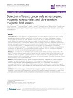
Figure 3 Effects of C3G on ethanol-mediated phosphorylation of ErbB2, cSrc, FAK and p130Cas. A: MCF7ErbB2 cells were treated with
ethanol (0 or 400 mg/dl) with/without C3G (10, 20 or 40 μM) for 48 h. Cells were seeded on fibronectin-coated culture wells for 3 h and then
harvested for analysis of the phosphorylation of ErbB2, FAK, p130Cas and cSrc with immunoblotting. The expression of actin served as a loading
control. B: The relative expression of phosphorylated ErbB2, FAK, p130Cas and cSrc was determined by densitometry and normalized to the
expression of actin. * denotes a statistically significant difference from untreated controls. # denotes a significant difference from ethanol-treated
groups. ε denotes a significant difference from ethanol- and C3G (10 μM)-treated groups. δ denotes a significant difference from ethanol- and
C3G (20 μM)-treated groups. C: The phosphorylation of ErbB2 and FAK in MDA-MB231 cells was analyzed as described above. The experiment
was replicated three times.
Xu et al. Molecular Cancer 2010, 9:285
/>
Page 7 of 14
Figure 4 Effects of C3G on the interaction among ErbB2, FAK, cSrc and p130Cas. MCF7ErbB2 cells were treated with ethanol (0 or 400 mg/
dl) with/without C3G (10, 20 or 40 μM) for 48 h. Cells were plated on fibronectin-coated culture wells. A: After 1 h of attachment on fibronectin,
cell lysates were collected and immunoprecipitated (IP) with an anti-ErbB2 antibody, then immunoblotted (IB) with either an anti-FAK or antiErbB2 antibody. B: After 3 h of attachment, cell lysates were IP with an anti-cSrc antibody and IB with either an anti-p130Cas, FAK or cSrc
antibody. C and D: The association between ErbB2 and FAK (panel A) and the association between FAK and p130Cas (panel B) was quantified by
densitometry. * denotes a statistically significant difference from untreated controls. # denotes a significant difference from ethanol-treated
groups. ε denotes a significant difference from ethanol- and C3G (10 μM)-treated groups. δ denotes a significant difference from ethanol- and
C3G (20 μM)-treated groups. E: After 3 h of attachment, cell lysates were IP with an anti-p130Cas antibody and IB with either an anti-FAK or antip130Cas antibody. The experiment was replicated three times.
Xu et al. Molecular Cancer 2010, 9:285
/>
Page 8 of 14
Figure 5 Effects of C3G on ethanol-induced activation of JNKs. MCF7ErbB2 cells were treated with ethanol (0 or 400 mg/dl) with/without
C3G (10, 20 or 40 μM) for 48 h. Cells were seeded on fibronectin-coated culture wells for 3 h. A: Cell lysates were collected and analyzed for the
phosphorylation/expression of JNKs with immunoblotting. B: Cell lysates were IP with an anti-JNK antibody and IB with either an anti-p130Cas or
anti-JNK antibody. The experiment was replicated three times. C and D: The phoshorylation of JNKs and the association between JNKs and
p130Cas were quantified by densitometry. * denotes a statistically significant difference from untreated controls. # denotes a significant difference
from ethanol-treated groups. ε denotes a significant difference from ethanol- and C3G (10 μM)-treated groups. δ denotes a significant difference
from ethanol- and C3G (20 μM)-treated groups.
at the leading edge/lamellipodia in ethanol-treated
MDA-MB231 cells. Ethanol caused an approximate 3fold increase in the number of lamellipodia (Figure 6B).
C3G inhibited ethanol-induced lamellipodia formation;
however, the inhibition was not concentration-dependent and C3G at 10 or 40 μM had a similar effect (Figure 6B). We demonstrated an accumulation of p-FAK
(Tyr861) at the leading area in ethanol-treated cells (Figures 6A and 7A). Ethanol also caused redistribution of
paxillin, and more paxillin was localized at the leading
edge following ethanol exposure (Figure 7A). Since the
activation of FAK leads to the recruitment of paxillin
and p130Cas to focal adhesion sites [27,30], we examined
the effect of ethanol on focal adhesions. Ethanol
enhanced the assembly of focal adhesions and C3G significantly inhibited ethanol-induced formation of focal
adhesions (Figure 7B).
C3G scavenges ethanol-induced accumulation of reactive
oxygen species (ROS)
Ethanol causes intracellular accumulation of reactive
oxygen species (ROS) and induces oxidative stress
[10,31]. Since C3G is a potent antioxidant, the inhibitory
effect of C3G on ethanol-induced migration/invasion
may be mediated by its antioxidant property. We evaluated the effect of other antioxidants at concentrations
Xu et al. Molecular Cancer 2010, 9:285
/>
Page 9 of 14
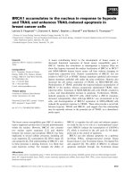
Figure 6 Effects of C3G on the development of lamellipodia. MDA-MB231 cells were treated with ethanol (0 or 400 mg/dl) with/without
C3G (10 or 40 μM) for 48 h. Cells were seeded on fibronectin-coated coverslips for 3 h. A: The expression of actin (Alexa Fluor 488 Phalloidin)
and phosphorylated FAK (Tyr 861) (Alexa Flour 594) were detected with immunofluorescent staining. The arrow indicates lamellipodia. Scale bar
= 5 μm. B: Cells with extended leading areas (lamellipodia) were counted in ten randomly selected fields in each treatment group. The
percentage of cells with lamellipodia was determined. The experiment was replicated three times. * denotes a significant difference from
untreated controls. # denotes a significant difference from ethanol-treated groups.
Xu et al. Molecular Cancer 2010, 9:285
/>
Page 10 of 14
Figure 7 Effects of C3G on ethanol-mediated formation of focal adhesions. MDA-MB231 cells were treated with ethanol (0 or 400 mg/dl)
with/without C3G (40 μM) for 48 h. Cells were seeded on fibronectin-coated coverslips for 3 h. A: The expression of paxillin (Alexa Fluor 488)
and phosphorylated FAK (Tyr861) (Alexa Fluor 594) were detected by immunofluorescent staining. Arrows indicate the co-localization of p-FAK
(Tyr861) and paxillin. Scale bar = 5 μm. B: Focal adhesions were counted randomly on 10 or more cells. The number of focal adhesions per cell
was calculated. Each datum point was the mean ± SEM of three independent experiments. * denotes a significant difference from untreated
controls. # denotes a significant difference from ethanol-treated groups.
Xu et al. Molecular Cancer 2010, 9:285
/>
Page 11 of 14
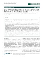
Figure 8 Effects of C3G and antioxidants on ethanol-induced ROS generation, cell invasion and ErbB2 phosphorylation. A: MCF7ErbB2
cells were exposed to ethanol (0 or 400 mg/dl) with/without C3G (10 μM), SOD (50 U/ml)/catalase (200 U/ml), NAC (5 mM) or vitamin C (20
μM) for 48 h. Intracellular ROS levels were measured by flow cytometry as described under the Materials and Methods. B: The invasive potential
of MCF7ErbB2 cells was evaluated as described above and expressed relative to untreated controls. C: The phosphorylation of ErbB2 in MCF7ErbB2
cells was analyzed with immunoblotting. The experiment was replicated three times. * denotes a statistically significant difference from
untreated controls. # denotes a significant difference from ethanol-treated groups. # denotes a significant difference from ethanol- and
C3G-treated groups.
that had a similar ROS scavenging capacity as C3G.
Superoxide dismutase (SOD) is a scavenger for O2• and
catalase is a scavenger for hydrogen peroxide (H2O 2).
N-aceytlcysteine (NAC) (5 mM), vitamin C (20 μM) and
SOD (50 U/ml) plus catalase (200 U/ml) had approximately the same antioxidant effect as C3G (10 μM) (Figure 8A). As shown in Figure 8B, C3G most effectively
inhibited ethanol-enhanced invasion of breast cancer
cells; NAC and vitamin C also provided significant inhibition, but to a lesser extent. On the other hand, SOD
plus catalase had little effect on ethanol-enhanced cell
invasion. A similar result regarding the effect of C3G
and other antioxidants on ethanol-induced ErbB2 phosphorylation was observed (Figure 8C).
Xu et al. Molecular Cancer 2010, 9:285
/>
Discussion
C3G as a potent agent to alleviate ethanol-induced cell
migration/invasion
In search for better chemopreventive or chemotherapeutic agents, we isolated a natural antioxidant cyanidin-3glucoside (C3G) from blackberries [14,32]. C3G is a
member of the anthocyanin family which is present in
various vegetables and fruits, especially edible berries.
We have confirmed C3G’s antioxidant property [14,23].
C3G has been implicated in some beneficial health
actions including reducing age-associated oxidative
stress, improving cognitive brain function, as well as
anti-diabetic, anti-inflammation, anti-atherogenic and
anti-obesity activity [33]. C3G exhibits anti-cancer properties in various in vitro and animal models of carcinogenesis and tumor development; the effects of C3G
include the inhibition of tumor cell proliferation and the
attenuation of cell migration/invasion as well as metastasis in vivo [13-18].
We demonstrate here that C3G inhibits ethanolmediated migration/invasion in cells expressing high
levels of ErbB2. C3G has a greater inhibitory effect on
the invasion of cells treated with 200 mg/dl ethanol
compared to 400 mg/dl (Figure 1B); the underlying
mechanism is unclear. C3G is effective at 10 μM, a concentration that is lower than previously reported for its
anti-cancer effects. C3G inhibits the migration/invasion
of A549 lung cancer cells at 40-100 μM [14,34]. It is
unlikely that C3G-mediated inhibition of tumor cell
migration/invasion in this study results from decreased
cell viability. C3G up to 40 μM does not affect the viability of breast cancer cells (Figure 1D). It is reported that
C3G at 100 μM fails to reduce the viability of A549
lung cancer cells [34]. However, at 20 μM, C3G significantly decreases the viability of HS578T human breast
cancer cells [13]. Thus, cells apparently display differential sensitivity to C3G. For BT474 cells, C3G does not
have a dose-dependent inhibitory effect. It is likely
BT474 cells are more sensitive to C3G and the concentration may need to be lower in order to see the dosedependent inhibition.
Consistent with its effects on migration/invasion, C3G
affects early events associated with cell motility. C3G
inhibits ethanol-mediated cell adhesion to the ECM, formation of focal adhesions and development of lamellipodia. These events are prerequisites for cell migration/
invasion. These results suggest that ethanol-induced
migration/invasion is initiated by tumor cell/ECM interaction and C3G blocks this interaction.
C3G and ethanol-stimulated cell signaling
Ethanol-stimulated tumor cell/ECM interaction may be
initiated by its effect on ErbB2 activity. Ethanol increases
Page 12 of 14
the phosphorylation of ErbB2 and enhances the adhesion of breast cancer cells with high levels of ErbB2 to
the ECM as well as the assembly of focal adhesions in
these cells [19]. These effects were not observed in
breast cancer cells expressing low levels of ErbB2.
FAK is a critical regulator of cell/ECM interaction and
is strongly implicated in tumor aggressiveness [35,36]. It
has been shown that FAK is essential for ErbB2/ErbB3induced oncogenesis and breast cancer invasion [37].
The phosphorylation of FAK at Tyr 861 plays an important role in the invasion of breast cancer cells [38]. FAK
is a substrate of cSrc. The activation of ErbB2 recruits
cSrc and FAK, resulting in the phosphorylation of cSrc
at Tyr 216 and FAK at Tyr 861 in breast cancer cells
[39,40]. An ErbB2 inhibitor Tyrphostin (AG825)
abolishes the ethanol-stimulated interaction between
ErbB2 and FAK, as well as the adhesion of breast cancer
cells to the ECM [41]. It appears C3G targets ErbB2
since C3G is able to inhibit ethanol-mediated phosphorylation of ErbB2, cSrc and FAK. Additionally, it inhibits
the ethanol-mediated interaction among these proteins.
Previously we have shown that JNK activation is
required in ethanol-induced migration/invasion of breast
cancer cells [10]. It was reported that JNK activation
during cell migration is mediated by p130Cas/Crk coupling or JSAP1 (JNK/stress-activated protein kinaseassociated protein 1)[42,43]. p130 Cas (Crk-associated
substrate) is an adaptor protein and it binds to and is
phosphorylated by FAK in a FAK/cSrc dependent manner [27]. Activation of p130Cas leads to recruitment of
Crk to form an adaptor complex which results in activation of Rac1 and JNKs. We show here that ethanol
increases the association of p130Cas with FAK/cSrc and
the phosphorylation of p130Cas. Ethanol also promotes
the association between p130Cas and JNKs. C3G inhibits
ethanol-mediated p130Cas /JNK interaction (Figure 5).
Together, our results indicate that ethanol activates
p130Cas and JNKs through the ErbB2/cSrc/FAK pathway. Blocking ErbB2/cSrc/FAK signaling by C3G inhibits ethanol-mediated activation of JNKs which is
necessary for cell migration/invasion. At times the effect
of C3G on cell signaling components, such as FAK, cSrc
and JNK, is not dose-dependent, which is not entirely
consistent with its effect on ethanol-induced cell migration/invasion. This suggests that the mechanism of ethanol-induced cell migration/invasion is complex and
probably multiple signaling pathways are involved.
Antioxidant property of C3G
Ethanol exposure causes the accumulation of intracellular ROS [10,23]. The antioxidant property of C3G is
confirmed in this study and we show that C3G effectively blocks ethanol-induced ROS production in breast
Xu et al. Molecular Cancer 2010, 9:285
/>
cancer cells. ROS is reported to be involved in the activation of EGFR [44]. To evaluate the involvement of
ROS, we compared the effect of C3G with other antioxidants. We have titrated antioxidants and identified concentrations for these antioxidants to produce a ROS
scavenging capacity similar to C3G at 10 μM. Although
these antioxidants have a similar capacity of scavenging
ROS, they are less effective in alleviating ethanolinduced cell invasion and ErbB2 phosphorylation. Broad
antioxidants, such as NAC and vitamin C, display modest inhibition on ethanol-induced cell invasion and
ErbB2 phosphorylation, but to a lesser extent compared
to C3G. More specific antioxidants, SOD (for superoxide) plus catalase (for hydrogen peroxide) fail to inhibit
ethanol-stimulated invasion and ErbB2 phosphorylation.
These data suggest that although the antioxidant property may be involved, C3G may also act through other
mechanisms to regulate ErbB2 signaling and subsequent
migration/invasion. We have previously shown that C3G
is able to reverse ethanol-induced inhibition of neurite
outgrowth in neuronal cells; however, its antioxidant
property is minimally involved [23].
C3G has drawn increasing attention because of its
potential anti-cancer properties. A recent animal study
investigates the pharmacokinetics of C3G and demonstrates that pharmacologically relevant concentrations of
C3G are achievable in vivo through oral administration
or intravenous injection in mice without apparent
adverse effects [45]. Further analysis suggests that concentrations required for in vivo action of C3G could be
much lower than that of in vitro [14,45]. In future studies, we will evaluate the effect of C3G on ethanolinduced tumor promotion in animal models. C3G may
offer a novel avenue for treating alcohol-related disorders.
Abbreviations
C3G: Cyanidin-3-glucoside; ECM: extracellular matrix; FAK: focal adhesion
kinase; IP: Immunoprecipitation; JNKs: c-Jun N-terminal kinases; NAC: Nacetyl-cysteine; ROS: reactive oxygen species; SOD: superoxide dismutase.
Acknowledgements
This research was supported by grants from the National Institutes of Health
(AA01540 and AA017226).
Author details
1
Department of Internal Medicine, University of Kentucky College of
Medicine, Lexington, KY 40536, USA. 2Pathophysiological Department, School
of Basic Medicine, Anhui Medical University, Hefei, Anhui, PR China 230032.
3
National Institute for Occupational Safety and Health, Morgantown, West
Virginia 26505, USA. 4Beltsville Agricultural Research Center, U. S. Department
of Agriculture, Beltsville, Maryland 20705, USA. 5Graduate Center for
Toxicology, University of Kentucky, 232 Health Sciences Research Building,
Lexington, Kentucky 40536, USA. 6Institute for Nutritional Sciences, Shanghai
Institutes for Biological Sciences, Chinese Academy of Sciences, Shanghai, PR
China 200031.
Authors’ contributions
MX carried out the biochemical studies and participated in all experiments
in this study. KB, JF and GC participated in assays for cell treatment,
Page 13 of 14
immunoblotting and ethanol exposure paradigm. MD, SW, XS, ZK and JL
conceived of the study, and participated in its design and coordination and
helped to draft the manuscript. All authors read and approved the final
manuscript.
Competing interests
The authors declare that they have no competing interests.
Received: 20 July 2010 Accepted: 29 October 2010
Published: 29 October 2010
References
1. Key J, Hodgson S, Omar RZ, Jensen TK, Thompson SG, Boobis AR,
Davies DS, Elliott P: Meta-analysis of studies of alcohol and breast cancer
with consideration of the methodological issues. Cancer Causes Control
2006, 17:759-70.
2. Seitz HK, Maurer B: The relationship between alcohol metabolism,
estrogen levels, and breast cancer risk. Alcohol Res Health 2007, 30:42-3.
3. Seitz HK, Becker P: Alcohol metabolism and cancer risk. Alcohol Res Health
2007, 30:38-7.
4. Tjonneland A, Christensen J, Olsen A, Stripp C, Thomsen BL, Overvad K,
Peeters PH, van Gils CH, Bueno-de-Mesquita HB, Ocke MC, Thiebaut A,
Fournier A, Clavel-Chapelon F, Berrino F, Palli D, Tumino R, Panico S,
Vineis P, Agudo A, Ardanaz E, Martinez-Garcia C, Amiano P, Navarro C,
Quiros JR, Key TJ, Reeves G, Khaw KT, Bingham S, Trichopoulou A,
Trichopoulos D, Naska A, Nagel G, Chang-Claude J, Boeing H, Lahmann PH,
Manjer J, Wirfalt E, Hallmans G, Johansson I, Lund E, Skeie G, Hjartaker A,
Ferrari P, Slimani N, Kaaks R, Riboli E: Alcohol intake and breast cancer
risk: the European Prospective Investigation into Cancer and Nutrition
(EPIC). Cancer Causes Control 2007, 18:361-73.
5. Visvanathan K, Crum RM, Strickland PT, You X, Ruczinski I, Berndt SI,
Alberg AJ, Hoffman SC, Comstock GW, Bell DA, Helzlsouer KJ: Alcohol
dehydrogenase genetic polymorphisms, low-to-moderate alcohol
consumption, and risk of breast cancer. Alcohol Clin Exp Res 2007,
31:467-76.
6. Vaeth PA, Satariano WA: Alcohol consumption and breast cancer stage at
diagnosis. Alcohol Clin Exp Res 1998, 22:928-34.
7. Weiss HA, Brinton LA, Brogan D, Coates RJ, Gammon MD, Malone KE,
Schoenberg JB, Swanson CA: Epidemiology of in situ and invasive breast
cancer in women aged under 45. Br J Cancer 1996, 73:1298-305.
8. Aye MM, Ma C, Lin H, Bower KA, Wiggins RC, Luo J: Ethanol-induced in
vitro invasion of breast cancer cells: the contribution of MMP-2 by
fibroblasts. Int J Cancer 2004, 112:738-46.
9. Ke Z, Lin H, Fan Z, Cai TQ, Kaplan RA, Ma C, Bower KA, Shi X, Luo J: MMP-2
mediates ethanol-induced invasion of mammary epithelial cells overexpressing ErbB2. Int J Cancer 2006, 119:8-16.
10. Ma C, Lin H, Leonard SS, Shi X, Ye J, Luo J: Overexpression of ErbB2
enhances ethanol-stimulated intracellular signaling and invasion of
human mammary epithelial and breast cancer cells in vitro. Oncogene
2003, 22:5281-90.
11. Paterson MC, Dietrich KD, Danyluk J, Paterson AH, Lees AW, Jamil N,
Hanson J, Jenkins H, Krause BE, McBlain WA: Correlation between c-erbB-2
amplification and risk of recurrent disease in node-negative breast
cancer. Cancer Res 1991, 51:556-67.
12. Slamon DJ, Clark GM, Wong SG, Levin WJ, Ullrich A, McGuire WL: Human
breast cancer: correlation of relapse and survival with amplification of
the HER-2/neu oncogene. Science 1987, 235:177-82.
13. Chen PN, Chu SC, Chiou HL, Chiang CL, Yang SF, Hsieh YS: Cyanidin 3glucoside and peonidin 3-glucoside inhibit tumor cell growth and
induce apoptosis in vitro and suppress tumor growth in vivo. Nutr
Cancer 2005, 53:232-43.
14. Ding M, Feng R, Wang SY, Bowman L, Lu Y, Qian Y, Castranova V, Jiang BH,
Shi X: Cyanidin-3-glucoside, a natural product derived from blackberry,
exhibits chemopreventive and chemotherapeutic activity. J Biol Chem
2006, 281:17359-68.
15. Shih PH, Yeh CT, Yen GC: Effects of anthocyanidin on the inhibition of
proliferation and induction of apoptosis in human gastric
adenocarcinoma cells. Food Chem Toxicol 2005, 43:1557-66.
16. Zhang Y, Vareed SK, Nair MG: Human tumor cell growth inhibition by
nontoxic anthocyanidins, the pigments in fruits and vegetables. Life Sci
2005, 76:1465-72.
Xu et al. Molecular Cancer 2010, 9:285
/>
17. Fukamachi K, Imada T, Ohshima Y, Xu J, Tsuda H: Purple corn color
suppresses Ras protein level and inhibits 7,12-dimethylbenz[a]
anthracene-induced mammary carcinogenesis in the rat. Cancer Sci 2008,
99:1841-6.
18. Zhang Y, Seeram NP, Lee R, Feng L, Heber D: Isolation and identification
of strawberry phenolics with antioxidant and human cancer cell
antiproliferative properties. J Agric Food Chem 2008, 56:670-5.
19. Xu M, Bower KA, Chen G, Shi X, Dong Z, Ke Z, Luo J: Ethanol enhances
the interaction of breast cancer cells over-expressing ErbB2 with
fibronectin. Alcohol Clin Exp Res 2010, 34:751-60.
20. Grimaldi C, Pisanti S, Laezza C, Malfitano AM, Santoro A, Vitale M,
Caruso MG, Notarnicola M, Iacuzzo I, Portella G, Di M, Bifulco M:
Anandamide inhibits adhesion and migration of breast cancer cells. Exp
Cell Res 2006, 312:363-73.
21. Wang F, Nohara K, Olivera A, Thompson EW, Spiegel S: Involvement of
focal adhesion kinase in inhibition of motility of human breast cancer
cells by sphingosine 1-phosphate. Exp Cell Res 1999, 247:17-28.
22. Xu M, Waters CL, Hu C, Wysolmerski RB, Vincent PA, Minnear FL:
Sphingosine 1-phosphate rapidly increases endothelial barrier function
independently of VE-cadherin but requires cell spreading and Rho
kinase. Am J Physiol Cell Physiol 2007, 293:C1309-C1318.
23. Chen G, Bower KA, Xu M, Ding M, Shi X, Ke ZJ, Luo J: Cyanidin-3-glucoside
reverses ethanol-induced inhibition of neurite outgrowth: role of
glycogen synthase kinase 3 Beta. Neurotox Res 2009, 15:321-31.
24. Vadlamudi RK, Sahin AA, Adam L, Wang RA, Kumar R: Heregulin and HER2
signaling selectively activates c-Src phosphorylation at tyrosine 215. FEBS
Lett 2003, 543:76-80.
25. Dolfi F, Garcia-Guzman M, Ojaniemi M, Nakamura H, Matsuda M, Vuori K:
The adaptor protein Crk connects multiple cellular stimuli to the JNK
signaling pathway. Proc Natl Acad Sci USA 1998, 95:15394-9.
26. Klemke RL, Leng J, Molander R, Brooks PC, Vuori K: Cheresh DA, CAS/Crk
coupling serves as a “molecular switch” for induction of cell migration. J
Cell Biol 1998, 140:961-72.
27. Cox BD, Natarajan M, Stettner MR, Gladson CL: New concepts regarding
focal adhesion kinase promotion of cell migration and proliferation. J
Cell Biochem 2006, 99:35-52.
28. Huang C, Jacobson K, Schaller MD: MAP kinases and cell migration. J Cell
Sci 2004, 117:4619-28.
29. Yamaguchi H, Wyckoff J, Condeelis J: Cell migration in tumors. Curr Opin
Cell Biol 2005, 17:559-64.
30. Turner CE: Paxillin interactions. J Cell Sci 2000, 113(Pt 23):4139-40.
31. Chen G, Ma C, Bower KA, Shi X, Ke Z, Luo J: Ethanol promotes
endoplasmic reticulum stress-induced neuronal death: involvement of
oxidative stress. J Neurosci Res 2008, 86:937-46.
32. Zheng W, Wang SY: Oxygen radical absorbing capacity of phenolics in
blueberries, cranberries, chokeberries, and lingonberries. J Agric Food
Chem 2003, 51:502-9.
33. Chen G, Luo J: Anthocyanins: are they beneficial in treating ethanol
neurotoxicity? Neurotox Res 2010, 17:91-101.
34. Chen PN, Kuo WH, Chiang CL, Chiou HL, Hsieh YS, Chu SC: Black rice
anthocyanins inhibit cancer cells invasion via repressions of MMPs and
u-PA expression. Chem Biol Interact 2006, 163:218-29.
35. Hao H, Naomoto Y, Bao X, Watanabe N, Sakurama K, Noma K, Motoki T,
Tomono Y, Fukazawa T, Shirakawa Y, Yamatsuji T, Matsuoka J, Wang ZG,
Takaoka M: Focal adhesion kinase as potential target for cancer therapy
(Review). Oncol Rep 2009, 22:973-9.
36. Schwock J, Dhani N, Hedley DW: Targeting focal adhesion kinase
signaling in tumor growth and metastasis. Expert Opin Ther Targets 2010,
14:77-94.
37. Benlimame N, He Q, Jie S, Xiao D, Xu YJ, Loignon M, Schlaepfer DD, AlaouiJamali MA: FAK signaling is critical for ErbB-2/ErbB-3 receptor
cooperation for oncogenic transformation and invasion. J Cell Biol 2005,
171:505-16.
38. Earley S, Plopper GE: Phosphorylation of focal adhesion kinase promotes
extravasation of breast cancer cells. Biochem Biophys Res Commun 2008,
366:476-82.
39. Vadlamudi RK, Sahin AA, Adam L, Wang RA, Kumar R: Heregulin and HER2
signaling selectively activates c-Src phosphorylation at tyrosine 215. FEBS
Lett 2003, 543:76-80.
Page 14 of 14
40. Vadlamudi RK, Adam L, Nguyen D, Santos M, Kumar R: Differential
regulation of components of the focal adhesion complex by heregulin:
role of phosphatase SHP-2. J Cell Physiol 2002, 190:189-99.
41. Xu M, Bower KA, Chen G, Shi X, Dong Z, Ke Z, Luo J: Ethanol Enhances
the Interaction of Breast Cancer Cells Over-Expressing ErbB2 With
Fibronectin. Alcohol Clin Exp Res 2010, 34(5):751-60.
42. Klemke RL, Leng J, Molander R, Brooks PC, Vuori K, Cheresh DA: CAS/Crk
coupling serves as a “molecular switch” for induction of cell migration. J
Cell Biol 1998, 140:961-72.
43. Takino T, Nakada M, Miyamori H, Watanabe Y, Sato T, Gantulga D,
Yoshioka K, Yamada KM, Sato H: JSAP1/JIP3 cooperates with focal
adhesion kinase to regulate c-Jun N-terminal kinase and cell migration. J
Biol Chem 2005, 280:37772-81.
44. von Montfort C, Fernau NS, Beier JI, Sies H, Klotz LO: Extracellular
generation of hydrogen peroxide is responsible for activation of EGF
receptor by ultraviolet A radiation. Free Radic Biol Med 2006, 41:1478-87.
45. Marczylo TH, Cooke D, Brown K, Steward WP, Gescher AJ:
Pharmacokinetics and metabolism of the putative cancer
chemopreventive agent cyanidin-3-glucoside in mice. Cancer Chemother
Pharmacol 2009, 64:1261-8.
doi:10.1186/1476-4598-9-285
Cite this article as: Xu et al.: Cyanidin-3-Glucoside inhibits ethanolinduced invasion of breast cancer cells overexpressing ErbB2. Molecular
Cancer 2010 9:285.
Submit your next manuscript to BioMed Central
and take full advantage of:
• Convenient online submission
• Thorough peer review
• No space constraints or color figure charges
• Immediate publication on acceptance
• Inclusion in PubMed, CAS, Scopus and Google Scholar
• Research which is freely available for redistribution
Submit your manuscript at
www.biomedcentral.com/submit