hcv genomic rna activates the nlrp3 inflammasome in human myeloid cells
Bạn đang xem bản rút gọn của tài liệu. Xem và tải ngay bản đầy đủ của tài liệu tại đây (2.22 MB, 10 trang )
HCV Genomic RNA Activates the NLRP3 Inflammasome in
Human Myeloid Cells
Wei Chen1., Yongfen Xu1., Hua Li1, Wanyin Tao1, Yu Xiang1, Bing Huang1, Junqi Niu2, Jin Zhong1*,
Guangxun Meng1*
1 Key Laboratory of Molecular Virology & Immunology, Institut Pasteur of Shanghai, Chinese Academy of Sciences, Shanghai, China, 2 Department of Hepatology, The
First Hospital of Jilin University, Changchun, China
Abstract
Background: Elevated plasma levels of IL-1b and IL-18 from patients with hepatitis C virus (HCV) infection indicate a
possible activation of inflammasome by HCV.
Methodology/Principal Findings: To demonstrate whether HCV infection activates the inflammasome, we investigated
inflammasome activation from HCV infected hepatic Huh7 cells, or monocytic cells and THP-1 derived macrophages
challenged with HCV virions, but no any inflammasome activation was detected in these cells. However, when we
transfected HCV genomic RNA into monocytes or macrophages, IL-1b was secreted in a dose-dependent manner. We also
detected ASC oligomerization and caspase-1 cleavage in HCV RNA transfected macrophages. Using shRNA-mediated gene
silencing or specific inhibitors, we found that HCV RNA-induced IL-1b secretion was dependent on the presence of
inflammasome components such as NLRP3, ASC and caspase-1. Furthermore, we also found that RIG-I was dispensable for
HCV RNA-induced NLRP3 inflammasome activation, while reactive oxygen species (ROS) production was required.
Conclusions: Our results indicate that HCV RNA activates the NLRP3 inflammasome in a ROS-dependent manner, and RIG-I
is not required for this process.
Citation: Chen W, Xu Y, Li H, Tao W, Xiang Y, et al. (2014) HCV Genomic RNA Activates the NLRP3 Inflammasome in Human Myeloid Cells. PLoS ONE 9(1): e84953.
doi:10.1371/journal.pone.0084953
Editor: Fayyaz S. Sutterwala, University of Iowa Carver College of Medicine, United States of America
Received May 30, 2013; Accepted November 20, 2013; Published January 6, 2014
Copyright: ß 2014 Chen et al. This is an open-access article distributed under the terms of the Creative Commons Attribution License, which permits
unrestricted use, distribution, and reproduction in any medium, provided the original author and source are credited.
Funding: This work was supported by grants from 100 Talent Program of the Chinese Academy of Sciences, Natural Science Foundation of China (91029707,
31170868), Novo Nordisk-CAS Research Foundation, SA-SIBS Scholarship Program, as well as grants from the National Key Programs on Infectious Disease
(2012ZX10002007-003). The funders had no role in study design, data collection and analysis, decision to publish, or preparation of the manuscript.
Competing Interests: The authors have declared that no competing interests exist.
* E-mail: (JZ); (GM)
. These authors contributed equally to this work.
taken as marker of the acute phase of HCV infection [8,11–15]. As
a special group of cytokines, the secretion of IL-1b and IL-18
involves a two step process: step 1 is the synthesis of pro-IL-1b and
pro-IL-18 (signal 1); step 2 is activation of caspase-1 (signal 2)
which cleaves pro-IL-1b and pro-IL-18 into mature IL-1b and IL18 [16–18]. Recently it was found that the activation of caspase-1
is mediated by the inflammasome, a protein complex composed of
PRRs including AIM2 (Absent In Melanoma 2) or NLRP3 (NODlike receptor family, pyrin domain containing 3), adaptor protein
ASC (apoptosis-associated specklike protein containing a CARD)
as well as pro-caspase-1 [16,19]. Other reported inflammasomes
include NLRP1-, NLRC4-, NLRP6-, NLRP7- as well as RIG-Iinflammasome [20–22]. Various microbes are able to activate
inflammasomes [23], and the NLRP3 and RIG-I inflammasomes
were reported to be activated by RNA viruses [24–27]. Thus,
elevated IL-1b and IL-18 levels in HCV-infected patients indicate
that HCV may trigger inflammasome activation.
Recently, Burdette et.al. reported that HCV (JFH-1) infection
induced NLRP3 inflammasome activation in the hepatoma cell
line Huh7.5 [28]. However, the expression of inflammasome
components was found to be prominent in Kupffer cells (KC) and
liver sinusoidal endothelial cells, moderate in periportal myofibro-
Introduction
Hepatitis C virus (HCV) infection tends to become persistent
and causes liver fibrosis and cirrhosis due to chronic inflammation
in humans [1]. The 9.6-kb genome of HCV ssRNA is composed of
a 59 untranslated region (59UTR), a single open reading frame
(ORF) and a 39UTR, as well as an internal ribosome entry site
(IRES) within the 59UTR, which directs translation of a
polyprotein precursor of about 3000 amino acids that is cleaved
into mature structural and non-structural proteins [2,3]. It was
reported that the HCV 59-ppp poly-U/UC RNA variants
stimulate strong retinoic acid-inducible gene I (RIG-I) activation
in vitro [4]. RIG-I was also reported to detect in vitro transcribed
HCV RNA, RNA without a 59-triphosphate end, 59-triphosphate
single-stranded RNA and short double-stranded RNA for type I
interferon production [5–7].
Besides the anti-viral type I interferon response, pro-inflammatory cytokines such as tumor necrosis factor (TNF)-a and
interleukin (IL)-6 can also be induced upon HCV infection [8–
10]. Recently, serum IL-18 and IL-1b levels have been observed to
be clearly higher in patients with chronic HCV infection and
HCV related cirrhosis than in healthy controls, and IL-18 was
PLOS ONE | www.plosone.org
1
January 2014 | Volume 9 | Issue 1 | e84953
HCV RNA Activates the NLRP3 Inflammasome
blasts and hepatic stellate cells, virtually absent in primary
hepatocytes [29], therefore, inflammasome activation in hepatocytes may not be the main origin of IL-b from HCV infected
patients. Instead, HCV virions or its components such as genomic
RNA may activate the inflammasome in KC or peripheral
myeloid cells, and this should be the main origin of IL-b.
Interestingly, a more recent study from Negash et al. revealed that
there was no appreciable IL-1b from HCV infected hepatoma
cells or primary hepatocytes, while robust IL-1b production was
induced by HCV virions in human macrophages [30].
In our present study, no inflammasome activation was observed
in HCV infected Huh7 or Huh7.5.1 cells. Moreover, we found
that HCV virions did not trigger IL-1b secretion in human
myeloid cells. However, we discovered that HCV RNA transfection in monocytes or macrophages induced robust IL-1b secretion,
which was dependent on the NLRP3 inflammasome. HCV RNA
transfection triggered ASC oligomerization and caspase-1 cleavage, suggesting that the HCV genome possesses the ability to
activate signal 2 directly. In addition, we found that neither IL-1b
secretion nor caspase-1 cleavage was dependent on RIG-I.
assessed for each sample by melting curve analysis. Relative
quantification was performed using standard curve analysis. The
quantification data are presented as a ratio to the control level.
The Homo sapiens (hs) gene specific primers used were as follows:
IFN-b, 59-GATTCATCTAGCACTGGCTGG-39 (forward)
and 59- CTTCAGGTAATGCAGAATCC-39 (reverse);
RIG-I, 59-CCTACCTACATCCTGAGCTACAT-39 (forward)
and 59-TCTAGGGCATCCAAAAAGCCA-39 (reverse);
IL-1b, 59-CACGATGCACCTGTACGATCA-39 (forward)
and 59-GTTGCTCCATATCCTGTCCCT-39 (reverse);
ASC, 59-AACCCAAGCAAGATGCGGAAG-39 (forward) and
59-TTAGGGCCTGGAGGAGCAAG-39 (reverse);
Actin, 59-AGTGTGACGTGGACATCCGCAAAG-39 (forward) and 59-ATCCACATCTGCTGGAAGGTGGAC-39 (reverse);
NLRP3, 59-AAGGGCCATGGACTATTTCC-39 (forward)
and 59-GACTCCACCCGATGACAGTT-39 (reverse);
Caspase-1, 59-TCCAATAATGCAAGTCAAGCC-39 (forward)
and 59-GCTGTACCCCAGATTTTGTAGCA-39 (reverse).
RNA Transfection into Myeloid Cells
Materials and Methods
RNA including negative control tRNA, positive control Poly
I:C, HCV 1–807 bp, 2406–3256 bp, 5626–6437 bp, HCV
39UTR, HCV full length (FL) RNA, ssRNA40, ssRNA41 and
ssPolyU (Invivogen, USA) were transfected with Lipofectamine
2000 (Invitrogen, USA) diluted in OptiMEM (Invitrogen, USA)
without nucleic acids according to the manufacturer’s protocol.
1 mg of nucleic acid were delivered into THP-1 cells or THP-1
derived macrophages with 2.5 ml of Lipofectamine 2000 unless
described otherwise.
Primary Monocyte Isolation and Cell Culture
Human PBMCs were obtained from the Shanghai Blood
Center (Shanghai, China). Human monocytes were separated by
PercollTM density-gradient centrifugation (G.E Healthcare, Biosciences, Sweden) from isolated PBMCs. Monocyte derived
macrophages (MDM) were generated by incubation of primary
monocytes with recombinant M-CSF (20 ng/ml) for a week as
described [30]. THP-1 cells were maintained in RPMI 1640
media, supplemented with 10% FBS, 100 IU/ml penicillin, 1 mg/
ml streptomycin, 0.25 mg/ml amphotericin B, non essential amino
acids, 1 mM sodium pyruvate, 10 mM HEPES buffer and 2 mM
glutamine. THP-1 cells were differentiated to macrophage-like
cells with 100 nM phorbol-12-myristate-13-acetate (PMA) for 3
hours and then rested for 48 hours before experiments. In some
indicated experiments, THP-1 cells were differentiated to macrophages by treatment with 40 nM of PMA overnight at 37uC as
described by Negash et al [30].
Generation of THP-1 Cells Expressing shRNAs Targeting
Genes of Interest
Three human RIG-I coding sequences were selected for
construction
of
specific
shRNA:
RIG-I-1,
ntGTGGAATGCCTTCTCAGAT;
RIG-I-2,
nt
GCTTCTCTTGATGCGTCAGTGATAGCAAC; RIG-I-3, nt
GATAGAGGAATGCCATTACACTGTGCTTG. Of them,
shRNA RIG-I-3 silenced cells were applied for function experiments. Similarly, three human AIM2 coding sequences were
selected for construction of specific shRNA: AIM2-1, nt
GCCTGAACAGAAACAGATG; AIM2-2, nt ATACAAGGAGATACTCTTGCTAACAGGCC; AIM2-3 nt CCCGAAGATCAACACGCTTCA. In this case, shRNA AIM2-1 silenced cells
were applied for function experiments. shRNA vectors against
human NLRP3, caspase-1, ASC, and their scramble vectors are
gifts from Dr. Jurg Tschopp [34]. Briefly, THP-1 cells stably
expressing shRNA were obtained as follows: ntGATGCGGAAGCTCTTCAGTTTCA of the human ASC coding sequence, ntCAGGTACTATCTGTTCT of the human NLRP3
coding sequence, ntGTGAAGAGATCCTTCTGTA of the
39UTR of the human caspase-1 were inserted into pSUPER.
The Pol III promoter shRNA cassettes from these vectors and
from a lamin A/C-specific pSUPER control construct were
inserted into the lentiviral vector pAB286.1, a derivative of pHR
that contains a SV40-puromycin acetyl transferase cassette for
antibiotic selection. Second-generation packaging plasmids
pMD2-VSVG and pCMV-R8.91 [35] were used for lentivirus
production.
HCVcc Preparation, Purification and HCV RNA Generation
The methods of HCVcc preparation had been described [31].
Harvested HCVcc was purified by sucrose density gradient
centrifugation and titrated [31]. To generate the full-length
genomic RNA, the 1–807 bp, 2406–3256 bp, 5626–6437 bp
and 39UTR of the HCV JFH-1 strain [32] and the pJFH-1
plasmids containing T7 promoter were linearized at the 39 of the
HCV cDNA by XbaI digestion [33], which was used as the
template for in vitro transcription (Ambion, Austin, TX, USA).
Quantification of IL-1b Secretion by ELISA
Supernatants were analyzed for cytokine IL-1b secretion by
ELISA (BD Biosciences, San Diego, CA) according to the
manufacturer’s instructions.
Quantitative Real-time PCR
RNA from human monocytes or Huh7 cells were extracted
using RNA Lyzol reagent (EXcell Bio, China). cDNA was
synthesized with the Rever TraAceHqPCR RT Kit (TOYOBO.CO, TLD, Japan). Quantitative real-time PCR was performed
on a 7900 Fast Real-Time PCR System (AB Applied Biosystems,
USA) using SYBRH Green Realtime PCR Master Mix (TOYOBO.CO, TLD, Japan). The specificity of amplification was
PLOS ONE | www.plosone.org
Immunoblotting
For immunoblotting, cells were lysed with buffer (10 mM Tris
pH 7.5, 1% NP-40, 150 mM NaCl, and protease inhibitor
2
January 2014 | Volume 9 | Issue 1 | e84953
HCV RNA Activates the NLRP3 Inflammasome
cocktail). Proteins were separated on sodium dodecyl sulphatepolyacrylamide gels and then transferred onto polyvinylidene
difluoride membranes. The membranes were blocked with 5%
milk in 1 X TBS with 0.5% Tween-20 and then probed with
primary antibodies as follows: rabbit anti-human mature (17 kDa)
IL-1b (D116, Cell Signaling, USA), goat anti-human pro-IL-1b
(31 kDa) (sc-1250, Santa Cruz, USA), rabbit anti-human caspase1 (sc-515, Santa Cruz, USA), and monoclonal mouse anti-human
b-actin (KM9001, Tianjin Sungene Biotech, China). Appropriate
HRP-conjugated secondary antibodies were used and signals were
detected using ECL reagent (Amersham, USA).
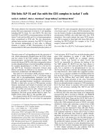
HCV RNA Induces IL-1b Secretion in Macrophages
Although we found that HCV virions did not activate the
inflammasome in hepatoma cell lines or myeloid cells, we believe
that some components instead of the HCV virion particle itself
could activate the inflammasome, because several reports showed
high plasma levels of IL-18 and IL-1b in HCV infected patients
[8,11–15]. Since HCV RNA is a well known PAMP in vivo and
in vitro [4,32,36], we evaluated the ability of HCV RNA in
triggering inflammasome activation in THP-1 derived macrophages. We transfected HCV RNA obtained from in vitro
transcription into macrophages, followed with IL-1b assay. In
this experiment, clear IL-1b mRNA up-regulation and IL-1b
protein secretion was observed (Figure 3A–B). In addition, HCV
RNA induced IL-1b production in a dose dependent manner
(Figure 3C). In a time kinetics test, IL-1b secretion was increased
from 3 h to 6 h post HCV RNA transfection and remained at a
steady level till 24 h after transfection (Figure 3D). Moreover,
genomic RNA extracted from purified HCV virions exhibited
similar induction of IL-1b (Figure 3E). To exclude the possibility of
contamination in the RNA preparation, we applied the unrelated
ApoE transcript as a control, which led to only background level of
IL-1b secretion compared with HCV RNA (Figure 3E). To further
exclude the possibility that some contamination might have caused
IL-1b induction, we digested the HCV RNA with RNase. The
result showed that it was the HCV RNA itself that accounted for
the IL-1b induction from myeloid cells, as RNase treated HCV
RNA lost the ability to induce IL-1b release (Figure 3F).
Moreover, we went a step further to demonstrate which part of
the HCV genome might have been accounting for the IL-1b
induction in macrophages. When different fragments of the HCV
genomic RNA was transfected under the same molar concentration (0.3 pM), we found that only the 39UTR contained the crucial
motif for IL-1b induction, although it was not as potent as the fulllength HCV genomic RNA (Figure 3G). It had been reported that
transfection with EMCV RNA fails to stimulate IL-1b secretion
[37], while uridine-rich single-stranded RNA40 (ssRNA40) from
the HIV-1 long terminal repeat is able to induce IL-1b production
[26]. Our study and others also confirmed that ssRNA40 but not
ssRNA41 nor Poly U was able to induce IL-1b secretion
(Figure 3H) [38]. These data suggest that not all virus RNA is
able to activate macrophages and certain specific sequence or
structure is critical for HCV RNA-induced IL-1b secretion.
Statistical Analysis
Data were analyzed for statistical significance by the two-tailed
student’s t test and values were shown as mean 6 standard
deviation (SD) if not described otherwise. Differences in P values
#0.05 were considered as statistically significant.
Results
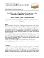
HCV Infection does not Induce IL-1b Secretion in Huh7
Cells
To demonstrate the possible production of IL-1b from HCVinfected hepatoma cells, cellular lysates and the supernatants (SNs)
from HCV virion-incubated Huh7 cells were collected at indicated
time points for analysis (Figure 1A–C). We found that the level of
IL-1b mRNA was not elevated in HCV (JFH-1) infected Huh7
cells (Figure 1A), nor was the IL-1b protein being detected in SNs
from these cells at day 1, day 2 or day 4 after virus infection
(Figure 1B), although the infection efficiency was found normal as
indicated by HCV RNA replication (Figure 1C). Moreover, in
another hepatoma cell line Huh7.5.1 cells, 4 days after HCV
infection, no IL-1b was detected either (Figure S1). To examine
the potential low level activation of the inflammasome in Huh7
cells, we treated the cells with LPS and ATP, but IL-1b production
was still not detected (Figure 1D–E). We next detected the
expression levels of the inflammasome components in HCV JFH1-infected Huh7 cells, and found that there was nearly no
inflammasome components expressed (Figure 1F), which was
similar to a previous report [29]. Therefore, we did not detect any
IL-1b secretion in HCV infected hepatoma cell lines.
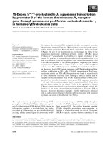
HCV Particles do not Induce IL-1b Secretion from Human
Monocytes and Macrophages
HCV RNA Transfection Activates the Inflammasome
Through NLRP3 but not RIG-I
Since clinical reports have shown that IL-1b and IL-18 were upregulated in HCV infected patients [8,11–15] and there exists
abundant expression of inflammasome components in monocytes
and macrophages [17], we speculated that HCV virion and/or its
components may activate the inflammasome in myeloid cells.
However, when we treated THP-1 monocytes (Figure 2A), THP-1
derived macrophages (Figure 2B), human primary monocytes
(Figure 2C) and macrophages (either unprimed or LPS primed)
(Figure 2D–E) with purified HCV virions at a multiplicity of
infection (MOI) from 0.001 to 2 as indicated, no any IL-1b
secretion was detected. Therefore, our results indicated that the
phagocytosis of HCV by monocytes or macrophages may not be
sufficient to activate the inflammasome. However, Negash et al.
found that HCV virions induced robust IL-1b secretion from
macrophages [30]. We speculated that the THP-1 differentiation
procedures between Negash’s and ours were different. However,
when we applied the exact same differentiation procedure, we still
could not detect any IL-1b in HCV treated macrophages (Figure
S2). Perhaps other differences in cell culture condition accounted
for the different observation.
PLOS ONE | www.plosone.org
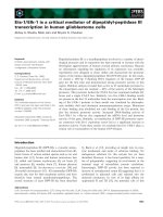
The robust IL-1b induction by HCV RNA from macrophages
mentioned above implied an activation of inflammasome. The IL1b mRNA and protein induction by HCV RNA indicated that
HCV RNA could provide both signal 1 and signal 2 for
inflammasome activation (Figure 3). Indeed, in LPS-primed
macrophages, HCV RNA induced as much IL-1b secretion as
exogenous ATP (Figure S3). As more direct evidence for
inflammasome activation [39], the cleavage of caspase-1 and
oligomerization of ASC in HCV RNA transfected cells was
examined. We found that HCV RNA triggered the cleavage of
caspase-1 and oligomerization of ASC as much as LPS+ATP in
macrophages (Figure 4A–B), indicating a typical activation of
inflammasome [40].
To further demonstrate the specificity of inflammasome
activation by HCV RNA, we transfected the HCV RNA into
macrophages derived from THP-1 cells with shRNA mediated
silencing for ASC, caspase-1, NLRP3 or AIM2 genes ([41,42] and
Figure S4A). It was found that IL-1b secretion induced by HCV
RNA was dependent on ASC, caspase-1 and NLRP3, but not
3
January 2014 | Volume 9 | Issue 1 | e84953
HCV RNA Activates the NLRP3 Inflammasome
Figure 1. HCV infection does not induce IL-1b secretion in Huh7 cells. Huh7 cells were incubated with HCV virions (MOI = 1) for 1, 2 or 4 days.
Total RNA was extracted for Q-PCR analysis (A, C, F) and supernatants were harvested for IL-1b ELISA testing (B). THP-1 derived macrophages and
Huh7 cells were incubated with LPS (200 ng/ml for 6 hours) followed by ATP pulsing (5 mM) for 30 minutes, the cells were then collected for IL-1b
mRNA detection by Q-PCR (D), and supernatants were harvested for IL-1b ELISA (E). Data shown here represent at least three independent
experiments performed with internal triplicates.
doi:10.1371/journal.pone.0084953.g001
RIG-I silenced cells compared with the control upon either HCV
RNA transfection or LPS stimulation (Figure 5C), while the
expression of type I interferon was clearly decreased in the absence
of RIG-I (Figure S5). These results indicated that in HCV RNA
transfected myeloid cells, neither pro-IL-1b synthesis nor caspase1 activation was dependent on RIG-I [25].
It is generally known that NLRP3 inflammasome-mediated
cytokine release requires two signals: signal 1 activation leads to
the synthesis of pro-IL-1b, pro-IL-18 and up-regulation of NLRP3
expression via NF-kB activity [48,49]; while signal 2 can be
triggered by agents or pathogens that cause potassium efflux,
mitochondria damage, mtDNA release, Reactive oxygen species
(ROS) production, intracellular calcium increase and cellular
cyclic AMP reduction [50–55], which induces activation of
caspase-1 and cleavage of pro-IL-1b as well as pro-IL-18. In
order to explore the mechanism of NLRP3 inflammasome
activation by HCV RNA, we investigated whether ROS was
involved in this process. In this experiment, we pretreated THP-1
derived macrophages with ROS inhibitor diphenyliodonium (DPI)
for 30 minutes, then transfected the HCV RNA into the cells
before conducting the IL-1b secretion assay 6 hours later. As
expected, DPI reduced HCV RNA-induced IL-1b release in a
dose dependent manner (Figure 5D). LPS treatment in parallel
AIM2 (Figure 4C). Similarly, ASC, caspase-1 and NLRP3 were all
required for caspase-1 activation induced by HCV RNA
(Figure 4D). Interestingly, the ASC oligomerization induced by
HCV RNA required the presence of NLRP3 and ASC, but
caspase-1 was dispensable (Figure 4D), which confirmed the recent
observation that caspase-1 is dispensable for ASC oligomerization
in murine cells [43]. These results thus indicated that HCV RNA
activated the NLRP3 inflammasome.
Mechanism Underlying NLRP3 Inflammasome Activation
Induced by HCV RNA
More and more studies reveal that NLRP3 may not be a direct
sensor for any PAMP [38,44]. HCV RNA was reported to be
recognized by RIG-I to activate IFN regulatory factor 3 and NFkB in HCV infected Huh7 cells [5,45–47]. We thus tested whether
RIG-I was involved in inflammasome activation upon HCV RNA
transfection. We generated shRNA targeting RIG-I in THP-1 cells
and confirmed that the knock-down efficiency was significant
(Figure S4B). However, when HCV RNA was transfected into
such cell derived macrophages, IL-1b mRNA expression and
protein secretion were not reduced in comparison with the control
(Figure 5A–B). Moreover, caspase-1 cleavage was also normal in
PLOS ONE | www.plosone.org
4
January 2014 | Volume 9 | Issue 1 | e84953
HCV RNA Activates the NLRP3 Inflammasome
Figure 2. HCV virion treatment does not trigger IL-1b secretion in human myeloid cells. THP-1 cells (A), THP-1 derived macrophages (B),
human primary monocytes (C), human primary unprimed (D) and LPS primed (E) macrophages were treated with purified HCV virions at different MOI
for 12 hours and the supernatants were harvested for IL-1b ELISA testing. Data shown here represent the mean 6 SD of at least three independent
experiments performed with internal triplicates.
doi:10.1371/journal.pone.0084953.g002
served as a positive control (Figure 5E). These results thus reveal
that HCV RNA-induced activation of the NLRP3 inflammasome
was ROS-dependent.
Negash et al. [30]. Burdette et al. performed their study in Huh7.5
cells that are RIG-I deficient [28]. However, Negash et al. did not
find appreciable IL-1b levels in HCV infected hepatoma cells and
primary hepatocytes (PH5CH8, IHH, Huh7 and Huh7.5 cells)
[30]. Although we conducted our study in Huh7 and Huh7.5.1
cells instead of Huh7.5 cells, these Huh7.5.1 cells were also RIG-I
deficient hepatoma cells alike Huh7.5 cells [30]. Some unknown
factor(s) in the Huh7.5 cells used by Burdette et al. may account for
their different findings in comparison with ours and that from
Negash et al.
Although a number of clinical discoveries provided clues that
HCV infection may activate the inflammasome [8,11–15], the fact
that HCV cannot infect macrophages or dendritic cells, and the
lack of availability of human primary hepatocytes or liver Kupffer
cells made the investigation rather difficult to perform. Nonetheless, Negash et al. found that HCV virions activate the NLRP3
inflammasome in macrophages upon phagocytosis and HCV
RNA was only responsible for pro-IL-1b synthesis, but not
caspase-1 activation [30]; while in our study, HCV virions could
not activate the inflammasome. Instead, we demonstrated that
Discussion
In the current study, we found that HCV RNA but not whole
virions activated the NLRP3 inflammasome in human myeloid
cells but not in hepatocytes. Recently, many studies on inflammasome activation mediated by viruses have been reported [24,56–
58]. Most viruses activate the inflammasome by infecting immune
cells such as macrophages and dendritic cells where inflammasome
components are well expressed [56]. Although some studies
indicated that NLRP3 is expressed in non-immune cells such as
keratinocytes and lung epithelial cells [59,60], its expression has
not been detected in primary hepatocytes [29]. We also found that
the expression level of NLRP3 in Huh7 cells was low, and was not
upregulated by HCV infection. It is interesting that Burdette et al.
found that HCV infection induced NLRP3 inflammasome
activation in Huh7.5 cells [28]. However, that result could not
be reproduced in our experimental system, nor in the study from
PLOS ONE | www.plosone.org
5
January 2014 | Volume 9 | Issue 1 | e84953
HCV RNA Activates the NLRP3 Inflammasome
Figure 3. HCV RNA induces IL-1b production in macrophages. THP-1 derived macrophages were stimulated with 2 mg/ml of yeast tRNA, poly
(I:C) and HCV genomic RNA for 6 hours, cells and supernatants were collected for IL-1b mRNA and protein detection by Q-PCR and ELISA, respectively
(A, B). Macrophages were stimulated with different doses of HCV RNA for 6 hours (C), or with 2 mg/ml HCV RNA for different time periods (D), and
then the supernatants were harvested for IL-1b ELISA. E, Macrophages were stimulated for 6 hours with different doses of in vitro transcribed HCV
RNA and HCV RNA extracted from purified HCV virions through a sucrose cushion, and the supernatants were harvested for IL-1b ELISA; ApoE served
as a negative control and LPS+ATP was set as a positive control. HCV RNA digested with RNase (F), different motifs of HCV RNA (G) and ssRNA40,
ssRNA41, polyU (H) were transfected into THP-1 derived macrophages, 6 hours later the supernatants were harvested for IL-1b ELISA. Data presented
are mean 6 SD of one representative of three independent experiments. B, ***represents P,0.001, **represents P,0.01 and *represents P,0.05 in
comparison with control during statistical analysis.
doi:10.1371/journal.pone.0084953.g003
PLOS ONE | www.plosone.org
6
January 2014 | Volume 9 | Issue 1 | e84953
HCV RNA Activates the NLRP3 Inflammasome
Figure 4. HCV RNA induces NLRP3 inflammasome activation. THP-1 derived macrophages were stimulated with HCV RNA for 6 hours, or LPS
(200 ng/ml) for 6 hours followed by 5 mM ATP pulsing for 30 minutes, then the whole cell lysates were harvested for immunoblotting (A, B). C, THP-1
cells expressing specific shRNAs targeting AIM2, NLRP3, ASC, or Caspase-1 genes were differentiated into macrophages, followed by stimulation with
2 mg/ml HCV RNA for 6 hours, and then the supernatants were harvested for IL-1b ELISA. D, Cells as in (A) were stimulated with HCV RNA for 6 hours,
and the supernatant and whole cell lysates were harvested for ASC specific immunoblotting. Data in C represent the means 6 SD of at least three
independent experiments performed with internal triplicates. A, B, D is one representative experimental result of at least three repeats, respectively.
***represents P,0.001 and **represents P,0.01 in comparison with controls during statistical analysis.
doi:10.1371/journal.pone.0084953.g004
human hepatoma Huh7.5 cells [62], which suggest that it could
also be transferred into monocytes or macrophages. Secondly,
non-neutralizing antibody may help macrophages engulf HCV
virions to promote HCV RNA delivery and recognition in vivo
[63,64].
Negash and colleagues demonstrated that HCV RNA is sensed
by TLR7 and induces the synthesis of pro-IL-1b through MyD88mediated NF-kB activation, while VISA is not involved in this
process. We have not investigated the possible role of TLR7 in
HCV RNA induced IL-1b production, and we identified that
HCV RNA induced pro-IL-1b synthesis was not RIG-I dependent. At present we could not exclude the possible involvement of
TLR7 in HCV RNA triggered IL-1b production, and whether
transfection of HCV RNA was able to activate the NLRP3
inflammasome in human myeloid cells. Our direct evidence for
HCV RNA induced NLRP3 inflammasome includes the formation of the ASC pyroptosome and the cleavage of caspase-1 in
macrophages. Furthermore, we found this process was dependent
on NLRP3, ASC and caspase-1.
Although we demonstrated that HCV RNA was responsible for
NLRP3 inflammasome activation by in vitro transfection, it would
be interesting to investigate how this happens in physiological
conditions. HCV RNA can be delivered into monocytes and/or
macrophages via the following routes. Firstly, HCV RNA was
reported to be delivered into human pDCs by exosomes when
HCV subgenome replicon cells or JFH-1 infected Huh7 cells are
co-cultured with pDCs [61], and it can be transmitted between
PLOS ONE | www.plosone.org
7
January 2014 | Volume 9 | Issue 1 | e84953
HCV RNA Activates the NLRP3 Inflammasome
Figure 5. Mechanisms underlying NLRP3 inflammasome activation triggered by HCV RNA. 2 mg/ml HCV RNA was transfected in RIG-I
silenced THP-1 cells, 6 hours later cells were harvested for IL1-b mRNA expression by Q-PCR (A), the supernatants were harvested for IL-1b ELISA (B). C,
Cells were stimulated with HCV RNA for 6 hours, and the supernatant and whole cell lysates were harvested for immunoblotting. D–E, THP-1 derived
macrophages were pretreated with ROS inhibitor DPI for half an hour, then challenged with HCV RNA (2 mg/ml) or LPS (1 mg/ml), 6 hours later the
supernatants were harvested for IL-1b ELISA. Data presented are the mean 6 SD of one representative figure out of three independent experiments.
***represents P,0.001, **represents P,0.01 and *represents P,0.05 in comparison with controls during statistical analysis.
doi:10.1371/journal.pone.0084953.g005
PLOS ONE | www.plosone.org
8
January 2014 | Volume 9 | Issue 1 | e84953
HCV RNA Activates the NLRP3 Inflammasome
VISA plays a role during the inflammasome activation process
awaits further study.
VISA was recently reported to promote NLRP3 inflammasome
activation, but the role of RIG-I was not included in that work
[65]. Interestingly, in our study HCV RNA did not activate
caspase-1 through RIG-I. It was reported that even different
strains of VSV appeared to be different in the activation of the
RIG-I inflammasome [25,56]. It could be that RIG-I inflammasome activation is specific for murine cells only upon certain virus
infection.
We have not elucidated the reason why HCV virions could not
induce inflammasome activation in our hands, a possible reason
could be that the macrophages in our hands are not as sensitive as
the cells in the study by Negash et al. It could also be due to some
yet unknown difference between the virions produced from these
two labs. As for the question of why phagocytosis of HCV virions
could not activate the inflammasome while transfection of HCV
RNA could, we speculate that in our system, the macrophages
require a larger amount of HCV RNA for inflammasome
activation, which can only be fulfilled through transfection.
Phagocytosis of virions might not provide sufficient amount of
HCV RNA for activation. However, this recognition of HCV
RNA may happen in physiologic conditions through exosomemediated delivery or non-neutralizing antibody-mediated engulfment.
Interestingly, we demonstrated that only certain portions of the
HCV RNA, which includes the 39UTR, could activate the
NLRP3 inflammasome efficiently. The other portions tested (1–
807 bp, 2406–3256 bp, 5626–6437 bp) were not able to do so.
However, the 39UTR was still not as potent as the full length HCV
genomic RNA in activating the inflammasome, indicating how
other motifs may also involved in the activation process. Negash
et al. speculated that transient production of p7 and other HCV
proteins might provide stimuli (such as signal 2) for inflammasome
activation [30], and during the revision of our study, Shrivastava
et al. published their observation that HCV P7 RNA induced IL1b secretion in macrophages in a way slightly weaker than HCV
genomic RNA [26]. It would be interesting to test whether there is
any synergistic effect when 39UTR and P7 RNA are cotransfected.
We verified that ROS was involved in HCV RNA-induced
inflammasome activation, and HCV RNA was able to activate
both signal 1 and signal 2 in human myeloid cells as many other
PAMPs and microbes do [41]. We have not studied whether other
mechanisms such as potassium efflux, calcium influx and
mitochondrial mtDNA release are related to HCV RNA-induced
NLRP3 inflammasome activation [50–55], which deserves further
investigation.
In summary, we have identified that HCV RNA but not virions
could activate the NLRP3 inflammasome. RIG-I was not required
for the activation, while ROS production was involved in this
process. Our study thus provided a novel route of inflammation
observed in HCV infected patients.
Supporting Information
Figure S1 HCV infection does not induce IL-1b secretion
from Huh7.5.1 cells. Huh7.5.1 cells were incubated with HCV
virions (MOI = 1) for 4 days, then supernatants were harvested for
IL-1b ELISA. LPS treated THP-1 mococytic cells was set as
positive control. Data are mean 6 SD of one representative out of
three independent experiments.
(TIF)
HCV infection does not induce IL-1b production from THP-1 derived macrophages. THP-1 cells were
differentiated to macrophages by treatment with 40 nM of PMA
overnight at 37uC as described by Negash et al [30]. These
macrophages were incubated with purified HCV virions with
indicated MOI for 12 hours and the supernatants were harvested
for IL-1b ELISA. Data presented are mean 6 SD of one
representative out of three independent experiments.
(TIF)
Figure S2
HCV RNA induces IL-1b from LPS-primed
macrophages. THP-1 derived macrophages primed or nonprimed with 100 ng/ml LPS for 6 hours were stimulated with
1 ug/ml LPS or transfected 2 mg/ml HCV RNA for 6 hours or
5 mM ATP for half an hour and the supernatants were harvested
for IL-1b ELISA. Data presented are mean 6 SD of one
representative out of three independent experiments.
(TIF)
Figure S3
Figure S4 The knock-down efficiency of AIM2 and RIG-I
in respective THP-1 cells. Q-PCR was applied to monitor the
expression of AIM2 or RIG-I in shRNA transfected THP-1
cells,AIM2-1 and RIG-I-3 were used for experiments in our study.
(TIF)
IFN-b induction by HCV RNA is dependent on
RIG-I. 2 mg/ml HCV RNA was transfected into macrophages
derived from THP-1 cells silenced for RIG-I, 6 hours later the cells
were harvested for IFN-b mRNA expression by Q-PCR. The
values represent mean value 6 SD of three independent
experiments. **represents P,0.01 in comparison with control in
statistic analysis.
(TIF)
Figure S5
Acknowledgments
We would like to thank Dr. Jurg Tschopp for providing the shRNA
constructs against NLRP3, Caspase-1, ASC and scramble. We thank Andy
Tsun for help with preparation of this manuscript.
Author Contributions
Conceived and designed the experiments: GM JZ. Performed the
experiments: WC YX HL. Analyzed the data: YX JZ GM. Contributed
reagents/materials/analysis tools: WT YX BH JN. Wrote the paper: YX
WC JZ GM.
References
5. Sumpter R Jr, Loo YM, Foy E, Li K, Yoneyama M, et al. (2005) Regulating
intracellular antiviral defense and permissiveness to hepatitis C virus RNA
replication through a cellular RNA helicase, RIG-I. J Virol 79: 2689–2699.
6. Kato H, Takeuchi O, Mikamo-Satoh E, Hirai R, Kawai T, et al. (2008) Lengthdependent recognition of double-stranded ribonucleic acids by retinoic acidinducible gene-I and melanoma differentiation-associated gene 5. J Exp Med
205: 1601–1610.
7. Kato H, Takeuchi O, Sato S, Yoneyama M, Yamamoto M, et al. (2006)
Differential roles of MDA5 and RIG-I helicases in the recognition of RNA
viruses. Nature 441: 101–105.
1. Pawlotsky JM (2004) Pathophysiology of hepatitis C virus infection and related
liver disease. Trends Microbiol 12: 96–102.
2. Bartenschlager R, Lohmann V (2000) Replication of hepatitis C virus. J Gen
Virol 81: 1631–1648.
3. Blight KJ, Kolykhalov AA, Rice CM (2000) Efficient initiation of HCV RNA
replication in cell culture. Science 290: 1972–1974.
4. Schnell G, Loo YM, Marcotrigiano J, Gale M Jr (2012) Uridine composition of
the poly-U/UC tract of HCV RNA defines non-self recognition by RIG-I. PLoS
Pathog 8: e1002839.
PLOS ONE | www.plosone.org
9
January 2014 | Volume 9 | Issue 1 | e84953
HCV RNA Activates the NLRP3 Inflammasome
37. Ito M, Yanagi Y, Ichinohe T (2012) Encephalomyocarditis virus viroporin 2B
activates NLRP3 inflammasome. PLoS Pathog 8: e1002857.
38. Mitoma H, Hanabuchi S, Kim T, Bao M, Zhang Z, et al. (2013) The DHX33
RNA helicase senses cytosolic RNA and activates the NLRP3 inflammasome.
Immunity 39: 123–135.
39. Wang H, Mao L, Meng G (2013) The NLRP3 inflammasome activation in
human or mouse cells, sensitivity causes puzzle. Protein Cell 4: 565–568.
40. Fernandes-Alnemri T, Wu J, Yu JW, Datta P, Miller B, et al. (2007) The
pyroptosome: a supramolecular assembly of ASC dimers mediating inflammatory cell death via caspase-1 activation. Cell Death and Differentiation 14: 1590–
1604.
41. Lei G, Chen M, Li H, Niu JL, Wu S, et al. (2013) Biofilm from a clinical strain of
Cryptococcus neoformans activates the NLRP3 inflammasome. Cell Res.
42. Li H, Wu S, Mao L, Lei G, Zhang L, et al. (2013) Human pathogenic fungus
Trichophyton schoenleinii activates the NLRP3 inflammasome. Protein Cell 4:
529–538.
43. Mao K, Chen S, Chen M, Ma Y, Wang Y, et al. (2013) Nitric oxide suppresses
NLRP3 inflammasome activation and protects against LPS-induced septic
shock. Cell Res 23: 201–212.
44. Meng G, Strober W (2010) New insights into the nature of autoinflammatory
diseases from mice with Nlrp3 mutations. Eur J Immunol 40: 649–653.
45. Meylan E, Curran J, Hofmann K, Moradpour D, Binder M, et al. (2005) Cardif
is an adaptor protein in the RIG-I antiviral pathway and is targeted by hepatitis
C virus. Nature 437: 1167–1172.
46. Foy E, Li K, Sumpter R Jr, Loo YM, Johnson CL, et al. (2005) Control of
antiviral defenses through hepatitis C virus disruption of retinoic acid-inducible
gene-I signaling. Proc Natl Acad Sci U S A 102: 2986–2991.
47. Yoneyama M, Kikuchi M, Natsukawa T, Shinobu N, Imaizumi T, et al. (2004)
The RNA helicase RIG-I has an essential function in double-stranded RNAinduced innate antiviral responses. Nat Immunol 5: 730–737.
48. Creagh EM, O’Neill LA (2006) TLRs, NLRs and RLRs: a trinity of pathogen
sensors that co-operate in innate immunity. Trends Immunol 27: 352–357.
49. Poeck H, Ruland J (2012) From virus to inflammation: mechanisms of RIG-Iinduced IL-1beta production. Eur J Cell Biol 91: 59–64.
50. Nakahira K, Haspel JA, Rathinam VA, Lee SJ, Dolinay T, et al. (2011)
Autophagy proteins regulate innate immune responses by inhibiting the release
of mitochondrial DNA mediated by the NALP3 inflammasome. Nat Immunol
12: 222–230.
51. Shimada K, Crother TR, Karlin J, Dagvadorj J, Chiba N, et al. (2012) Oxidized
mitochondrial DNA activates the NLRP3 inflammasome during apoptosis.
Immunity 36: 401–414.
52. Zhou R, Yazdi AS, Menu P, Tschopp J (2011) A role for mitochondria in
NLRP3 inflammasome activation. Nature 469: 221–225.
53. Lee GS, Subramanian N, Kim AI, Aksentijevich I, Goldbach-Mansky R, et al.
(2012) The calcium-sensing receptor regulates the NLRP3 inflammasome
through Ca2+ and cAMP. Nature 492: 123–127.
54. Murakami T, Ockinger J, Yu J, Byles V, McColl A, et al. (2012) Critical role for
calcium mobilization in activation of the NLRP3 inflammasome. Proc Natl Acad
Sci U S A 109: 11282–11287.
55. Rossol M, Pierer M, Raulien N, Quandt D, Meusch U, et al. (2012)
Extracellular Ca(2+) is a danger signal activating the NLRP3 inflammasome
through G protein-coupled calcium sensing receptors. Nat Commun 3: 1329.
56. Rajan JV, Rodriguez D, Miao EA, Aderem A (2011) The NLRP3 inflammasome detects encephalomyocarditis virus and vesicular stomatitis virus infection.
J Virol 85: 4167–4172.
57. Barlan AU, Griffin TM, McGuire KA, Wiethoff CM (2011) Adenovirus
membrane penetration activates the NLRP3 inflammasome. J Virol 85: 146–
155.
58. Lawrence TM, Hudacek AW, de Zoete MR, Flavell RA, Schnell MJ (2013)
Rabies Virus Is Recognized by the NLRP3 Inflammasome and Activates
Interleukin-1beta Release in Murine Dendritic Cells. J Virol 87: 5848–5857.
59. Watanabe H, Gaide O, Petrilli V, Martinon F, Contassot E, et al. (2007)
Activation of the IL-1beta-processing inflammasome is involved in contact
hypersensitivity. J Invest Dermatol 127: 1956–1963.
60. Hirota JA, Hirota SA, Warner SM, Stefanowicz D, Shaheen F, et al. (2012) The
airway epithelium nucleotide-binding domain and leucine-rich repeat protein 3
inflammasome is activated by urban particulate matter. J Allergy Clin Immunol
129: 1116–1125 e1116.
61. Dreux M, Garaigorta U, Boyd B, Decembre E, Chung J, et al. (2012) Shortrange exosomal transfer of viral RNA from infected cells to plasmacytoid
dendritic cells triggers innate immunity. Cell Host Microbe 12: 558–570.
62. Ramakrishnaiah V, Thumann C, Fofana I, Habersetzer F, Pan Q, et al. (2013)
Exosome-mediated transmission of hepatitis C virus between human hepatoma
Huh7.5 cells. Proc Natl Acad Sci U S A 110: 13109–13113.
63. Halstead SB, O’Rourke EJ (1977) Dengue viruses and mononuclear phagocytes.
I. Infection enhancement by non-neutralizing antibody. J Exp Med 146: 201–
217.
64. Mady BJ, Kurane I, Erbe DV, Fanger MW, Ennis FA (1993) Neuraminidase
augments Fc gamma receptor II-mediated antibody-dependent enhancement of
dengue virus infection. J Gen Virol 74 (Pt 5): 839–844.
65. Subramanian N, Natarajan K, Clatworthy MR, Wang Z, Germain RN (2013)
The Adaptor MAVS Promotes NLRP3 Mitochondrial Localization and
Inflammasome Activation. Cell 153: 348–361.
8. Lapinski TW (2001) The levels of IL-1beta, IL-4 and IL-6 in the serum and the
liver tissue of chronic HCV-infected patients. Arch Immunol Ther Exp (Warsz)
49: 311–316.
9. Nishitsuji H, Funami K, Shimizu Y, Ujino S, Sugiyama K, et al. (2013) HCV
infection induces inflammatory cytokines and chemokines mediated by the crosstalk between hepatocytes and stellate cells. J Virol.
10. Aroucha DC, do Carmo RF, Moura P, Silva JL, Vasconcelos LR, et al. (2013)
High tumor necrosis factor-alpha/interleukin-10 ratio is associated with
hepatocellular carcinoma in patients with chronic hepatitis C. Cytokine 62:
421–425.
11. Vecchiet J, Falasca K, Cacciatore P, Zingariello P, Dalessandro M, et al. (2005)
Association between plasma interleukin-18 levels and liver injury in chronic
hepatitis C virus infection and non-alcoholic fatty liver disease. Ann Clin Lab Sci
35: 415–422.
12. Ghany MG, Strader DB, Thomas DL, Seeff LB, American Association for the
Study of Liver D (2009) Diagnosis, management, and treatment of hepatitis C:
an update. Hepatology 49: 1335–1374.
13. Sharma A, Chakraborti A, Das A, Dhiman RK, Chawla Y (2009) Elevation of
interleukin-18 in chronic hepatitis C: implications for hepatitis C virus
pathogenesis. Immunology 128: e514–522.
14. Yoneda S, Umemura T, Katsuyama Y, Kamijo A, Joshita S, et al. (2011)
Association of serum cytokine levels with treatment response to pegylated
interferon and ribavirin therapy in genotype 1 chronic hepatitis C patients.
J Infect Dis 203: 1087–1095.
15. Chattergoon MA, Levine JS, Latanich R, Osburn WO, Thomas DL, et al.
(2011) High plasma interleukin-18 levels mark the acute phase of hepatitis C
virus infection. J Infect Dis 204: 1730–1740.
16. Schroder K, Tschopp J (2010) The inflammasomes. Cell 140: 821–832.
17. van de Veerdonk FL, Netea MG, Dinarello CA, Joosten LA (2011)
Inflammasome activation and IL-1beta and IL-18 processing during infection.
Trends Immunol 32: 110–116.
18. Dinarello CA (2009) Immunological and inflammatory functions of the
interleukin-1 family. Annu Rev Immunol 27: 519–550.
19. Franchi L, Munoz-Planillo R, Nunez G (2012) Sensing and reacting to microbes
through the inflammasomes. Nat Immunol 13: 325–332.
20. Strowig T, Henao-Mejia J, Elinav E, Flavell R (2012) Inflammasomes in health
and disease. Nature 481: 278–286.
21. Franchi L, Eigenbrod T, Munoz-Planillo R, Nunez G (2009) The inflammasome: a caspase-1-activation platform that regulates immune responses and
disease pathogenesis. Nat Immunol 10: 241–247.
22. Kinoshita T, Wang Y, Hasegawa M, Imamura R, Suda T (2005) PYPAF3, a
PYRIN-containing APAF-1-like protein, is a feedback regulator of caspase-1dependent interleukin-1beta secretion. J Biol Chem 280: 21720–21725.
23. Kanneganti TD (2010) Central roles of NLRs and inflammasomes in viral
infection. Nat Rev Immunol 10: 688–698.
24. Ichinohe T, Pang IK, Iwasaki A (2010) Influenza virus activates inflammasomes
via its intracellular M2 ion channel. Nat Immunol 11: 404–410.
25. Poeck H, Bscheider M, Gross O, Finger K, Roth S, et al. (2010) Recognition of
RNA virus by RIG-I results in activation of CARD9 and inflammasome
signaling for interleukin 1 beta production. Nat Immunol 11: 63–69.
26. Shrivastava S, Mukherjee A, Ray R, Ray RB (2013) Hepatitis C Virus Induces
IL-1beta/IL-18 IN Circulatory and Resident Liver Macrophages. J Virol.
27. Thomas PG, Dash P, Aldridge JR Jr, Ellebedy AH, Reynolds C, et al. (2009)
The intracellular sensor NLRP3 mediates key innate and healing responses to
influenza A virus via the regulation of caspase-1. Immunity 30: 566–575.
28. Burdette D, Haskett A, Presser L, McRae S, Iqbal J, et al. (2012) Hepatitis C
virus activates interleukin-1beta via caspase-1-inflammasome complex. J Gen
Virol 93: 235–246.
29. Boaru SG, Borkham-Kamphorst E, Tihaa L, Haas U, Weiskirchen R (2012)
Expression analysis of inflammasomes in experimental models of inflammatory
and fibrotic liver disease. J Inflamm (Lond) 9: 49.
30. Negash AA, Ramos HJ, Crochet N, Lau DT, Doehle B, et al. (2013) IL-1beta
production through the NLRP3 inflammasome by hepatic macrophages links
hepatitis C virus infection with liver inflammation and disease. PLoS Pathog 9:
e1003330.
31. Zhong J, Gastaminza P, Cheng G, Kapadia S, Kato T, et al. (2005) Robust
hepatitis C virus infection in vitro. Proc Natl Acad Sci U S A 102: 9294–9299.
32. Saito T, Owen DM, Jiang F, Marcotrigiano J, Gale M Jr (2008) Innate
immunity induced by composition-dependent RIG-I recognition of hepatitis C
virus RNA. Nature 454: 523–527.
33. Kato T, Date T, Miyamoto M, Furusaka A, Tokushige K, et al. (2003) Efficient
replication of the genotype 2a hepatitis C virus subgenomic replicon.
Gastroenterology 125: 1808–1817.
34. Petrilli V, Papin S, Dostert C, Mayor A, Martinon F, et al. (2007) Activation of
the NALP3 inflammasome is triggered by low intracellular potassium
concentration. Cell Death Differ 14: 1583–1589.
35. Naldini L, Blomer U, Gallay P, Ory D, Mulligan R, et al. (1996) In vivo gene
delivery and stable transduction of nondividing cells by a lentiviral vector.
Science 272: 263–267.
36. Dental C, Florentin J, Aouar B, Gondois-Rey F, Durantel D, et al. (2012)
Hepatitis C virus fails to activate NF-kappaB signaling in plasmacytoid dendritic
cells. J Virol 86: 1090–1096.
PLOS ONE | www.plosone.org
10
January 2014 | Volume 9 | Issue 1 | e84953