Báo cáo Y học: Expression pattern in the antennae of a newly isolated lepidopteran Gq protein a subunit cDNA potx
Bạn đang xem bản rút gọn của tài liệu. Xem và tải ngay bản đầy đủ của tài liệu tại đây (887.93 KB, 10 trang )
Expression pattern in the antennae of a newly isolated
lepidopteran Gq protein a subunit cDNA
Emmanuelle Jacquin-Joly
1
, Marie-Christine Franc¸ois
1
, Michael Burnet
2
, Philippe Lucas
1
,
Franck Bourrat
3
and Rosario Maida
4
1
INRA, Unite
´
de Phytopharmacie et Me
´
diateurs Chimiques, Route de Saint-Cyr, Versailles cedex, France;
2
Sympore GmbH, Reutlingen, Germany;
3
UPR 2197 DEPSN, Institut de Neurosciences A. Fessard, CNRS, Gif-sur-Yvette, France;
4
Max-Planck-Institut fu
¨
r Verhaltensphysiologie, Seewiesen, Germany
From the antennae of the moth Mamestra brassicae, we have
identified a lepidopteran G protein a subunit belonging to
the Gq family, through immunological detection in crude
antennal extract and antennal primary cell cultures, followed
by molecular cloning. The complete cDNA sequence
(1540 bp) contains an open reading frame encoding a
protein of 353 amino acids. This deduced sequence possesses
all of the characteristics of the Gq family and shares a very
high degree of amino-acid sequence identity with vertebrate
(80% with mouse or human Gqa) and invertebrate subunits
(varying between 60 and 87% for Gqa from organisms as
diverse as sponge and Drosophila). The expression pattern of
the Gq subunit in adult antennae was associated with the
olfactory sensilla suggesting a specific role in olfaction. These
data provide molecular evidence for a component of the
phosphoinositide signaling pathway in moth antennae: this
Gproteina subunit may be involved in the olfaction trans-
duction process through interaction with G-protein-coupled
receptors, stimulating the phospholipase C mediated second
messenger pathway.
Keywords: G protein; a subunit; olfaction; Lepidoptera;
in situ hybridization.
For insects, olfaction plays an essential role in processing
chemical signals from the environment, leading to the
detection of food, reproductive partners, oviposition sites,
hosts, prey or predators. In particular, pheromone percep-
tion in moths has become a model for a growing number
of studies on the mechanisms of olfactory reception and
transduction. Although invertebrate chemosensory systems
show a great diversity across phyla, there are strong
similarities at the cellular level. The pheromone sensing
system of moths is morphologically very close to olfactory
systems from organisms as diverse as flies, nematodes or
lobsters. In moths, pheromone receptor cells are localized
in specialized sensory organs, the sensilla trichodea,
distributed on the antennae. Pheromone molecules, usually
emitted by the female, enter the sensilla of the male
antennae and are bound by specialized soluble proteins
that traffic through the extracellular lymph to the dendrite
membrane where they are recognized by specific olfactory
receptors. The transduction events following binding of the
receptor have been recently clarified by the discovery of the
first putative invertebrate odorant receptor genes in
Drosophila [1–3]. The receptor proteins appear to belong
to the seven-transmembrane G protein coupled receptor
multigene family that also include vertebrate odorant
receptor molecules [4]. These receptors relay signals from
cell surface to intracellular effectors through guanine
nucleotide-binding proteins: the G proteins. G proteins
play a central role in a wide variety of signal transduction
pathways, mediating the perception of environmental cues
in all higher eukaryotic organisms. In particular, G pro-
teins have been implicated in signal-transduction events
underlying olfaction and vision (reviewed in [5]). They have
been classified into different subtypes depending on which
second messenger they predominantly control. Although
these distinctions are not absolute, Gs frequently activates
adenylate cyclase whereas G
i
inhibits it, Gq mediates the
stimulation of phospholipase C and hence phosphoinosi-
tide turnover, and G
12
regulates Na
+
/K
+
exchanges [6].
All G proteins consist of three subunits, a, b and c,with
the nucleotide-binding and hydrolyzing a subunit defining
the protein’s identity. The a subunit is believed to confer
receptor and effector specificity on the heterotrimer. After
its activation, different secondary pathways can occur:
adenylate cyclase catalyses the formation of cAMP,
whereas phospholipase C hydrolyses membrane phospha-
tidylinositol, liberating inositol 1,4,5-triphosphate (InsP
3
)
and diacylglycerol. Although cAMP and InsP
3
cascades
appear to be active as two alternative pathways in
vertebrate olfaction [7], mechanisms of olfactory signal
transduction in insects seem to involve the InsP
3
pathway.
Experiments on the rapid kinetics of second messengers in
antennal homogenates of insects demonstrated an elevation
of InsP
3
upon stimulation with pheromones [8–10] and
nonpheromonal compounds [11] and it has been shown
that G proteins are functionally active in signal transduc-
tion of different sensory systems of invertebrates [12].
Additionally, a phospholipase C b and a protein kinase C
were recently identified in pheromone receptor neurons of
the moth Antheraea polyphemus [13].
Correspondence to E. Jacquin-Joly, Phytopharmacie, INRA, Route
de Saint-Cyr, 78026 Versailles cedex, France.
Fax: + 33 1 30 83 31 19, Tel.: + 33 1 30 83 32 12,
E-mail:
Abbreviations:InsP
3
, inositol 1,4,5-triphosphate.
(Received 28 November 2001, revised 28 February 2002, accepted 4
March 2002)
Eur. J. Biochem. 269, 2133–2142 (2002) Ó FEBS 2002 doi:10.1046/j.1432-1033.2002.02863.x
G proteins from different families have been studied in
several invertebrate species including locust Go [14], Dro-
sophila Gq [15], the Lymnaea stagnalis Gq [16] or lobsters
Gq [17–19] and Gs [20]. The presence of different G proteins
was reported in lepidopteran antennae in toxin sensitivity
studies [8,21,22]. However, using antibodies raised against
different G proteins, Laue et al. [23] could detect positive
stain only with an antiserum raised against the asubunit of
a G protein belonging to the Gq/11 G protein family.
So far, no G a subunit sequence is available in Lepidop-
tera, except a Go a cloned in the moth Manduca sexta [24].
In order to develop a better understanding of all the
elements of the olfactory signaling pathway in insects, we
report here characterization, molecular cloning and expres-
sion localization in the antennae of the first lepidopteran G
protein a subunit belonging to the Gq family.
MATERIALS AND METHODS
Insects
Animals were reared in Domaine du Magneraud (INRA,
France) on a semiartificial diet [25] at 20 °C, 60% relative
humidity, exposed to a 16-h/8-h light/dark photoperiod and
sexed as pupae. Antennae from 3-day-old adults were
dissected and stored at )80 °C until use.
Preparation of extracts, gel electrophoresis
and immunoblotting
Two hundred whole antennae from either male and female
adults were homogenized in 1 mL of 20 m
M
Tris/HCl,
pH 7.3, with a home-made moto-driven homogenizer, and
centrifuged at 10 000 g for 30 min The supernatant, con-
taining soluble proteins and membrane vesicles, was used in
the further experiments.
PAGE was performed at a concentration of 10% of
polyacrylamide in the presence of 5% SDS, according to the
procedure of Laemmli [26]. Protein bands were detected with
Coomassie Brilliant Blue R-250 (Serva). After electropho-
retic separation, proteins were electrotransferred onto
nitrocellulose membranes (Schleicher & Schuell, Germany)
and were treated with 2.5% BSA, 2.5% gelatin, 1% goat
serum and 0.05% Tween 20 in NaCl/P
i
for2hinorderto
prevent unspecific binding and incubated overnight with a
Gq/11 a antiserum (Calbiochem), at a dilution of 1 : 1000.
Bound antibodies were detected with goat anti-rabbit
1
Ig
conjugated with alkaline phosphatase (dilution 1 : 10 000),
using 5-bromo-4-chloroindolyl phosphate/nitroblue tetra-
zolium as substrate. The affinity purified Gq/11 a antiserum
was raised against a synthetic decapeptide corresponding to
the C-terminal of a G protein a subunit and cross-reacts
with the a subunits of Gq and G
11
(Calbiochem).
Primary cultures of antennal neurons
Cultures were prepared as previously described [27]. Briefly,
antennal flagella from 3-day-old male pupae were dissected
in 3 + 2 medium (three parts of Leibovitz’s L15 medium
and two parts of Grace’s medium supplemented with
lactalbumine hydrolysate and yeastolate). Flagella were
disrupted by incubation in
L
-cysteine-activated papain
(1 mgÆmL
)1
) followed by trituration with a fire-polished
Pasteur pipette. The resulting cell suspension was then
plated onto uncoated Falcon Petri dishes. Two hours after
plating the cells, the culture medium was replaced by a
3 + 2 medium supplemented with 5% fetal bovine serum.
The cultures were then inverted to form a Ôhanging columnÕ
and were maintained for 2–4 weeks at 22 °C in humid
atmosphere.
Antennal cells were grown in culture for 2–3 weeks prior
to harvesting in 20 m
M
Tris/HCl, pH 7.3 buffer, and
extracting into Laemmli sample buffer.
RNA extraction and cDNA synthesis
Total RNA was extracted from 200 antennae with the
Tri-Reagent (Euromedex). Single stranded cDNA was
synthesized from 1 lg of total RNA with M-MLV (USB),
using buffer and protocol supplied with the enzyme. The
reaction mixture contained dNTP mix (Pharmacia), Rnasin
(Promega), oligodT
18
with an anchor: CATGCATGCGGC
CGCAAGCT
18
VN (synthesized by Isoprim, Toulouse,
France), sterile water and template RNA to a final volume
of 50 lL. The mix was heated at 68 °C for 5 min and chilled
on ice before adding the M-MLV (600 U), then incubated 1
hat37°C and finally the reverse transcriptase was
inactivated at 95 °C for 5 min. For the 3¢ RACE, reverse
transcription was performed on 1 lg of total RNA accord-
ing to the manufacturer’s instructions (3¢-AmpliFIND-
ER
TM
RACE Kit, Clontech), using a 20-lLreaction
mixture. For the 5¢ RACE, cDNA was synthesized from
1 lg of male antennae total RNA at 42 °Cfor1.5husing
the SMART
TM
RACE cDNA Amplification Kit (Clontech)
with 200 U of Superscript II (Gibco BRL), 5¢ CDS-primer
and SMART II oligonucleotide, according to the manufac-
turer’s instructions.
Internal amplification
Two degenerate primers were designed according to
consensus regions of several G protein a subunit sequences
from different species, including the mollusk Lymnaea
stagnalis sequence [16]. The nucleotide sequence of the sense
primer is based on the amino-acid motif FIKQMR (5¢-CG
C
GAATTCNTTYATHAARCARATGMG-3¢)andthe
antisense primer is based on the amino-acid sequence
ATDTENL (5¢-TGT
GGATCCTTITTYTCIGTRTCIG
TNGC-3¢). EcoRI and BamHI restriction sites (indicated
by underlining), respectively, have been included to facilitate
subcloning. Approximately 1 ng of cDNA was used for
polymerase chain reaction carried out with Taq polymerase
(1 U) (Promega) in 10 m
M
Tris/HCl, pH 9.0, 50 m
M
KCl,
0.1% Triton X-100, 1.5 m
M
MgCl
2
,0.2m
M
of each dNTP.
A 900-bp PCR product was generated after 40 cycles
consisting of 1 min at 94 °C, 1 min at 44 °Cand1minat
72 °C in a Hybaid thermocycler. Subcloning in pZERO
(Invitrogen) using EcoRI and BamHI resulted in loss of a
part of the amplified product due to a BamHI internal site.
So, cloning was then performed using a TA vector from
Invitrogen, pCR
TM
II, using the TOPO cloning kit.
3¢ RACE-PCR
For the 3¢ RACE-PCR, two amplifications were con-
ducted as described in the manufacturer’s instructions
2134 E. Jacquin-Joly et al. (Eur. J. Biochem. 269) Ó FEBS 2002
(3¢-AmpliFINDER
TM
RACE Kit, Clontech). The first one
was conducted on 1 lLofthe3¢ reverse transcription
reaction, with a primary sense gene-specific primer deduced
from the sequence obtained after the internal amplification
(5¢-GCATTATAGAATACCCATTTGACCTG-3¢) and
with an antisense Anchor Primer (furnished in the kit). It
consisted of 30 cycles of 1 min at 94 °C, 1 min at 55 °Cand
1minat 72°C. The second amplification consisted of a
nested PCR and was carried out on 1 lL of the first
amplified product, using a second sense gene-specific primer
(5¢-GACCTGGAAGAAATACGATTTAGAATGG-3¢)
and the Anchor Primer from the kit, and consisted of 30
cycles of 1 min at 94 °C, 1 min at 55 °C and 1 min at 72 °C.
A 600-bp amplification product was obtained.
5¢ RACE-PCR
Amplification was performed on 2.5 lLof5¢-RACE-ready
cDNA using Universal Primer Mix (Clontech) as a sense
primer and an antisense gene-specific primer, designed
according to the cDNA sequence obtained from the
internal amplification (5¢-TCGCCTGCCGTCGTAGCAC
TCCTG -3¢). The 50-lL amplification mix was prepared
according to the SMART
TM
RACE cDNA Amplification
kit instructions using the Advantage 2 Polymerase mix
(Clontech). Touchdown PCR was performed using hot-
start as follows: after 1 min at 94 °C,5cyclesof30sat
94 °C and 3 min at 72 °C, then 5 cycles of 30 s at 94 °C,
30 s at 70 °C and 3 min at 72 °C, then 25 cycles of 30 s at
94 °C, 30 s at 68 °C and 3 min at 72 °C, then 5 min at
72 °C.
Cloning and sequencing
The amplified cDNAs were ligated into the plasmid
pCR
TM
-II using the TOPO cloning kit from Invitrogen
(the Netherlands). Recombinant plasmids were isolated
using Plasmid Midi kit from Qiagen and both strands were
subjected to automated sequencing by ESGS (Evry,
France). Database searches were performed with the
BLAST
program (NCBI) and sequence alignment with the
CLUSTALW
(NPS @IBCP).
In situ
hybridization
RNA sense and antisense probes (900 bp long) were in vitro
transcribed from linearized pCRII-cDNA plasmid, result-
ing from the cloning of the internal amplification, using T7
and SP6 RNA polymerase (Promega) following recom-
mended protocol and in the presence of 1.5 U of Rnasin
(Promega). Probe quality was confirmed under denaturing
conditions by formaldehyde agarose gel electrophoresis and
the probes stored at )80 °C until use.
For hybridization, antennae were removed from adult
head, cut into pieces and fixed overnight at 4 °Cin4%
paraformaldehyde in NaCl/P
i
. Fixed tissues were dehydra-
ted in 100% methanol and stored at )20 °C. The hybrid-
ization protocol was performed on whole-mount pieces of
antennae as previously described [28]. Hybridization was
detected using alkaline-phosphatase-conjugated anti-
digoxygenin Ig (1 : 4000) and stained with Nitro blue
tetrazolium chloride/5-bromo-4-chloro-3-indolyl phosphate,
toluidine salt (Boehringer Mannheim). After sufficient
staining, specimens were washed in NaCl/P
i
and fixed in
4% paraformaldehyde for 20 min, then dehydrated through
a graded series of ethanol and wax-embeded. Six-micro-
meter longitudinal sections were cut and counter-stained
with acridine orange. Sections were photographed, then
pictures were digitized and processed using
ADOBE PHOTO-
SHOP
5.0.
RESULTS
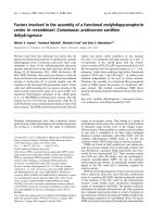
Immunodetection of the Gq/11 a subunit.
Proteins extracted from male and female antennae and
from primary antennal cell culture of M. brassicae were
separated by SDS/PAGE and analysed by Western-blot
using a Gq/11 a antiserum (Fig. 1). Crude homogenates of
male and female antennae contained an immunoreactive
band with an apparent molecular mass of 40 kDa (Fig. 1,
left, A,B). In the sample of primary cell culture of
M. brassicae, a band with the same apparent molecular
weight was labeled by the antiserum, indicating that the
protein is also present in the in vitro cell cultures (Fig. 1,
left, C).
Cloning and cDNA sequencing
A 900-bp cDNA product was amplified with RT-PCR
using degenerate oligonucleotide primers. After cloning
and sequencing, this product was translated and the
deduce amino-acid sequence was compared with sequences
in the GenBank database. This product appeared to be
very similar to a subunits from G proteins belonging to
the Gq family. It was then extended to the 5¢ and the
3¢ untranslated regions by 5¢ and 3¢ RACE, respectively.
This allowed us to obtain the sequence of a full length
Fig. 1. Biochemical detection of Gq/11 a. M, molecular markers. Left
(A,B,C) Coomassie stain after 10% SDS/PAGE of antennal and cell
culture homogenates. (A) Male M. brassicae (4 antennae equivalent),
(B) female M. brassicae (6 antennae equivalent), (C) primary cell cul-
tures of M. brassicae male antennae (15 Petri dishes equivalent to 15
antennae). Right (A,B,C) Western-blot after SDS/PAGE of antennal
and cell culture homogenates using Gq/11 a antiserum (dilution
1 : 1000). The antiserum cross-reacted only with a single band of about
40 kDa in both male (A) and female (B) antennal extracts as well as in
the primary cell culture extracts (C).
Ó FEBS 2002 A lepidopteran Gq protein a subunit (Eur. J. Biochem. 269) 2135
cDNA of 1541 bp (Fig. 2). This sequence has been
deposited in the GenBank database with accession
number AF448447. Nucleotide sequence analysis revealed
that the cDNA contains a putative coding region of
1059 bp, encoding a 353 amino-acid protein with a
theoretical molecular mass of 41 400 Da and an isoelectric
point of 5.35, as determined using
MWCALC
(Infobiogen)
(Fig. 2). There are several upstream ATG codons but
soon followed by stop codons. The ATG at position 199
has a favorable sequence context for translation initiation
[29] and could be proposed to be the start of the protein
coding domain. Sequence analysis of the 3¢ end cDNA
revealed that there is a polyadenylation signal upstream of
the poly(A).
Analysis of the primary structure of
M. brassicae
Gqa
The putative protein product encoded by the cloned cDNA
was aligned with different G proteins from invertebrates
and vertebrates retrieved from blast search (Fig. 3). This
putative protein showed a high degree of identity to other
known Gqa proteins from invertebrates (Drosophila, 87%;
Limulus, 83%; lobster, 85%) but also from vertebrates
(mouse, 80%; human, 80%), and is less similar to other Ga
types (47.5% with Go of the Lepidoptera M. sexta,for
example) (Table 1).
Furthermore, the M. brassicae Gqa subunit exhibits
important characteristics of other Gqa proteins, namely: the
amino-acid sequence G40TGESGKST
FI typical of the
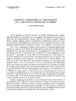
Fig. 2. cDNA and deduced amino-acid
sequence of the M. brassicae Gq a subunit
(GenBank accession number no. AF448447).
The suggested start ATG and stop TGA
codons are in bold italics. Positions of the
primers for the internal amplification are
underlined (solid line), as are gene specific
primers and nested primer for the 3¢ RACE
amplification (dashed lines) and the gene
specific primer used for the 5¢RACE amplifi-
cation (dotted line). Palmitoylation sites
C3C4, G40TGES box and putative cholera
toxin site Arg177 are in boxes.
2136 E. Jacquin-Joly et al. (Eur. J. Biochem. 269) Ó FEBS 2002
A domain, with the characteristic residues underlined [30], a
N-terminal cysteine doublet (Cys3, Cys4) in a MXCC motif
that represent putative sites for palmitoylation [31], a
putative cholera toxin ADP-ribosylation site (Arg177) and a
G40TGES ÔGAG boxÕ sequence that is present in the GTP-
binding domain of other Gqa proteins (Fig. 2).
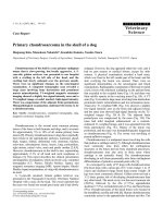
Expression pattern in male antennae
In situ hybridization experiments were performed using
digoxigenin incorporated antisense and sense RNA probes
against adult male antennae. The M. brassicae antenna is
filiform, 1 cm long and comprises about 72 segments [32].
Each segment exhibits the same general organization: the
dorsal side is covered with two rows of scales and the
olfactory hairs (the sensilla) are located on the ventral side
as can be seen using scanning electron microscopy
(Fig. 4A). In males, the olfactory hairs are distributed in
two classes according to their length. The long ones (long
sensilla trichodea) are located on the lateral part of the
ventral area and are arrayed in four to five parallel rows [32]
(Fig. 4A, white arrows). Short sensilla trichodea are located
medio-ventrally and are not arranged in rows.
Sense strand controls gave no signals (not shown)
whereas antisense probe hybridization is restricted to the
sensilla (ventral) side of the antennae (Fig. 4B,E). Close
examination revealed hybridization in cells at the bases of
the sensilla hairs (Fig. 4C,D) and sometimes two labeled
somata can be seen at the base of one sensillum (Fig. 4E).
On longitudinal sections through the antennae, it is difficult
to distinguish between long and short sensilla as only parts
of the sensilla are visible (Fig. 4B,E). However, sections
through the cuticle permitted the observation of labeled
spots distributed in the ventro-lateral region with a row
pattern consistent with the distribution of the long sensilla
trichodea (Fig. 4F, white arrows). Typical structures of
Fig. 3. Alignment using
CLUSTAL W
of G protein a subunit of the q family from different species, including invertebrates and vertebrates. Amino-acid
identities are in bold. Sequences compared to M. brassicae Gqa sequence are from Drosophila melanogaster (GenBank accession numbers M58016;
M30152; U31092), Homarus americanus (U89139), Panulirus argus (AF201328), the mouse Mus musculus (P21279), the dog Canis familiaris
(Q28294) and human (P50148). Several motifs indicative of this Ga family are conserved: N-terminal cysteines, arginine177, and a GAG box.
Ó FEBS 2002 A lepidopteran Gq protein a subunit (Eur. J. Biochem. 269) 2137
sensilla coeloconica, that resemble flowers, can be observed
on sections through the cuticle, without any associated
labeling (Fig. 4G, black arrows), whereas on the same
section other sensilla without any particular distribution are
labeled, that correspond to short sensilla trichodea.
DISCUSSION
Several studies have previously suggested that G-protein-
mediated signal transduction pathway may occur in
pheromone-sensitive receptor cells in insects. For example,
it has been shown that nerve impulse activity of phero-
mone receptor cells increased significantly after G protein-
activating natrium fluoride application to their outer
dendrite in single sensilla trichodea of the moth Bombyx
mori [23]. Additionally, using a Gq/11 antiserum, the
same authors revealed the presence of a protein that is
likely to belong to the Gq family in antennae of both
B. mori and Antheraea pernyi. In this context, we report
here the immunodetection of a protein with the same
characteristics, the molecular cloning of the corresponding
cDNA to get the total amino-acid sequence of the protein,
and the expression pattern of the corresponding mRNA
as a first step to clarify the role of G protein a subunit in
olfaction.
Immunodetection of Gq/11 a subunit in antennae
homogenate and in neuron primary culture
The molecular mass of the immunoreactive band observed
is consistent with the molecular mass of other G protein a
subunits, and may represent the a subunit(s) of one or
several proteins belonging to the Gq/11 family.
The visualization of such proteins in our olfactory cell
culture is consistent with the occurrence of a Gq protein in
antennal primary cell culture already observed in lobster
[17], which mediates excitatory odor transduction in olfac-
tory receptor neurons in this species.
Molecular cloning of a cDNA coding for a Gqa subunit
in male antennae
Because of the strong conservation of the G protein a
subunit throughout evolution, we decided to use the
PCR technique to identify a cDNA encoding M. brass-
icae Gqa-like protein. We then identified a putative
transcript encoding a G protein a subunit homologous to
invertebrate and vertebrate Gqa, suggesting the presence
of a specific Gqa gene in M. brassicae. The molecular
mass of the predicted protein is consistent with that
determined by Western blot after SDS/PAGE (Fig. 1).
Table 1. G protein a subunit characterized in insects, including the Gq of M. brassicae described here, and in some other invertebrate groups. Databank
accession numbers, references and expression pattern/putative role are also given. ORN, olfactory receptor neurons.
Species
G protein a
subunit class
Accession
no. Ref.
% identity with Gq
of M. brassicae Possible function
Insecta
Lepidoptera Mamestra brassicae Gq AF448447 This paper 100 Expression in ORN
Manduca sexta Go Z49080 [24] 47.5 Developmental role in
embryonic neurons
Diptera Drosophila
melanogaster
DGq1 M58016 [54] 81 retinal expression
DGq2 M30152 [55] 81 Expression in nervous
system and ovaries
DGq3
G
U31092
M23094
[15]
[56]
87
48.6
Expression in
chemosensory cells and
central nervous system
Expression in embryos
and pupae
Calliphora vicina G AJ250443 [57] 81.5 Visual protein of the
compound eyes
Orthoptera Locusta migratoria Go A61035 [14] 49 Expressed in
nervous tissues
Crustacea Panulirus argus Gq/11 AF201328 [19] 85 Expression in ORN
Homarus
americanus
Gq U89139 [18] 85 Expression in neurons of
olfactory organs
and brain
Chelicerata Limulus polyphemus Gq U88586 [58] 83 Expression in eyes
Mollusca Patinopecten
yessoensis
Gq AB006456 [59] 80 Expression in visual cells
Lymnaea stagnalis Gq Z23106 [16] 78 Expression in neurons
Octopus vulgaris Gq AB025782 [60] 76 Expression in
photoreceptor cells
Loligo forbesi Gq L10289 [61] 75 Visual G protein a
subunit
Echinodermata Asterina pectinifera G(I) X66378 [62] 59 ?
Demospongiae Geodia cydonium Gq Y14248 [63] 50.1 Oocyte maturation
2138 E. Jacquin-Joly et al. (Eur. J. Biochem. 269) Ó FEBS 2002
The antibody used for the Western-blot is directed to the
decapeptide QSALKEFNLA that corresponds to a
defined C-terminal sequence found in both Gqa and
G
11
a (Calbiochem). The deduced amino-acid sequence
shares high C-terminal identity with this sequence
(QLNLKEYNLV) and thus should have been detected
with such antibodies. Although proteins of this class are
highly conserved in sequence and molecular mass, the
observation of only a single band on the Western blot,
combined with the molecular cloning data, suggests that
the cloned cDNA probably encodes the protein detected
using commercial antibodies.
The predicted protein we obtained shares high identities
with other already known Gq protein a subunits and
therefore can be placed within this family. Indeed,
M. brassicae Gqa possesses all the characteristics observed
in Gqa-like proteins. In particular, the cysteine doublet at
the N-terminal part probably serves as a site for post-
translational attachment of a palmitoyl group (16-carbon,
saturated fatty acid) through a labile, reversible thioester
linkage [33,34], and which could serve as a membrane
anchor.
It is noteworthy that proteins from Gq family are
highly conserved throughout evolution: the putative
M. brassicae Gq sequence is 80% identical to mouse,
dog and human Gq; however, it shares only 47%
identity with a Go sequence from M. sexta, another
lepidopteran (Table 1). In particular, the M. brassicae G
subunit is 87% identical to the dGqa-3 of Drosophila
[15]. It differs only in two domains: 70–130 and the
C-terminal region that is important for receptor interac-
tions [35,36].
Expression pattern in the adult male antennae
In situ hybridization revealed that this G protein subunit is
expressed in both long and short sensilla trichodea
(Fig. 4F,G) in cells that could be neurons because of
several observations. On Fig. 4C, for instance, the labeled
cell is located at the base of the cuticular hair and
protrusions emanating from the soma that could corres-
pond to the dendrite are seen entering the base of sensillum
hair (Fig. 4C). Such protrusions have already been
observed after in situ hybridization in labeled neurons of
M. sexta antennae [37]. Furthermore, two somata can be
seen that are labeled at the base of the same sensilla
(Fig. 4E), possibly corresponding to the two receptor
neurons observed in all sensilla. The shape, size and
position of the stained cells also suggest their identity as
olfactory neurons.
The Gqa appeared to be associated only with sensilla
trichodea, devoted to pheromone reception [32], with no
expression in sensilla coeloconica. These latter structures
have been shown to be involved in plant-related volatile
detection, at least in B. mori [38]. We can then suppose that
although both sensilla types are implicated in olfaction, they
do not express same G protein a subunits, maybe according
to the ligands they are tuned to. Such a phenomenon has
already been observed in the vertebrate vomeronasal organ,
an organ responsible for detecting pheromones. Two
G protein subtypes are selectively activated by different
classes of compounds [39]: some neurons express receptors
encoded by one multigene family and the G protein a
subunit a
i
, whereas some others express receptors encoded
by another multigene family and the G protein a subunit a
o
.
Fig. 4. Expression pattern of M. brassicae G
protein a subunit revealed by in situ hybridiza-
tion to mRNA in longitudinal sections of male
antennae. (A) Scanning electron microscopy of
a male antennae. The ventral surface is cov-
ered by short and long sensilla, the last being
arranged in parallel rows (white arrows). (B,E)
expression of G protein a subunit on the sen-
silla side of the antennae. (C,D) sensilla
trichodea at higher magnification. (F) Section
through the cuticule in the ventro-lateral
region of the antennae showing G protein a
subunit expression in row pattern (white
arrows) consistent with the distribution of the
long sensilla trichodea devoted to pheromone
reception. (G) Sensilla coeloconica are not
labeled (black arrows) whereas the surrounded
sensilla (short sensilla) are labeled. Scale:
(A,B,E,F) 50 lm; (C,D,G) 10 lm.
Ó FEBS 2002 A lepidopteran Gq protein a subunit (Eur. J. Biochem. 269) 2139
Recently, the a subunit of a G protein of the Gq family
has been immunolocalized in olfactory sensilla preparations
of the silkmoth Antherea pernyi [23]. Using immunocytol-
ogy, the authors were able to show that all types of olfactory
sensilla are labeled. However, labeling is not restricted to
sensillar cells and can be observed in auxiliary cells,
epidermal cells and subcuticular extracellular space. This
is not in contradiction with our observations given that the
tools and the organisms used in the two studies are different:
using antibodies they visualized the localization of the
protein whereas by using in situ hybridization to mRNA we
revealed only the expressing cells that are likely to be
olfactory neurons. The immunological study [23] suggests
that Gq plays a role in olfactory signal transduction as long
as the protein predominates in the dendrites of olfactory
receptor cells.
Implication of Gq in lepidoptera olfaction
Gqa have been frequently presumed to play a role in
olfaction in invertebrates. For example, the protein dGqa-3
of Drosophila was detected in the third antennal segment,
maxillary palps, the tip of the proboscis and in the brain
[15]. Some of the immunoreactive cells have been identified
as antennal olfactory neurons, non-neuronal accessory cells,
or gustatory neurons, suggesting that this protein is involved
in olfactory and gustatory responses in Drosophila. Several
studies on lobsters support Gq involvement in odor
transduction in olfactory receptor neurons. An anti-Gq/11
Ig has been shown to selectively block odor-evoked inward
current in voltage-clamped cultured neurons and immuno-
labeled a band of 45-kDa in Western-blot analyses [17].
Similarly, a Gqa protein has been cloned in two lobster
species, Homarus americanus [18] and Panulirus argus [19],
that is expressed in olfactory receptor neurons, suggesting
that one function of Gqa is to mediate olfactory transduc-
tion. In our study, expression of proteins in olfactory
sensilla trichodea, apparently in neurons, leads us to
hypothesize that, in M. brassicae, this G protein subunit is
involved in pheromone reception. In addition, our data
demonstrated that the olfactory organ of this species
expresses a gene that is critical for the phosphoinositide
signaling pathway: the fact that this protein belongs to the
Gqa subunits suggests that a phospholipase C second
messenger pathway may be implicated in transduction of
olfactory signals in lepidoptera. Such a hypothesis has
already been proposed for insects in a variety of species
using kinetics based methodology (reviewed in [40]) and
immunological detection of Gq/11a subunits in antennae
[23]. Additionally, a phospholipase C b and a protein kinase
C, two enzymes involved in the InsP
3
transduction pathway,
were identified by using specific antibodies directed against
molecules involved in intracellular olfactory signalling [13].
The two enzymes were detected after Western blot with
homogenates of isolated pheromone-sensitive sensilla
trichodea, containing no other cellular elements than the
outer dendrites of pheromone receptor neurons. In lobsters,
several recent studies showed that phospholipase C b
mediates olfactory transduction as well [41]. Molecular
evidence for two components of the phosphoinositide
signaling pathway in lobster olfactory receptor neurons
has been provided [19]: a G protein a subunit of the Gq
familyandanInsP
3
-gated channel or an InsP
3
receptor. In
addition, the authors showed that the InsP
3
receptor is
associated with the plasma membrane, suggesting a novel
mechanism for regulating intracellular ions within restricted
cellular compartments of neurons [19]. Interestingly, InsP
3
receptors have also been immunolocalized within the
dendritic membrane of olfactory sensilla of moths [42].
Elevation of InsP
3
and InsP
3
-gated-Ca
2+
influx in phero-
mone-stimulated cell cultured olfactory neurons has also
been shown [43].
Here, we provide molecular evidence that support the
previous findings and the first lepidopteran sequence of a
Gqa subunit.
The Caenorhabditis elegans genome project has revealed
20 genes encoding a-subunits of G proteins, 14 of which are
expressed almost exclusively in subsets of chemosensory
neurons [44,45]. Then it seems likely that this nematode uses
multiple Ga subunits per cell, leading us to hypothesize that
neurons mediating more than one sensory modality can do
so via distinct intracellular pathways [46], each mediating a
particular response to a specific class of chemical stimuli
[47]. However, C. elegans expresses multiple chemosensory
receptors per olfactory neurons, which is not the case in
Drosophila where neurons are likely to express only a single
olfactory receptor gene, although sometimes along with a
broadly expressed receptor of unknown function [48]. In
moths, the lack of any information on putative olfactory
receptors does not permit such considerations. However, it
cannot be excluded that different types of Ga subunits may
be involved in olfactory transduction in Lepidoptera.
Different G proteins are found in specialized tissue and
they have there different functions, although they all share
structural properties such as the heterotrimeric composition
with a, b, c subunits. However, a subunits are distinct
whereas b subunits are quite similar [49]. The similarity of
our a subunit sequence with others implicated in olfactory
transduction further supports our hypothesis that this
subunit is involved in odor transduction cascade in moth
antennae.
Our identification of a Gqa subunit expressed in olfactory
sensilla supports the hypothesis that G-protein-coupled
olfactory receptors are functional in insects. In insects, seven
transmembrane domain proteins coupled to G-protein-
mediated second messenger cascades have been found to
date only in Drosophila [1–3] and Anopheles gambie [50] and
attempts to find similar receptor proteins in other insects
have failed. An olfactory-specific protein (SNMP for
sensory neuron membrane protein) of two transmembrane
domains uniquely expressed in olfactory receptor neurons
has been characterized in the silkmoth A. polyphemus
[51,52] as well as in the moths B. mori, Heliothis virescens
and M. sexta [37]. In this latter species, a second SNMP
homologue was also identified [37,53]. These proteins are
homologous with the CD36 receptor family, which pre-
dominately recognizes proteinaceous ligands. One could
then not exclude a possible role as olfactory receptor,
considering the probable interaction with odorant binding
proteins carrying the odorant molecule to the receptor.
Although no olfactory receptor has been identified in
Lepidoptera, the discovery of a Gqa subunit expressed in
olfactory neurons and sharing high identities with the
olfactory/gustatory Drosophila Gqa subunits suggests that
seven transmembrane domain receptor proteins should exist
in moth antennae and are involved in olfaction.
2140 E. Jacquin-Joly et al. (Eur. J. Biochem. 269) Ó FEBS 2002
ACKNOWLEDGEMENTS
This study was supported by founds from Institut National de la
Recherche Agronomique, including Rosario Maida fellowship during
his stay in Versailles.
REFERENCES
1. Clyne, P.J., Warr, C.G., Freeman, M.R., Lessing, D., Kim, J. &
Carlson, J.R. (1999) A novel family of divergent seven-trans-
membrane proteins: candidate odorant receptors in Drosophila.
Neuron 22, 327–338.
2. Gao, Q. & Chess, A. (1999) Identification of candidate Drosophila
olfactory receptors from genomic DNA. Genomics 60, 31–39.
3. Vosshall, L.B., Amrein, H., Morozov, P.S., Rzhetsky, A. & Axel,
R. (1999) A spatial map of olfactory receptor expression in the
Drosophila antennae. Cell 96, 725–736.
4. Buck, L. & Axel, R. (1991) A novel multigene family may encode
odorant receptors: a molecular basis for odor recognition. Cell 65,
175–187.
5. Stryer, L. & Bourne, H.R. (1986) G proteins: a family of signal
transducers. Annu.Rev.CellDev.Biol.2, 391–419.
6. Neer, E.J. (1995) Heterotrimeric G proteins: organizers of trans-
membrane signals. Cell 80, 249–257.
7. Breer, H., Raming, K. & Krieger, J. (1994) Signal recognition and
transduction in olfactory neurons. Biochim. Biophys. Acta 1224,
277–287.
8. Breer, H., Boekhoff, I. & Tareilus, E. (1990) Rapid kinetics of
second messenger formation in olfactory transduction. Nature
345, 65–68.
9. Boekhoff, I., Seifert, E., Go
¨
ggerle,S.,Lindemann,M.,Kru
¨
ger,
B.W. & Breer, H. (1993) Pheromone-induced second-messenger
signaling in insect antennae. Insect Biochem. Mol. Biol. 23, 757–
762.
10. Kaissling, R.E. & Boekhoff, I. (1993) Transduction in intracellular
messengers in pheromone receptor cell of the moth Antherea
polyphemus. In Sensory Systems of Arthropods (Wiese, K., ed.), pp.
489–502. Birkha
¨
user, Basel.
11. Wegener, J.W., Boekhoff, I., Tareilus, E. & Breer, H. (1993)
Olfactory signaling in antennal receptor neurons of the locust
(Locusta migratoria). J. Insect. Physiol. 39, 153–163.
12. Krieger, J. & Breer, H. (1999) Olfactory reception in Invertebrates.
Science 286, 720.
13. Maida, R., Redkozubov, A. & Ziegelberger, G. (2000) Identifi-
cation of PLCb and PKC in pheromone receptor neurons of
Antheraea polyphemus. Neuroreport 11, 1773–1776.
14. Raming, K., Krieger, J. & Breer, H. (1990) Molecular cloning,
sequencing and expression of cDNA encodage a Go-protein from
insect. Cell Signalling 2, 311–321.
15. Talluri, S., Bhatt, A. & Smith, D.P. (1995) Identification of a
Drosophila G protein alpha subunit (dGq alpha-3) expressed in
chemosensory cells and central neurons. Proc. Natl Acad. Sci.
USA 92, 11475–11479.
16. Knol,J.C.,Ramnatsingh,S.,vanKesteren,E.R.,vanMinnen,J.,
Planta, R.J., van Heerikhuizen, H. & Vreugdenhil, E. (1995)
Cloning of a molluscan G protein alpha subunit of the Gq class
which is expressed differentially in identified neurons. Eur.
J. Biochem. 230, 193–199.
17. Fadool,D.A.,Estey,S.J.&Ache,B.W.(1995)Evidencethat
Gq-protein mediates excitatory odor transduction in lobster
olfactory receptor neurons. Chem. Senses 20, 489–498.
18. McClintock, T.S., Xu, F., Quintero, J., Gress, A.M. & Landers,
T.M. (1997) Molecular cloning of a lobster G alpha (q) protein
expressed in neurons of olfactory organ and brain. J. Neurochem.
68, 2248–2254.
19. Munger,S.D.,Gleeson,R.A.,Aldrich,H.C.,Rust,N.C.,Aches,
B.W. & Greenberg, R.M. (2000) Characterization of a phos-
phoinositide-mediated odor transduction pathway reveales plasma
membrane localization of an inositol 1,4,5-triphosphate receptor
in lobster olfactory receptor neurons. J. Biol. Chem. 27, 20450–
20457.
20. Xu, F., Hollins, B., Gress, A.M., Landers, T.M. & McClintock,
T.S. (1997) Molecular cloning and characterization of a lobster G
alphaS protein expressed in neurons of olfactory organ and brain.
J. Neurochem. 69, 1793–1800.
21. Breer, H., Raming, K. & Boekhoff, I. (1988) G-proteins in the
antennae of insects. Naturwissenschaften 75, 627.
22. Boekhoff, I., Raming, K. & Breer, H. (1990) Pheromone-induced
stimulation of inositol-triphosphate formation in insect antennae
is mediated by G-protein. J. Comp. Physiol. B 160, 99–103.
23. Laue, M., Maida, R. & Redkozubov, A. (1997) G-protein acti-
vation, identification and immunolocalization in pheromone-sen-
sitive sensilla trichodea of moths. Cell Tissue Res. 288, 149–158.
24. Horgan, A.M., Lagrange, M.T. & Copenhaver, P.F. (1995) A
developmental role for the heterotrimeric G protein Go alpha in a
migratory population of embryonic neurons. Dev. Biol. 172, 640–
653.
25. Poitout, S. & Bues, R. (1974) Elevage de 28 espe
`
ces de le
´
pidopte
`
res
noctuidae et de 2 espe
`
ces d’arctiidae sur milieu artificiel simplifie
´
.
Particularite
´
selon les espe
`
ces. Ann. Zool. Ecol. Anim. 6, 431–441.
26. Laemmli, U.K. (1970) Cleavage of structural proteins during the
assembly of the head of bacteriophage T4. Nature 227, 680–685.
27. Lucas, P. & Nagnan-Le Meillour, P. (1997) Primary culture of
antennal cells of Mamestra brassicae: morphology of cell types and
evidence for biosynthesis of pheromone-binding proteins in vitro.
Cell Tissue Res. 289, 375–382.
28. Jacquin-Joly, E., Bohbot, J., Franc¸ ois, M.C., Cain, A.H. &
Nagnan-Le Meillour, P. (2000) Characterisation of the general
odourant-binding protein 2 in molecular coding of odourants in
Mamestra brassicae. Eur. J. Biochem. 267, 6708–6714.
29. Kozak, M. (1989) The scanning model for translation: an update.
J. Cell Biol. 108, 229–241.
30. Halliday, K.R. (1984) Regional homology in GTP-binding proto-
oncogene products and elongation factors. J. Cyclic Nucleotide
Protein Phosphorylation Res. 9, 435–448.
31. Wedegaertner, P.B., Wilson, P.T. & Bourne, H.R. (1995) Lipid
modifications of trimeric G proteins. J. Biol. Chem. 270, 503–506.
32. Renou, M. & Lucas, P. (1994) Sex pheromone reception in
Mamestra brassicae L. (Lepidoptera): responses of olfactory
receptor neurons to minor components of the pheromone blend.
J. Insect Physiol. 40, 75–85.
33. Linder, M.E., Middleton, P., Heppler, J.R., Taussig, R., Gilman,
A.G. & Mumby, S.M. (1993) Lipid modifications of the G pro-
teins: a subunits are palmitoylated. Proc. Natl Acad. Sci. USA 90,
3675–3679.
34. Parenti, M., Vigano
´
,A.,Newman,C.M.H.,Milligan,G.&
Magee, A.I. (1993) A novel N-terminal motif for palmitoylation of
G-protein a subunits. Biochem. J. 291, 349–353.
35. Noel, J.P., Hamm, H.E. & Sigler, P.B. (1993) The 2.2 A
˚
crystal
structure of transducin-a complexed with GTP
c
S. Nature 366,
654–663.
36. Coleman, D.E., Berghuis, A.M., Lee, M.E., Linder, M.E.,
Gilman, A.G. & Sprang, S.R. (1994) Structures of active con-
formations of Gi alpha 1 and the mechanism of GTP hydrolysis.
Science 265, 1405–1412.
37. Rogers, M.E., Krieger, J. & Vogt, R.G. (2001) Antennal SNMPs
(sensory neuron membrane proteins) of Lepidoptera define a
unique family of invertebrate CD36-like proteins. J. Neurobiol. 49,
47–61.
38. Pophof, B. (1997) Olfactory responses record from sensilla
coeloconica of the silkmoth Bombyx mori. Physiol. Entomol. 22,
239–248.
39. Krieger,J.,Schmitt,A.,Lobell,D.,Gudermann,T.,Schultz,G.,
Breer, H. & Boekhoff, I. (1999) Selective activation of a G protein
Ó FEBS 2002 A lepidopteran Gq protein a subunit (Eur. J. Biochem. 269) 2141
subtypes in the vomeronasal organ upon stimulation with urine-
derived compounds. J. Biol. Chem. 274, 4655–4662.
40. Krieger, J., Mameli, M. & Breer, H. (1997) Elements of the
olfactory signaling pathways in insect antennae. Invert. Neurosci.
3, 137–144.
41. Xu, F. & McClintock, T.S. (1999) A lobster phospholipase C-beta
that associates with G-proteins in response to odorants. J. Neu-
rosci. 19, 4881–4888.
42. Laue, M. & Steinbrecht, R.A. (1999) Topochemistry of moth
olfactory sensilla. Int. J. Morph. Embryol. 26, 217–228.
43. Stengl, M. (1994) Inositol-triphosphate-dependent calcium cur-
rents precede cation currents in insect olfactory receptor neurons
in vitro. J. Comp. Physiol. A 174, 187–194.
44. Bargmann, C.I. (1998) Neurobiology of the Caenorhabditis elegans
genome. Science 282, 2028–2033.
45.Jansen,G.,Thijssen,K.L.,Werner,P.,vanderHorst,M.,
Hazendonk, E. & Plasterk, R.H.A. (1999) The complete family of
genes encoding G proteins of Caenorhabditis elegans. Nat. Genet.
21, 414–419.
46. Hart, A.C., Kass, J., Shapiro, J.E. & Kaplan, J.M. (1999) Distinct
signaling pathways mediate touch and osmosensory responses in a
polymodal sensory neuron. J. Neurosci. 19, 1952–1958.
47. Lessing, D. & Carlson, J.R. (1999) Chemosensory behavior:
the path from stimulus to response. Curr. Opin. Neurobiol. 9,
766–771.
48. Chiappe, M.E. & Vosshall, L.B. (2001) A Broadly Expressed
Odorant Receptor Gene in Drosophila. 7th European Symposium
for Insect Taste and Olfaction, September 22–28, Villasimius
(Cagliari), Italy.
49. Gilman, A.G. (1987) G-proteins: transducers of receptor-gener-
ated signals. Annu. Rev. Biochem. 56, 615–649.
50. Fox,A.N.,Pitts,R.J.,Robertson,H.M.,Carlson,J.R.&Zwiebel,
L.J. (2001) Candidate odorant receptors from the malaria vector
mosquito Anopheles gambiae and evidence of down-regulation in
response to blood feeding. Proc.NatlAcad.Sci.USA98, 14693–
14697.
51. Rogers, M.E., Sun, M., Lerner, M.R. & Vogt, R.G. (1997)
SNMP-1, a novel membrane protein of olfactory neurons of
the silkmoth Antheraea polyphemus with homology to the
CD36 family of membrane proteins. J. Biol. Chem. 272, 14792–
14799.
52. Rogers,M.E.,Steinbrecht,R.A.&Vogt,R.G.(2001)Expression
of SNMP-1 in olfactory neurons and sensilla of male and female
antennae of the silkmoth Antheraea polyphemus. Cell Tissue Res.
303, 433–446.
53. Robertson, H.M., Martos, R., Sears, C.R., Todres, E.Z., Walden,
K.K.O. & Nardi, B. (1999) Diversity of odorant binding proteins
revealed by an expressed sequence tag project on male Manduca
sexta moth antennae. Insect Mol. Biol. 8, 501–518.
54. Lee, Y.J., Dobbs, M., Verardi, M.L. & Hyde, D.R. (1990) dGq: a
Drosophila gene encoding a visual system-specific G-alpha mole-
cule. Neuron 5, 889–898.
55. de Sousa, S.M., Hoveland, L.L., Yarfitz, S. & Hurley, J.B. (1989)
The Drosophila G-0-alpha-like G protein gene produces multiple
transcripts and is expressed in the nervous system and in ovaries.
J. Biol. Chem. 264, 18544–18551.
56. Provost, N.M., Somers, D.E. & Hurley, J.B. (1988) A Drosophila
melanogaster G protein alpha-subunit gene is expressed primarily
in embryos and pupae. J. Biol. Chem. 263, 12070–12076.
57. Schulz, S., Huber, A., Schwab, K. & Paulsen, R. (1999) A novel
Gc isolated from Drosophila constitutes a visual G protein c
subunit of the fly compound eye. J. Biol. Chem. 274, 37605–37610.
58. Munger, S.D., Schremser-Berlin, J L., Brink, C.M. & Battelle,
B.A. (1996) Molecular and immunological characterization of a
Gq Protein from ventral and lateral eye of the horseshoe crab
Limulus polyphemus. Invert. Neurosci. 2, 175–182.
59. Kojima,D.,Terakita,A.,Ishikawa,T.,Tsukahara,Y.,Maeda,A.
& Shichida, Y. (1997) A novel Go-mediated phototransduction
cascade in scallop visual cells. J. Biol. Chem. 272, 22979–22982.
60. Iwasa, T., Yanai, T., Nakagawa, M., Kikkawa, S., Obata, S.,
Usukura, J. & Tsuda, M.G. (1999) Protein alpha subunit genes in
octopus photoreceptor cells. Direct GenBank Submission, acces-
sion no. AB025782.
61. Ryba, N.J.P., Findlay, J.B.C. & Reid, J.D. (1993) The molecular
cloning of the squid (Loligo forbesi) visual Gq-alpha subunit and
its expression in Saccharomyces cerevisiae. Biochem. J. 292,333–
341.
62. Chiba, K., Tadenuma, H., Matsumoto, M., Takahashi, K.,
Katada, T. & Hoshi, M. (1992) The primary structure of the alpha
subunit of a starfish guanosine-nucleotide-binding regulatory
protein involved in 1-methyladenine-induced oocyte maturation.
Eur. J. Biochem. 207, 833–838.
63. Seack, J., Kruse, M. & Muller, W.E. (1998) Evolutionary analysis
of G-proteins in early metazoans: cloning of alpha- and beta-
subunits from the sponge Geodia cydonium. Biochim. Biophys.
Acta 1401, 93–103.
2142 E. Jacquin-Joly et al. (Eur. J. Biochem. 269) Ó FEBS 2002