Báo cáo Y học: Functional analysis of the rat bile salt export pump gene promoter Regulation by bile acids, drugs and endogenous compounds potx
Bạn đang xem bản rút gọn của tài liệu. Xem và tải ngay bản đầy đủ của tài liệu tại đây (393.99 KB, 9 trang )
Functional analysis of the rat bile salt export pump gene promoter
Regulation by bile acids, drugs and endogenous compounds
Thomas Gerloff
1
, Andreas Geier
2
, Ivar Roots
1
, Peter J. Meier
3
and Carsten Gartung
2
1
Institute of Clinical Pharmacology, Charite
´
University Medical Center, Humboldt University, Berlin, Germany;
2
Department of Internal Medicine, Aachen University of Technology, Aachen, Germany;
3
Division of Clinical Pharmacology
and Toxicology, Department of Medicine, University Hospital, Zurich, Switzerland
The 5¢ flanking region of the bile salt export pump (Bsep)
gene was systematically analysed to provide the basis for
understanding the mechanisms which regulate Bsep tran-
scription. In addition substrates and drugs were investigated
for their ability to alter Bsep promoter activity. Bsep pro-
moter function was restricted to hepatocyte derived HepG2
cells. The 5¢ deletional analysis revealed a biphasic shape of
reporter gene activities, indicating a suppressive element
between nucleotides )800 and )512. Two consensus sites for
the farnesoid X receptor (FXR) were located at nucleotides
)473 and )64. The latter was characterized as functionally
active in bile acid-mediated feed-back regulation of Bsep
transcription. Bsep promoter activity was reduced by
rifampin and b-estradiol. The anti-estrogen tamoxifen
stimulated promoter activity. Dexamethasone, hydrocorti-
sone and phenobarbital had no effect on Bsep promoter
activity. In conclusion, the data suggest that transcriptional
regulation of the Bsep gene can be modulated by a number of
endogenous compounds and xenobiotics. FXR was a major
regulatory factor, mediating bile acid feed-back stimulation
of Bsep transcription.
Keywords: bile flow; drug-induced cholestasis; transcrip-
tional regulation.
Bile secretion by vertebrate liver is caused by the continuous
vectorial excretion of bile acids and other osmotically active
substrates across the canalicular pole of hepatocytes. Bile
acid-dependent bile flow provides the major driving force for
the generation and maintenance of liver excretory processes
and is therefore essential for proper hepatic clearance of
endogenous compounds and xenobiotics. Canalicular secre-
tion of bile acids is predominantly mediated by the
transmembrane transporter system BSEP (human)/Bsep
(rodents) [1]. Rat Bsep belongs to the superfamily of ATP-
binding cassette (ABC) transporters and is closely related to
P-glycoprotein, the gene product of Mdr1a/1b. The Bsep
gene encodes a 160-kDa polypeptide which is highly, and
almost exclusively, expressed in the canalicular membrane of
hepatocytes. Impairment of the Bsep transporter system
results in cholestasis due either to inherited mutations of its
gene [2], or secondary to dysfunction caused by biliary
obstruction, xenobiotics or systemic inflammation.
Regulation of protein expression of hepatic transport
systems plays an important role in the production of bile in
normal and diseased liver. Major goals in the regulation of
hepatocellular transporters are to prevent intracellular
accumulation of toxic bile acids and to maintain biliary
flow for ongoing hepatic clearance. A differential expression
of the basolateral bile acid uptake system Na
+
-taurocholate
cotransporting polypeptide (Ntcp) and the canalicular Bsep
could clearly be demonstrated in animal models of choles-
tasis and liver regeneration [3,4]. Whereas Ntcp was down-
regulated in these models, Bsep expression was sustained,
thus protecting hepatocytes from damage caused by toxic
bile acids and metabolites. Observations from farnesoid X
receptor (FXR)-deficient mice [5] fed a diet of cholic acid
(CA) demonstrated that FXR is a critical transcription
factor that controls differential expression of the key liver
cell basolateral and canalicular bile acid transporters Ntcp
and Bsep. While CA feeding resulted in a large increase of
Bsep mRNA levels in the livers of FXR wild-type mice, no
increase was observed in FXR-null mice [5]. In contrast
Ntcp mRNA was down-regulated following CA feeding but
remained unchanged in FXR-deficient mice. Recent studies
on the human BSEP and rat Ntcp promoters support this
concept [6,7].
Treatment with drugs frequently results in impairment
of liver function [8]. A variety of mechanisms have been
described to cause drug-induced cholestasis, including
decreased hepatocellular bile secretion [9]. Steroid hormones
like estradiol have been demonstrated to down-regulate
Ntcp and Bsep mRNA levels [3] or to inhibit Bsep transport
function [10]. However, effects of drugs on Bsep promoter
function have not been studied yet.
To analyse systematically the rat Bsep promoter and to
investigate the regulation of Bsep transcription by bile acids,
drugs and endogenous compounds we identified the
5¢ flanking region of the rat Bsep gene and analysed its
nucleotide sequence with respect to putative transcription
Correspondence to T. Gerloff, Institute of Clinical Pharmacology,
Charite
´
, University Medical Center, Humboldt University Berlin,
Schumannstrasse 20/21, D-10098 Berlin, Germany.
Fax: +49 30 94063329, Tel.: +49 30 94062845,
E-mail: thomas.gerloff@gmx.de
Abbreviations: Bsep, bile salt export pump; ABC, ATP-binding
cassette; FXR, farnesoid X receptor; Ntcp, Na
+
taurocholate
cotransporting polypeptide; CA, cholic acid; FXRE, FXR response
element; TC, taurocholic acid, TUDCA, tauroursodeoxycholic acid;
CDCA, chenodeoxycholic acid; DCA, deoxycholic acid;
LCA, lithocholic acid; OATP: organic anion transporting
polypeptide; RXR, retinoid X receptor.
(Received 9 April 2002, accepted 30 May 2002)
Eur. J. Biochem. 269, 3495–3503 (2002) Ó FEBS 2002 doi:10.1046/j.1432-1033.2002.03030.x
factor binding sites. We determined the minimal Bsep
promoter region capable to mediate basal Bsep expression
and provide evidence for a liver-specific function of the Bsep
promoter. Our data demonstrate FXR-mediated bile acid
feed-back regulation of Bsep promoter activity in HepG2
cells transfected with a Bsep-luciferase-reporter gene con-
struct, without any cotransfection of FXR expression
plasmids, as in a previous report [6]. Furthermore the
present study indicates the ability of drugs and endogenous
compounds to affect Bsep transcription, thus altering
hepatic bile flow and clearance.
MATERIALS AND METHODS
Genomic cloning and sequence analysis of the 5¢ flanking
region
We amplified rat genomic DNA and Bsep cDNA by PCR
using the oligonucleotide primers 5¢-AACTGTTCTGGT
GTGGATTCC-3¢ and 5¢-ATAGAAGATCTCTTGGTC
CTG-3¢ designed from the known rat Bsep cDNA clone [1].
As the two PCR products had the same length these
oligonucleotide primers were used for screening a rat P1
genomic DNA library (Genome Systems) by PCR. Each
strand of the 5¢ flanking region of the Bsep gene was directly
sequenced (Epidauros, Bernried, Germany) by automated
sequencing. The sequence was analysed for putative
cis-acting regulatory elements by using the transcription
factor database
TRANSFAC
4.0 (MatInspector V2.2, http://
transfac.gbf.de).
Construction of plasmids
Aseriesof5¢ deletions of the flanking region ranging from
nucleotides )1453 to )27 of upstream sequence and
extending downstream to nucleotide +80 were created by
PCR using a genomic P1 Bsep clone as a template. The
forward primers were designed with a 5¢ flanking MluI
restriction site and the reverse primers contained a 5¢ flank-
ing BglII site. The PCR amplicons were cloned into the
EcoRV site of the pMOSBlue vector (Amersham Pharmacia
Biotech) and subsequently excised with MluIandBglII. The
DNA fragments were then cloned directionally into the
MluI–BglII sites of the pGL3-Basic (Promega) luciferase
reporter gene vector. The plasmid constructs were verified
by sequencing. Mutations of the FXR response element
(FXRE) adjacent to the TATA box in the rat Bsep
promoter were generated using the Quickchange
TM
Site-
Directed Mutagenesis Kit (Stratagene). The deletion plas-
mid extending to nucleotide )126 served as a template. An
antisense (5¢-CACTGTTTGCTTATATTTCAATGGAA
TAAAGTCCAGCTCTAGC-3¢; exchanged bases in bold)
and sense (5¢-GCTAGAGCTGGACTTTATTCCATT
GAAATA-TAAGCAAACAGTG-3¢) oligonucleotide of
the IR-1 element was used in a temperature cycling reaction
as described in the manufacturer’s protocol to produce the
mutated plasmid m-126.
Cell culture, transient transfections and reporter gene
assays
Human hepatoblastoma HepG2 (HB-8065, ATCC), colon
carcinoma CaCo2 (ACC169, DSMZ), Madin–Darby
canine kidney (MDCK; Dr Birchmeier, Max Delbru
¨
ck
Center of Molecular Medicine, Berlin, Germany) and
mouse fibroblast NIH 3T3 cells (Dr Blankenstein, Max
Delbru
¨
ck Center of Molecular Medicine, Berlin, Germany)
were cultured in Dulbecco’s modified Eagle medium
containing 10% fetal bovine serum, 1% nonessential amino
acids, 1 mmolÆL
)1
sodium pyruvate, and 2 mmolÆL
)1
glutamine. Cells were transferred to six-well plates at
50–60% confluency and incubated at 37 °C24hpriorto
transient transfections. Using Tfx
TM
-20 (Promega) as
cationic liposome 2.7 lg of reporter gene construct and
0.3 lg of the control reporter gene plasmid pRL-TK
(Promega) were cotransfected. The DNA–liposome mixture
was removed after 2 h, and cells were incubated with
Dulbecco’s modified Eagle medium containing 10% fetal
bovine serum for an additional 48 h. Cells were lysed, and
cell extracts were assayed for luciferase activities in a Turner
Designs TD-20/20 luminometer (Promega) using the Dual
luciferase assay system (Promega). Relative reporter gene
activities were expressed as the ratio of the firefly luciferase
activity (reporter) and the renilla luciferase (transfection
control) activity.
Mapping of the transcriptional start site
Two 5¢ RACE products were isolated from 1 lgtotalRNA
from rat liver using gene-specific oligonucleotide primers
corresponding to nucleotides 29–56 and 150–177 of the Bsep
cDNA sequence [1] and subcloned into the pMosBlue
vector (Amersham Pharmacia Biotech). Based on the
sequence of the 5¢ RACE products an antisense oligonucle-
otide extending 100 bp from nucleotide 62 of the Bsep
cDNA was radiolabeled at the 5¢endwith[c-
32
P]ATP
(Ambion, AMS Biotechnology, Germany) to perform an
S1-nuclease assay for the exact localization of the transcrip-
tion initiation site. Total RNA from rat liver (100 lg) was
hybridized with the labelled antisense probe at 42 °Cfor2 h
(Ambion, Germany) and subsequently digested with S1
nuclease for 30 min at 37 °C. The remaining DNA–RNA
hybridized fragments were electrophoresed on a 7% acryl-
amide sequencing gel. Sequencing reactions of the
M13mp18 cloning vector were run in parallel and used as
a size marker. After transferring the gel to 3 MM Whatman
paper the dried gel was scanned using a phosphorimager
(Raytest, Germany).
EMSA
Preparation of nuclear extracts and EMSAs were performed
as described previously [11]. Protein concentrations were
determined according to Bradford [12]. Nuclear extracts
(5–10 lg protein) were incubated on ice for 30 min
with a specific
32
P-end-labeled oligonucleotide probe
(2 · 10
4
c.p.m.) in a 20-lL reaction containing 8 lLwater,
4 lL5· binding buffer (25 m
M
Hepes pH 7.6, 50 m
M
KCl,
0.5 m
M
dithiothreitol, 5 m
M
MgCl
2
,0.5m
M
EDTA, 10%
glycerol) and 2 lg poly(dI-dC)-poly(dI-dC) (Amersham).
For competition assays, 100-fold molar excess of specific
unlabeled over labeled oligonucleotides were added to the
binding reaction. Samples were electrophoresed through a
nondenaturing 6% polyacrylamide gel. A double-stranded
oligonucleotide containing the putative FXRE of the
Bsep promoter sequence was used (sense strand sequence
3496 T. Gerloff et al. (Eur. J. Biochem. 269) Ó FEBS 2002
5¢-GACTTTAGGCCATTGACCTATAAG-3¢). For su-
pershift experiments nuclear extracts were preincubated
for 30 min on ice with 1 lg of polyclonal antibodies (Santa
Cruz) either against FXRa,RXRa or both prior to addition
of the labelled oligonucleotide probe.
Statistical analysis
All values are given as mean ± SD of triplicate transfec-
tions. Student’s t-test was used to compare promoter
activities with controls. P values < 0.05 were considered
to be statistically significant.
RESULTS
Determination of the transcriptional start site
of the
Bsep
gene
The transcription initiation site of the Bsep gene was located
by both 5¢ RACE amplification and S1 nuclease digestion.
Two oligonucleotides corresponding to nucleotides 29–56
and 150–177 of the published Bsep cDNA [1] were used for
RACE. Sequencing of the RACE products revealed that the
amplified 5¢ flanking regions started at nucleotide 6 and
nucleotide 8 of the cDNA sequence, respectively. To exactly
map the transcription start site an oligonucleotide extending
100 bp from nucleotide 62 of the Bsep cDNA and including
the start region of the RACE products was subsequently
used in an S1 nuclease digestion assay. Two protected
fragments of 73 and 64 bp were observed with total RNA
from rat liver (Fig. 1). The larger fragment gave the
strongest signal intensity and was thus used to designate
nucleotide +1 in the Bsep promoter sequence. No protected
fragments were identified using yeast tRNA as template
(data not shown).
Analysis of the 5¢ flanking region sequence
of the
Bsep
gene
A total of 2488 bp upstream from the 5¢-end of the Bsep
cDNA was sequenced in both directions; 1583 bp are
shown in Fig. 2. Identity with the Bsep cDNA sequence
begins at nucleotide +96. Several consensus matches for
potential transcription factor binding sites were identified
by searching the TRANSFAC 4.0 transcription factor-
binding site database (
MATINSPECTOR V
2.1). General DNA
elements containing motifs for a TATA box at nucleotide
)52, a CAAT box at nucleotide )66, multiple octamer
binding sites (nucleotides )708, )684, )667, )417, )396
and )197), and several NF1 sites (nucleotides )407, )237,
)211 and )73) could be detected. Multiple binding sites
for the liver enriched transcription factor HNF3b were
found at nucleotides )718, )685, )661, )632 and )567.
Interestingly there was only one HNF1 site (nucleotide
)839), and no consensus elements were found for the liver
enriched factor HNF4. Multiple AP1 sites are noted at
nucleotides )468, )447, )347, )338, )277, )243, )169,
)98 and ) 59. Three consensus motifs for C/EBP-b
binding elements are located at nucleotides )200, )582
and )757. Strikingly two binding sites for the FXR/9-cis-
retinoic acid receptor heterodimer at nucleotides )64 and
)473 could be identified. These motifs are comprised of
two inverted repeats separated by one nucleotide (IR-1)
and have recently been demonstrated to function as bile
acid responsive elements [13].
5¢ Deletional analysis of the Bsep promoter
in transfected HepG2 cells
The 5¢ flanking regions of the rat Bsep promoter capable
of conferring basal activity was assessed by a series of
deletion constructs cloned into the pGL3–luciferase
reporter plasmid. The Bsep promoter sequence inserted
in reverse orientation served as a negative control. HepG2
cells were transfected with the reporter-plasmids and
luciferase activities were determined relative to the level
of renilla luciferase cotransfected by the expression vector
pRL-TK (Fig. 3). Luciferase activity with the longest
construct (p-1453) was set 100%. Removal of the region
between nucleotides )1453 and )800 resulted in a
dramatic reduction of promoter activity by 90%. How-
ever, further deletion of the sequence to nucleotide )187
increased the activity to a maximum of 170%. At
Fig. 1. Determination of the transcriptional start site. In a nuclease
protection assay, an antisense oligonucleotide extending 100 bp from
nucleotide 62 of the Bsep cDNA was hybridized with total RNA from
rat liver and subsequently digested by S1 nuclease. The major pro-
tected fragment displayed in lane S (arrow) had a size of 73 bp. The
minor protected fragment was 64 bp in length. Lanes A, C, G and T
are sequence reactions of the M13mp18 cloning vector used as a size
marker.
Ó FEBS 2002 Rat Bsep promoter analysis (Eur. J. Biochem. 269) 3497
nucleotide )126 the luciferase activity reached nearly the
same levels as the p-1453 construct. When the deletion
was extended further to nucleotide )27, the values
dropped to basal levels obtained with the promoterless
pGL3-Basic vector (data not shown) or with the construct
in reverse orientation (Fig. 3). The minimal element
maintaining full promoter activity was identified as the
construct extending from nucleotide )126 to nucleotide
+80.
Functional analysis of the Bsep promoter in cell lines
We investigated the tissue specificity of the Bsep promo-
ter activity by transfecting several nonhepatic cell lines
with the reporter constructs p-1453, p-187, p-126 and
p+80–1453. In contrast with HepG2 cells, no luciferase
reporter gene activation was observed with any of the
plasmids in NIH 3T3 or MDCK cells (Fig. 4). Only
slight activation with the constructs p-187 and p-126
was observed in CaCo2 cells. Thus, the full length
(p-1453) as well as the minimal (p-126) promoter
constructs were able to mediate liver restricted luciferase
reporter expression.
Effect of bile acids on Bsep promotor function
in HepG2 cells
Bile acid dependancy of rat Bsep promoter activity was
analysed by transfecting the full length (p-1453) and the
minimal (p-126) promotor constructs into HepG2 cells in
the presence of various bile acids (Fig. 5). Expressed
luciferase activities were higher with the minimal p-126
(Fig. 5B) as compared with the full length p-1453
(Fig. 5A) promoter construct. While most of the uncon-
jugated bile acids exerted positive effects on Bsep
reporter activity, the taurine conjugated bile salts tauro-
cholic acid and tauroursocholic acid (TUDCA) had
virtually no effect. The strongest reporter gene activation
occurred with the primary bile acid chenodeoxycholic
acid (CDCA) in p-126 transfected cells (230 ± 18% of
controls). This CDCA-mediated stimulation of luciferase
activities was concentration dependent for both reporter
Fig. 2. Nucleotide sequence of the 5¢ flanking
region of the rat Bsep gene. A total of 2488 bp
has been sequenced. For better understanding
only part of this sequence is presented.
Nucleotides are numbered relative to the
major transcription initiation site (nucleotide
+1; arrow). Potential binding sites for
cis-acting elements are underlined with their
names indicated above. The TATA motif is
boxed. The minor transcription start site at
nucleotide +10 is marked by an asterisk.
The complete nucleotide sequence of the Bsep
promoter is available in the GenBank and
EMBL databases under the accession number
AF452071.
3498 T. Gerloff et al. (Eur. J. Biochem. 269) Ó FEBS 2002
gene constructs. Weaker activation of the Bsep promoter
was observed with the secondary bile acids deoxycholic
acid (DCA) and lithocholic acid (LCA) with maximal
effects in p-126 transfected cells at 100 l
M
(170 ± 12%)
and 50 l
M
(160 ± 11%), respectively. The taurine-con-
jugated dihydroxylated bile acids taurodeoxychenocholic
acid (130 ± 5%, p-126) and taurochenodeoxycholic acid
(135 ± 6%, p-126) exhibited only slight stimulations of
the Bsep promoter, and the trihydroxylated conjugates
taurocholic acid (98 ± 10%, p-126) and tauroursode-
oxycholic acid (90 ± 12%, p-126) had no stimulatory
effects at all. Mutation of the FXRE-motif immediately
upstream of the TATA box in the p-126 minimal
promoter construct completely abolished the stimulation
of luciferase activity by CDCA in HepG2 cells (Fig. 6).
Interestingly CDCA reduced the activity of the
mutant m-126 Luc even below that of the wild-type
p-126 Luc.
Analysis of the binding affinity of the putative
FXRE site for the orphan nuclear receptor FXR
To characterize the FXRE site identified in the Bsep
promoter EMSA were carried out using an oligonucleotide
corresponding to the nucleotide sequence of the first FXRE
Fig. 3. Deletional analysis of the rat Bsep promoter activity in trans-
fected HepG2 cells. Varying lengths of the 5¢ region of the Bsep gene,
from )1453 to )27 bp relative to the transcription start site and
extending to nucleotide +80, were amplified by PCR using the
genomic clone as a template and then inserted into the promoterless
luciferase vector pGL3 basic. The plasmid p+80–1453 containing the
same nucleotide sequence in antisense orientation served as a control.
The constructs were transiently cotransfected with a renilla luciferase
expression plasmid (pRL-TK) into HepG2 cells as described in
Materials and methods. Luciferase activity of each construct was
determined as relative light units of firefly luciferase per relative light
units of renilla luciferase (luc/ren). All values were expressed relative to
the longest construct p-1453, which was assigned 100%. Transfections
were carried out in triplicate, and repeated three times. Data are
the means ± 1 SD.
Fig. 4. Functional analysis of the Bsep promotor in cell lines. The
constructs p-1453, p-187, p-126 and p+80–1453 were cotransfected
with pRL-TK into four different cell lines given on the left of the panel.
Activities are expressed as relative light units of luciferase activity per
relative light units of renilla luciferase activity. Transfections were
carried out in triplicate. Data are expressed as means ± SD of three
individual experiments.
Fig. 5. Effect of bile acids on Bsep promotor function in HepG2 cells.
(A) Reporter gene activity after transfection in triplicate with p-1453,
the longest construct containing all upstream regulatory elements.
Cells were subsequently incubated for 48 h with the indicated bile acids
at the concentrations stated in Materials and methods. Promotor
activities were determined relative to untreated controls which were set
100%. Data are the means ± SD of at least three individual experi-
ments. (B) Transfection with p-126, the minimal Bsep promoter con-
taining one FXRE close to the TATA motif. Incubation with bile acids
and assays of promotor activities were carried out as in (A).
*P <0.05.
Ó FEBS 2002 Rat Bsep promoter analysis (Eur. J. Biochem. 269) 3499
element immediately adjacent to the TATA box (Fig. 7).
The labelled probe was incubated with nuclear extracts from
rat liver. A specific slowly migrating complex was formed
(lanes 1 and 2). No complex could be detected in the
presence of excess unlabelled oligonucleotide as specific
competitor (lane 9) whereas formation of the complex was
unaffected by excess of a nonspecific competitor (lane 10).
Addition of either a specific antibody against the
nuclear receptor FXR (lanes 3 and 4), or against retinoid
X receptor (RXR) (lanes 5 and 6) or a combination of both
(lanes 7 and 8) resulted in a reduction of signal intensities of
the specific band. Supershifted bands were rather weak and
only detectable after overexposure of the autoradiograph.
These findings are consistent with the observation that for
transcriptional regulation FXR together with RXR form a
heterodimer that subsequently binds to FXRE elements of
bile acid-sensitive genes.
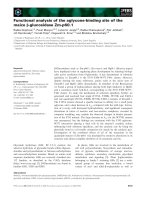
Regulation of Bsep promoter function by drugs
The ability of drugs to regulate Bsep gene transcription was
assessed by transfection of the full length construct p-1453
into HepG2 cells and subsequent treatment of these cells
with various compounds. The promoter activity was
significantly reduced by rifampin (77 ± 7%) and b-estra-
diol (77 ± 6%) (Fig. 8). In contrast, the estrogen antag-
onist tamoxifen induced Bsep promoter activity (134 ±
15%). No significant effect was observed following treat-
ment with the steroids dexamethasone (98 ± 9%) and
hydrocortisone (91 ± 13%) or with the narcotic pheno-
barbital (111 ± 24%).
DISCUSSION
Understanding mechanisms which preserve continuous bile
flow under physiologic and cholestatic conditions requires
knowledge of Bsep promoter function. Recently a sophis-
ticated network has been revealed comprised of orphan
nuclear receptors as feed-back regulators for the synthesis,
hepatocellular uptake and excretion of bile acids [6,7,13–15].
Furthermore transcriptional regulation of Bsep might play a
critical role in drug-induced cholestasis. Thus, we present
the nucleotide sequence, as well as a systematic structural
and functional analysis of the 5¢ flanking region of the Bsep
gene.
The 5¢ deletional analysis of the Bsep promoter revealed a
region from nucleotide )126 to nucleotide )27 providing
basal activity at similar levels as compared with the longest
reporter construct extending to nucleotide )1453 upstream
from the transcription start site. The former region was
Fig. 7. Binding activity of hepatic nuclear extracts to an oligonucleotide
containing the FXRE. Hepatic nuclear extracts were prepared from
untreated rats. Nuclear extracts (5–10 lg protein) were incubated with
a radiolabelled oligonucleotide containing the FXRE binding site,
electrophoresed through a 6% nondenaturing polyacrylamide gel and
autoradiographed. For supershift experiments nuclear extracts were
preincubated with polyclonal antibodies against either FXRa (lane 3
and 4), RXRa (lane 5 and 6) or both (lane 7 and 8) prior to incubation
with the specific oligonucleotides. Samples represented in lanes 9 and
10 were incubated in the presence of unlabelled specific (SC) and
nonspecific (NSC) competitor DNA at 100-fold molar excess.
Fig. 6. Decrease of basal activity and loss of CDCA-mediated stimu-
lation of the minimal Bsep promoter after mutation of the FXRE. The
wild-type (p-126) or the mutated (m-126) minimal Bsep reporter
plasmid were transfected into HepG2 cells, as described in Fig. 5.
Luciferase activities were determined following a 48 h incubation in
the presence or absence of 100 l
M
CDCA. *P <0.05.
Fig. 8. Effects of endogenous substrates and drugs on Bsep promoter
function. The full length Bsep reporter plasmid (p-1453) was trans-
fected into HepG2 cells and luciferase activities were measured after a
48 h incubation period in the presence of the indicated compounds.
The following substrate concentrations were used: 5 lmolÆL
)1
each of
dexamethasone, hydrocortisone, and b-estradiol; 50 lmolÆL
)1
each of
phenobarbital, rifampin, and tamoxifen. Assay conditions were the
same as described in Fig. 5. *P < 0.05.
3500 T. Gerloff et al. (Eur. J. Biochem. 269) Ó FEBS 2002
therefore designated the minimal Bsep promoter, appar-
ently containing all binding sites required for basal
transcription. The minimal promoter and larger constructs
were only functional in the human hepatoblastoma derived
cell line HepG2, indicating a liver-specific activity. Trans-
acting factors directing liver-specific gene expression include
a set of liver-enriched factors, such as HNF1, HNF3b,
HNF4, and C/EBPb [16,17]. Among these factors only a
putative binding site for HNF3-b overlapping with the
TATA box of transcription initiation could be found within
the minimal Bsep promoter. Additional consensus motifs of
the minimal promoter include three AP1 sites and a
CCAAT box. As opposed to the apparent importance of
HNF1a in liver-specific expression of the basolateral
transporters Ntcp and organic anion transporting polypep-
tide (OATP)-C [11,18] and other genes, including cyto-
chromes P450 [19], albumin and a
1
-antitrypsin [20] a
consensus motif for HNF1a could not be detected in the
minimal rat Bsep promoter. Basal transcription of the major
canalicular organic anion exporter Mrp2, another member
of hepatocellular ABC transporters, was also not dependent
on HNF1 [21]. Thus, minimal promoter activity and liver-
specific transcription of canalicular ABC transporters
appear not to require HNF1 but seem to be controlled by
other factors.
Further upstream of the minimal promoter several
putative binding sites for liver enriched and ubiquitously
expressed transcription factors were located. The function
of the general DNA elements, including sites for the
octamer binding proteins, NF1 and SP1 need further
evaluation. Of note were five HNF3-b motifs in close
vicinity between nucleotide )718 and nucleotide )550
preceeded by a HNF1a site. In this study liver specificity
of Bsep transcription was not dependent upon the combined
action of HNF1 and HNF3 as was reported for the human
glucose transporter type 2 isoform gene [22]. Therefore the
close distribution of these sites seems to play a different role,
e.g. in developmental regulation of Bsep expression.
Interestingly, the 5¢ deletional analysis resulted in repor-
ter gene activities that were distributed in a bimodal manner
(Fig. 3). Peak luciferase activities were obtained with the
constructs extending to nucleotide )1453 and nucleotide
)187, respectively, while transfection of the p-800 plasmid
resulted in < 10% relative promoter activity but rapidly
increased with further progressive deletions. This suggests
the influence of a strong inhibitory cis-acting element
located between nucleotide )800 and nucleotide )512.
Among known inhibitory consensus motifs, including AP-2,
PuF, CREB and Ets, only a MyoD site was detected
between nucleotide )797 and nucleotide )787 that could
potentially mediate a repressive effect on Bsep gene
transcription. Apart from its function as an activator in
skeletal muscle differentiation [23] MyoD has been des-
cribed as an inhibitor of cell proliferation [24] and repressor
of the myogenic HLH Myf-5 gene expression [25].
Bile secretion and enterohepatic circulation of bile acids
are critically dependent on two key transport systems of
hepatocytes: The Ntcp (sodium-taurocholate cotransport-
ing polypeptide) mediates basolateral uptake of bile acids
whereas Bsep mediates their excretion into the biliary tract.
There is good evidence from studies on animal models that
these transporters are inversely regulated in response to
cholestatic conditions and during liver regeneration [3,4]
with a pattern of diminished Ntcp expression and sustained
or even up-regulated Bsep expression. The purpose of this
regulatory scheme is to protect hepatocytes against accu-
mulation of toxic bile acids by preventing their further
uptake and preserving or enhancing their excretion. The
discovery of bile acids as ligands of the orphan liver receptor
FXR [14] led to the concept of FXR-mediated regulation of
Ntcp and Bsep. Indeed, studies in mice, deficient for FXR
clearly demonstrated a failure to appropriately adjust Ntcp
and Bsep transcription after feeding with cholic acid [5] and
thus these mice rapidly developed liver injury. While Ntcp
down-regulation by bile acid-bound FXR was mediated
indirectly requiring the additional action of a small
heterodimeric partner [7], binding of FXR to an IR-1
element within the 5¢ flanking region of the human BSEP
gene was recently confirmed [6]. In the present study we
found a similar IR-1 element at an identical position
(nucleotide )52 to nucleotide ) 64) in the rat Bsep promoter
and demonstrated binding of the RXRa/FXR heterodimer
to this element in an electrophoretic retardation assay. In
both species the IR-1 motifs were adjacent to or in the case
of the rat even overlapping with a TATA consensus
sequence. Furthermore a binding site for HNF3-b or
HNF-5 was also located immediately nearby in the rat and
human Bsep promoter, respectively. The conserved local-
ization within a similar surrounding indicates the functional
importance of the IR-1 element in mammalian Bsep/BSEP
expression and suggests its interaction with members of the
HNF transcription factor family and general factors of the
transcription initiation complex. In addition, the IR-1
element obviously contributes to basal activity of the Bsep
minimal promoter, because mutagenesis of this site signifi-
cantly reduces the relative reporter gene activity to 80% of
wild-type controls (Fig. 6). The intrinsic bile acid synthesis
in HepG2 cells [26], namely of CDCA and CA further
supports this concept. The full length Bsep promoter was
stimulated by a number of conjugated and unconjugated
bile acids in HepG2 cells. The primary bile acid CDCA was
the most potent stimulator, followed by DCA and LCA. As
expected from the localization of the IR-1 element stimu-
lation of reporter gene activity could also be observed with
the minimal promoter extending 126 bp upstream of the
transcription initiation site. Using this construct stimulation
of luciferase activities by the unconjugated bile acids CDCA
and DCA was clearly concentration dependent. In contrast
treatment with LCA, a hydrophobic secondary bile acid
resulted in lower promoter activities above 50 l
M
, due to its
known cytotoxic effects [27]. Mutation of the IR-1 element
within the minimal promoter resulted in a loss of CDCA
reporter stimulation, indicating the functional importance
of this binding site for bile acid-mediated Bsep regulation.
Although conjugated bile acids are the predominant form
found in bile [28] they were either weak stimulators of the
Bsep minimal promoter (taurodeoxychenocholic acid) or
even failed to enhance reporter activity (TC, TUDCA).
Previous studies showed that conjugated bile acids could
only activate FXR after they have been transported into the
cells by a membrane carrier [14]. As HepG2 cells express
the human liver organic anion transporting polypeptide
OATP-A (previously called OATP) in their plasma mem-
brane they are capable of taking up taurin-conjugated bile
acids [29] that can bind to FXR. The weak Bsep promoter
stimulation by conjugated bile acids could be explained by
Ó FEBS 2002 Rat Bsep promoter analysis (Eur. J. Biochem. 269) 3501
lower intracellular concentrations compared with their
conjugated forms, despite the carrier-mediated uptake.
Drug-induced cholestasis is a common phenomenon in
medical treatment. The molecular mechanisms of drug-
induced liver injury include impairment of hepatocellular
bile secretion, obstructive cholangiolitis and sclerosing
cholangitis [30]. Bile secretion is dependent on the amount
of Bsep expressed in hepatocellular canalicular membranes.
As substrates, such as b-estradiol and rifampin inhibit Bsep
promoter activity they are capable of causing cholestasis by
the reduction of canalicular bile acid transport capacity. In
contrast with b-estradiol the anti-estrogen tamoxifen stimu-
lated Bsep promoter activity (Fig. 7). Estrogen binds to
intracellular receptor proteins that subsequently act on
target genes either by binding to-specific estrogen response
elements or by stimulating the activity of factors of the AP1
complex [31,32]. Both elements are located on the Bsep
promoter and could therefore potentially be involved in
estrogen-mediated modification of Bsep gene transcription.
In analogy to the human choline acetyltransferase and the
lipoprotein lipase gene the inhibition of the Bsep promoter
activity by estrogens may also be mediated by AP1 or AP1-
like recognition sites [33,34].
In summary, we have identified the rat Bsep gene
promoter and characterized its activity in different cell lines.
Bsep promoter function was restricted to hepatocyte-
derived HepG2 cells. Comparative sequence analysis of
the 5¢ flanking region of the rat Bsep gene revealed binding
sites for liver enriched and ubiquitously expressed tran-
scription factors. We have located FXRE consensus sites
and demonstrated the functional importance of the FXRE
immediately upstream of the TATA box in bile acid
negative feed-back regulation. There is evidence for differ-
ential modulation of Bsep gene transcription by various
compounds. Bsep promoter analysis in the presence of
drugs could serve as a useful tool in predicting drug-induced
cholestasis caused by impaired gene transcription.
ACKNOWLEDGEMENTS
Supported in part by grants from the Deutsche Forschungsgemein-
schaft to T.G. (Ge 812/2-1) and C.G. (SFB 542, Teilprojekt C1).
REFERENCES
1. Gerloff, T., Stieger, B., Hagenbuch, B., Madon, J., Landmann, L.,
Roth, J., Hofmann, A.F. & Meier, P.J. (1998) The sister of
P-glycoprotein represents the canalicular bile salt export pump of
mammalian liver. J. Biol. Chem. 273, 10046–10050.
2. Strautnieks, S.S., Bull., L.N., Knisely, A.S., Kocoshis, S.A., Dahl,
N., Arnell, H., Sokal, E., Dahan, K., Childs, S., Ling, V., Tanner,
M.S., Kagalwalla, A.F., Nemeth, A., Pawlowska, J., Baker, A.,
Mieli-Vergani, G., Freimer, N.B., Gardiner, R.M. & Thompson,
R.J. (1998) A gene encoding a liver-specific ABC transporter is
mutated in progressive familial intrahepatic cholestasis. Nat.
Genet. 20, 233–238.
3. Lee, J.M., Trauner, M., Soroka, C.J., Stieger, B., Meier, P.J. &
Boyer, J.L. (2000) Expression of the bile salt export pump is
maintained after chronic cholestasis in the rat. Gastroenterology
118, 163–172.
4. Gerloff, T., Geier, A., Stieger, B., Hagenbuch, B., Meier, P.J.,
Matern, S. & Gartung, C. (1999) Differential expression of
basolateral and canalicular organic anion transporters during
regeneration of rat liver. Gastroenterology 117, 1408–1415.
5. Sinal,C.J.,Tohkin,M.,Miyata,M.,Ward,J.M.,Lambert,G.&
Gonzales, F.J. (2000) Targeted disruption of the nuclear receptor
FXR/BAR impairs bile acid and lipid homeostasis. Cell 102,
731–744.
6. Ananthanarayanan, M., Balasubramanian, N., Makishima, M.,
Mangelsdorf, D.J. & Suchy, F.J. (2001) Human bile salt export
pump promoter is transactivated by the farnesoid X receptor/bile
acid receptor. J. Biol. Chem. 276, 28857–28865.
7. Denson, L.A., Sturm, E., Echevarria, W., Zimmerman, T.L.,
Makishima, M., Mangelsdorf, D.J. & Karpen, S.J. (2001) The
orphan nuclear receptor, shp, mediates bile acid-induced inhibi-
tion of the rat bile acid transporter, Ntcp. Gastroenterology. 121,
140–147.
8. Kaplowitz, N. (2001) Drug-induced liver disorders: implications
for drug development and regulation. Drug Safety 24, 483–490.
9. Kullak-Ublick, G.A. & Meier, P.J. (2000) Mechanisms of
cholestasis. Clin. Liver Dis. 4, 357–385.
10. Stieger, B., Fattinger, K., Madon, J., Kullak-Ublick, G.A. &
Meier, P.J. (2000) Drug- and estrogen-induced cholestasis through
inhibition of the hepatocellular bile salt export pump (Bsep) of rat
liver. Gastroenterology 118, 422–430.
11. Karpen, S.J., Sun, A.Q., Kudish, B., Hagenbuch, B., Meier, P.J.,
Ananthanarayanan, M. & Suchy, F.J. (1996) Multiple factors
regulate the rat liver basolateral sodium-dependent bile acid
cotransporter gene promoter. J. Biol. Chem. 271, 15211–15221.
12. Bradford, M.M. (1976) A rapid and sensitive method for the
quantitation of microgram quantities of protein utilizing the
principle of protein-dye binding. Anal. Biochem. 72, 248–254.
13. Goodwin, B., Jones, S.A., Price, R.R., Watson, M.A., McKee,
D.D., Moore, L.B., Galardi, C., Wilson, J.G., Lewis, M.C., Roth,
M.E., Maloney, P.R., Willson, T.M., Koller, B.H. & Kliewer, S.A.
(2000) A regulatory cascade of the nuclear receptors FXR, SHP-1,
and LRH-1 represses bile acid biosynthesis. Mol. Cell 6, 517–526.
14. Parks, D.J., Blanchard, S.G., Bledsoe, R.K., Chandra, G.,
Consler, T.G., Kliewer, S.A., Stimmel, J.B., Willson, T.M.,
Zavacki, A.M., Moore, D.D. & Lehmann, J.M. (1999) Bile
acids: natural ligands for an orphan nuclear receptor. Science 284,
1365–1368.
15. Chiang, J.Y.L., Kimmel, R., Weinberger, C. & Stroup, D. (2000)
Farnesoid X receptor responds to bile acids and represses cho-
lesterol 7a-hydroxylase gene (CYP7A1) transcription. J. Biol.
Chem. 275, 10918–10924.
16. Cereghini, S. (1996) Liver-enriched transcription factors and
hepatocyte differentiation. FASEB J. 10, 267–282.
17. Xanthopoulos, G. & Mirkovitch, J. (1993) Gene regulation
in rodent hepatocytes during development, differentiation and
disease. Eur. J. Biochem. 216, 353–360.
18. Jung, D., Hagenbuch, B., Gresh, L., Pontoglio, M., Meier, P.J. &
Kullak-Ublick, G.A. (2001) Characterization of the human
OATP-C (SLC21A6) gene promoter and regulation of liver-
specific OATP genes by hepatocyte nuclear factor 1 alpha.
J. Biol. Chem. 276, 37206–37214.
19. Padgham, C.R., Boyle, C.C., Wang, X.J., Raleigh, S.M., Wright,
M.C. & Paine, A.J. (1993) Alteration of transcription factor
mRNAs during the isolation and culture of rat hepatocytes sug-
gests the activation of a proliferative mode underlies their ded-
ifferentiation. Biochem. Biophys. Res. Commun. 197, 599–605.
20. Courtois, G., Morgan, J.G., Campbell, L.A., Fourel, G. &
Crabtree, G.R. (1987) Interaction of a liver-specific nuclear factor
with the fibrinogen and alpha 1-antitrypsin promoters. Science
238, 688–692.
21. Kauffmann, H.M. & Schrenk, D. (1998) Sequence analysis and
functional characterization of the 5¢-flanking region of the rat
multidrug resistance protein 2 (MRP2) gene. Biochem. Biophys.
Res. Commun. 245, 325–331.
22. Cha,J.Y.,Kim,H.,Kim,K.S.,Hur,M.W.&Ahn,Y.(2000)
Identification of transacting factors responsible for the tissue-
3502 T. Gerloff et al. (Eur. J. Biochem. 269) Ó FEBS 2002
specific expression of human glucose transporter type 2 isoform
gene. J. Biol. Chem. 275, 18358–18365.
23. Weintraub, H., Dwarki, V.J., Verma, I., Davis, R., Hollenberg, S.,
Snider, L., Lassar, A. & Tapscott, S.J. (1991) Muscle-specific
transcriptional activation by MyoD. Genes Dev. 5, 1377–1385.
24. Crescenzi, M., Fleming, T.P., Lassar, A.B., Weintraub, H. &
Aaronson, S.A. (1990) MyoD induces growth arrest independent
of differentiation in normal and transformed cells. Proc. Natl
Acad.Sci.USA87, 8442–8446.
25. Rudnicki, M.A., Braun, T., Hinuma, S. & Jaenisch, R. (1992)
Inactivation of MyoD in mice leads to up-regulation of the
myogenic HLH gene Myf-5 and results in apparently normal
muscle development. Cell 71, 383–390.
26. Levy, J., Budai, K. & Javitt, N.B. (1994) Bile acid synthesis in
HepG2 cells: effect of cyclosporin. J. Lipid Res. 35, 1795–1800.
27. Takikawa, H., Tomita, J., Takemura, T. & Yamanaka, M. (1991)
Cytotoxic effect and uptake mechanism by isolated rat hepato-
cytes of lithocholate and ist glucuronide and sulfate. Biochim.
Biophys. Acta 1091, 173–178.
28. Matoba, N., Une, M. & Hoshita, T. (1986) Identification of
unconjugated bile acids in human bile. J. Lipid Res. 27, 1154–1162.
29. Kullak-Ublick, G.A., Beuers, U. & Paumgartner, G. (1996)
Molecular and functional characterization of bile acid transport
in human hepatoblastoma HepG2 cells. Hepatology 23, 1053–
1060.
30. Erlinger, S. (1997) Drug-induced cholestasis. J. Hepatol. 26, 1–4.
31. Webb, P., Lopez, G.N., Uht, R.M. & Kushner, P.J. (1995)
Tamoxifen activation of the estrogen receptor/AP-1 pathway:
potential origin for the cell-specific estrogen-like effects of anties-
trogens. Mol. Endocrinol. 9, 443–456.
32. Webb, P., Nguyen, P., Valentine, C., Lopez, G.N., Kwok, G.R.,
McInerney, E., Katzenellenbogen, B.S., Enmark, E., Gustafsson,
J.A., Nilsson, S. & Kushner, P.J. (1999) The estrogen receptor
enhances AP-1 activity by two distinct mechanisms with different
requirements for receptor transactivation functions. Mol.
Endocrinol. 13, 1672–1685.
33. Schmitt, M., Bausero, P., Simoni, P., Queuche, D., Geoffroy, V.,
Marschal, C., Kempf, J. & Quirin-Stricker, C. (1995) Positive and
negative effects of nuclear receptors on transcription activation by
AP-1 of the human choline acetyltransferase proximal promoter.
J. Neurosci. Res. 40, 152–164.
34. Homma,H.,Kurachi,H.,Nishio,Y.,Takeda,T.,Yamamoto,T.,
Adachi, K., Morishige, K., Ohmichi, M., Matsuzawa, Y. &
Murata, Y. (2000) Estrogen suppresses transcription of lipoprotein
lipase gene. Existence of a unique estrogen response element on the
lipoprotein lipase promoter. J. Biol. Chem. 275, 11404–11411.
Ó FEBS 2002 Rat Bsep promoter analysis (Eur. J. Biochem. 269) 3503