Báo cáo Y học: A functional role of the membrane-proximal extracellular domains of the signal transducer gp130 in heterodimerization with the leukemia inhibitory factor receptor pot
Bạn đang xem bản rút gọn của tài liệu. Xem và tải ngay bản đầy đủ của tài liệu tại đây (385.9 KB, 11 trang )
A functional role of the membrane-proximal extracellular domains
of the signal transducer gp130 in heterodimerization
with the leukemia inhibitory factor receptor
Andreas Timmermann, Andrea Ku¨ ster, Ingo Kurth, Peter C. Heinrich and Gerhard Mu¨ ller-Newen
Institut fu
¨
r Biochemie, Rheinisch-Westfa
¨
lische Technische Hochschule Aachen, Germany
gp130 is the common signal transducing receptor subunit of
interleukin (IL)-6-type cytokines. gp130 either homodimer-
izes in response to IL-6 and IL-11 or forms heterodimers
with the leukemia inhibitory factor (LIF) receptor (LIFR) in
response to LIF, oncostatin M (OSM), ciliary neurotrophic
factor (CNTF), cardiotrophin-1 (CT-1) or cardiotrophin-
like cytokine resulting in the onset of cytoplasmic tyrosine
phosphorylation cascades. The extracellular parts of both
gp130 and LIFR consist of several Ig-like and fibronectin
type III-like domains. The role of the membrane-distal
domains of gp130 (D1, D2, D3) and LIFR in ligand binding
is well established. In this study we investigated the func-
tional significance of the membrane-proximal domains of
gp130 (D4, D5, D6) in respect to heterodimerization with
LIFR. Deletion of each of the membrane-proximal domains
of gp130 (D4, D5andD6) leads to LIF unresponsiveness.
Replacement of the gp130 domains by the corresponding
domains of the related GCSF receptor either restores weak
LIF responsiveness (D4-GCSFR), leads to constitutive
activation of gp130 (D5-GCSFR) or results in an inactive
receptor (D6-GCSFR). Mutation of a specific cysteine in D5
of gp130 (C458A) leads to constitutive heterodimerization
with the LIFR and increased sensitivity towards LIF
stimulation. Based on these findings, a functional model of
the gp130–LIFR heterodimer is proposed that includes
contacts between D5 of gp130 and the corresponding
domain D7 of the LIFR and highlights the requirement for
both receptor dimerization and adequate receptor orienta-
tion as a prerequisite for signal transduction.
Keywords: cytokines; receptors; signal transduction; leuke-
mia inhibitory factor; gp130.
Secretion of mediators by cells that are recognized by
specific receptors on target cells is a basic mechanism of
intercellular communication. The molecular mechanism by
which binding of the ligand to the receptor on the plasma
membrane leads to the onset of cytoplasmic signal trans-
duction cascades has gained considerable attention during
recent years. In the case of receptors that span the
membrane only once, ligand induced receptor dimerization
has been accepted as the main mechanism for receptor
activation [1]. Only recently, several reports suggested that
some receptors may exist as preformed dimers or multimers
that switch from an inactive to an active conformation upon
ligand binding [2,3].
Hematopoietic cytokine receptors [4] consist of an
extracellular part, a single transmembrane region, and a
cytoplasmic part that is devoid of any intrinsic enzymatic
activity but constitutively associates with tyrosine kinases of
the Janus kinase (Jak) family. Upon ligand binding the
associated Jaks become activated by transphosphorylation
and phosphorylate tyrosine residues in the cytoplasmic part
of the receptor. These phosphotyrosines serve as docking
sites for signalling molecules that, in most cases, also
become phosphorylated. Most importantly, STAT (signal
transducer and activator of transcription) factors are
recruited to the receptor, dimerize upon phosphorylation
and translocate into the nucleus to induce expression of
target genes [5].
Based on the architecture of the extracellular part,
hematopoietic cytokine receptors can be subdivided into
two groups. The extracellular parts of short cytokine
receptors like erythropoetin recepter (EpoR), growth
hormone receptor (GHR), prolactinR, IL-2Rb or IL-4R
consist of only a single cytokine binding module (CBM).
The CBM is made up of two fibronectin type III-like
(FNIII) domains containing some characteristic conserved
motifs in their primary structures. Several structures of
CBMs of short cytokine receptors bound to their ligands
have been solved showing that in the active receptor dimer
the membrane-proximal domains are juxtaposed in a well-
defined orientation [6,7].
The extracellular parts of complex cytokine receptors like
gp130, LIFR, leptinR or GCSFR contain at least one CBM
and additional FNIII- and Ig-like domains. The cytokine
receptor gp130 consists of an Ig-like domain (D1), followed
by a CBM (D2, D3) and three FNIII-like domains (D4, D5,
and D6) (Fig. 1) [8]. The role of the membrane-distal
domains (D1–D3) in ligand binding has been well estab-
lished by functional and structural studies. In response
Correspondence to G. Mu
¨
ller-Newen, Institut fu
¨
r Biochemie,
Rheinisch-Westfa
¨
lische Technische Hochschule Aachen,
Pauwelsstr. 30, D-52057 Aachen, Germany.
Fax: + 49 241 8082428, Tel.: + 49 241 8088860,
E-mail:
Abbreviations: CBM, cytokine binding module; FNIII, fibronectin
type III-like; GCSF, granulocyte colony stimulating factor; GH,
growth hormone; IL, interleukin; Jak, Janus kinase; LIF, leukemia
inhibitory factor; OSM, oncostatin M; STAT, signal transducer and
activator of transcription.
Note: a web site is available at
(Received 28 February 2002, accepted 18 April 2002)
Eur. J. Biochem. 269, 2716–2726 (2002) Ó FEBS 2002 doi:10.1046/j.1432-1033.2002.02941.x
to cytokines like IL-6 and IL-11 that lead to gp130
homodimerization, two different epitopes of gp130 are
involved in ligand binding: the Ig-like domain and the CBM
[9–11]. In the case of LIF-induced heterodimerization of
LIFR with gp130, the cytokine first binds to the Ig-like
domain of the LIFR [12,13]. gp130 is recruited to the LIF–
LIFR complex via its CBM without involvement of its
Ig-like domain [14,15]. The cytokine oncostatin M (OSM)
first binds to gp130 and then induces heterodimerization
with LIFR [12] or OSMR [16].
Besides the CBM and Ig-like domains both gp130 and
LIFR share three further membrane-proximal FNIII-like
domains as a common structural feature [17]. In a previous
study we established a functional role for each of the
membrane-proximal domains of gp130 for receptor activa-
tion in response to ligands that lead to gp130 homodime-
rization. We proposed a particular role for D5 of gp130 in
respect to proper receptor spacing and orientation [18]. In
this study, using gp130 deletion mutants, point mutants and
chimeric receptors, the role of the individual membrane-
proximal domains of gp130 in heterodimerization with the
LIFR is evaluated. A cysteine to alanine mutation in D5 of
gp130 in combination with the LIFR leads to a weak
constitutive activity and an elevated response to stimulation
with LIF. A model for the gp130/LIFR interaction is
proposed, in which D5 of gp130 contacts domain 7 of the
LIFR.
MATERIALS AND METHODS
Enzymes, proteins, antibodies, chemicals,
and cell culture media
Enzymes were purchased from Roche (Mannheim, Ger-
many) and protein A–Sepharose was obtained from
Amersham (Freiburg, Germany). Fugene was obtained
from Roche (Mannheim, Germany). DMEM and antibi-
otics were obtained from Life Technologies (Eggenstein,
Germany); fetal bovine serum was provided by Seromed
(Berlin, Germany). [a-
32
P]deoxyATP was purchased from
Hartmann Analytic (Braunschweig, Germany). Human
rIL-6 was expressed in Escherichia coli, refolded, and
purified as described by Arcone et al.[19].Thespecific
activity was 10
8
units per mg of protein in the B9 cell
proliferation assay [20]. Soluble IL-6R (sIL-6R) [21] was
expressed in insect cells as described previously. The gp130
mAbs B-P4 and B-P8 and the LIFR mAb 10B2 were
generated as described elsewhere [22,23]. The polyclonal
LIFR antiserum sc-659 was obtainded from New England
Biolabs (Frankfurt/Main, Germany). All other Abs were
purchased from Dako (Hamburg, Germany). NaCl/P
i
buffer contained 200 m
M
NaCl, 2.5 m
M
KCl, 8 m
M
Na
2
HPO
4
,and1.5m
M
KH
2
PO
4
.
Cell culture
BaF3-cells, a murine pro-B lymphocyte line, were cultured in
RPMI 1640 containing 10% fetal bovine serum, 100 mgÆL
)1
streptomycin, 60 mgÆL
)1
penicillin and 5% conditioned
medium from X63Ag-653 BPV-mIL-3 myeloma cells as a
source of IL-3. Simian monkey kidney cells (COS7) were
cultured in DMEM supplemented with 10% fetal bovine
serum, 100 mgÆL
)1
streptomycin, and 60 mgÆL
)1
penicillin.
Cells were grown at 37 °C in a water-saturated atmo-
sphere at 5% CO
2
. BaF3 transfectants were cultured in the
presence of 0.5 lgÆmL
)1
hygromycin if transfected with the
LIFR expression vector pSBC1/2-LIFR/Hygro and
1mgÆmL
)1
G418 if transfected with a pSVL-gp130-expres-
sion vector together with pSV2-Neo.
All cells were regularly checked for the absence of
mycoplasma infection using PCR detection of mycoplasma
DNA.
Plasmid construction
Construction of gp130 wild-type and domain mutant
expression vectors D4, D5, D6 and D5 has been described
elsewhere [18]. The domain exchange mutants gp130 D4
and D6 were cloned analogously after amplifying the DNA
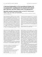
Fig. 1. Schematic representation of gp130, gp130c and LIFR. The
predicted structural organizations of gp130, gp130c and LIFR are
shown. Black lines in the CBM indicate conserved cysteine residues,
black bars the WSXWS motifs. The Ig-like domains and the mem-
brane-proximal FNIII domains are labelled. The extracellular domains
of gp130 and LIFR are numbered from domain 1 (D1) to domain 6
(D6) or domain 1 (D1) to domain 8 (D8), respectively. In the cyto-
plasmic part of gp130c, the amino-acid residues following the Jak
binding sites (box1 and box2, gray boxes) were replaced by the strongly
and specifically STAT1-activating motif YDKPH of the interferon-c
receptor.
Ó FEBS 2002 Heterodimerization of gp130 with LIFR (Eur. J. Biochem. 269) 2717
encoding domains 4 and 6 of GCSFR using the oligo-
nucleotides: 5¢-ACTACCGAACGGGCC
CCCGGGGTC
AGACTGGACACATGG-3¢ and 5¢-TCGGGCCATGGC
ATG
CCCGGGGGTCAGAGCTGGG-3¢ for amplifica-
tion of D4 of GCSFR and 5¢-TACTCTCAAGAAATG
CCCGGGTCCCATGCCCCAGAG-3¢ and 5¢-GCCCAG
GATGATGTGTAGCTC
CCCGGGCTCTGGGGTCAA
GGT-3¢ for D6 of GCSFR (the XmaI sites are underlined)
as PCR primers. Starting point for cloning of gp130 C458A,
C466A and C491A was the full length human gp130 cDNA
cloned into the XhoIandBamHI site of the eukaryotic
expression vector pSVL lacking the EcoRI site (gp130-
pSVLDEco). Using this vector as template, for each point
mutant two fragments were amplified. In a first reaction, the
DNA was amplified using the primer pSVL(sense) and an
antisense primer containing the mutation. A second PCR-
fragment was generated using the primers pSVL(antisense)
and a sense primer with the corresponding mutation. These
fragments were isolated, mixed and served as templates for a
fusion PCR using the primers pSVL(sense) and pSVL(anti-
sense). The reaction products were digested with the
restriction enzymes Xho IandBstEII and cloned into the
expression vector gp130-pSVLDEco. The primers used for
the PCR reactions were: pSVL(sense) 5¢-GTGTTACTT
CTGCTCT-3¢; pSVL(antisense) 5¢-TCTAGTTGTGGTT
TGT-3¢; C458A(sense) 5¢-ATACTTGAGTGGGCTGTG
TTATCAG-3¢; C458A(antisense) 5¢-ATCTGATAACAC
AGCCCACTCAAGTAT-3¢; C466A(sense) 5¢-GATAAA
GCACCCGCTATCACAGACTGG-3¢; C466A(antisense)
5¢-CCAGTCTGTGATAGCGGGTGCTTTATCTG-3¢;
C491A(sense) 5¢-GCAGAGAGCAAAGCCTATTTGAT
AACAG-3¢ and C491(antisense) 5¢-TGTTATCAAATAG
GCTTTGCTCTCTG-3¢.
PCRs were performed applying standard procedures. All
plasmids were sequenced using an ABI Prism Automated
sequencer (Applied Biosystems).
The full-length human LIFR cDNA was cloned into
pSBC-1 to yield the mammalian expression vector pSBC-
LIFR as previously described [15]. For the transfection of
BaF3-cells, the bicistronic expression vector pSBC1/2-
LIFR/Hygro was used [15,24].
Transfection of cells
Plasmid DNA was transfected into BaF3-cells by electro-
poration. Thirty micrograms of the bicistronic LIFR
expression vector pSBC1/2-LIFR-Hygro were electropo-
rated into 3.5 · 10
6
cells in 0.8 mL medium applying a
single 70-ms pulse at 200 V. Selection with hygromycin
(0.5 mgÆmL
)1
) was initiated 24 h after transfection. Selected
BaF3 clones were screened for the presence of membrane-
bound LIFR proteins by flow cytometry. For transfection
of gp130 constructs, 28 lg of the gp130 expression vector
were coelectroporated with 2 lg of pSV2neo as described
above. Selection with G418 (3 mgÆmL
)1
) was initiated 24 h
after transfection. For transfection, either untransfected
BaF3-cells or cells previously transfected with pSBC1/2-
LIFR-Hygro were used. Selected BaF3 clones were screened
for the presence of membrane-bound gp130 proteins by
flow cytometry.
COS7 cells were transiently transfected using the Fugene
method. Efficiency of transfection was analysed by flow
cytometry.
Flow cytometry
Cells were collected, washed and resuspended in cold NaCl/
P
i
containing 5% fetal bovine serum and 0.1% sodium
azide. Subsequently, cells were incubated on ice with
10 lgÆmL
)1
gp130antibodiesB-P4orB-P8or10lgÆmL
)1
LIFR antibody 10B2. Cells were washed with cold NaCl/P
i
/
azide and incubated with R-phycoerythrin-conjugated anti-
(mouse IgG) Fab-fragment at a 1 : 50 dilution. Again, cells
were washed with cold NaCl/P
i
/azide and then resuspended
in 400 lLNaCl/P
i
/azide followed by flow cytometry
analysis using a FACScalibur (Beckton Dickinson).
Electrophoretic mobility shift assay (EMSA)
Cells were incubated at 37 °C for 15 min in the presence of
IL-6/sIL-6R, LIF, OSM or left unstimulated. BaF3-cells
were stimulated with 25 ngÆmL
)1
IL-6 and 1 lgÆmL
)1
sIL-
6R or 50 ngÆmL
)1
LIF or 50 ngÆmL
)1
OSM. COS7 cells
were stimulated with 12.5 ngÆmL
)1
IL-6 and 500 ngÆmL
)1
sIL-6R, 20 ngÆmL
)1
LIF and 4 ngÆmL
)1
OSM. Where
indicated, cells were preincubated for 2 h in the presence of
500 l
M
2-mercaptoethanol prior to stimulation. Prepar-
ation of nuclear extracts and EMSAs were performed as
described previously [25]. A double stranded size-inducible
element (SIE) oligonucleotide derived from the c-fos
promoter (m67SIE; 5¢-GATCCGGGAGGGATTTACGG
GGAAATGCTG-3¢) was used as
32
P-labeled probe [26].
The protein–DNA complexes were separated on a 4.5%
polyacrylamide gel containing 7.5% glycerol. The electro-
phoresis was performed using 0.25 · NaCl/Tris/borate
buffer at 20 VÆcm
)1
.
Coimmunoprecipitation of LIFR/gp130 complexes
Transiently transfected COS7 cells were stimulated for
15 min with IL-6/sIL-6R, LIF or OSM as described or left
unstimulated. Where indicated, cells were preincubated for
2 h in the presence of 500 l
M
2-mercaptoethanol prior to
stimulation. Immediately after stimulation, cells were washed
twice with ice-cold NaCl/P
i
containing 100 l
M
vanadate.
After addition of 600 lL lysis buffer (10% glycerol, 0.25%
Brij-96, 50 m
M
Tris/HCl, 50 l
M
Na
3
VO
4
,100l
M
EDTA,
1m
M
phenylmethanesulfonyl fluoride, 1 mgÆL
)1
aprotinin,
1mgÆL
)1
leupeptin, pH 8.0) the cells were collected and lysed
for 30 min in a microcentrifuge tube. The lysate was
centrifuged for 1 min at 3000 r.p.m. in an Eppendorf
centrifuge and the supernatant was transferred into a new
centrifuge tube. Following incubation of the lysate with
1.6 lg sc-659 antiserum for 12 h at 4 °C15mgproteinA–
sepharose was added. After incubation for 12 h at 4 °C, the
complexes were washed twice with NaCl/Tris/borate/Non-
idet P40 buffer, resuspended in Laemmli-buffer, incubated at
95 °C for 5 min and separated on a 7% SDS polyacrylamide
gel under reducing conditions followed by electroblotting.
Immunoblotting and enhanced
chemiluminescence (ECL) detection
Immunoprecipitated proteins separated by SDS/PAGE
were transferred to a poly(vinylidene difluoride) membrane
by a semidry electroblotting procedure [27]. Poly(vinylidene
difluoride) membranes were blocked in a solution of 20 m
M
2718 A. Timmermann et al. (Eur. J. Biochem. 269) Ó FEBS 2002
Tris/HCl (pH 7.6), 137 m
M
NaCl, 0.1% Nonidet-P40
containing 10% bovine serum albumin and probed with
antibody, followed by incubation with horseradish peroxi-
dase conjugated secondary antibody. Immunoreactive pro-
teins were detected by chemiluminescence using the ECL-kit
(Amersham, UK) following the manufacturer’s instruc-
tions.
RESULTS AND DISCUSSION
Each of the three membrane-proximal domains
of gp130 is required for signal transduction
in response to LIF and OSM
To investigate the role of the membrane-proximal FNIII-
domains of gp130 in signal transduction through hetero-
dimeric complexes with the LIFR, mutants of gp130, in
which single FNIII-domains are deleted were generated
lacking either D4 (gp130-D4), D5 (gp130-D5) or D6 (gp130-
D6) [18]. These gp130 mutants were coexpressed with the
LIFR in different cell types. The STAT-activation after
stimulation with the cytokines IL-6, LIF or OSM was used
as a measure of signal transduction through the analysed
complexes.
Cells of the murine pre-B cell line BaF3 do not express
endogenous gp130 or LIFR. After stably transfecting these
cells with the respective cDNAs, cell surface expression of
both receptors was detected. After stable transfection of the
deletion constructs gp130D4, gp130D5orgp130D6 together
with the LIFR expression vector in BaF3-cells, the surface
expressions of both receptors were similar to those detected
for wild-type gp130/LIFR transfected cells (Fig. 2A, upper
panel).
After stimulation of these cells with IL-6/sIL-6R, none of
the analysed mutants showed a STAT activation similar to
wild-type receptors (Fig. 2A, lower panel, right). This
confirms the previously reported inactivity of the deletion
mutants in response to the gp130-homodimerizing cytokine
IL-6 [18]. Interestingly, also the formation of active hetero-
dimers with wild-type LIFR in response to LIF or OSM is
strongly reduced or abolished by deletion of individual
membrane-proximal domains of gp130. Thus, in BaF3-cells,
each of the membrane-proximal domains of gp130 is
necessary for the efficient formation of a signal transducing
heterodimeric complex of gp130 and the LIFR.
To ensure that the measured receptor activation after
cytokine stimulation does not depend on the analysed
cellular environment, the deletion mutants were expressed
together with the LIFR in COS7 cells. In a previous report
[15], we established a system that allows the study of gp130
mutants together with the LIFR in COS7 cells despite the
presence of low amounts of endogenous wild-type receptors.
To achieve this, the cytoplasmic tyrosine motifs of gp130
that predominantly recruit STAT3 were replaced by the
STAT1 recruiting motif of the interferon-c receptor result-
ing in a chimeric protein designated gp130c.Inorderto
investigate the role of the membrane-proximal domains of
gp130 in receptor activation, the FNIII-domain deletions
were introduced into the gp130c-construct (gp130-D4c,
gp130-D5c or gp130-D6c) [18]. Each of these constructs was
cotransfected with the LIFR into COS7 cells. Enhanced
receptor surface expression was detected by flow cytometry
(Fig. 2B, left panel).
Transfected cells were stimulated with IL-6/sIL-6R, LIF
or OSM and subsequently activation of STAT1 was
analysed by EMSA (Fig. 2B, right panel). In mock-trans-
fected cells only a weak response to IL-6/sIL-6R and OSM
was observed due to endogenously expressed receptors
resulting in a low-level activation of mainly STAT3. Cells
transfected with gp130c showed an increased STAT1
response to IL-6, and less pronounced to OSM. The LIF
response remained unchanged. Transfection of LIFR alone
did not significantly change the sensitivity of the cells to any
of the cytokines. Transfection of both gp130c and LIFR led
to a prominent response of the cells to all three cytokines.
No STAT1-activation after cytokine stimulation could be
detected when one of the gp130c deletion constructs was
coexpressed with the LIFR. In COS7 cells under conditions
of receptor overexpression, as in BaF3-cells, each of the
membrane-proximal domains of gp130 is necessary for the
formation of signal transducing heterodimeric complexes of
gp130 and LIFR.
Two explanations for this finding can be discussed. The
first is based on the identical domain architecture of LIFR
and gp130 in the membrane-proximal six domains. This is
likely to result in the same distance between the cell surface
and the ligand-binding epitopes of both receptors. Deletion
of a single domain in the membrane-proximal part of gp130
leads to a shift of the receptor areas involved in ligand
binding closer to the membrane, resulting in the inability of
the receptor chains to form an active receptor dimer.
Additionally, the membrane-proximal domains can act as
contact sites between the signal transducing receptor chains
or can permit the signal competent conformation of gp130
homo- or heterodimers by adjusting a defined position
towards each other. Thus, deletion of a membrane-proximal
domain of gp130 may be without consequence on ligand
binding but lead to a larger distance or a twist of the
cytoplasmic parts of the receptors responsible for signal
transduction.
Replacement of single membrane-proximal
FNIII-domains of gp130 by corresponding domains of
GCSFR leads to different effects on signal transduction
To investigate, if the function of the membrane-proximal
domains of gp130 is limited to ensure the correct spacing
between the CBM and the membrane, each of the domains
was replaced by the corresponding domain of the GCSFR.
The replacement is assumed to compensate for the shift of
the ligand-binding epitopes of the receptors. The domain
architecture of GCSFR is identical to that of gp130;
moreover these receptors share 46% sequence homology.
The gp130 constructs with exchanged individual FNIII-
domains were introduced into the gp130c-construct result-
ing in the mutants gp130D4c and gp130D6c, respectively.
The construction of the corresponding mutant gp130D5c
has been previously described [18].
Upon co-expression of the gp130 chimeras with the LIFR
in COS7 cells, both receptors were expressed on the cell
surface as detected by flow cytometry in amounts similar to
those of gp130 wild-type (data not shown). Signal trans-
duction was measured by STAT1 activation in an EMSA
(Fig. 3). The exchange of individual membrane-proximal
FNIII-domains of gp130 by the corresponding domains of
the GCSFR resulted in a complex signal transduction
Ó FEBS 2002 Heterodimerization of gp130 with LIFR (Eur. J. Biochem. 269) 2719
pattern. Cells transfected with gp130D5c together with the
LIFR showed a prominent STAT1-activation independ-
ently of stimulation. This activation was not enhanced by
stimulation with cytokines. These results are in line with
observations previously made in COS7 cells transfected
with the gp130D5c-construct alone [18]. When coexpressed
2720 A. Timmermann et al. (Eur. J. Biochem. 269) Ó FEBS 2002
with the LIFR, the gp130D6c chimera showed no
STAT1-activation after stimulation with cytokines. After
stimulation with IL-6/sIL-6R or OSM, cells that express
LIFR together with gp130D4c showed no STAT1-activa-
tion. In contrast, these cells showed a pronounced STAT1
activation upon stimulation with LIF.
In the heterodimeric LIFR/gp130 receptor system,
different demands are posed to the individual FNIII
domains for signal transduction. Because domain 4 of
gp130 can be replaced by a similar domain of a different
receptor without abrogation of signal transduction, this
points to a spacer role of the domain. Intriguingly, signal
transduction of the D4-GCSFR chimera occurs only after
stimulation with LIF, while after OSM stimulation no
STAT activation can be found in the transfected cells. In
previous experiments [15] we were able to show that
different epitopes in the gp130 CBM are required for after
LIF- and OSM-induced STAT activation. The difference in
signal transduction of the D4-GCSFR chimera after
stimulation with LIF and OSM could point to further
epitopes positioned C-terminally to the CBM that play
specific roles in activation of the receptor by these two
cytokines.
The ligand independent activation of the D5-GCSFR
chimera occurs in both the absence and the presence of
LIFR with identical intensities (compare with [18]). This
suggests that the constitutive activation of this mutant
receptor is due to the formation of homomeric gp130D5
complexes without involvement of LIFR. The observation
of constitutive gp130 activation after replacement of D5 led
to the proposal of a model for gp130 activation [18]. In this
model, D5 is the site for direct contact of two gp130
molecules.
Analysis of dimerization of gp130 mutants:
constructs lacking domains 5 or 6 do not
heterodimerize with the LIFR in response to LIF
In addition to the ability of an extracellular receptor mutant
to transduce a signal, its propensity to form a complex with
a second ligand binding or signal transducing receptor chain
is a substantial point to judge its biological activity. To
investigate the LIF dependence and structural requirements
for dimerization of the signal transducing receptor chains
gp130 and LIFR, a coprecipitation assay was established.
The amounts of cell surface expressed receptors in stably
transfected BaF3-cells were too small to achieve a copre-
cipitation of gp130 with LIFR independently of the lysis
buffer used for the disintegration of the cells (data not
shown). Because of the high expression levels of the
transiently transfected receptors in COS7 cells, they were
used for the analysis of dimerization between LIFR and
gp130 or gp130 mutants (Fig. 4A). In mock-transfected
COS7 cells, a weak coprecipitation of LIFR and gp130
could be detected after stimulation of the cells with LIF
(lane 2). This is due to the endogenous expression of both
receptor chains on these cells. After transfection of both
Fig. 3. STAT1 activation in COS7 cells transiently transfected with
LIFR and either gp130D4c, gp130D5c or gp130D6c in response to
various cytokines. Forty-eight hours after transfection cells were sti-
mulated as described in Fig. 1B as indicated. Nuclear extracts were
prepared and activated STAT1 homodimers were detected by EMSA.
A representative of three independent experiments is shown.
Fig. 2. STAT activation in cells expressing gp130 deletion mutants in response to various cytokines. (A) STAT activation in BaF3-cells stably
transfected with LIFR and either gp130, gp130D4, gp130D5 or gp130D6 in response to various cytokines. (Upper panel) Cells were analysed for
receptor surface expression by flow cytometry. Cells were incubated with gp130 antibody B-P8 (light gray histograms) or with LIFR antibody 10B2
(dark gray histograms) followed by phycoerythrin-conjugated secondary antibody. As a negative control, mock-transfected cells were treated in the
same way (black histograms). The receptor surface expressions of the cells used for the EMSA in the lower panel are shown. After transfection of
the cells with pSVL-gp130 or pSBC1/2-LIFR/Hygro, the encoded proteins can be detected on the cell surface in similar amounts. (Lower panel)
Stably transfected cells were stimulated for 15 min with IL-6 (25 ngÆmL
)1
in the presence of 1 lgÆmL
)1
sIL-6R), LIF (50 ngÆmL
)1
)orOSM
(50 ngÆmL
)1
) or left unstimulated (–) as indicated. Nuclear extracts were prepared and activated STAT3 and STAT1 homodimers as well as
STAT1/3 heterodimers were detected by EMSA after binding to a labelled oligonucleotide probe (m67SIE). A representative of three independent
experiments is shown. (B) STAT activation in COS7 cells transiently transfected with gp130c, LIFR, LIFR/gp130c, LIFR/gp130D4c,LIFR/
gp130D5c or LIFR/gp130D6c in response to various cytokines. (Left) Forty-eight hours after transfection cells were analysed for receptor surface
expression by flow cytometry. Cells were incubated with gp130 antibody B-P8 (gp130) or with LIFR antibody 10B2 (LIFR) followed by
phycoerythrin-conjugated secondary antibody (black histograms). As a negative control, mock-transfected cells were treated in the same way (gray
histograms). The receptor surface expressions of the cells used for the EMSA in the right panel are shown. Surface expression of gp130c after
transfection of pSVL-gp130c does not influence the LIFR surface expression. Transfection of the LIFR expression vector results in increased LIFR
surface expression without affecting gp130 expression (upper row, left and central histograms). Consequently, transfection of both, gp130c and
LIFR led to strongly increased surface expression of both receptors (upper row, right histograms). The surface expression of the gp130 deletion
constructs was similar with the one of gp130c and did not interfere with LIFR-expression (lower histograms). (Right) Forty-eight hours after
transfection cells were stimulated for 15 min with IL-6 (12.5 ngÆmL
)1
in the presence of 500 ngÆmL
)1
sIL-6R), LIF (20 ngÆmL
)1
)orOSM
(4 ngÆmL
)1
) or left unstimulated (–) as indicated. Nuclear extracts were prepared and activated STAT1 homodimers were detected by EMSA after
binding to a labelled oligonucleotide probe (m67SIE). A representative of three independent experiments is shown.
Ó FEBS 2002 Heterodimerization of gp130 with LIFR (Eur. J. Biochem. 269) 2721
LIFR and gp130, a prominent signal of coprecipitated
gp130 was seen, when the cells were stimulated with LIF
(lane 4). Even after overexpression of both receptor chains,
in unstimulated cells gp130 did not coprecipitate with LIFR
(lane 3). To distinguish between complexes of LIFR with
the endogenous gp130 and those of LIFR and transfected
gp130, gp130c was used for cotransfection with the LIFR in
COS7 cells. After LIF stimulation of the LIFR/gp130c
transfected cells, two gp130 bands could be seen in the
Western blot following LIFR precipitation. The upper band
was due to endogenous gp130, while the lower one
represented the cytoplasmically truncated gp130c.
To control LIFR precipitation, the blots were also
probed with LIFR antibody. In all precipitations performed
with LIFR transfected cells, two LIFR bands of apparent
molecular mass of 190 and 175 kDa appeared on the
Western blot. They represented differently glycosylated
forms of the LIFR in different steps of protein maturation.
The slower migrating form (190 kDa) corresponds to the
reported molecular mass of LIFR and is assumed to be the
cell surface expressed receptor [12].
In order to analyse the ability of gp130 domain mutants
to form heterodimers with LIFR, the deletion mutants
gp130D5c and gp130D6c and the chimeric receptors
gp130D5c and gp130D6c were coexpressed with the LIFR
in COS7 cells (Fig. 4B). The only monoclonal antibody with
sufficient sensitivity for the detection of gp130 in a
coprecipitation experiment maps to domain 4 of the
extracellular part of the receptor [28]. Therefore, analysis
of the ability of the mutants gp130D4 and gp130D4 to form
complexes with the LIFR by coprecipitation was not
possible with the experimental procedure used in this study.
As the formation of a high affinity complex of LIF, LIFR
and gp130 is a prerequisite for signal transduction upon LIF
stimulation, a coprecipitation analysis of LIFR and
gp130D4 is not meaningful as this chimeric receptor is able
to transduce a signal in response to LIF.
Stimulation-independent formation of complexes with
the LIFR could be not detected for any of the gp130
mutants analysed. All gp130c domain mutants showed a
decreased coprecipitation with the LIFR compared to wild-
type gp130c. Deletion of domain 6 in gp130 led to a
complete loss of coprecipitation of this mutant with the
LIFR. Compared with the deletion mutants gp130D5and
gp130D6, the respective domain replacement mutants
gp130D5 and gp130D6 showed an increased coprecipitation
of LIFR. In all cells transfected with gp130 mutants,
endogenous wild-type gp130 was coprecipitated with LIFR
after LIF stimulation. This served as a control for proper
coprecipitation conditions.
In the case of LIF, ligand binding is a two step process.
First, the cytokine is bound by the specific LIFR with low
affinity (K
d
¼ 1–3 · 10
)9
M
). Then, engagement of gp130
to this low affinity complex leads to the formation of a
signal transducing trimer, in which LIF is bound with high
affinity (K
d
¼ 1–20 · 10
)11
M
) to both receptor chains
[12]. Under the conditions used for the coprecipitation
experiments there is no indication of ligand-independent
preassociation of the receptor chains. Therefore, the dime-
rization of LIFR and gp130 can be used as a measure of
high affinity ligand binding.
Deletion of individual membrane-proximal FNIII
domains of gp130 leads to the abrogation of ternary
complex formation. Therefore the ligand cannot be bound
with high affinity when one of the domains is missing, even
though the receptor epitopes involved in ligand binding are
unchanged.
Replacement of D6 of gp130 by the corresponding
domain of the GCSFR leads to the abrogation of signal
transduction of all tested cytokines. As coprecipitation of
this gp130 mutant and LIFR after stimulation with LIF is
still possible, it seems very unlikely that the high affinity
binding of the ligand is abrogated in this chimera, indicating
that the reason for the missing signal transduction is not an
incorrect spacing between the ligand binding epitopes of
gp130 and the membrane. Domain 6 therefore is believed to
play a role in the formation of specific contacts between
gp130 and the LIFR.
Fig. 4. Coprecipitation of LIFR with wild-type and mutant gp130. (A)
Coprecipitation of gp130 with the LIFR is ligand dependent. COS7
cells were transfected with LIFR and wild-type gp130 or LIFR and
gp130c or were left untransfected. Forty-eight hours after transfection
cells were stimulated for 15 min with LIF (50 ngÆmL
)1
)orleft
unstimulated (–). After lysis of the cells with Brij buffer the LIFR was
precipitated by addition of 1 lgÆmL
)1
of the specific antiserum sc-659
that is directed against the 19 C-terminal amino acids of the receptor
and thus does not interfere with ligand binding and extracellular
receptor dimerization. The immunoprecipitated proteins were separ-
ated by gel electrophoresis on a 7% SDS/PAGE gel followed by
transfer to a poly(vinylidene difluoride) membrane. Detection of gp130
was performed with the monoclonal antibody B-P4. After removing of
the antibodies from the blot, LIFR was detected using the specific
LIFR-antiserum. (B) gp130 mutants lacking domains 5 or 6 do not
heterodimerize with LIFR in response to LIF. Forty-eight hours after
transfection of COS7 cells with LIFR and the gp130 chimeras copre-
cipitation and detection of LIFR and gp130 was performed as des-
cribed in (A).
2722 A. Timmermann et al. (Eur. J. Biochem. 269) Ó FEBS 2002
Based on our model for gp130 activation [18], we propose
here a model for a ternary gp130/LIFR/LIF complex that is
consistent with the data presented in this work (Fig. 5). The
formation of a signal transducing heterocomplex of gp130
and LIFR depends on the binding of a cytokine (e.g. LIF).
Site II of the ligand contacts the CBM of gp130 [14,15],
while site III contacts the Ig-like domain of the LIFR [13].
In the resulting complex, D3 of gp130 and D5 of the LIFR
show the greatest distance between individual receptor
domains. This distance does not allow domain contacts in
activated receptor complexes. gp130 D4 and LIFR D6
point towards each other but have no contact, thus
diminishing the distance of the C-terminally located
domains. D5 of gp130 serves as a contact site with D7 of
the LIFR. The membrane-proximal D6 of gp130 and D8 of
the LIFR, respectively, get into close proximity, ensuring
the correct spacing and orientation of the cytoplasmic parts
of the receptors for signalling. An exchange of gp130 D6
with a domain that does not allow interactions with the
corresponding domain 8 of the LIFR abrogates the
receptor’s signalling capacity.
The complex architecture of the three membrane prox-
imal domains of gp130 and LIFR in the signal transducing
complex correlates well with the observation that the
exchange of all three membrane-proximal gp130 domains
(D4–D6) with the corresponding domains of the GCSFR
inhibits the formation of LIFR/gp130 complexes and high
binding affinity for LIF [29].
A Cys to Ala point mutation in the domain 5 of gp130
leads to a weak constitutive activation of gp130/LIFR
heterodimers and increased sensitivity towards LIF
The domain 5 of gp130 proposed to contact domain 7 of
LIFR contains three cysteine residues. Based on a structural
model of D5, these have been suggested to be involved in the
dimerization and activation of gp130 by formation of
disulfide bonds [30]. The analysis of cysteine residues in
gp130 led to the finding that the two N-terminal cysteines of
D5 (C458 and C466) form an intramolecular disulfide bond,
while the third (C-terminal) cysteine (C491) in this domain
contains a free thiol group. The latter was proposed to be
protected against solvent contact by a loop of eight amino
acids of D5, which is positioned by the disulfide bond
between cysteines C458 and C466 [30].
To analyse the role of the three cysteines in D5 of gp130
in heterodimerization with the LIFR, each of the cysteines
was mutated to alanine. Considering the disulfide bond
between C458 and C466, mutation of one of these amino
acids leads to a free thiol group in the domain (e.g. C466-SH
in the construct gp130C458A). These mutants were intro-
duced into the gp130c construct and together with the LIFR
transiently transfected into COS7 cells. The surface expres-
sion of the mutant receptors were similar to that of wild-
type gp130 (data not shown). Cotransfection of the gp130
mutants C466Ac and C491Ac together with the LIFR did
not lead to a constitutive signal transduction in COS7 cells
(Fig. 6A, upper panel). Also, stimulation of these cells with
OSM did not result in STAT1 activation. In both cases,
stimulation with LIF led to signal transduction, while a
significant STAT1 activation after stimulation with IL-6
was detectable only for the C491A mutant. However,
cotransfection of LIFR and the C458Ac mutant in COS7
cells lad to a weak STAT1 activation independent of
cytokine stimulation. This activation was dramatically
increased after stimulation of the cells with LIF, but not
after stimulation with OSM. Signal transduction after
stimulation with IL-6/sIL-6R was similar to that of wild-
type gp130.
To investigate whether the C458A mutation leads to the
formation of gp130-homodimers or the LIFR is required
for stimulation-independent signalling, the gp130C458Ac-
construct was transfected into COS7 cells alone or together
with LIFR (Fig. 6A, lower panel). Without LIFR expres-
sion, no constitutive STAT activation was detectable.
Stimulation with LIF or IL-6/sIL-6R did not lead to a
STAT activation. Interestingly, after stimulation with OSM
the C458A mutant can transduce a signal. Thus, the
constitutive signal transduction and increased sensitivity
towards LIF of the C458A mutant depends on the
coexpression of LIFR on the cell surface.
In the study of Moritz et al. [30], cysteine C458 was
proposed to become part of an intermolecular disulfide
bond between gp130 chains upon receptor activation.
Fig. 5. Model of gp130/LIFR dimerization.
After LIF (white cylinder) binding, gp130
(dark gray ovals) and LIFR (white ovals)
dimerize and transduce a signal. In our model,
the membrane proximal domains function to
bring the cytoplasmic parts of the receptors in
close proximity. The model is based on
experiments performed with chimeric recep-
tors, in which individual domains of gp130 are
replaced by the corresponding domains of the
GCSFR (black). The receptor binding sites of
LIF are labelled with II and III.
Ó FEBS 2002 Heterodimerization of gp130 with LIFR (Eur. J. Biochem. 269) 2723
Exchange of this amino acid to alanine leads to the
abrogation of IL-6 signal transduction in COS7 cells
transiently transfected with this mutant. In contrast, upon
coexpression of gp130C458A together with LIFR IL-6
signal transduction is not impaired. Additionally, the
gp130C458A mutant is able to transduce a signal in
response to OSM. Therefore, there does not seem to be an
absolute requirement for this cysteine in gp130 signal
transduction.
To investigate whether the constitutive activity of the
C458A mutant relies on the formation of a disulfide bond
not present in the wild-type receptor, the signal transduction
of this mutant was measured under reducing conditions
(Fig. 6B). These experiments were performed in analogy to
activation and dimerization studies of different receptors
[31,32]. Cells cotransfected with LIFR and gp130C458Ac or
with wild-type gp130c were preincubated in the presence of
2-mercaptoethanol prior to stimulation with LIF. After
incubation of the cells with 2-mercaptoethanol, the surface
expression of both transfected receptors were similar to that
of cells incubated under nonreducing conditions (data not
shown). While the reducing conditions did not lead to a
change in signal transduction in mock (lanes 1–4) and
LIFR/gp130 transfected cells (lanes 5–8), the constitutive
activity and increased sensitivity to LIF of the C458Ac
mutant was abrogated. Instead, the mutant receptor
behaved like wild-type gp130c (lanes 9–12). These findings
point to the formation of a new disulfide bond after
mutation of cysteine 458 to alanine in gp130, that gives rise
to constitutively active LIFR/gp130C458Ac heterodimers.
How is this new disulfide bond positioned in the
heterodimeric complex? One possibility is an intramolecular
bond between the remaining cysteines C466 and C491 of
gp130. This could result in a conformational change within
the receptor chain enabling the activating interaction with
the LIFR. Another explanation is that of an intermolecular
bond between gp130C458Ac and the LIFR. The preformed
complex would than enable the stimulation-independent
STAT1 activation.
To distinguish between these possibilities, coprecipita-
tions of LIFR with the gp130C458Ac mutant were
performed (Fig. 6C). LIFR was expressed in COS7 cells
either alone, together with gp130c or gp130C458Ac.In
contrast to the LIF-dependent coprecipitation of LIFR and
gp130c (lanes 1 and 2), coexpression of LIFR with
gp130C458Ac led to the ligand-independent coprecipitation
of both receptor chains independently of stimulation (lanes
3 and 4, respectively). A stable complex of LIFR and
Fig. 6. Analysis of signal transduction and coprecipitation of gp130 cysteine mutants in domain 5. (A) STAT1 activation in COS7 cells transiently
transfected with LIFR and either gp130C458Ac, gp130C466Ac or gp130C491Ac in response to various cytokines. Forty-eight hours after
transfection cells were stimulated as described in Fig. 1B as indicated. Nuclear extracts were prepared and activated STAT1 homodimers were
detected by EMSA. A representative of three independent experiments is shown. (B) STAT1 activation in COS7 cells transiently transfected with
LIFR and gp130c or gp130C458Ac in response to LIF in absence or presence of 2-mercaptoethanol. Forty-eight hours after transfection cells were
incubated for 2 h in medium containing 500 l
M
2-mercaptoethanol as indicated. Cells were then stimulated for 15 min with LIF (20 ngÆmL
)1
)or
left unstimulated as indicated. Nuclear extracts were prepared and activated STAT1 homodimers were detected by EMSA as decribed in legend to
Fig. 1B. (C) The gp130C458A mutant ligand-independently coprecipitates with the LIFR. Forty-eight hours after transfection coprecipitation of
LIFR and gp130 was performed as described in legend to Fig. 3A. While in LIFR/gp130 transfected cells the coprecipitation of the receptors
depends on stimulation with LIF, it is independent of stimulation in LIFR/gp130C458A transfected cells.
2724 A. Timmermann et al. (Eur. J. Biochem. 269) Ó FEBS 2002
gp130C458Ac is therefore formed independently of cytoki-
ne stimulation. This constitutive complex formation points
to a covalent bonding between the two receptor chains on
the cell surface, as indicated in the activation model based
on these results (Fig. 7). The complex does not have the
conformation necessary for the effective induction of
signalling pathways, although a weak constitutive receptor
activation can be observed, probably due to the increased
proximity of the cytoplasmic parts of the receptors. This
finding highlights the importance of proper receptor orien-
tation in addition to dimerization for receptor activation.
Upon ligand binding, the complex adopts a conformation
able to effectively induce signalling. The complex might
function as a trap for the cytokine, as the dissociation of the
ligand from the receptors is diminished. This would lead to a
prolonged signal transduction via the receptor complex,
resulting in the observed prominent STAT activation.
The elevated signal transduction of the gp130C458A-
mutant together with the LIFR depends on ligand binding
to the preformed receptor complex, which in case of LIF is a
two step process of defined order. This binding order can
explain the observed cytokine specificity of the gp130C458A
mutant, as it can be assumed that also in the preformed
receptor complex LIF first interacts with the LIFR via a site
III, Ig-like domain contact and is than transferred into the
position where it also contacts gp130 via a site II, CBM
interaction. IL-6, which can also induce signal transduction
via the gp130C458A mutant, is recruited by the signal
transducer only after binding to its specific a-receptor
IL-6Ra. The sIL-6Ra, which is not involved in signal
transduction in the cytoplasm, was preincubated with IL-6
prior to stimulation. Therefore, the IL-6/sIL-6R complexes
used in the discussed experiments could bind to the
gp130C458A/LIFR complex and induce a conformational
change of this receptor chain sufficient for the induction of a
cytoplasmic signal. In contrast, the first step of OSM signal
transduction is binding of the cytokine to gp130. The
discussed experiments suggest that this binding is inhibited
in the preformed LIFR/gp130C458A complex, thereby
abrogating OSM signalling.
In a recently published paper by Chow et al.[11]the
solution structure of the membrane-distal three domains of
gp130 (D1–D3) in complex with viral IL-6 was reported. In
this complex, gp130 is believed to adopt the same three
dimensional structure as in the complex with IL-6 and
IL-6Ra. The presented structure revealed that the two D3
domains of the gp130 fragments point away from each
other, as was proposed in our previously published model
for gp130 activation by Kurth et al. [18]. The membrane-
proximal FNIII domains of gp130 are therefore assumed to
be arranged in such a way that the C-terminal parts of the
membrane-proximal domain D6 are positioned in close
vicinity to each other. In analogy, our current model based
on the presented data proposes a similar domain architec-
ture in the gp130/LIFR heterodimeric complex. Further-
more, we propose that in all cytokine receptors that share
structural homology with gp130 and LIFR like OSMR,
GCSFR and IL-12R, ligand binding leads to a receptor
dimer, in which the C-terminal domains of the CBM are
separated from each other. The three membrane-proximal
FNIII domains function in bringing the transmembrane
and cytoplasmic regions in close proximity in order to
enable signal transduction to occur. More structural data
are required to substantiate our proposed model of gp130/
LIFR activation.
ACKNOWLEDGEMENTS
We thank Dr John Wijdenes (DIACLONE, Besanc¸ on, France) for
providing the gp130 mAbs B-P4 and B-P8 and Dr Vincent Pitard
(CNRS-UMR 5540, Universite
´
de Bordeaux 2, Bordeaux, France)
for providing the LIFR mAb 10B2 used in this study. This work was
supported by grants from the Deutsche Forschungsgemeinschaft
(SFB 542) and the Fonds der Chemischen Industrie (Frankfurt,
Germany).
REFERENCES
1. Heldin, C H. (1995) Dimerization of cell surface receptors in
signal transduction. Cell 80, 213–223.
2. Ballinger, M.D. & Wells, J.A. (1998) Will any dimer do? Nat.
Struct. Biol. 5, 938–940.
3. Jiang, G. & Hunter, T. (1999) Receptor signaling: when dimer-
ization is not enough. Curr. Biol. 9, R568–R571.
4. Wells, J.A. & de Vos, A.M. (1996) Hematopoietic receptor com-
plexes. Annu. Rev. Biochem. 65, 609–634.
5. Ihle, J.N. (1995) Cytokine receptor signaling. Nature 377, 591–594.
6. deVos,A.M.,Ultsch,M.&Kossiakoff,A.A.(1992)Human
growth hormone and extracellular domain of its receptor: crystal
structure of the complex. Science 255, 306–312.
7. Syed, R.S., Reid, S.W., Li, C., Cheetham, J.C., Aoki, K.H., Liu,
B., Zhan, H., Osslund, T.D., Chirino, A.J., Zhang, J., Finer-
Moore, J., Elliott, S., Sitney, K., Katz, B.A., Matthews, D.J.,
Wendoloski, J.J., Egrie, J. & Stroud, R.M. (1998) Efficiency of
signalling through cytokine receptors depends critically on
receptor orientation. Nature 395, 511–516.
8. Hibi, M., Murakami, M., Saito, M., Hirano, T., Taga, T. &
Kishimoto, T. (1990) Molecular cloning and expression of an IL-6
signal transducer, gp130. Cell 63, 1149–1157.
9. Kurth, I., Horsten, U., Pflanz, S., Dahmen, H., Ku
¨
ster, A.,
Gro
¨
tzinger, J., Heinrich, P.C. & Mu
¨
ller-Newen, G. (1999)
Activation of the signal transducer gp130 by both interleukin-6
Fig. 7. Position of the proposed disulfide bond (-S–S-) between LIFR
and gp130C458A leading to constitutive dimerization of the receptors.
Without LIF stimulation, only a weak signal occurs. After ligand
binding, the receptor dimer adopts the conformation required for
efficient signal transduction. Domain 7 of LIFR contains four
cysteines that might function as binding partners.
Ó FEBS 2002 Heterodimerization of gp130 with LIFR (Eur. J. Biochem. 269) 2725
and interleukin-11 requires two distinct binding epitopes.
J. Immunol. 162, 1480–1487.
10. Pflanz, S., Kurth, I., Gro
¨
tzinger, J., Heinrich, P.C. & Mu
¨
ller-
Newen, G. (2000) Two different epitopes of the signal transducer
gp130 sequentially cooperate on IL-6-induced receptor activation.
J. Immunol. 165, 7042–7049.
11. Chow, D., He, X., Snow, A.L., Rose-John, S. & Garcia, K.C.
(2001) Structure of an extracellular gp130 cytokine receptor
complex. Science 291, 2150–2155.
12. Gearing, D.P., Comeau, M.R., Friend, D.J., Gimpel, S.D., Thut,
C.J., McGourty, J., Brasher, K.K., King, J.A., Gillis, S., Mosley,
B., Ziegler, S.F. & Cosman, D. (1992) The IL-6 signal transducer,
gp130: an oncostatin M receptor and affinity converter for the LIF
receptor. Science 255, 1434–1437.
13. Owczarek, C.M., Zhang, Y., Layton, M.J., Metcalf, D., Roberts,
B. & Nicola, N.A. (1997) The unusual species cross-reactivity of
the leukemia inhibitory factor receptor alpha-chain is determined
primarily by the immunoglobulin-like domain. J. Biol. Chem. 272,
23976–23985.
14. Hammacher, A., Richardson, R.T., Layton, J.E., Smith, D.K.,
Angus, L.J.L., Hilton, D.J., Nicola, N.A., Wijdenes, J. & Simp-
son, R.J. (1998) The immunoglobulin-like module of gp130 is
required for signaling by interleukin-6 but not by leukemia
inhibitory factor. J. Biol. Chem. 273, 22701–22707.
15. Timmermann, A., Pflanz, S., Gro
¨
tzinger, J., Ku
¨
ster, A., Kurth, I.,
Pitard, V., Heinrich, P.C. & Mu
¨
ller-Newen, G. (2000) Different
epitopes are required for gp130 activation by interleukin-6,
oncostatin M and leukemia inhibitory factor. FEBS Lett. 468,
120–124.
16. Mosley, B., De Imus, C., Friend, D., Boiani, N., Thoma, B., Park,
L.S. & Cosman, D. (1996) Dual oncostatin M (OSM) receptors.
Cloning and characterization of an alternative signaling subunit
conferring OSM-specific receptor activation. J. Biol. Chem. 271,
32635–32643.
17. Gearing, D.P., Thut, C.J., VandenBos, T., Gimpel, S.D., Delaney,
P.B., King, J., Price, V., Cosman, D. & Beckmann, M.P. (1991)
Leukemia inhibitory factor receptor is structurally related to the
IL-6 signal transducer, gp130. EMBO J. 10, 2839–2848.
18. Kurth, I., Horsten, U., Pflanz, S., Timmermann, A., Ku
¨
ster, A.,
Dahmen, H., Tacken, I., Heinrich, P.C. & Mu
¨
ller-Newen, G.
(2000) Importance of the membrane-proximal extracellular
domains for activation of the signal transducer gp130. J. Immunol.
164, 273–282.
19. Arcone, R., Pucci, P., Zappacosta, F., Fontaine, V., Malorni, A.,
Marino, G. & Ciliberto, G. (1991) Single-step purification and
structural characterization of human interleukin-6 produced in
Escherichia coli from a T7 RNA polymerase expression vector.
Eur. J. Biochem. 198, 541–547.
20. Aarden, L.A., De Groot, E.R., Schaap, O.L. & Lansdorp, P.M.
(1987) Production of hybridoma growth factor by human
monocytes. Eur. J. Immunol. 17, 1411–1416.
21. Weiergra
¨
ber, O., Hemmann, U., Ku
¨
ster, A., Mu
¨
ller-Newen, G.,
Schneider, J., Rose-John, S., Kurschat, P., Brakenhoff, J.P.J.,
Hart, M.H.L., Stabel, S. & Heinrich, P.C. (1995) Soluble human
interleukin-6 receptor: expression in insect cells, purification and
characterization. Eur. J. Biochem. 234, 661–669.
22. Wijdenes, J., Heinrich, P.C., Mu
¨
ller-Newen, G., Roche, C., Zong-
Jiang, G., Clement, C. & Klein, B. (1995) Interleukin-6 signal
transducer gp130 has specific binding sites for different cytokines
as determined by antagonistic and agonistic anti-gp130 mono-
clonal antibodies. Eur. J. Immunol. 25, 3474–3481.
23. Pitard, V., Taupin, J L., Miossec, V., Blanchard, F., Cransac, M.,
Jollet, I., Vernallis, A., Hudson, K., Godard, A., Jacques, Y. &
Moreau, J F. (1997) Production and characterization of
monoclonal antibodies against the leukemia inhibitory factor
low affinity receptor, gp190. J. Immunol. Methods 205, 177–190.
24. Thiel, S., Behrmann, I., Timmermann, A., Dahmen, H., Mu
¨
ller-
Newen,G.,Schaper,F.,Tavernier,J.,Pitard,V.,Heinrich,P.C.&
Graeve, L. (1999) Identification of a Leu-Ile interalization motif
within the cytoplasmic domain of the leukaemia inhibitory factor.
Biochem. J. 339, 15–19.
25. Wegenka, U.M., Lu
¨
tticken, C., Buschmann, J., Yuan, J.,
Lottspeich, F., Mu
¨
ller-Esterl, W., Schindler, C., Roeb, E., Hein-
rich, P.C. & Horn, F. (1994) The interleukin-6-activated acute-
phase response factor is antigenically and functionally related to
members of the signal transduction and transcription (Stat) factor
family. Mol. Cell. Biol. 14, 3186–3196.
26. Wagner, B.J., Hayes, T.E., Hoban, C.J. & Cochran, B.H. (1990)
The SIF binding element confers sis/PDGF inducibility onto the
c-fos promoter. EMBO J. 9, 4477–4484.
27. Kyhse-Anderson, J. (1984) Electroblotting of multiple gels: a
simple apparatus without buffer tank for rapid transfer of proteins
form polyacrylamide to nitrocellulose. J. Biochem. Biophys.
Methods 10, 203–209.
28. Pflanz, S., Kernebeck, T., Giese, B., Herrmann, A., Pachta-Nick,
M., Stahl, J., Wollmer, A., Heinrich, P.C., Mu
¨
ller-Newen,G.&
Gro
¨
tzinger, A. (2001) The signal transducer gp130: Biochemical
characterization of the three membrane-proximal extracellular
domains and studies on their oligomerization potential. Biochem.
J. 356, 605–612.
29. Hammacher, A., Wijdenes, J., Hilton, D.J., Nicola, N.A.,
Simpson, R.J. & Layton, J.E. (2000) Ligand-specific utilization of
the extracellular membrane-proximal region of the gp130-related
signalling receptors. Biochem. J. 345, 25–32.
30. Moritz, R.L., Hall, N.E., Connolly, L.M. & Simpson, R.J. (2001)
Determination of the disulfide structure and N-glycosylation sites
of the extracellular domain of the human signal transducer gp130.
J. Biol. Chem. 276, 8244–8253.
31. Robertson, S.C., Meyer, A.N., Hart, K.C., Galvin, B.D., Webster,
M.K. & Donoghue, D.J. (1998) Activating mutations in the
extracellular domain of the fibroblast growth factor receptor 2
function by disruption of the disulfide bond in the third
immunoglobulin-like domain. Proc. Natl Acad. Sci. USA 95,
4567–4572.
32. Siegel, P.M. & Muller, W.J. (1996) Mutations affecting conserved
cysteine residues within the extracellular domain of Neu promote
receptor dimerization and activation. Proc.NatlAcad.Sci.USA
93, 8878–8883.
2726 A. Timmermann et al. (Eur. J. Biochem. 269) Ó FEBS 2002