- Trang chủ >>
- Khoa Học Tự Nhiên >>
- Vật lý
detection of organic gases using tio2 nanotube - based gas sensors
Bạn đang xem bản rút gọn của tài liệu. Xem và tải ngay bản đầy đủ của tài liệu tại đây (262.91 KB, 4 trang )
www.elsevier.com/locate/procedia
Proceedings of the Eurosensors XXIII conference
Detection of organic gases using TiO
2
nanotube-based gas sensors
Min-Hyun Seo
a
, Masayoshi Yuasa
b
, Tetsuya Kida
b
, Jeung-Soo Huh
c
, Noboru Yamazoe
b
,
Kengo Shimanoe
b,
*
a
Department of Molecular and Material Sciences, Interdisciplinary Graduate School of Engineering Sciences, Kyushu University,
Fukuoka 816-8580, Japan
b
Department of Energy and Material Sciences, Faculty of Engineering Sciences, Kyushu University, Fukuoka 816-8580, Japan
c
Department of Materials Science and Metallurgy, Kyungpook National University, Daegu 702-701, South Korea
Abstract
We fabricated porous gas sensing films composed of TiO
2
nanotubes prepared by a hydrothermal treatment for the detection of
organic gases, such as alcohol and toluene. The morphology of the sensing films was controlled with a ball-milling treatment and
calcination at high temperature to improve the sensitivity of the films. The sensor using nanotubes with the ball-milling treatment
exhibited the improved sensor responses to toluene at 500
o
C. The results obtained indicated the importance of the microstructure
control of sensing layers in terms of particle packing density, pore size distribution, and particle size and shape for detecting large
sized organic gas molecules.
Keywords; Organic gas, Gas sensor, TiO
2
nanotube, Microstructure control, Ball-milling
1. Introduction
Metal-oxide semiconductors such as SnO
2
, TiO
2
, WO
3
and ZnO are extensively used as gas sensors owing to their
sensitive conductivity changes upon gas reaction and adsorption. SnO
2
and ZnO have been the most widely used
materials for gas sensing applications. Recently, gas sensing performances of TiO
2
have also attracted great interest
and many efforts have been devoted for developing titania based gas sensors.
TiO
2
nanostructures such as nanoparticles, nanowires, nanosheets, and nanotubes have attracted much attention
for a range of electrochemical applications such as photocatalysts, battery, and solar cells because of their good
photo- and electrochemical-activities, low costs, and good physicochemical stability. For gas sensor applications, it
has been reported that TiO
2
with a large surface area shows good sensing properties to CO, H
2
and NOx. The
important feature of TiO
2
-based gas sensors is that they can be operated at high temperature because of the good
chemical stability of TiO
2
[1-3].
For semiconductor gas sensors, the porosity of sensing films is an important parameter; porous sensing films can
facilitate ga
s diffusion deep inside of the films and give high gas sensitivity. In particular, the microstructure control
is important to detect large organic molecules like toluene gas [4]. Also, there has recently been a strong demand for
* Corresponding author. Tel.:+81-92-583-7876; fax:+81-92-583-7538.
E-mail address: sim
Procedia
Chemistry
1876-6196/09/$– See front matter © 2009 Published by Elsevier B.V.
doi:10.1016/j.proche.2009.07.048
Procedia Chemistry 1 (2009) 192–195
a compact gas sensor capable of detecting various organic gases due to the increasing need for air quality monitoring
in the environment and the workplace [5].
In this work, we fabricated porous gas sensing films composed of TiO
2
nanotubes prepared by a hydrothermal
treatment and studied the organic gas sensing properties of the porous TiO
2
nanotubular films. The investigation was
carried out with a particular emphasis on the promotion of the gas sensitivity through the porosity control of gas
sensing films. The porosity was controlled by changing the calcination temperature and the condition of ball-milling
treatment.
2. Experimental
TiO
2
nanotubes were prepared by a hydrothermal method as reported in the literature [6, 7]. A TiO
2
commercial
powder was hydrothermally treated with a NaOH solution (10 mol/L) at 230
o
C for 24 h in a Teflon-lined autoclave.
After the treatment, the TiO
2
powder was washed with an HCl solution (0.2 mol/L) under ultrasonic irradiation for 1
h. Then, the obtained products were filtered and dried to recover TiO
2
nanotubes. The resulting nanotubes were
calcined at 600 or 700
o
C for 1 h, and subjected to a ball milling treatment for 3 h.
For the measurement of sensing properties, TiO
2
thick films were fabricated by a screen-printing method. By
using a binary dispersant mixed α-terpineol (95 mass%) with ethyl cellulose (5 mass%), the sensor material was
converted into a paste, which was screen-printed on an alumina substrate attached with a pair of Au electrodes. After
screen-printing, the fabricated sensor devices were calcined at 600
o
C for 1 h. The resulting products were
characterized by X-ray diffraction (XRD) with Cu-Kα radiation, scanning electron microscopy (SEM), transmission
electron microscopy (TEM), and porosity of the films by mercury porosimetry.
The gas sensing properties of the films were measured in a conventional gas-flow apparatus equipped with
heatin
g facility. CO, H
2
, ethanol and toluene were used as target gases. The rate of gas-flow was fixed at 0.1
dm
3
/min and the device temperature was set at 500
o
C. The sensor response was defined as R
air
/ R
gas
, where R
air
and
R
gas
are the electric resistances in air and in a test gas, respectively.
3. Results and
Discussion
3.1 Characterization of nanotubular TiO
2
films
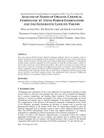
Figure 1 shows SEM images of the surface of TiO
2
thick films composed of nanoparticles (a) and nanotubes (b-d).
Uniform TiO
2
nanotubes of 600 nm in length and 70 nm in diameter were formed by the hydrothermal treatment of
the nanoparticles (b). Clearly, the morphology of the particles changed and the porosity of the films was increased
by the hydrothermal treatment. Uniform nanotubes were formed by the hydrothermal treatment. On the other hand,
after 3 h ball-milling, the nanotube length was decreased. Moreover, more intimate contact was achievied by the
ball-milling treatment as shown in Fig. 1 (c). However, the calcination at higher temperature resulted in the sintering
of the nanotubes and a decrease in the porosity as shown in Fig. 1 (d).
(a)
200nm
(c)
200nm
(b)
200nm
(d)
200nm
(a)(a)
200nm200nm
(c)(c)
200nm200nm
(b)(b)
200nm200nm
(d)(d)
200nm200nm
Fig. 1. SEM images of (a) P-25 commercial particles, (b) TiO
2
nanotubes obtained by hydrothermal treatments for 24 h at 230
o
C, (c) TiO
2
nanotubes ball milled for 3 h, and (d) TiO
2
nanotubes after calcination at 700
o
C. (a)-(c) films were calcined at 600
o
C.
M H. Seo et al. / Procedia Chemistry 1 (2009) 192–195
193
Figure 2 shows the pore size distribution of the films composed of TiO
2
nanoparticles and nanotubes after
calcination at 600 or 700
o
C and ball-milling. The distribution of pores peaks at approximately 36 and 201 nm for the
films composed of commercial TiO
2
particles and TiO
2
nanotubes obtained at 230
o
C, respectively, indicating that
the porosity of the film was increased by using the nanotubular particles. The ball-milling treatment didn’t give a
great influence on the pore size distribution, but calcination at higher temperature resulted in the decrease in the pore
volume.
0.01 0.1 1
0.00
0.02
0.04
0.06
0.08
0.10
0.12
0.14
Pore size / µm
Pore volume ( cc / g )
(a) 36 nm
(b) 201 nm
(c) 165 nm
(d) 140 nm
Fig. 2. Pore size distribution of the sensing films composed of (a) P-25 commercial particles, (b) TiO
2
nanotubes obtained by hydrothermal
treatments for 24 h at 230
o
C, (c) TiO
2
nanotubes ball milled for 3 h, and (d) TiO
2
nanotubes after calcination at 700
o
C. (a)-(c) films were calcined
at 600
o
C.
Figure 3 shows TEM images of TiO
2
nanoparticles (a) and nanotubes after calcination at 600 (b) and 700
o
C (d)
and ball-milling (c). Uniform TiO
2
nanotubes of 600 nm in length and 70 nm in diameter were formed by the
hydrothermal treatment of the nanoparticles and the wall thickness of nanotubes prepared by hydrothermal treated at
230
o
C is estimated to be ca. 7 nm from the TEM images (b). Furthermore, as shown in the TEM images (b, c), we
found that the structure of TiO
2
nanotubes was stable even after calcination at 600
o
C. However, TiO
2
nanotubes
calcined at 700
o
C had heavily-aggregated particles and the tubelar structure was lost as shown in Fig. 3 (d).
(
a
)
30nm
(
d
)
100nm
30nm
50nm
(
c
)
(
b
)
Fig. 3. TEM images of (a) P-25 commercial particles, (b) TiO
2
nanotubes obtained by hydrothermal treatments for 24 h at 230
o
C, (c) TiO
2
nanotubes ball milled for 3 h, and (d) TiO
2
nanotubes after calcination at 700
o
C. (a)-(c) films were calcined at 600
o
C.
3.2 Gas sensing properties of nanotubular TiO
2
films
Figure 4 shows the sensor response of the films to H
2
, CO, ethanol and toluene gases at 500
o
C. The sensor using
the TiO
2
nanotubes (b) exhibited good sensor responses as compared with the sensor using the commercial TiO
2
M H. Seo et al. / Procedia Chemistry 1 (2009) 192–195
194
nanoparticles (a). Furthermore, the sensor using the nanotubes with the ball-milling treatment (c) exhibited the
improved sensor responses to toluene.
This is probably because of the improvement of th
e particle packing density of the film as a result from the
decrease in the tube length after ball milling. In contrast, the sensor using the nanotubes calcined at 700
o
C (d)
showed lower sensitivity to all gases because of the decrease in the porosity.
Thus, the results obtained suggests that the microstructure control of sensing layers in terms of particle packing
de
nsity and pore size distribution is quite effective for improving the sensitivity of TiO
2
-nanotube based gas sensors.
0
10
20
30
40
50
60
( a ) ( b ) ( c ) ( d )
Sensor response ( R
air
/ R
gas
)
Type of sensing films
500 ppm H
2
500 ppm CO
47 ppm Ethanol
50 ppm Toluene
O.T. 500
o
C
Fig. 4. Sensor responses to H
2
(500 ppm), CO (500 ppm), ethanol (47 ppm), and toluene (50 ppm) gases at 500
o
C for the devices using (a) P-25
commercial particles, (b) TiO
2
nanotubes obtained by hydrothermal treatments for 24 h at 230
o
C, (c) TiO
2
nanotubes ball milled for 3 h, and (d)
TiO
2
nanotubes after calcination at 700
o
C. (a)-(c) films were calcined at 600
o
C.
4. Conclusion
The structure of TiO
2
nanotubes was stable after calcinations for 1 h at 600
o
C. The sensor using TiO
2
nanotubes
prepared by the hydrothermal treatment exhibited high sensitivity to toluene rather than CO and H
2
. The ball-milling
treatment shorten the tube length and significantly improved the gas sensitivity probably because of the improved
particle packing density in the sensing film. Thus, the results obtained indicate the importance of the microstructure
control of sensing layers in terms of tube length, pore size distribution, and particle size in tubes for detecting large
sized organic gas molecules.
References
1. N. Yamazoe, G. Sakai, K. Shimanoe, Oxide semiconductor gas sensors, Catal. Surveys from Asia, 1 (2003) 63-75.
2. D. E. Williams, Semiconducting oxides as gas-sensitive resistors, Sens. Actuators, B, Chem 57 (1999) 1-16.
3. A. M. Ruiz, A. Corneta, K. Shimanoe, J. R. Morante, N. Yamazoe, Effects of various metal additives on the gas sensing performances of
TiO
2
nanocrystals obtained from hydrothermal treatments, Sens. Actuators, B, Chem 108 (2005) 34-40.
4. Min-Hyun Seo, Masayoshi Yuasa, Tetsuya Kida, Jeung-Soo Huh, Kengo Shimanoe,
Noboru Yamazoe, “Gas sensing characteristics and
porosity control of nanostructured films composed of TiO
2
nanotubes”, Sens. Actuators B, Chem 137 (2009) 513-520.
5. Tetsuya Kida, Takuya Minami, Shotaro Kishi, Masayoshi Yuasa, Kengo Shimanoe, Noboru Yamazoe, “Planar-type BiCuVOx solid
electrolyte sensor for the detection of volatile organic compounds”, Sens. Actuators, B, Chem 137 (2009) 147–153.
6. Z. Miao, D. Xu, J. Ouyang, G. Guo, X. Zhao, Y. Tang, Electrochemically Induced Sol-Gel Preparation of Single-Crys
talline TiO
Nanowires,
2
Nano Lett. 2 (2002) 717-720.
7. T. Kasuga, M. Hiramatsu, A. Hoson, T. Sekino, K. Niihara, Formation of titanium oxide nanotube, Langmuir 14 (1998) 3160-3163.
M H. Seo et al. / Procedia Chemistry 1 (2009) 192–195
195