- Trang chủ >>
- Khoa Học Tự Nhiên >>
- Vật lý
electrochromics of single crystalline wo3 · h2o nanorods
Bạn đang xem bản rút gọn của tài liệu. Xem và tải ngay bản đầy đủ của tài liệu tại đây (581.33 KB, 6 trang )
Electrochromics of single crystalline WO
3
Æ H
2
O nanorods
Xiaolan Wei
1
, Pei Kang Shen
*
State Key Laboratory of Optoelectronic Materials and Technologies, School of Physics and Engineering, Sun Yat-Sen University,
135 Xingang Road, Guangzhou, Guangdong 510275, China
Received 20 November 2005; received in revised form 9 December 2005; accepted 9 December 2005
Available online 18 January 2006
Abstract
Single crystalline WO
3
Æ H
2
O nanorods have been prepared using a facile solution route for the first time, in which PVA-124 is used as a
structure-directing agent and glacial acetic acid as a stabilizer. The results prove that the x value in the fully reduced form of
M
x
W
6+
(1Àx)
W
5+
x
O
3
is close to 1 for the rod-type WO
3
Æ H
2
O nanocrystals instead of 0.7 for the platelet-type WO
3
Æ H
2
O nanocrystals.
The electrochromic performance of the rod-type WO
3
Æ H
2
O nanocrystals is significantly improved in terms of fast response time and high
contrast due to the plane with wider lattice spacing of d = 5.36 A
˚
is parallel to the growth direction and faces towards the electrolyte.
Ó 2005 Elsevier B.V. All rights reserved.
Keywords: WO
3
Æ H
2
O nanorods; Single crystals; Electrochromics; One-dimensional oxides
1. Introduction
It is well known that tungsten oxide (WO
3
) has promis-
ing properties for applications in electrochromic devices
[1–6], gas sensors [7,8], photoelectrochemistry [9] and elect-
rocatalysis [10,11]. As an electrochromic material, WO
3
is
the first and most extensivel y studied compound and
remains the most promising candidate for electrochromic
devices, such as antidazzle car rear-view mirrors and smart
windows [2–6]. A great deal of effort has been placed on
improving the performance of WO
3
films [2–6,12,13],
including the efforts to obtain nanoscale porous tungsten
oxide films [14,26]. However, there has been no momentous
breakthrough for decades. In the latest reports, new and
interesting applications of existing or prospective tungsten
oxide monohydrate based devices have come to light [15].
An all-plastic WO
3
Æ H
2
O/polyaniline flexible electrochro-
mic device was reported recently [16].
Both amorphous and crystalline electrochromic WO
3
films were prepared and compared [17]. Crystalline WO
3
showed pronounced coloring persistence relative to amor-
phous. However, intercalation and deintercalation
appeared to be faster for a-WO
3
due to the slower kinetics
for c-WO
3
which possesses denser structure and smaller
diffusion pathway. Livage and Guzman [18] synthesized
hydrous WO
3
Æ H
2
O crystals aimed to improve the response
time. WO
3
Æ H
2
O crystals belong to orthorhombic crystal
system. WO
3
Æ H
2
O is formed of layers built up by corner
sharing [WO
6
] octahedra, with water molecules between
these planes. Intercalated water molecules lead to
WO
3
Æ H
2
O layers with a basal spacing d = 5.36 A
˚
, exhibit-
ing enhanced intercalation properties towards cationic spe-
cies such as H
+
,Li
+
, even long-chain alkylammonium ions
comparing with anhydrous WO
3
[18]. If a surface with wide
spacing, d = 5.36 A
˚
in WO
3
Æ H
2
O is exposed to electrolyte,
the efficiency of intercalation towards cationic species
should be enhanced, and the redox performance would
be improved. One-dimensional WO
3
Æ H
2
O nanocrystals
would meet this expectation. One-dimensional nanostruc-
tures may offer opportunities for investigating the effect
of size and dimensionality on their comprehensive optical,
magnetic, and electronic properties [19] .
1388-2481/$ - see front matter Ó 2005 Elsevier B.V. All rights reserved.
doi:10.1016/j.elecom.2005.12.008
*
Corresponding author. Tel.: +86 20 84036736; fax: +86 20 84113369.
E-mail address: (P.K. Shen).
1
Present address: Department of Applied Chemistry, South China
University of Technology, Guangzhou, China.
www.elsevier.com/locate/elecom
Electrochemistry Communications 8 (2006) 293–298
One-dimensional oxides can be prepared by different
methods [20–23]. However, the controllable growth of the
selective crystal plane is extremely difficult. In this paper,
we report a facile solution route to single crystalline
WO
3
Æ H
2
O shaped with rod and platelet. Poly(vinyl alco-
hol) (PVA-124) was used as a structure-directing agent
and glacial acetic acid as a stabilizer. The effect of the mor-
phologies of WO
3
Æ H
2
O for the same crystalline phase and
the different intercalation degrees of cations into the WO
3
films on the electrochromic performance was investigated.
2. Experimental
2.1. WO
3
Æ H
2
O nanocrystal preparation
The rod-type WO
3
Æ H
2
O was prepared by adding 0.4 g
PVA-124 (average polymerization degree 2400–2500, Kura-
ray, Japan) in 40 ml distilled–deionized water and stirring at
100 °C for 10 min. 5 ml HAc and 4 g Na
2
WO
4
Æ 2H
2
O were
added after the solution had been cooled to 60 °C. The mix-
ture was stirred at 60 °C for 30 min to form a transparent
solution. Afterwards , 15 ml 5 mol L
À1
HNO
3
was added
into the solution very slowly (0.4 mL per minute) at the
same temperature, resulting in a pale yellow solution. Keep-
ing the solution at 60 °C for 4 h, the yellow precipitates
formed increasingly and then were aged at room tempera-
ture for another 4 days. The precipi tates were separated
and washed carefully using distilled-deionized water until
the sodium ions are undetectable. Finally, the precipitates
were washed by acetone and dried in air. The platelet-type
WO
3
Æ H
2
O was prepared by the same procedure. The differ-
ence was that the 5 ml concentrated HNO
3
solution instead
of 15 mL 5 mol L
À1
HNO
3
was added at fast speed and
without aging. The precipitates were formed during the
addition of the concentrated HNO
3
. The purified rod-type
and platelet-type WO
3
Æ H
2
O was, respectively, suspended
into distilled-deionized water under an ultrasonic stirring
for 1 h to form a colloidal solution (5 g L
À1
WO
3
Æ H
2
O).
WO
3
Æ H
2
O films were formed by dropping 20 lL of the col-
loidal solution onto pre-cleaned 1 cm
2
ITO glass substrates
(15 X/h) and then drying in air at room temperature.
2.2. WO
3
Æ H
2
O nanocrystal characterization
The microstructure of WO
3
Æ H
2
O was examined with
a high-resolution transmission electron microscope
(HRTEM, JEOL JEM–2010, operated at 200 kV). An X-
ray diffractometer D/Max-IIIA (Rigaku Co., Japan) using
Cu K a1(k = 1.540056 A
˚
) as the radiation source, which
was used to examine the crystalline structure. The measure-
ment of the electrochromic properties of WO
3
Æ H
2
O films
was carried out in situ on VoltaLab 80 Universal Electro-
chemical Laboratory (Radiometer Analytical Laboratory,
France) and a UV-2102PC Spectrometer (UNICO Instru-
ment Co., USA) in a standard three-electrode cell. A plat-
inum wire was used as the counter electrode and Ag|AgCl
as the reference electrode, respectively. Cyclic voltammetric
experiments were carried out in the potential range of
À0.6 V to 0.6 V (vs. Ag|AgCl) at different scan rates.
3. Results and discussion
Either rod-type or platelet-type WO
3
Æ H
2
O nanocrystals
could be prepared, depending on the preparation condi-
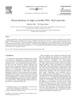
tions and the aging time. Fig. 1 shows the TEM images
of the prepared rod-type WO
3
Æ H
2
O nanocrystals
(Fig. 1a and c) and platelet -type WO
3
Æ H
2
O nanocrystals
(Fig. 1 b). The corresponding selected area electron diffrac-
tion (SAED) patterns shown in the insets of Fig. 1a and b
indicate that both of the two nanocrystals are single crys-
tals. On the top of rod-type WO
3
Æ H
2
O nanocrystals, the
lattice spacing of d = 5.36 A
˚
is parallel to the growth direc-
tion and is faced towards the electrolyte. The wider spacing
(5.36 A
˚
) towards the surface would be of benefit to the H
+
intercalation and deintercalation, resulting in an increased
electrochromic speed. While for the platelet-type WO
3
Æ
H
2
O nanocrystals, the plane faced towards the electrolyte
is the plane with the lattice spacing of d = 3.47 A
˚
in two-
dimensional directions.
The crystalline phase of the prepared WO
3
was identi-
fied by X-ray powder diffraction (Fig. 2). XRD patterns
of the products were exclusively orthorhombic WO
3
Æ H
2
O
(Pmnb, a = 5.249 A
˚
, b = 10.71 A
˚
, c = 5.133 A
˚
, JCPDS-
ICDD 84-0886). Three strongest lines in the Fig. 2a (rod-
type sample) are 3.4795 A
˚
, 5.3875 A
˚
and 2.5742 A
˚
and in
Fig. 2b (platelet-type sample) are 3.4793 A
˚
, 5.3833 A
˚
and
2.5714 A
˚
. These values are consistent with the standard
values of 3.47 A
˚
, 5.36 A
˚
and 2.56 A
˚
in JCPDS-ICDD 84-
0886, corresponding to (111), (020) and (1 31) planes.
The results suggest that the rod-type and the platelet-type
nanocrystals are the same in c rystalline phase. The inten-
sity ratio of the peak (020) to peak (111) for rod-type sam-
ples is larger than that of for platelet-type samples,
suggesting that the abundant planes in rod-type
WO
3
Æ H
2
O are (0 2 0) planes, and vice versa. No diffraction
peaks due to WO
3
Æ 2H
2
O or other tungsten oxides were
discerned, which indicates the high purities of the nanorods
and nanoplatelets. The confirmation of the WO
3
Æ H
2
O
structure was further conducted by the thermogravimetric
(TG) analyses (Fig. 3). It is obv ious that the weight loss
before 60 ° C is the evaporation of water moisture. After-
wards, the WO
3
Æ H
2
O started to decompose and lose the
coordinated water at higher temperature. The weight loss
measured was 6.5% for sample (a) and 6.7% for sample
(b), respectively, which are in keeping with the theoretical
calculation (7.2%) for the WO
3
Æ H
2
O structure.
WO
3
Æ H
2
O is formed of layers built up by corner shar-
ing [WO
6
] octahedral, with water molecules between these
planes. Hydrous oxides are precipitated upon the acidifi-
cation of tungsten [WO
4
]
2À
(Na
2
WO
4
as precursors in this
study). Coordi nation expansion leads to the formation of
sixfold coordinated W
VI
via the nucleophilic addition of
two water molecules [18]. One water molecule is bonded
along the z-axis opposite to the W@O bond while the four
294 X. Wei, P.K. Shen / Electrochemistry Communications 8 (2006) 293–298
OH groups are in the equatorial xy plane. Oxolation along
equivalent x and y directions leads to the formation of the
layered WO
3
Æ H
2
O nanocrystals (Fig. 4a). The structure of
the oxide network depends on the acidification rate and the
aging time. The preparation of platelet WO
3
Æ H
2
O has
been reported by different methods. Bala
´
zsi and Pfeifer
[24] prepared microscaled platelet WO
3
Æ H
2
O under the
quick addition of strong acid without the control. Stucky
and co-workers [25] used benzyl alcohol and WCl
6
synthe-
sized platelet WO
3
Æ H
2
O. In our case, nanoscaled platelet
WO
3
Æ H
2
O crystals were prepared in the presence of glacial
acetic acid and PVA-124 under the quick addition of con-
centrated HNO
3
. However, by controlling the adding rate
of the dilute HNO
3
and the aging time we synthesized
the nanorod-type WO
3
Æ H
2
O for the first time. PVA-124
is a structure-directing agent in this study. It is estimated
that the distance between two hydroxyl oxygen is
0.251 nm since the C–C bond is 0.154 and \CCC is 109°.
The hydrogen of hydroxyl group will tilt due to the influ-
ence of lone pare electrons to fit the narrow spacing of
W–O–W (3.47 A
˚
) by the formation of hydrogen bonds
(Fig. 4b). The crystals growth substantially favors the
direction of (0 20) plane instead of (111) plane. However,
Fig. 1. TEM images of the rod-type (a) and platelet-type (b) WO
3
Æ H
2
O nanocrystals. (c) HRTEM image of rod-type WO
3
Æ H
2
O nanocrystals. The
selected area electron diffraction patterns of the rod-type (inset in a) and platelet-type (inset in b) WO
3
Æ H
2
O nanocrystals are shown as well.
10 20 30 40 50 60 70 80
0
200
400
600
0
200
400
600
131
111
020
b
2theta(deg.)
131
111
020
a
Intensity/cps
Fig. 2. XRD patterns of WO
3
Æ H
2
O nanorod (a) and nanoplatelet (b).
X. Wei, P.K. Shen / Electrochemistry Communications 8 (2006) 293–298 295
the PVA-124 might lose the effect if the crystal growth is
too fast.
The electrochromic properties of the nanorod and nano-
platelet WO
3
Æ H
2
O were characterized as a typical applica-
tion. Fig. 5a shows the cyclic voltammograms of
WO
3
Æ H
2
O films on indium-doped tin oxide (ITO) glass
substrates. The CV curves shaped as previous reported
[26–28]. It is worth noting that the second anodic peak is
remarkable and shows evidence of the existence of meso-
porous structure [26,27]. Comparing the current density
of the platelet WO
3
Æ H
2
O and the rod-type WO
3
Æ H
2
Oat
the same loading (in mg cm
À2
of WO
3
Æ H
2
O), we found
that more than double the current density was observed
on the latter sample. For example, the reduction current
0 50 100 150 200 250 300
b
-24
-20
-16
-12
-8
-4
Heat flow/mW
0 50 100 150 200 250 300
80
84
88
92
96
100
104
Temperature/
o
C
a
Weight/%
Fig. 3. Thermogravimetric (TG) analyses of (a) rod-type WO
3
Æ H
2
O and (b) platelet-type WO
3
Æ H
2
O.
W
O
O
H
2
O
W
O
O
H
2
O
W
O
O
W
O
H
2
O
O
H
3
O
£«
OH
2
C
C
CH
2
C
CH
2
HO
O
O
H
H
O
O
H
O
O
H
W
W
O
O
W
W
O
O
~0.35nm
~109
o
¡« 0.2 5
0
.
1
5
4
O
O
ab
Fig. 4. Structures of (a) layered WO
3
Æ H
2
O and (b) hydrogen bond between WO
3
Æ H
2
O and PVA-124.
-600 -400 -200 0 200 400 600
-40
-30
-20
-10
0
10
20
30
2
1
a
Potential/mV
uCrrneedtsntiy/mA mc
2-
gm
1-
050250
75
80
85
90
95
b
2
1
Trans/%
Time/s
Fig. 5. Cyclic voltammograms of platelet-type (1) and rod-type WO
3
Æ H
2
O (2) in 0.5 mol L
À1
H
2
SO
4
with the scan rate of 100 mV s
À1
(a) and the in situ
transmittance changes (b).
296 X. Wei, P.K. Shen / Electrochemistry Communications 8 (2006) 293–298
density of rod-type WO
3
Æ H
2
O was 40 mA cm
À2
mg
À1
and
only 6 mA cm
À2
mg
À1
for the platelet-type sample at
À0.6 V. The in situ UV–Vis transmittance responses at
650 nm during the potential cycling were recorded and
shown in Fig. 5b. When the WO
3
Æ H
2
O films were cathod-
ically polarized, the films turned blue in color and the color
intensified with the increase in the cathodic potential. The
blue films were bleached under the anodic polarization.
The optical responses were symmetrical upon the symmet-
ric potential cycling, indicating electrochromic reversibility.
Also, the change in transmittance was more than doubled
for rod-type compared with that of platelet-type
WO
3
Æ H
2
O.
The possible explanation of why the current density or
the contrast of the two samples is different at the same
potential with the same loading is that the amount of
inserted cations is different. The color of WO
3
film switches
reversibly from transparent to blue upon electrochemical
redox reactions
WO
3
þ xðM
þ
þ e
À
Þ!M
x
W
6þ
ð1ÀxÞ
W
5þ
x
O
3
M ¼ H; Li; Na;
When a negative potential is suppli ed to the WO
3
Æ H
2
O,
there is an influx of conducting electrons into the tungsten
oxide film. To keep the system electrically neutral, cations
(H
+
,Li
+
) should move into the film from the surrounding
electrolyte. Without this charge compensation, the elec-
trons could not be injected into the film. The diffusion of
the cations into the oxide layer is slow which determines
the response time. We presum ed that the nano WO
3
Æ H
2
O
with the larger lattice spacing toward the electrolyte would
be faster in coloration/bleaching rate.
The cathodic charges were integrated and compared at
different scan rates. These charges corresponded to the for-
mation of a H
x
WO
3
compound with different x values,
assuming all the charge intercalated contributed to interca-
lation of H ions. The stoichiometric charge for one electron
reduction of WO
3
is 387 mC mg
À1
. Therefore, the x values
can be calculated. Table 1 compares the electrochromic effi-
ciencies of two nano WO
3
Æ H
2
O.
Fig. 6 shows the plots of x against scan rate. In the case
of rod-type WO
3
Æ H
2
O, the x value closes 1 at fully
reduced state and is still over 0.9 at scan rate as high as
20 mV s
À1
. While, platelet-type WO
3
Æ H
2
O could not be
fully reduced and the x value is only less than 0.3 at scan
rate of 5 mV s
À1
. The results indicate that the rod-type
WO
3
Æ H
2
O can be deeply colored by the insertion of more
cations. Moreover, the coloration/b leaching rate is very
fast as a result of the fast mass transport due to the direct
connection of electrolyte with larger lattice spacing.
The be tter redox performance of the rod-type
WO
3
Æ H
2
O nanocrystals is related to its structure. The
hydrogen ions in the solution are normally hydrated and
intercalate into the oxide matrix as hydrous ions. The inser-
tion of the hydrous ions is easier along with the larger lat-
tice spacing. The (020) plane of the rod-type WO
3
Æ H
2
O
faced to the solution and the hydrous hydrogen ions
inserted into this plane by relaying water molecules in the
layer spacing one by one as shown in Fig. 4b. The short
depth and larger width of the rod-type WO
3
Æ H
2
O struc-
ture resulted in a fast coloration/bleaching speed and fully
intercalation. The hydrous hydrogen ions inserted into the
oxide matrix along with the (1 1 1) plane that is perpendic-
ular against the (0 20) plane in the case of platelet-type
WO
3
Æ H
2
O. The intercalation and deintercalation speed
of the cations are slower due to the small lattice spacing
and without the relay by the bonded water.
4. Conclusion
A facile solution route prepared single crystalline
WO
3
Æ H
2
O nanorods for the first time, in which PVA-
124 was used as a structure-directing agent and glacial
acetic acid as a stabilizer. For the rod-type WO
3
Æ H
2
O
nanocrystals, the lattice spacing of d = 5.36 A
˚
is parallel
Table 1
Comparison of the electrochromic efficiencies of two nano WO
3
Æ H
2
O at different scan rates
Rod-type WO
3
Æ H
2
O (mV s
À1
) 100 50 20 5 1
Charge, mC 9.1 9.5 18.7 19.3 19.5
Charge, mC mg
À1
172 179 352 364 368
x value 0.44 0.46 0.91 0.94 0.95
Platelet-type WO
3
Æ H
2
O (mV s
À1
) 100 50 20 5 1
Charge, mC 6.9 7.1 9.4 9.6 24.9
Charge, mC mg
À1
77 79 104 107 276
x value 0.198 0.204 0.27 0.28 0.71
0 20 40 60 80 100
0.0
0.2
0.4
0.6
0.8
1.0
x
Scan rate / mV s
-1
rod
platelet
Fig. 6. Plots of the normalized amount of inserted ions against the scan
rate.
X. Wei, P.K. Shen / Electrochemistry Communications 8 (2006) 293–298 297
to the growth direction and is faced toward the electrolyte.
The wider spacing (5.36 A
˚
) toward the surface would be of
benefit to the H
+
intercalation and deintercalation, result-
ing in an increased electrochromic speed, whereas for the
platelet-type WO
3
Æ H
2
O nanocrystals, the plane faced
toward the electrolyte is the plane with the lattice spacing
of d = 3.47 A
˚
in two-dimensional directions. The results
prove that the x value in the fully reduced form of
M
x
W
6+
(1Àx)
W
5+
x
O
3
is close to 1 for the rod-type
WO
3
Æ H
2
O nanocrystals, while it is 0.7 for the platelet-type
WO
3
Æ H
2
O nanocrystals. The fast response time and high x
value of the rod-type WO
3
Æ H
2
O nanocrystals improved
significantly the redox performance. Our results also point
to the fundamentally impor tant possibility of fine-tuning
the electrochromic performance and catalytic activity of
tungsten oxide and other transition metal oxides.
Acknowledgments
This study was supported by the National Natural Sci-
ence Foundation of China (20476108), Guangdong Prov-
ince Natural Science Foundation (04105500), Guangdong
Science and Technology Key Project (2004A11004001,
2005A11001002).
References
[1] C. Santato, M. Odziemkowski, M. Ulmann, J. Augustynski, J. Am.
Chem. Soc. 123 (2001) 10639.
[2] S.K. Deb, Appl. Opt. (Suppl. 3) (1969) 192.
[3] S.K. Deb, Philos. Mag. 27 (1973) 801.
[4] C.G. Granqvist, E. Avendano, A. Azens, Thin Solid Films 442 (2003) 201.
[5] A. Jonsson, M. Furlani, G.A. Niklasson, Sol. Energ. Mat. Sol. C 84
(2004) 361.
[6] E. Avendano, L. Berggren, G.A. Niklasson, C.G. Granqvist, A.
Azens, Thin Solid Films 496 (2006) 30.
[7] J.L. Solis, S. Saukko, L. Kish, C.G. Granqvist, V. Lantto, Thin Solid
Films 391 (2001) 255.
[8] Y.J. Li, P.P. Tsai, Solid State Ionics 86 (1996) 1001.
[9] L.Y. Su, L.G. Zhang, J.H. Fang, M.H. Xu, Z.H. Lu, Sol. Energ. Mat.
Sol. C 58 (1999) 133.
[10] A.F. Pe
´
rez-Cadenas, C. Moreno-Castilla, F.J. Maldonado-Ho
´
dar,
J.L.G. Fierro, J. Catal. 217 (2003) 30.
[11] C.D. Baertsch, K.T. Komala, Y.H. Chua, E. Iglesia, J. Catal. 205
(2002) 44.
[12] P.K. Shen, J. Syed-Bokhari, A.C.C. Tseung, J. Electrochem. Soc. 138
(1991) 2778.
[13] N.R. Tacconi, C.R. Chenthamarakshan, K.L. Wouters, F.M. Mac-
Donnell, K. Rajeshwar, J. Electroanal. Chem. 566 (2004) 249.
[14] E. Ozkan, S H. Lee, P. Liu, C.E. Tracy, F.Z. Tepehan, J.R. Pitts,
S.K. Deb, Solid State Ionics 149 (2002) 139.
[15] C. Bala
´
zsi, J. Pfeifer, Sol. Energ. Mat. Sol. C 76 (2003) 577.
[16] C. Marcel, J M. Tarascon, Solid State Ionics 143 (2001) 89.
[17] H. Kamal, A.A. Akl, K. Abdel-Hady, Physica B 349 (2004) 192.
[18] J. Livage, G. Guzman, Solid State Ionics 84 (1996) 205.
[19] K. Lee, W. Seo, J.T. Park, J. Am. Chem. Soc. 125 (2003) 3408.
[20] R.P. Greta, F. Krumeich, R. Nesper, Angew. Chem. Int. Ed. 41
(2001) 2446.
[21] J. Hu, T.O. Wang, C.M. Lieber, Acc. Chem. Res. 32 (1999) 435.
[22] X. Wang, Y. Li, J. Am. Chem. Soc. 124 (2002) 2880.
[23] Y. Zhu, H. Li, Y. Koltypin, Y.R. Hacohen, A. Gedanken, Chem.
Commun. 24 (2001) 2616.
[24] Cs. Bala
´
zsi, J. Pfeifer, Solid State Ionics 124 (1999) 73.
[25] M. Niederberger, M.H. Bartl, G. Stucky, J. Am. Chem. Soc. 124
(2002) 13642.
[26] W. Chen, E. Baudrin, B. Dunn, J.I. Zink, J. Mater. Chem. 11 (2001)
92.
[27] S H. Baeck, K S. Choi, T.F. Jaramillo, G.D. Stucky, E.W.
McFarland, Adv. Mater. 15 (2003) 1269.
[28] D J. Kim, S I. Pyun, Solid State Ionics 99 (1997) 185.
298 X. Wei, P.K. Shen / Electrochemistry Communications 8 (2006) 293–298