- Trang chủ >>
- Khoa Học Tự Nhiên >>
- Vật lý
facile large scale synthesis of ws2 nanotubes from wo3 nanorods prepared by a hydrothermal route
Bạn đang xem bản rút gọn của tài liệu. Xem và tải ngay bản đầy đủ của tài liệu tại đây (606.94 KB, 6 trang )
Solid State Sciences 7 (2005) 67–72
www.elsevier.com/locate/ssscie
Facile large scale synthesis of WS
2
nanotubes from WO
3
nanorods
prepared by a hydrothermal route
Helen Annal Therese
a
, Jixue Li
b
,UteKolb
b
, Wolfgang Tremel
a,∗
a
Institut für Anorganische Chemie und Analytische Chemie der Johannes Gutenberg-Universität, Duesbergweg 10-14, 55099 Mainz, Germany
b
Institut für Physikalische Chemie, Welderweg 11, 55099 Mainz, Germany
Received 17 June 2004; received in revised form 27 July 2004; accepted 8 October 2004
Available online 13 December 2004
Abstract
Hexagonal WO
3
nanorods of5–50 nmin diameter and 150–250 nmin length have been synthesised in gram quantitiesby a low temperature
hydrothermal route using citric acid as a structural modifier and hexadecylamine as a templating agent. The ratio of [A]/[W] play an important
role on WO
3
nanorods formation. These WO
3
nanorods were found highly suitable as a precursor for the synthesis of a good yield of
multiwalled WS
2
nanotubes by reducing them with H
2
S at 840
◦
C for 30 min. The length and the wall thickness of the WS
2
nanotubes could
be altered by controlled reduction of the oxide precursor. The morphology, structure and the composition of the WO
3
nanorods and WS
2
nanotubes were characterised by X-ray diffraction (XRD), high resolution transmission electron microscopy (HRTEM), energy dispersive
X-ray analysis (EDX) and by selected area electron diffraction techniques (SAED).
2004 Elsevier SAS. All rights reserved.
Keywords: WO
3
; Nanorods; Hydrothermal route; WS
2
; Nanotubes
1. Introduction
Following the discovery of carbon nanotubes in 1991 [1],
nanostructured inorganic materials and their syntheses have
attracted tremendous attention due to their superior mechan-
ical properties, their unique electronic behaviour and their
high potential in making technologically advanced nanode-
vices. The interesting properties of these nanomaterials arise
from their enormous surface area, strength and the quan-
tum size. Among nanomaterials, nanowires and nanotubes
are good candidates for studying the phenomena such as
electrical resistivity, strength, magnetic and optical proper-
ties in one dimension. During the past few years, nanotubes
of various materials with graphite-like layered structures
has been synthesised successfully using techniques such as
arc discharge [2], laser ablation [3], electron beam irradi-
ation [4], sonochemical [5], hydrothermal reaction [6] and
*
Corresponding author.
E-mail address: (W. Tremel).
iodine transport [7], etc. Among the various classes of non-
carbon nanotubes, transition metal chalcogenides of the gen-
eral formula MQ
2
(M = W,Mo,V,Nb,Ta,Zr;Q= S,
Se) are significant materials and reveal interesting electronic
and optical properties [8–10]. Inorganic fullerene like MoS
2
nanoparticles and MoS
2
nanotubes exhibit excellent lubri-
cating properties [11] and show high scope as tips for scan-
ning probemicroscopes[12]. Recent electrochemicalstudies
on MoS
2
nanotubes revealed that the nanotubes can store
relatively large amounts of gaseous hydrogen by electro-
chemical storage [13].MoS
2
nanotubes of up to 5 µm length
and 10–20 nm in diameter were first synthesised by Tenne
and co-workers by reducing MoO
3
in the atmosphere of a
mixture of H
2
/N
2
and H
2
Sgas[14].
Recently, WO
3
nanorods were found to be a versatile
precursor in the synthesis of WS
2
nanotubes apart from
its utility in electrochromic devices [15], gas sensors [16],
rechargeable lithium batteries [17], memory devices [18],
etc. For example WS
2
nanotubes were synthesised by an-
nealing in a H
2
S stream a nanostructured tungsten oxide pre-
cursor, which was produced by heating tungsten filaments
1293-2558/$ – see front matter 2004 Elsevier SAS. All rights reserved.
doi:10.1016/j.solidstatesciences.2004.10.006
68 H.A. Therese et al. / Solid State Sciences 7 (2005) 67–72
under reduced pressure in the presence of water vapour [19,
20]. Walton and co-workers have synthesised WS
2
tubes
from W
18
O
49
rods produced by heating tungsten foil at high
voltages under Ar in the presence of SiO
2
[21].
Various strategies have been employed for the synthe-
sis of WO
3
nanorods as synthesising large quantities WO
3
nanostructures of the required size becomes a greater task.
WO
3
nanorods were produced by heating the tungsten fil-
ament in an inert atmosphere either in the presence of ox-
ides like SiO
2
[22],B
2
O
3
[23] or in the presence of water
vapour [20] or by IR irradiation of W in the presence of
air [24]. Electrochemical etching followed by heating also
yielded WO
3
nanorods [25]. Controlled removal of the sur-
factants from the mesolamellar precursors resulted in WO
3
nanorods in large quantity [26]. Recently, the synthesis of
WO
3
nanorodsat a large scale [27] has been described by the
oxidation of W(CO)
6
with an amine oxide in a low-volatile
solvent(oleylamine)at 270
◦
C. Most of the abovementioned
synthetic techniques are very tedious and require expensive
experimentalsetup or inert atmosphere. Above all, obtaining
a single phase compound of similar morphology and dimen-
sions in considerable quantity still is a difficult task. In this
article we report the synthesis of WO
3
nanorods of 5–50 nm
in diameter and 150–250 nm in length, in gram quantities
by a simple hydrothermal method. WO
3
nanorods were re-
duced with H
2
S to obtain a very high yield of multiwalled
WS
2
nanotubes.
2. Experimental
2.1. Synthesis of WO
3
nanorods
An aqueous solution containing a mixture of 1.32 g of
(NH
4
)
10
W
12
O
41
·7H
2
O and 2.10 g of citric acid was heated
around 120
◦
C under constant stirring for 4–5 h until a gel
was formed, which was allowed to stand overnight. 2.45 g of
hexadecyl amine dissolved in ethanol was added as an addi-
tive to the gel and stirred for 10 h. The resulting mixture was
transferred to a Teflon autoclavewith a stainless steel protec-
tive outer body and heated at 180
◦
C for 7 days. The product
obtained was washed with ethanol, cyclohexene, water and
finally with ethanol and dried at room temperature. The im-
portance of citric acid as a structural modifier was studied
by replacing citric acid with hydrochloric acid, while main-
taining the pH of the reactant same as the corresponding
experiment carried with citric acid.
2.2. Conversion of WO
3
nanorods to WS
2
nanotubes
An alumina crucible containing WO
3
nanorods was
placed in a tubular furnace and heated up to 840
◦
CinArgas
flow, then switched to H
2
S gas for 30 min at 840
◦
C to allow
the complete conversion of oxide nanorods to tungsten sul-
phide nanotubes and finally cooled to room temperature in
the presence of Ar. A heating rate of 5
◦
Cmin
−1
was main-
tained during the heating/cooling process.
2.3. Characterisation
The products obtained from hydrothermal reactions after
washing and drying were analysed by X-ray powder diffrac-
tion in θ/2θ reflection geometry using Siemens D8 powder
diffractometer equipped with a position sensitive detector.
Data was collected between 2θ = 5
◦
and 60
◦
(using Cu-Kα
radiation), at an operation potential of 40 kV and a current
of 40 mA. The morphology of the WO
3
nanorods and WS
2
nanotubeswas characterisedbyhigh-resolutiontransmission
electron microscopy (FEI Tecnai F30 ST operated at an ex-
traction voltage of 300 kV, equipped with an EDXA energy
dispersive X-ray spectrometer) and by selected area electron
diffraction techniques (SAED).
For transmission electron microscopic(TEM) studies, the
sample was prepared by crushing them mechanically with
a mortar and pestle followed by dispersing the powder ul-
trasonically in absolute ethanol and placing a drop of this
suspension on to a copper grid coated with a holey carbon
films.
3. Results and discussion
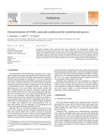
Representative TEM images of the tungsten oxide sam-
ples obtained from two different trials of hydrothermal reac-
tions are given in Fig. 1 (with m and w given in the caption
of Fig. 1). Fig. 1ashowsapartofaWO
3
particle with
many nonseparable rods protrudingout. TEM image of these
rods are shown at a slightly higher magnification in the in-
set of Fig. 1a. This sample consists of particles exclusively
grown in a bunch-like fashion. Fig. 1b shows very well sep-
arated WO
3
nanorods aggregated together due to the high
surface energy owing to their nanosize. The nanorod lengths
in Fig. 1b range from 150 to 250 nm and their diameters
vary from 5 to 50 nm. A calculation of the particle size dis-
tribution of these samples shows that more than 85% of the
tungsten oxide rods are within the range of 15–50 nm in di-
ameter.
The powder X-ray diffraction pattern of the WO
3
nano-
rods (shown in Fig. 2) could be well indexed based on a
hexagonalcell of WO
3
with lattice constants a = 7.37 Å and
c = 3.77 Å. The lattice parameters of the WO
3
rods reported
here vary slightly from the reported (ICSD code = 32,001;
a = 7.298 Å, c = 3.899 Å) values for the hexagonal WO
3
.
The reflection half-widths indicate the presence of nanoscale
WO
3
rods. The particle size of the nanorods calculated from
the XRD pattern using Scherrer’s formula varies between 50
and 185 nm.
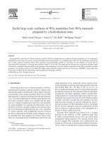
Fig. 3 shows experimental and simulated HRTEM im-
ages and SAED diffraction patterns of a WO
3
nanorod. The
lattice parameters of 0.38 and 0.63 nm correspond to the
d-spacings of (001) and (100) of the WO
3
hexagonal cell.
H.A. Therese et al. / Solid State Sciences 7 (2005) 67–72 69
Fig. 1. TEM images of WO
3
nanorods synthesised by hydrothermal reactions at different mole percentages m =[W]/([A]+[W]+[HDA]) (%) and ratios
of w =[W]/[HAD] (%) ([W], [A] and [HAD] correspond to the number of moles of ammonium tungstate, citric acid and hexadecylamine). (a) Depicts the
bunch-like morphology of the WO
3
particle grown at m = 3.42 and w = 18.6, whereas (b) shows the morphology of WO
3
rods obtained at grown at m = 1.8
and w = 1.6. The WO
3
rods are shown at higher magnification in the inset. w values < 5andm values 1 resulted in the formation of nanorods.
Fig. 2. Powder X-ray diffraction pattern of WO
3
nanorods. All reflections
are indexed based on a hexagonal WO
3
cell with a = 7.37 Å, c = 3.77 Å.
These values are also in agreement with the lattice parame-
ters obtained from the powder XRD pattern. All rods tend
to grow along the ‘c’ direction. The high resolution image
filtered via FFT with DM3.6 (see inset (a)) is in good agree-
ment with the image of zone [010] (see inset (b)) simulated
by multislice method [28,29] (thickness of 37 Å, defocus
−855 Å, C
s
= 1.2 mm) using Cerius [30]. The same holds
for the dynamically calculated diffraction pattern compared
with the experimental SAED pattern. Selected area energy
dispersive X-ray analyses (EDX) of individual nanorods ex-
hibit the existence of tungsten and oxygen in an atomic ratio
of 1:3.
Orthorhombic WO
3
·1/3H
2
O needles has been synthe-
sised by Figlarz and co-workers [31] by hydrothermal syn-
thesis of tungstic gel at 120
◦
C. These needles on heating at
250
◦
C yielded hexagonal WO
3
, but there was nothing men-
tioned about the yield of such needles. In the present report,
it is important to mention that samples prepared for m 1
and w<5 contain exclusively hexagonal WO
3
nanorods.
Recently, nanotubes of VO
x
have been synthesised [32–34]
by low temperature sol–gel techniques. In both cases of VO
x
nanotube synthesis, hexadecyl amine used as a template gets
Fig. 3. Experimental HRTEM image of a WoO
3
nanorod along b axis (top);
the filtered image is shown as inset (a) the corresponding simulated image as
inset (b) together with experimental SAED pattern (bottom, left-hand side)
and dynamically calculated ED pattern for zone [010] (bottom, right-hand
side).
intercalated into the vanadium oxide structure, resulting in
larger d-spacings (∼ 3nm). From the lattice spacings of the
WO
3
rods reported here one could see that the hexadecyl
amine is not intercalated into the WO
3
nanorods. This also
helps us in availing this material as a precursor for WS
2
–NT
synthesis. Synthesis of WO
3
nanorods was also carried out
using hydrochloric acid instead of citric acid, while main-
taining the pH similar to the reaction which yielded WO
3
nanorods. This reaction resulted in a product containing a
70 H.A. Therese et al. / Solid State Sciences 7 (2005) 67–72
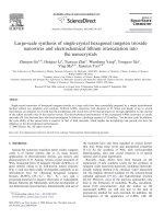
Fig. 4. A low resolution TEM image of the WS
2
nanotubes (a) indicates the yield of nanotubes during the conversion process. TEM image of hollow WS
2
nanotubes (b). The sheet-like appearance in (b) are due to Moire patterns. The open end of a nanotube is indicated by an arrow. HRTEM micrograph of a
typical MWNT (c) along with its SAED pattern (d). The chiral angle of the nanotubes could be calculated as ∼ 10
◦
based on the SAED.
mixture of longer rods of various thickness and unevenly
shaped crystalline particles. Similarly synthesis carried out
for m<1andw<5 resulted in a mixed product with more
of highly crystalline WO
3
particles and a few bunch-like
WO
3
particles.
In our study, the role of citric acid as a structural mod-
ifier and the mechanism involved in the growth of WO
3
is
not clear. But from the above study we could infer that the
amount of citric acid plays an important role in the forma-
tion of WO
3
nanorods. One could speculate that at higher m
values, the 3 carboxylategroup of each citric acid could bind
to more than one WO
6
octahedron and helping in the olation
of WO
6
in a directed manner while hindering the oxolation
in all directions due to steric effects. This could lead to the
formation of shorter and thinner rods. On the other hand, at
lower m values, two or more oxygens of the WO
6
octahe-
dra could be contributed from a single citric acid molecule,
hence hindering both the olation and oxolation to a large
extent. During hydrothermal reaction at 180
◦
C, when citric
acid decomposes the hydrophobic hexadecyl amine template
could be helping in preserving the rod like structure of the
tungsten oxides. For w values > 5 the surface coverage of
the growing WO
3
nanorods is not sufficient to limit the par-
ticle growth and WO
3
bunches and rods are obtained. When
citric acid is fully replaced by hydrochloric acid condensa-
tion of WO
6
takes place rather very quickly resulting in a
mixture of longer and thicker rods and crystals.
The nanorods were converted to WS
2
nanotubes by heat-
ing the WO
3
nanorods in Ar gas up to 840
◦
Candthen
treating them in hydrogen disulphide atmosphere for 30 min.
TEM images of WS
2
nanotubes obtained after H
2
S reduc-
tion (Fig. 4a) show the high yield of WS
2
nanotubes. How-
ever a manifold increase in the diameter and the length of
the WS
2
tubes compared to the WO
3
starting material was
H.A. Therese et al. / Solid State Sciences 7 (2005) 67–72 71
Fig. 5. The various defects formed in a nanotube during the reduction process (a). HRTEM micrograph of a typical open ended WS
2
nanotube encapsulated
by a WS
2
mantle (b). The shorter nanotubes obtained when the nanorods were reduced for a duration of 10 min under H
2
S are shown in (c) and (d).
observed. The nanotube thicknesses range broadly from 20
to 200 nm and their length varies approximately from 1
to 8 µm. A large fraction of the nanotubes has open ends
(Fig. 4b). Studies on these nanotubes by HRTEM combined
with EDX analyses reveal the complete conversion of oxide
rods to sulphide tubes during the reduction process which
allows the synthesis of large amounts of multiwalled nan-
otubes (MWNTs). A HRTEM image of one such represen-
tative MWNT is shown in Fig. 4c. The interlayer spacing
of 0.65 nm between the tubular walls is consistent with the
(002) d-spacing of 2H–WS
2
lattice. The helicity of the nan-
otube (Fig. 4c) could be calculated as ∼ 10
◦
based on the
selected area diffraction(SAED) pattern (Fig. 4c) of the mul-
tiwalled WS
2
nanotube [35].
The synthesis of WS
2
nanotubes by reduction of WO
x
nanorods has been reported earlier, where tungsten disul-
phide tubes were produced by heating WO
x
particles first
in flowing H
2
/N
2
mixture and then in flowing H
2
Sgas.
In consistency with WS
2
tubes reported by Tenne and co-
workers [20] the WS
2
nanotubes reported in this contribu-
tion also contain both open (see the tubes marked by an
arrow in Fig. 4b) and closed ends (Fig. 5b), and exhibit
plenty of defects. One such nanotube with defects is shown
in Fig. 5a. A mechanism for the growth of nanotubes from
oxide whiskers and rods has been proposed previously [19,
20]. According to this mechanism the growth of WS
2
layer
starts by encapsulating the WO
x
particle anisotropically in
the initial phase of the reaction with the H
2
/N
2
and H
2
S
flow. During the course of the reaction this embryonic WS
2
layer starts growing inward as well as slowly converting the
oxide, which is continuously growing on the other end of
the particles by the condensation of WO
x
from the vapour
state. A similar mechanism is plausible in the present nano-
tube synthesis, where the role of the reducing H
2
/N
2
gas has
been replaced by the pretreatment of the oxide with Ar gas.
A TEM analysis of the oxiderodsafter the pretreatmentwith
72 H.A. Therese et al. / Solid State Sciences 7 (2005) 67–72
Ar shows the formation of an intermediate tungsten oxide
with many defects while the appearance of the rods was re-
tained. This could be related to a higher surface activity of
the oxide nanorods.
We have also observed many interesting structures such
as open multiwalled WS
2
tubes coated with a WS
2
mantle
(Fig. 5b). It appears that the outer tubes have grown on top of
the inner MWNT. This could be explained by the formation
of WS
2
in the vapour phase followed by the growth of WS
2
layers on the preexisting WS
2
nanotubes. When the reduc-
tion period of WO
3
nanorods was decreased to 10 min we
obtained MWNTs with a length of 30 nm (Fig. 5c and 5d)
with 3–5 tube walls. Some of these particles also can be de-
scribed as a slightly elongated fullerene structured WS
2
.
4. Conclusions
In conclusion, we have synthesised WO
3
nanorods in
large quantities by a low temperature sol–gel route. These
nanorods were then converted by reduction with H
2
Stoget
WS
2
nanotubes in large quantities. This simple and inexpen-
sive approach might be extended to the synthesis of other
MS
2
nanotubes. It was also possible to control the wall
thickness and the length of the nanotubes by selecting ap-
propriate reaction times for the reduction.
Acknowledgement
We are grateful to the Federal Ministry for Research and
Technology (BMBF) for the support of this research within
the program “Multifunctional Materials and Miniaturized
Devices” at the University of Mainz and the Deutsche
Forschungsgmeinschaft (DFG, SFB 625).
References
[1] S. Iijima, Nature 354 (1991) 56.
[2] N.G. Chopra, R.J. Luyken, K. Cherrey, V.H. Crespi, M.L. Cohen, S.G.
Louie, A. Zettl, Science 269 (1995) 966.
[3] T. Guo, P. Nikolaev, A. Thess, D.T. Colbert, R.E. Smalley, Chem.
Phys. Lett. 243 (1995) 49.
[4] D. Golberg, Y. Bando, K. Kurashima, T. Sasaki, Appl. Phys. Lett. 72
(1998) 2108.
[5] M.M. Mdleleni, T. Hyeon, K.S. Suslick, J. Amer. Chem. Soc. 120
(1998) 6189.
[6] F. Krumeich, H J. Muhr, M. Niederberger, F. Bieri, B. Schnyder, R.
Nesper, J. Amer. Chem. Soc. 121 (1999) 8324.
[7] M. Remskar, A. Mrzel, Z. Skraba, A. Jesih, M. Ceh, J. Demšar, P.
Stadelmann, F. Lévy, D. Mihailovic, Science 292 (2001) 479.
[8] G. Seifert, H. Terrones, M. Terrones, G. Jungnikel, T. Frauenheim,
Phys. Rev. Lett. 85 (2000) 146.
[9] G. Seifert, H. Terrones, M. Terrones, T. Frauenheim, Solid State Com-
mun. 115 (2000) 635.
[10] M. Kociak, O. Stephan, L. Henrard, V. Charbois, A. Rothschild, R.
Tenne, C. Colliex, Phys. Rev. Lett. 87 (2001) 075,501.
[11] R. Tenne, M. Homyonfer, Y. Feldman, Chem. Mater. 10 (1998) 3225.
[12] A. Rothschild, S.R. Cohen, R. Tenne, Appl. Phys. Lett. 75 (1999)
4025.
[13] J. Chen, S.L. Li, Z.L. Tao, J. Alloys Compd. 356 (2003) 413.
[14] Y. Feldman, E. Wasserman, D.J. Srolovitz, R. Tenne, Science 267
(1995) 222.
[15] C. Santato, M. Odziemkowski, M. Ulmann, J. Augustynski, J. Amer.
Chem. Soc. 123 (2001) 10,639.
[16] W.M. Qu, W. Wlodarski, Sens. Actuators B 64 (2000) 42.
[17] P. Poizot, S. Grugeon, L. Dupont, J M. Tarascon, Nature 407 (2000)
496.
[18] I. Turyan, U.O. Krasovec, B. Orel, T. Saraidorov, R. Reisfeld, D. Man-
dler, Adv. Mater. 12 (2000) 330.
[19] A. Rothschild, G.L. Frey, M. Homyonfer, R. Tenne, M. Rappaport,
Mater. Res. Innovat. 3 (1999) 145.
[20] A. Rothschild, J. Sloan, R. Tenne, J. Amer. Chem Soc. 122 (2000)
5169.
[21] Y.Q. Zhu, W.K. Hsu, N. Grobert, B.H. Chang, M. Terrones, H. Ter-
rones, H.W. Kroto, D.R.M. Walton, Chem. Mater. 12 (2000) 1190.
[22] Y.Q. Zhu, W. Hu, W.K. Hsu, M. Terrones, N. Grobert, J.P. Hare, H.W.
Kroto, D.R.M. Walton, H. Terrones, Chem. Phys. Lett. 309 (1999) 327.
[23] Z. Liu, Y. Bando, C. Tang, Chem. Phys. Lett. 372 (2003) 179.
[24] Y.B. Li, Y. Bando, D. Golberg, K. Kurashima, Chem. Phys. Lett. 367
(2003) 214.
[25] G. Gu, B. Zheng, W.Q. Han, S. Roth, J. Liu, Nano Lett. 2 (2002) 849.
[26] X L. Li, J F. Liu, Y D. Li, Inorg. Chem. 42 (2003) 921.
[27] K. Lee, W.S. Seo, J.T. Park, J. Amer. Chem. Soc. 125 (2003) 3408.
[28] J.M. Cowley, A.F. Moodie, Acta Crystallogr. 10 (1957) 609.
[29] W.O. Saxton, M.A. O’Keefe, D.J.H. Cockayne, M. Wilkens, Ultrami-
croscopy 12 (1983) 75.
[30] Cerius2, version 4.6MS, Molecular modeling environment, Accelrys
Inc., 9685 Scranton Road, San Diego, CA 92121-3752, USA.
[31] M. Figlarz, Rev. Chim. Miner. 22 (1985) 177.
[32] F. Krumeich, H J. Muhr, M. Niederberger, F. Bieri, B. Schnyder, R.
Nesper, J. Amer. Chem. Soc. 121 (1999) 8324.
[33] M. Niederberger, H J. Muhr, F. Krumeich, F. Bieri, D. Günther, R.
Nesper, Chem. Mater. 12 (2000) 1995.
[34] G.T. Chandrappa, N. Steunon, S. Cassaignon, C. Bauvais, P.K. Biswas,
J. Livage, J. Sol-Gel Sci. Technol. 26 (2003) 593.
[35] R. Rosentsveig, A. Margolin, Y. Feldman, R. Popovitz-Biro, R. Tenne,
Appl. Phys. A 74 (2002) 367.