- Trang chủ >>
- Khoa Học Tự Nhiên >>
- Vật lý
glucose - mediated hydrothermal synthesis and gas sensing characteristics of wo3 hollow microspheres
Bạn đang xem bản rút gọn của tài liệu. Xem và tải ngay bản đầy đủ của tài liệu tại đây (1.35 MB, 7 trang )
Sensors and Actuators B 142 (2009) 236–242
Contents lists available at ScienceDirect
Sensors and Actuators B: Chemical
journal homepage: www.elsevier.com/locate/snb
Glucose-mediated hydrothermal synthesis and gas sensing characteristics of
WO
3
hollow microspheres
Choong-Yong Lee, Sun-Jung Kim, In-Sung Hwang, Jong-Heun Lee
∗
Department of Materials Science and Engineering, Korea University, Anam-Dong, Sungbuk-Gu, Seoul 136-713, Republic of Korea
article info
Article history:
Received 21 May 2009
Received in revised form 29 July 2009
Accepted 18 August 2009
Available online 21 August 2009
Keywords:
WO
3
NO
2
sensor
Hollow microspheres
Carbon template
abstract
Tungsten-coated carbon microspheres were prepared by one-pot hydrothermal reaction of an aqueous
solution containing glucose and sodium tungstate. The spheres were converted into WO
3
hollow micro-
spheres by the decomposition of their core carbon. The [glucose]/[sodium tungstate] ratio of the stock
solution determined not only the morphology of the precursors but also the phase of the powders after
calcination. The WO
3
hollow microspheres showed a higher gas response and more selective detec-
tion of 0.5–2.5 ppm NO
2
than WO
3
solid and nano-porous microspheres did. The enhanced NO
2
sensing
characteristics are explained in relation to the surface area, pore volume, and hollow morphology.
© 2009 Elsevier B.V. All rights reserved.
1. Introduction
Oxide semiconductor nanostructures with hollow morphology
have a variety of functional applications such as photocatalysts,
energy storage, piezoelectric materials, and gas sensors [1–3].In
particular, when the shells of hollow spheres are thin and nano-
porous, the gas can diffuse toward the entire sensing surface, which
enhances the gas sensing behaviors [4]. In contrast, when the pri-
mary particles are agglomerated into dense and large secondary
particles, the gas sensing reaction involving the resistance change
occurs only on the outer part of the agglomerates, which decreases
the gas responses.
The chemical routes used to prepare oxide hollow structures
are divided into two groups according to the use of templates. The
representative approaches to prepare hollow structures without
template are the hydrothermal self-assembly reaction [5], ultra-
sonic spray pyrolysis [6], Ostwald ripening of porous secondary
particles [7], and outward oxidation of metal spheres by Kirkendall
effect [8]. Although one-step reaction is simple and convenient,
the precise control of wall thickness and/or hollow morphology
remains a challenging issue. By contrast, the thinness and porosity
of the wall and the size of the hollow spheres can be manipulated by
optimizing the use of well-defined templates. The chemical routes
include layer-by-layer assembly [9], heterocoagulation [10], and
controlled hydrolysis [11].
∗
Corresponding author. Tel.: +82 2 3290 3282; fax: +82 2 928 3584.
E-mail address: (J H. Lee).
Sun and Li [12] suggested the synthetic route to prepare
monodisperse carbon microsphereswith hydrophilic surface by the
hydrothermal reaction of glucose and demonstrated the reactiv-
ity of carbon spheres through the uniform coating of Ag. Titirichi
[13] prepared various metal oxide (Fe
2
O
3
, NiO, Co
3
O
4
, MgO, CuO)
hollow spheres by one-pot hydrothermal reaction of a solution con-
taining metal ions and glucose and subsequent decomposition of
core carbon. In addition, the hollow structures of SnO
2
and ZnO
[14,15] have been prepared by similar chemical routes. However,
to the best of our knowledge, the preparation of WO
3
hollow struc-
tures using one-pot hydrothermal reaction of a solution containing
the precursors of carbon templates and tungsten component has
not been reported.
In this contribution, WO
3
hollow microspheres are prepared
by one-pot hydrothermal reaction and their gas sensing char-
acteristics are investigated. The study focuses on the formation
mechanism of tungsten-precursor-coated carbon spheres and hol-
low WO
3
microspheres and the effect of particle morphology on
the NO
2
sensing characteristics.
2. Experimental
2.1. Preparation of sample
d(+)-Glucose monohydrate (C
6
H
12
O
6
·H
2
O, 99.5% Fluka Chemi-
cal Co., Ltd.) and sodium tungstate (IV) (Na
2
WO
4
·2H
2
O, 99% Kanto
Chemical Co., Ltd.) were used as carbon precursor and source
material, respectively. Table 1 shows the composition of the stock
solution for hydrothermal reaction. Glucose (3.963 g, 0.02 mol) was
0925-4005/$ – see front matter © 2009 Elsevier B.V. All rights reserved.
doi:10.1016/j.snb.2009.08.031
C Y. Lee et al. / Sensors and Actuators B 142 (2009) 236–242 237
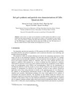
Fig. 1. (a) Electrode configuration, (b) sensor structure, and (c) schematic diagram of testing system.
dissolved in 20 ml of distilled water and a designated amount of
Na
2
WO
4
·2H
2
O was dissolved in 20 ml of distilled water. After mix-
ing the two solutions and subsequent mild stirring, the solutions
were transferred toa Teflon-lined, stainless steelautoclave (volume
100 cm
3
), sealed, and then heated at 200
◦
C for 24 h. After cooling,
the reaction products were washed with water and ethanol and
dried at 60
◦
C for 24 h. The as-prepared precursors were converted
into WO
3
by the decomposition of core carbon via heat treatment
(HT) at 450
◦
C. The samples were designated according to the molar
ratio between glucose and Na
2
WO
4
·2H
2
O (denoted as ‘R’) in the
stock solution. For example, R30 precursor and R30 powders mean
the precursors and WO
3
powders prepared from the stock solution
with R = 30, respectively. In order to investigate the effect of HT
on the morphology of the WO
3
microspheres, the heat-treatment
temperature (HTT) and duration at HTT were varied.
2.2. Characterization of sample
The morphologies of the precursors and powders were analyzed
by scanning electron microscopy (SEM, Hitachi S-4300) and trans-
mission electron microscopy (TEM, FEITecnai 20). The crystal phase
was analyzed using X-ray diffraction (XRD, Rigaku D/MAX-2500
V/PC). The surface areas of the powders were investigated using
Brunauer–Emmett–Teller (BET) method (Tristar 3000, Micromerit-
ics Co., Ltd.).
2.3. Gas sensing characteristics
The WO
3
spherical powders after HT were prepared in a paste
form and applied to an alumina substrate having two Au electrodes
(Fig. 1(a) and (b), substrate: 1.5 mm × 1.5 mm, spacing between
two electrodes: 0.2 mm). The sensor element was heat treated at
400
◦
C for 30 min to decompose the organic component of the paste.
The films after HT were ∼15 m thick. The sensor was installed
in a quartz tube and the furnace temperature was stabilized at a
constant sensing temperature (300
◦
C) (Fig. 1(c)). The gas concen-
tration was controlled by changing the mixing ratio of the parent
gases (5ppm NO
2
, 200ppm C
2
H
5
OH, 200ppm CH
3
COCH
3
, 100ppm
CO, 100 ppm C
3
H
8
, 1000 ppm CH
4
, all in air balance) and dry syn-
thetic air. A flow-through technique with a constant flow rate
(500 ml/min) was used. The gas response (S = R
a
/R
g
or R
g
/R
a
) was
measured at 400
◦
C by comparing the resistance of the sensor in
Table 1
Sample specification and composition of the stock solution for hydrothermal
reaction.
Specification [Glucose]/[Na
2
WO
4
·2H
2
O] Glucose Na
2
WO
4
·2H
2
O
R30 30 0.5 M 0.0166 M
R5 5 0.5 M 0.1 M
R0.5 0.5 0.5 M 1 M
high-purity air (R
a
) with that in the target gases (R
g
). The electri-
cal resistance sensor was monitored using a Picoammeter/Voltage
Source (Keithley 6487) interfaced with a computer.
3. Results and discussion
3.1. X-ray diffraction analysis
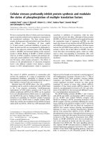
Fig. 2 shows the XRD patterns of the R30, R5, and R0.5 powders
prepared by HT of precursors at 450
◦
C for 1 h. The R30 powders
Fig. 2. X-ray diffraction patterns of the powders prepared by heat treatment of (a)
R30, (b) R5, and (c) R0.5 precursors at 450
◦
C for 1 h.
238 C Y. Lee et al. / Sensors and Actuators B 142 (2009) 236–242
showed a pure monoclinic phase of WO
3
, while the R5 and R0.5
powders were identified as Na
2
W
2
O
7
and Na
2
WO
4
, respectively.
The [Na]/[W] ratio in the Na
2
WO
4
powders prepared at R = 0.5 was
2, which was 2 times higher than the same ratio in the Na
2
W
2
O
7
powders prepared at R = 5. The Na content of the stock solution at
R =0.5 was 10 times higher than that at R = 5. Accordingly, the high
Na
2
WO
4
·2H
2
O concentration of the stock solution was thought
to have increased the Na content of the precursors and powders,
despite the removal of a large portion of Na by washing.
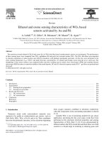
3.2. Particle morphology
The morphology of the precursors was closely dependent on
the R values. At R = 0.5 and 5, irregular precipitate morphologies
were prepared (Fig. 3(c) and (e)). In contrast, nearly mono-disperse,
spherical precursors with clean surface were prepared (Fig. 3(a)).
The morphology remained similar after HT at 450
◦
C for 1 h
(Fig. 3(b), (d) and (f)). Thus, the morphology of the powders could
be designed in the stage of hydrothermal reaction. The WO
3
micro-
spheres after HT (R30 powders) consisted of many small primary
particles (10–20 nm) (inset in Fig. 3(b)), whereas the average pri-
mary particle sizes of R5 and R0.5 powders were ∼400 nm and
∼1 m (insets in Fig. 3(d) and (f)). These results indicated that the R
values determined the phase, morphology and the primary particle
sizes of the powders.
The scale bar of Fig. 3(b) is 10 times smaller than that of Fig. 3(a).
The sizes of more than 300 spheres were measured using the SEM
observation of well-dispersed precursors and powders. The aver-
age particle diameters of the R30 precursors and R30 powders were
1300 ± 249 nm and 226 ± 41 nm, respectively, indicating that the
spherical precursors shrank to 17% of their precursor’s diameter
during HT. The significant decrease of sphere sizes during HT in
the present study can be attributed to the decomposition of the
core carbon and subsequent sintering between primary particles.
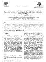
To confirm this, the R30 precursors were heat treated at a more
rapid heating rate for a shorter duration at HTT (Fig. 4). The sphere
diameters decreased with increasing thermal energy input dur-
ing HT. At a high heating rate (20
◦
C/min) and short duration at
HTT (10 min), the powders retained their black color after HT, indi-
cating the incomplete decomposition of core carbon (R30-RH-SD
Fig. 3. Scanning electron micrographs of (a) R30 precursors, (b) R30 powders, (c) R5 precursors, (d) R5 powders, (e) R0.5 precursors, and (f) R0.5 powders. The powders (b),
(d), and (f) were prepared by heat treatment of the precursors (a), (c), and (e) at 450
◦
C for 1 h, respectively.
C Y. Lee et al. / Sensors and Actuators B 142 (2009) 236–242 239
Fig. 4. The sphere diameters according to heat treatment schedules and transmission electron micrographs of (a) R30 precursors, (b) R30-RH powders heat treated at 450
◦
C
for 40 min (heating rate: 20
◦
C/min) and (c) R30 powders heat treated at 450
◦
C for 1h (heating rate: 5
◦
C/min) (RH-SD: rapid heating and short duration at heat treatment
temperature; RH: rapid heating).
powders). However, whitish yellow powders could be prepared
by increasing the duration at HTT from 10 min to 40 min (R30-RH
powders).
The TEM image of the R30 precursor showed a very clean sur-
face (Fig. 4(a)). Even the small spheres showed a completely black
contour, indicating that the precursors were not hollow but solid
and consisted of carbon and W precursors. After HT with grad-
ual heating rate and long duration at HTT (R30 powders), most
of the powders showed a solid and nano-porous morphology but
a few spheres showed a hollow morphology (arrow and inset in
Fig. 4(c)). In contrast, most of the spheres showed a hollow mor-
phology due to the rapid heating to HTT (R30-RH powders and
Fig. 4(b)), although solid and nano-porous spheres were also found.
The hollow shell was ∼30 nm thin (inset in Fig. 4(b)).
Sun and Li [12] suggested that monodisperse carbon micro-
spheres with hydrophilic surface can be prepared by the
hydrothermal reaction of glucose. The polymerization of glucose
into aromatic compounds and oligosaccharides and their subse-
quent carbonization into spheres were suggested as the formation
mechanism. The OH and CHO groups bonded to the surfaces of
the carbon spheres are advantageous for the reaction with metal
cations. Titirichi [13] prepared metal-ion-coated carbon spheres by
one-pot hydrothermalreaction of a solution containing glucose and
metal salts and transformed these spheres into metal oxide hollow
spheres by the decomposition of the core carbon.
In the present study, the hollow morphologies were found at
the R30-RH powders. This indicates that the carbon spheres with
hydrophilic surfaces are formed by the cross linking between glu-
cose in the beginning stage of reaction and W ions are coated on the
negatively charged surface of carbon spheres in the later stage of
reaction. Thus, the precursors in Fig. 4(a) can be regarded as carbon
spheres loosely coated with W-precursors. The precursor spheres
were transformed to hollow WO
3
spheres by the decomposition
of the core carbon (Fig. 4(b)) and then further transformed into
solid and nano-porous spheres by the sintering between the WO
3
primary particles within the spheres (Fig. 4(c)).
3.3. Surface area and porosity
The nitrogen adsorption–desorption isotherms plots and corre-
sponding pore-size distribution plots of R30-RH, R30, R5, and R0.5
powders are given in Fig. 5. The volumes of pores in the size range
of 2–70 nm for the R30-RH and R30 powders were markedly larger
that those for the R5 and R0.5 powders. The surface area of the R30-
RH, R30, R5, and R0.5 powders were 19.0, 12.7, 7.22, and 1.27 m
2
/g,
respectively, indicating that the spherical WO
3
powders prepared
at R = 30 (R30 and R30-RH powders) were advantageous to achieve
high gas responses. Indeed, the gas responses of the R5 and R0.5
powders to NO
2
,C
2
H
5
OH, CH
3
COCH
3
, CO, C
3
H
8
, and CH
4
were very
low (not shown), while the R30 and R30-RH powders showed very
high gas responses (as presented below). The large pore volume
of the R30 powders (Fig. 5), despite their apparently solid interior
structure (Fig. 4(c)), was attributed to their evolution from hollow
spheres. The further increase of pore volumes in the size range of
5–20 nm by the employment of rapid heating indicates that the
hollow morphology of R30-RH powders is advantageous to achieve
both of high surface area and large pore volume.
3.4. Gas sensing characteristics
As stated above, the gas responses of the R5 and R0.5 powders
were negligible. Thus, the gas responses of the R30-RH and R30
240 C Y. Lee et al. / Sensors and Actuators B 142 (2009) 236–242
Fig. 5. Nitrogen adsorption–desorptionisotherm plots and corresponding pore-size
distribution plots of R30-RH, R30, R5, and R0.5 powders.
powders at 300
◦
C were measured and the results are shown in
Fig. 6. Note that the gas response to NO
2
is the R
g
/R
a
value and
those to other gases are the R
a
/R
g
values. The increase of resis-
tance upon exposure to NO
2
in WO
3
can be explained by the
adsorption of the more negatively charged oxygen on the surface
(NO
2
(g)+e
−
→ NO(g)+ O
−
(surf)). The gas response (R
g
/R
a
)ofR30
powders to 1 ppm NO
2
was 20.1, while the responses (R
a
/R
g
)to
100 ppm CH
3
COCH
3
, CO, C
3
H
8
,CH
4
, and C
2
H
5
OH were very low
(1.1–3.3) (Fig. 6(b)). The concentration of NO
2
was 1/100 of those of
the other gases. These results demonstrated the high gas response
and selective detection of WO
3
nano-porous microspheres to NO
2
.
The selective detection and gas responses were enhanced further
by the use of hollow R30-RH powders. The R
g
/R
a
value to 1 ppm NO
2
was increased to 53.9 whereas those to the other gases decreased
to 1.1–1.9 (Fig. 6(a)).
Fig. 6. Gas responses to 1 ppm NO
2
and 100 ppm CH
3
COCH
3
,CO,C
3
H
8
,CH
4
, and
C
2
H
5
OH at 300
◦
C in air: (a) R30-RH powders heat treated at 450
◦
C for 40 min (heat-
ing rate: 20
◦
C/min) and (b) R30 powders heat treated at 450
◦
C for 1 h (heating rate:
5
◦
C/min). The gas response to NO
2
is the R
g
/R
a
value and those to other gases are
the R
a
/R
g
values.
The sensing transients to 0.5–2.5 ppm NO
2
of the R30-RH and
R30 powders at 300
◦
C are shown in Fig. 7(a) and (b), respectively.
The sensor resistance in air (R
a
) was rather high (40–50 M). When
exposed to NO
2
, the sensor resistance greatly increased up to a few
G level. The gas responses of the R30-RH sensor to 0.5–2.5 ppm
NO
2
were 38.6–81.5, which were 2.2–2.9 times higher than those
of the R30 sensor (13.4–36.5) (Fig. 7(c)).
Table 2 summarizesthe NO
2
responses of theundoped WO
3
sen-
sors estimated from the literature data [16–27]. Most of the WO
3
sensors were operated at 100–300
◦
C and the response to NO
2
var-
ied greatly according to the morphology and preparation method
of the sensor materials. Our result of R
g
/R
a
= 53.9 to 1 ppm NO
2
is
compares very favorably to the values reported in the literature
[16–27], although a few studies [19,20,22,25] have reported higher
gas responses.
The higher gas response of the R30 microspheres than those
of the R5 and R05 powders was attributed to the higher surface
area for gas sensing reaction and the large pore volume for the
effective gas diffusion (Fig. 5). The gas response was improved fur-
Fig. 7. Dynamic sensing transients of (a) R30-RH and (b) R30 sensors and (c) gas responses (R
g
/R
a
) of R30-RH and R30 sensors to 0.5–2.5 ppm NO
2
.
C Y. Lee et al. / Sensors and Actuators B 142 (2009) 236–242 241
Table 2
Gas responses to NO
2
in the present study and those reported in the literature [16–27].
WO
3
sensing materials (preparation) [NO
2
] R
g
/R
a
Sensing temperature (
◦
C) Reference
Hollow microspheres 1 ppm 53.9 300 Present study
Thin film (thermal evaporation) 0.5 ppm ∼22 100 [16]
Thin film (rf sputtering) 1 ppm ∼5 370 [17]
Thin film (vacuum thermal deposition) 1 ppm ∼4 200 [18]
Thin film (Aerosol-assisted CVD) 0.2 ppm ∼80 150 [19]
Thin film (reactive magnetron sputtering) 1 ppm ∼450 200 [20]
Nanocrystalline powders 5 ppm ∼75 150 [21]
Lamellar structures (acidification) 1 ppm ∼280 200 [22]
Nanostructures (thermal evaporation) 5 ppm ∼3 250 [23]
WO
3
nanopetals (dealloying of W–Al alloy and thermal oxidation) 5 ppm ∼4 250 [24]
NWs network (thermal evaporation) 1 ppm ∼150 300 [25]
NWs array (thermal evaporation) 1 ppm ∼10 180 [26]
Mesoporous nanostructures 1 ppm ∼10 230 [27]
ther by the employment of a more hollow morphology through
the rapid decomposition of the core carbon (R30-RH powders).
Although the R30 powders are relatively porous, the diffusion of
NO
2
to the centerof the WO
3
secondary spheres canbe hampered in
part by thetortuousconfiguration of the nanopores,which becomes
more significant when the secondary spheres become larger. This
decreases the gas response because only the surface region of the
microspheres changes the resistance by the gas sensing reaction.
In contrast, a hollow morphology with a thin shell configuration
and large pore volume facilitates the in-diffusion of the sensing gas
and counter-diffusion of the reactant gas. With effective gas diffu-
sion, the resistance of all the primary particles within the secondary
hollow spheres is affected upon NO
2
exposure, which increases the
gas response. Thus, the WO
3
hollow microspheres examined in the
present study are promising sensing materials to detect NO
2
with
a high sensitivity and selectivity.
4. Conclusion
WO
3
hollow microspheres were prepared by the glucose-
mediated, hydrothermal synthesis of W-coated carbon spheres and
their calcination at 450
◦
C. With the input of increasing thermal
energy during calcination, the hollow WO
3
microspheres were
gradually transformed into solid microspheres. Both hollow and
solid WO
3
microspheres showed high response and selective detec-
tion to 0.5–2.5 ppm NO
2
. In particular, the responses to NO
2
were
increased 2.2–2.9 times by using hollow morphology, which was
attributed to the high surface area for gas sensing and the effec-
tive diffusion of NO
2
toward all the primary particles through the
nano-porous and thin shell layers.
Acknowledgements
This work was supported by KOSEF NRL program grant funded
by the Korean Government (MEST) (No. R0A-2008-000-20032-0)
and a grant from the Fundamental R&D program for Core Tech-
nology of Materials (M2008010013) funded by the Ministry of
Knowledge Economy, Republic of Korea.
References
[1] F. Caruso, Nanoengineering of particle surfaces, Adv. Mater. 13 (2001) 11–22.
[2] R. Meyer Jr., H. Weitzing, Q. Xu, Q. Zhang, R.E. Newnham, Lead zirconate titanate
hollow-sphere transducers, J. Am. Ceram. Soc. 77 (1994) 1669.
[3] S. Han, B. Jang, T. Kim, S.M. Oh, T. Hyeon, Simple synthesis of hollow tin dioxide
microspheres and their applications to lithium-ion battery anodes, Adv. Funct.
Mater. 15 (2005) 1845–1850.
[4] J H. Lee, Gas sensors using hierarchical and hollow oxide nanostructures:
overview, Sens. Actuators B. 140 (2009) 313–336.
[5] Q. Zhao, Y. Gao, X. Bai,C.Wu,Y. Xie,FacilesynthesisofSnO
2
hollow nanospheres
and applications in gas sensors and electrocatalysts, Eur. J. Inorg. Chem. (2008)
1643–1648.
[6] V. Jokanovi
´
c, A.M. Spasi
´
c, D. Uskokovi
´
c, Designing of nanostructured hollow
TiO
2
spheres obtained by ultrasonic spray pyrolysis, J. Colloid Interface Sci. 278
(2004) 342–352.
[7] X.W. Lou, Y. Wang, C. Yuan, J.Y. Lee, L.A. Archer, Template-free synthesis of
SnO
2
hollow nanostructures with high lithium capacity, Adv. Mater. 18 (2006)
2325–2329.
[8] Y. Yin, R.M. Rioux, C.K. Erdonmez, S. Hughes, G.A. Somorjai, A.P. Alivisatos, For-
mation of hollow nanocrystals through the nanoscale Kirkendall effect, Science
30 (2004) 711–714.
[9] F. Caruso, X. Shi, R.A. Caruso, A. Susha, Hollow titania spheres from layered
precursor deposition onsacrificial colloidal core particles, Adv.Mater. 13 (2001)
740–744.
[10] N. Kawahashi, E. Matijevi
´
c, Preparation and properties of uniform coated col-
loidal particles, J. Colloid Interface Sci. 138 (1990) 534–542.
[11] J Y. Lee, J H. Lee, S H. Hong, Y.K. Lee, J Y. Choi, Coating of BaTiO
3
nano-layer
on spherical Ni powders for MLCC, Adv. Mater. 15 (2003) 1655–1658.
[12] X. Sun, Y. Li, Colloidal spheres and their core/shell structures with noble-metal
nanoparticles, Angew. Chem. 116 (2004) 607–611.
[13] M M. Titirici, M. Antonietti, A. Thomas, A generalized synthesis of metal
oxide hollow spheres using a hydrothermal approach, Chem. Mater. 18 (2006)
3808–3812.
[14] H.X. Yang, J.F. Qian, Z.X. Chen, X.P. Ai, Y.L. Cao, Multilayered nanocrystalline
SnO
2
hollow microspheres synthesized by chemically induced self-assembly
in the hydrothermal environment, J. Phys. Chem. C 111 (2007) 14067–
14071.
[15] J. Yu, X. Yu, Hydrothermal synthesis and photocatalytic activity of zinc oxide
hollow spheres, Environ. Sci. Technol. 42 (2008) 4902–4907.
[16] A. Ponzoni, E. Comini, M. Ferroni, G. Sberveglieri, Nanostructured WO
3
deposited by modified thermal evaporation for gas-sensing applications, Sens.
Actuators B 490 (2005) 81–85.
[17] M. Stankova, X. Vilanova, E. Llobet, J. Calderer, C. Bittencourt, J.J. Pireaux, X.
Correig, Influence of the annealing and operating temperatures on the gas-
sensing properties of rf sputtered WO
3
thin-film sensors, Sens. Actuators B 105
(2005) 271–277.
[18] G. Xie, J. Yu, X. Chen, Y. Jiang, Gas sensing characteristics of WO
3
vacuum
deposited thin films, Sens. Actuators B 123 (2007) 909–914.
[19] S. Ashraf, C.S. Blackman, R.G. Palgrave, S.C. Naisbitt, I.P. Parkin, Aerosol assisted
chemical vapour deposition of WO
3
thin films from tungsten hexacarbonyl and
their gas sensing properties, J. Mater. Chem. 17 (2007) 3708–3713.
[20] Y. Shen, T. Yamazaki, Z. Liu,D. Meng, T. Kikuta, N. Nakatani, Influence of effective
surface area on gas sensing properties of WO
3
sputtered thin films, Thin Solid
Films 517 (2009) 2069–2072.
[21] H. Xia, Y. Wang, F. Kong, S. Wang, B. Zhu, Z. Guo, J. Zhang, Y. Wang, S. Wu,
Au-doped WO
3
-based sensor for NO
2
detection at low operating temperature,
Sens. Actuators B 134 (2008) 133–139.
[22] T. Kida, A. Nishiyama, M. Yuasa, K. Shimanoe, N. Yamazoe, Highly sensitive NO
2
sensors using lamellar-structured WO
3
particles prepared by an acidification
method, Sens. Actuators B 135 (2009) 568–574.
[23] T. Sicilliano, A. Tepore, G. Micocci, S.D. Manno, E. Filippo, WO
3
gas sensors
prepared by thermal oxidation of tungsten, Sens. Actuators B 133 (2008) 321–
326.
[24] Z. Liu, T. Yamazaki, Y. Shen, D. Mang, T. Kikuta, N. Nakatani, T. Kawabata, Deal-
loying derived synthesis of W nanopetal films and their transformation into
WO
3
, J. Phys. Chem. C 112 (2008) 1391–1395.
[25] A. Ponzoni, E. Comini, G. Sberveglieri, Z. Zhou, Z. Deng, N.S. Xu, Y. Ding, Z.L.
Wang, Ultrasensitive and highly selective gas sensors using three-dimensional
tungsten oxide nanowires networks, Appl. Phys. Lett. 88 (2006) 203101.
[26] B. Cao, J. Chen, X. Tang, W. Zhou, Growth of monoclinic WO
3
nanowire array
for highly selective NO
2
detection, J. Mater. Chem. 19 (2009) 2323–2327.
242 C Y. Lee et al. / Sensors and Actuators B 142 (2009) 236–242
[27] Rossinyol, A. Prim, E. Pellicer, J. Arbiol, F. Hernández-Ramírez, F. Peiró, A. Cornet,
J.R. Morante, L.A. Solovyov,B. Tian, T. Bo, D. Zhao,Synthesis and characterization
of chromium-doped mesoporous tungsten oxide for gas sensing applications,
Adv. Funct. Mater. 17 (2007) 1801–1806.
Biographies
Choong-Yong Lee studied materials science and engineering and received his BS
degree from Korea University in 2007. He is currently a master course student at
Korea University. His research involves the preparation of oxide nanostructures for
gas sensor applications.
Sun-Jung Kim studied materials science and engineering and received his BS and
MS degrees in 2006 and 2008, respectively, at Korea University in Korea. He is cur-
rently studying for PhD degree at Korea University. His research interests are oxide
nanostructures for chemical sensor applications and the combinatorial design of gas
sensing materials.
In-Sung Hwang studied materials science and engineering and received his BS from
Kumoh National University, Korea, in 2004. In 2006, he received his MS degree from
Korea University. He is currently studying for a PhD at Korea University. His research
interest is oxide nanostructure-based electronic devices.
Jong-Heun Lee has been a Professor at Korea University since 2008. He received
his BS, MS, and PhD degrees from Seoul National University in 1987, 1989, and
1993, respectively. Between 1993 and 1999, he developed automotive air–fuel-
ratio sensors at the Samsung Advanced Institute of Technology. He was a Science
and Technology Agency of Japan (STA) fellow at the National Institute for Research
in Inorganic Materials (currently NIMS, Tsukuba, Japan) from 1999 to 2000, a
research professor at Seoul National University from 2000 to 2003, and an asso-
ciate professor at Korea University from 2003 to 2008. His current research
interests include chemical sensors, functional nanostructures, and solid oxide
electrolytes.