- Trang chủ >>
- Khoa Học Tự Nhiên >>
- Vật lý
synthesis of single-crystalline hollow b-feooh nanorods via a controlled
Bạn đang xem bản rút gọn của tài liệu. Xem và tải ngay bản đầy đủ của tài liệu tại đây (490.74 KB, 8 trang )
Brief communication
Synthesis of single-crystalline hollow b-FeOOH nanorods via a controlled
incomplete-reaction course
Haiyun Yu, Xinyu Song, Zhilei Yin, Weiliu Fan, Xuejie Tan, Chunhua Fan and Sixiu Sun*
Key Laboratory of Colloid and Interface Chemistry, Shandong University, Ministry of Education, Jinan,
250100, People’s Republic of China; *Author for correspondence (Tel.: +86-0531-88364879; Fax: +86-
0531-88564464; E-mail: )
Received 6 August 2005; accepted in revised form 21 October 2005
Key words: FeOOH, nanorod, hollow, dissolution-recrystallization, colloids
Abstract
The single-crystalline b-FeOOH hollow nanorods with a diameter ranging from 20$30 nm and length in
the range of 70–110 nm have been successfully synthesized through a two-step route in the solution. The
phase transformation and the morphologies of the hollow b-FeOOH nanorods were investigated with
X-ray powdered diffraction (XRD) , scanning electron microscopy (SEM), transmission electron micros-
copy (TEM), selected area electric diffraction (SAED), high-resolution transmission electron microscopy
(HRTEM), infrared spectrum (IR) and thermo-gravimetric analysis (TGA). These studies indicate that the
first step is an incomplete-reaction course. Furthermore, The formation mechanism of the hollow nanorods
has been discussed. It is found that the mixed system including chitosan and n-propanol is essential for the
final formation of the hollow b-FeOOH nanorods.
Introduction
Recently, much effort has been devoted to syn-
thesis of hollow inorganic materials because of
their low density and high surface area compared
with bulk materials (Sun & Xia, 2004; Wang et al.,
2004). These materials may be found a wide range
of potenti al applications in many areas, such as
catalysts, potential drug carriers, coatings, low-
density materials and nanoreactor (Mathlowitz
et al., 1997; Caruso et al., 1998; Huang et al.,
1999; Fowler et al., 2001). Many hollow inorganic
materials including metals, non-oxides and metal
oxides have been synthesized (Sun & Xia, 2002;
Peng et al., 2003; He et al., 2004; Liu & H.C.
Zeng, 2004; Yang & Zeng, 2004). The general
approach for synthesizing such materials is based
on the use of hard-template or soft-template such
as polystyrene beads, colloid particles, emulsions,
vesicles and droplets. Moreover, most of products
are polycrystalline submicrometer spheres aggre-
gated by nanoparticles. To our best knowledge,
only several non-spheres and single-crystallin e
hollow structures have been prepared (Chen et al.,
2003; Jiang et al., 2004; Sun & Xia, 2004).
The b-FeOOH has a large tunnel-type structure
where iron atoms are strongly bonded to the
framework. Lithium can be intercalated and
extracted freely in the tunnels during discharge
and charge processes. As a promising candidate
for an electrode material, b-FeOOH exhibits good
electrochemical performance with a high theoreti-
cal discharge capacity (Flynn, 1984; Kanno et al.,
1996; Amine et al., 1999). Recently, a self sup-
ported-pattern of oriented alignment of b-FeOOH
nanowires fabricated through means of a
Journal of Nanoparticle Research (2007) 9:301–308 Ó Springer 2007
DOI 10.1007/s11051-005-9054-5
low-temperature solution route was reported by
Xiong and his co-workers (Xiong et al., 2003). The
b-FeOOH is also used as an iron source to prepare
other iron compounds with special morphologie s.
Peng et al. reported that single-crystal magnetite
nanorods could be formed by hydrothermal
reduction of b-FeOOH nanorods (Peng et al.,
2005).
In this paper, we present a novel controlled
incomplete-reaction course for fabricating single-
crystalline b-FeOOH hollow nanorods with length
in the range of 70–110 nm and width in the range
of 20–30 nm. In particular, a process mechanism
has been revealed for synthesis of single-crystalline
b-FeOOH hollow nanorods: (i) formation of
b-FeOOH nanorods by aggregation-dehydration
of most amorphous Fe(OH)
3
; (ii) decomposition
of residual Fe(OH)
3
inside the nanorods to H
2
O
and b-FeOOH; (iii) crystal aging and hollowing of
b-FeOOH nanorods by a dissolution-recrystalli-
zation process.
Experimental details
A chitosan (the degree of deacetylateion is 55%)
solution (CS) was prepared by mixing 1.5 g
chitosan into 100 ml 3% acetic acid solution.
Other agents used in this work were analytic
grades. In a typical experiment, 2 ml CS was
added into 15 ml 0.3 M FeCl
3
solution, followed
by an addition of 15 ml n-propanol and 0.408 g
urea. In the first step, the mixed solution was put
into a three-necked flask, which was heated and
maintained at 82°C for 5 h under stirring. After
centrifugalized, a yellow precipitate was obtained.
The product was repeatedly washed with anhy-
drous ethanol. In the second step, the washed
precipitate was dispersed into 20 ml anhydrous
ethanol, and then transferred into a stainless
autoclave with a PTFE (polytetrafluoroethylene)
container of 25 ml and maintained at 180°C for
15 h. Subsequently the autoclave was allowed to
cool down naturally. The yellow precipitates were
collected, and washed with anhydrous ethanol
several times. Finally, the product was dried at
60°C in air.
XRD measurements of the as-prepared sample
were carried on a Japan Rigaku D/max-c A 200
X-ray diffractometer with CuKa radiation
(k=1.54178 A
˚
). SEM images were obtained on a
JSM-6700F scanning electric microscope (JEOL).
TEM images were taken on a JEM-100CXII
transmitting electric microscope (JEOL), operat-
ing at 80 kV. TEM analysis was prepared by
placing a drop of colloid al solution onto the
formvar-covered copper grid. HRTEM images
were obtained on Technai F30 at 300 kV. FT-IR
spectra of all the samples were measured with a
Bio- Rad model FTS-165 IR spectrometer. TGA
was conducted on Mettler Toledo SDTA851e
under a N
2
atmosphere and a heating rate of
20°C min
)1
.
Results and discussion
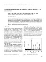
The XRD pattern of the final product is shown in
Figure 1b. All the diffraction peaks can be indexed
as b-FeOOH crystals with a monoclinic structure
(JCPDS Card No. 80–1770, Fe
8
O
8
(OH)
8
Cl
1.35
,a
kind of b-FeOOH, Akaganeite, a=1.060 nm,
b=0.3034 nm and c=1.051 nm). SEM, TEM and
HRTEM images of the final product are shown in
Figure 2. The center portion of structure is lighter
than that the edge, confirming the hollow interiors
of the nanorods in Figure 2b. It can be seen that
b-FeOOH hollow nanorods have an average
diameter of 20$30 nm and an aspect ratio above
3$4. Almost there is only one big cavity in each
particle with length of 50$70 nm and width above
10 nm. The Fast Fourier Transform (FFT) image
in the inset indicates the single crystalline nature of
the single hollow nanorod and the nanorods
growth along the [110] direction.
The intermediate products at different reaction
periods were used as the samples for the TEM and
SAED characterizations (Figure 3) to track the
formation of the hollow b-FeOOH nanorods.
After maintained at 82°C for 20 min, amorphous
nanoparticles of Fe(OH)
3
can be observed in the
TEM image (Figure 3a). Along with the longer of
the heated-time, solid nanorods are obtained
(Figure 3b and e). According to the XRD pattern
of these solid nanorods (Figure 1a), the crystallo-
graphic phase is b-FeOOH. Apparently, these
nanorods are formed by an aggregation-dehydra-
tion process of amorphous Fe(OH)
3
nanoparticles
(Sugimoto & Muramatsu, 1996). From the SAED
parrtens (there are many particles included in the
selected area) shown in the corner, the crystalline
of b-FeOOH solid nanorods is not well, which
302
implies these particles including the component of
Fe(OH)
3
(It can be proved by IR spectra and TG
in the following text). Figure 3c shows the transi-
tion state of nanorods in the ethanol-thermal
reaction at 180°C for 2 h. It is clearly that solid
nanorods begin to change into porous nanorods.
Figure 3d and f show the morphologies of the last
products, which demonstrate that the small inter-
spaces in the nanorod have coalesced into a single
void and the size of products is smaller than that in
Figure 3e. The SAED pattern in Figure 3f indi-
cates that the crystalline of products is better than
that of Figure 3e. The SAED patterns also show
the crystallographic phase of nanorods is still
b-FeOOH in Figure 3e and f, which can be vali-
dated by XRD patterns in Figure 1.
FTIR spectroscopy and TGA were employed to
investigate the information of the inter mediate
products and final products, which could be
helpful to research the formation mechanism of
the hollow structures. As shown in IR spectrum
(Figure 4), the absorption at 672.46 cm
)1
in Fig-
ure 4a is the characteristic vibration of Fe(OH)
3
.
The absorptions at 692.27 and 632.92 cm
)1
in
Figure 4b are the characteristic vibrations of Fe–O
in b-FeOOH (Sugimoto et al., 1998). These
information implies the presence of Fe(OH)
3
in the
b-FeOOH before an ethanol-thermal process. The
band at 1630 cm
)1
is attributed to the N–H
vibration and the band at 1555 cm
)1
is assigned to
the vibration of acidamide in chitosan (Guan &
Cheng, 2004), which indicate the existence of
chitosan in the intermediate products.
The results from TGA (Figure 5) are in good
agreement with the data from IR spectra. The first
weight loss in Figure 5a and b may be attributed
to the emission of absorbed alcohol and H
2
O. The
last weight loss in two samples may be ascribed to
the decomposition of the residual chitosan. The
second weight loss with 10.11 wt% in Figure 5b is
attributed the transition from b-FeOOH to Fe
2
O
3
(the theoretical calculation is 10.11 wt%). In Fig-
ure 5a, the middle weight loss can be divided three
steps and the weight loss rate is 24.22 wt% that
exceeds the decomposition of the pure b-FeOOH.
Therefore, these weight losses are ascribed to the
decomposition of Fe(OH)
3
and b-FeOOH and the
empietement of these two decompositions.
On the basis of the above results, we proposed
the formation mechanism of the hollow structures.
The information about the intermediate products
showed that the first step was an incomplete
reaction course. During the aggregation-dehydra-
tion, not all of the amorphous Fe(OH)
3
nanopar-
ticles formed b-FeOOH solid nanorods. There was
still some amorphous Fe(OH)
3
remained within
the b-FeOOH solid nanorods. At the same time,
chitosan and n-propanol absorbed onto the sur-
faces of Fe(OH)
3
and b-FeOOH nanoparticles
by their interaction. In the second step of the
Figure 1. XRD patterns of b-FeOOH nanorods: (a) b-FeOOH nanorods gained by maintained 5 h at 82°C before ethanol-
thermal reaction, (b) hollow b-FeOOH nanorods after ethanol-thermal reaction.
303
preparation, with a longer ethanol-thermal process
time, the remained Fe(OH)
3
began to decompose
into H
2
Oandb-FeOOH. As shown in Figure 3c,
the solid nanorods changed into porous nanorods.
The chitos an and n-propanol absorbed on the
surface of the b-FeOOH nanorods could coact
with each other and produce a more compact resist
(Cason et al., 2001). H
2
O from the decomposition
of Fe(OH)
3
was restricted in the b-FeOOH nano-
rods interior by this resist to avoid entering into
the bulk solution. Under the ethanol-thermal
condition, the existence of H
2
O led to a dissolu-
tion-recrystallization process of b-FeOOH (Su-
gimoto & Muramatsu, 1996):
H
2
O Ð OH
À
þ H
þ
ð1Þ
Fe
8
O
8
ðOHÞ
8
Cl
1:35
þ H
þ
Ð Fe
3þ
þ H
2
O þ Cl
À
ð2Þ
This process was restricted within the nanorods
due to the existence of H
2
O only within the nano-
rods. Because the equilibrium solute concentration
Figure 2. SEM, TEM and HRTEM images of b-FeOOH hollow nanorods prepared as above experiment: (a) SEM image of
b-FeOOH hollow nanorods, (b) TEM image of b-FeOOH hollow nanorods, (c) HRTEM image and its related Fourier
transform electron diffraction pattern of a single hollow Nanorod.
304
near a small void is higher than near a large void,
as described by the Gibbs–Thompson equation.
Along with the process of the dissolution-recrys-
tallization, small voids will coalesce into a large
void (Yin et al., 2004). If under the conditions of
the absence of chitosan or the presence of H
2
Oin
the second step solution, the mass transfer could
be found between the b-FeOOH nanorods through
the dissolution of H
2
O in the bulk solution. As a
result shown in Figure 6a and b, large a-Fe
2
O
3
particles were obtained in the second step (Sha
et al., 2004), which could be proved in our further
experiments (Table 1). The synergism of chitosan
and n-propanol prohibited the transition from
b-FeOOH to a-Fe
2
O
3
through restricting H
2
O
only within the nanorods. Furthermore, the
aggregation of b-FeOOH nanorods was prevented
by this synergism too. Therefore, it is necessary
that the synergism of chitosan and n-propanol for
the preparation of hollow b-FeOOH nanorods. In
addition, the similar hollow b-FeOOH nanoparti-
cles also can be gained by changing n-propanol
with ethanol or isopropanol in the first step
(Figure 6c and d), which indicated that the syn-
ergism also occurred between ethanol and chitosan
or between isopropanol and chitosan.
In order to examine the processing parameters
that control the morphology, size and structural
properties of the hollow b-FeOOH nanorods, the
factors including the amount of chitosan, reaction
Figure 3. Schematic illustration of the cavity forming process. (TEM images) Evolution of b-FeOOH hollow nanorods: (a)
maintained 0.2 h at 82°C, (b) and (e) maintained 5 h at 82°C, (c) ethanol-thermal 2 h at 180°C, (d) and (f) ethanol-thermal
15 h at 180°C.
305
Figure 4. IR spectra of the b-FeOOH nanorods: (a) b-FeOOH nanorods gained by maintained 5 h at 82°C before ethanol-
thermal reaction, (b) hollow b-FeOOH nanorods after ethanol-thermal reaction.
Figure 5. Thermo-gravimetric analysis (TGA) of the b-FeOOH nanorods: (a) b-FeOOH nanorods gained by maintained 5 h
at 82°C before ethanol-thermal reaction, (b) hollow b-FeOOH nanorods after ethanol-thermal reaction.
306
temperature and time were investigated. The sizes
of the final products can be controlled by changing
the first step reaction-time from 2 to 7 h and
reaction temperature from 70 to 88°C. The shorter
of the time and the higher of the temperature are
chosen, the smaller of the product size will be.
Conclusion
In summary, a new method for preparation of
single-crystal b-FeOOH nanorods with hollow
interiors by controlling the phase transition
degree from Fe(OH)
3
to b-FeOOH without any
template has been demonstrated. In this experi-
ment, a dissolution-recrystallization process has
been conduced within the nanorods by small
quantity of water that comes from the decom-
pose of residual Fe(OH)
3
in the b-FeOOH
nanorods. This concept may be applicable to
fabricate other hollow inorganic structures, and
these hollow nanoparticles may be used as pri-
mary building blocks to fabricate curved archi-
tectures.
Figure 6. TEM images of the a-Fe
2
O
3
particles and hollow b-FeOOH nanorods under the different conditions in Table1: (a)
a-Fe
2
O
3
particles gained by direct maintained the first step suspension at 180°C for 15 h without a water removal process, (b)
a-Fe
2
O
3
particles gained at the absence of the chitosan in the first step, (c) hollow b-FeOOH nanorods prepared by changing
n-propanol with ethanol in the first preparation step, (d) hollow b-FeOOH nanorods prepared by changing n-propanol with
isopropanol in the first preparation step.
Table 1. Experimental conditions used in the control experiments
Sample Raw material
in the first step
Solvent in
the first step
Mixed system
in the second step
Final
product
Morphology
a FeCl
3
, urea, CS 15 ml n-propanol, 15 ml H
2
O The first step suspension aFe
2
O
3
Quasi-cubic submicroparticles
b FeCl
3
, urea 15 ml n-propanol, 15 ml H
2
O b-FeOOH, ethanol a-Fe
2
O
3
Sphere submicroparticles
c FeCl
3
, urea, CS 15 ml ethanol, 15 ml H
2
O b-FeOOH, ethanol b-FeOOH Hollow nanorods
d FeCl
3
, urea, CS 15 ml isopropanol, 15 ml H
2
O b-FeOOH, ethanol b-FeOOH Hollow nanorods
307
References
Amine K., H. Yasuda & M. Yamachi, 1999. b-FeOOH, a new
positive electrode material for lithium secondary batteries. J.
Power Sources 81, 221–223.
Caruso F., R.A. Caruso & H. Mo
¨
hwald, 1998. Nanoengineer-
ing of inorganic and hybrid hollow spheres by colloidal
templating. Science 282, 1111–1114.
Cason J.P., M.E. Miller, J.B. Thompson & C.B. Roberts, 2001.
Solvent effects on copper nanoparticle growth behavior in
AOT reverse micelle systems. J. Phys. Chem. B. 105, 2297–
2302.
Chen D., D. Chen, X. Jiao & Y. Zhao, 2003. Hollow-structured
hematite particles derived from layered iron (hydro)oxyhy-
droxide–surfactant composites. J. Mater. Chem. 13, 2266–
2270.
Flynn C.M. Jr, 1984. Hydrolysis of inorganic iron(III) salts.
Chem. Rev. 84, 31–41.
Fowler C.E., D. Khushalani & S. Mann, 2001. Facile synthesis
of hollow silica microspheres. J. Mater. Chem. 11, 1968–
1971.
Guan H. & X. Cheng, 2004. Study of cobalt(II)-chitosan
coordination polymer and its catalytic activity and selectivity
for vinyl monomer polymerization. Polym. Adv. Technol. 15,
89–92.
He T., D. Chen, X. Jiao, Y. Xu & Y. Gu, 2004. Surfactant-
assisted solvothermal synthesis of Co
3
O
4
hollow spheres with
oriented-aggregation nanostructures and tunable particle
size. Langmuir 20, 8404–8408.
Huang H., E.E. Remsen, T. Kowalewski & L. Karen, 1999.
Nanocages derived from shell cross-linked micelle templates.
J. Am. Chem. Soc. 121, 3805–3806.
Jiang Z.Y., Z.X. Xie, X.H. Zhang, S.C. Lin, S.Y. Xie, R.B.
Huang & L.S. Zheng, 2004. Synthesis of single-crystalline
ZnO polyhedral submicrometer-sized hollow beads using
laser-assisted growth with ethanol droplets as soft templates.
Adv. Mater. 16, 904–907.
Kanno R., T. Shirane, Y. Kawamoto, Y. Takeda, M. Takano,
M. Ohashi & Y. Yamaguchi, 1996. Synthesis, structure, and
electrochemical properties of a new lithium iron oxide,
LiFeO
2
, with a corrugated layer structure. J. Electrochem.
Soc. 143, 2435–2442.
Liu B. & H.C. Zeng, 2004. Mesoscale organization of CuO
nanoribbons: formation of ‘‘Dandelions’’. J. Am. Chem. Soc.
126, 8124–8125.
Mathlowitz E., J.S. Jacob, Y.S. Jong, G.P. Carino, D.E.
Chickering, P. Chaturvedl, C.A. Santos, K. Vijayaraghavan,
S. Montgomery, M. Bassett & C. Morrell, 1997. Biologically
erodable microspheres as potential oral drug delivery sys-
tems. Nature 386, 410–414.
Peng Q., Y. Dong & Y. Li, 2003. ZnSe semiconductor hollow
microspheres. Angew. Chem. Int. Ed. 42, 3027–3230.
Peng Z.M., M. Wu, Y. Xiong, J. Wang & Q. Chen, 2005.
Synthesis of magnetite nanorods through reduction of
b-FeOOH. Chem. Lett. 34, 636–634.
Sha G., T. Wang, J. Xiao & C. Liang, 2004. A mild
solvothermal route to a-Fe
2
O
3
nanoparticles. Mate. Res.
Bull. 39, 1917–1921.
Sugimoto T. & A. Muramatsu, 1996. Formation mechanism of
monodispersed a-Fe
2
O
3
particles in dilute FeCl
3
solutions.
J. Colloid Interf. Sci. 184, 626–638.
Sugimoto T., H. Itoh & T. Mochida, 1998. Shape control of
monodisperse hematite particles by organic additives in the
gel–sol system. J. Colloid Interf. Sci. 205, 42–52.
Sun Y. & Y. Xia, 2002. Shape-controlled synthesis of gold and
silver nanoparticles. Science 298, 2176–2179.
Sun Y. & Y. Xia, 2004. Mechanistic study on the replace-
ment reaction between silver nanostructures and chloroau-
ric acid in the aqueous medium. J. Am. Chem. Soc. 126,
3892–3901.
Wang L., Y. Ebina, K. Takada & T. Sasaki, 2004. Ultrathin
hollow nanoshells of manganese oxide. Chem. Commun.
1074–1075.
Xiong Y.J., Y. Xie, S. Chen & Z. Li, 2003. Fabrication of self-
supported patterns of aligned b-FeOOH nanowires via a
low-temperature solution reaction. Chem. Eur. J. 9, 4991–
4996.
Yang H.G. & H.C. Zeng, 2004. Preparation of hollow anatase
TiO
2
nanospheres via Ostwald ripening. J. Phys. Chem. B.
108, 3492–3495.
Yin Y., R.M. Rioux, C.K. Erdonmez, S. Hughes, G.A.
Somorjai & A. PaulAlivisatos, 2004. Formation of hollow
nanocrystals through the nanoscale Kirkendall effect. Science
304, 711–714.
308