- Trang chủ >>
- Khoa Học Tự Nhiên >>
- Vật lý
tem study on the formation process of tio2 nanotubes
Bạn đang xem bản rút gọn của tài liệu. Xem và tải ngay bản đầy đủ của tài liệu tại đây (68.87 KB, 4 trang )
Chinese Chemical Letters Vol. 14, No. 4, pp 419 – 422, 2003
419
TEM Study on the Formation Process of TiO
2
Nanotubes
Jing Wei ZHANG, Xin Yong GUO, Zhen Sheng JIN*, Shun Li ZHANG,
Jing Fang ZHOU, Zhi Jun ZHANG*
Lab of Special Functional Materials, Henan University, Kaifeng 475001
Abstract: The process, that the polycrystalline TiO
2
powders were converted into TiO
2
nanotubes,
was observed with transmission electron microscope. The results obtained indicated that in
concentrated NaOH aqueous solution, anisotropic swelling appears on the polycrystalline TiO
2
granula at first, and then the nanotubes are formed.
Keywords: TiO
2
nanotubes, anisotropic swelling, TEM.
Since the carbon nanotubes were discovered by Iijima
1
, the tube-shaped nano-structured
material attracted extensive attention, owing to their novel properties. Various methods,
such as electric arc discharging of graphite or pyrolysis of small molecule hydrocarbon
was used for the preparation of carbon nanotubes
2-4
. In 1998, Kasuga et al. found that
the TiO
2
nanotubes can be obtained through the treatment of the powdered
polycrystalline TiO
2
in concentrated NaOH solution
5
. Afterwards, we studied its
morphological structure and physicochemical properties
6
. Obviously, the formation
process and mechanism of TiO
2
nanotube is different from that of carbon nanotubes.
Study on its formation process will helpful to understand the mechanism of formation of
TiO
2
nanotubes in the solution.
In this paper, using transmission electron microscope (TEM), the formation process
of TiO
2
nanotubes in the concentrated NaOH solution was investigated. The results
indicated that TiO
2
nanotubes are formed in the stage of alkali treatment of polycry-
stalline TiO
2
, and not in the stage of the acid treatment following the alkali treatment.
This conclusion differs from Kusuga
7
.
100 mL of 9 mol/L NaOH aqueous solution was placed in a PTFE bottle, equipped
with a reflux condenser. Then, the bottle was placed in an oil bath. When the aqueous
solution was heated up to 110
o
C, 10 g anatase TiO
2
was added and stirred magnetically.
An aliquot of sample was withdrawn after 2, 12, 30 min, and their morphological
structures were observed using TEM. After 20 h, the reaction was ceased. When the
reaction mass was cooled down to room temperature, the solid was separated and washed
with deionized water repeatedly until the conductivity of the supernatant reached 0.8
µs/cm. Then a half of the solid was soaked in 0.1 mol/L HCl solution for 10 min, and
*
E-mail:
Jing Wei ZHANG et al.
420
washed with deionized water again until the conductivity reached 5.0 µs/cm. The
morphology and Na
+
content were determined by TEM and EDS.
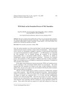
Figure 1a and 1b show the TEM images of the raw TiO
2
and the sample treated
with alkali for 2 min respectively. Obviously, they are different in size, the latter is
bigger than the former, which reveals that the alkali treatment leads to the granular
crystals swelling. Figure 2a and 2b show the high-resolution TEM
images of the
sample treated with alkali for 12 min. From Figure 2a, the swelling stripes can be seen
on the side pointed by arrow, but not observed on the side perpendicular to it. This
phenomenon hints that the swelling is anisotropic. In Figure 2b, there are fragments
peeling off from the granular crystal pointed by arrow. After treating for 30 min, the
tube-shaped TiO
2
emerges (see Figure 3). The TEM images of the sample treated with
alkali for 20h and washed with deionized water are shown in Figure 4a, 4b and 4c. In
Figure 4a, only TiO
2
nanotubes are present, the particles disappear. In Figure 4b, a
four-layered nanotube with inner diameter 6.4 nm, outer diameter 9.3 nm and distance
Figure 1 TEM images
Figure 2 HRTEM images (After reaction with alkali for 12 min)
a) Raw TiO
2
; b) After treatment of alkali reaction for 2min.
a) Swelling side (pointed by arrow); b) Fragments peeling off from granular TiO
2
.
TEM Study on the Formation Process of TiO
2
Nanotubes
421
Figure 3 TEM image (After reaction with alkali for 30 min)
Figure 4 TEM images (After reactiion with alkali for 20 h)
0.8 nm
outer inner
Figure 5 The schematic model of nanotube TiO
2
a), b), c) Washed by deionized water;
d) Treated by 0.1 mol/L HCl and washed with deionized water.
Jing Wei ZHANG et al.
422
between adjacent layers 0.8 nm is observed. In Figure 4c an eight-layered nanotube
with inner and outer diameter 6.4 and 18.6 nm, and distance between adjacent layers
0.8nm is observed. Figure 4d shows the image of sample, treated with 0.1mol/L HCl
and washed with deionized water again. Comparing 4a and 4d, it can be seen that there
is no evident morphological difference, but the EDS analysis indicates the sample(4a)
contains 5.8 atomic percent of Na and the latter(4d) contains only trace of Na, it shows
that the treatment with HCl solution is helpful for the removing of Na
+
ion from TiO
2
nanotubes.
Why the distance between adjacent layers is 0.8 nm? Because the adjacent layers
of nanotube are kept apart by wall of OH
-
groups (shown as Figure 5). Theoretically,
the distance of the layers of the wall should be 4
−
OH
γ
+2
+4
Ti
γ
=4×0.12 nm+2×0.075
nm=0.63 nm
8
. Owing to the repulsive force between OH
-
groups, the distance of the
layers of the wall broadens to 0.8 nm.
Based on the above results, it can be concluded that the anisotropic swelling took
place first, then the structured fragments peeled off from the particles. The TiO
2
nanotubes are assembled with the structured fragments. Further investigations on
formation mechanism are now in progress.
Acknowledgment
This project was supported by the National Natural Science Foundation of China (20071010).
References
1. S. Iijima, Nature, 1991, 56, 354.
2. S. Iijima, T. Ichihashi, Nature, 1993, 363, 603.
3. M. Endo, H. W. Kroto, J. Phys. Chem., 1992, 96, 6941.
4. W. Z. Li, S. S. Xie, L. X. Qian et al., Science, 1996, 274, 1701.
5. T. Kasuga, M. Hiramatsu, A. Hoson et al., Langmuir, 1998, 14, 3160.
6. Zhang Shunli, Zhou jingfang, Zhang Zhijun et al., Chinese Science Bulletin, 2000, 45, 1533.
7. T. Kusuga, M. Hiramatsu, A. Hoson et al., Adv. Mater., 1999, 11, 1307.
8. R. D. Shannon, Acta Crystallogr. A, 1976, 32, 751.
Received 29 October, 2001
Revised 1 November, 2002