optical coherence tomography versus intravascular ultrasound to evaluate stent implantation in patients with calcific coronary artery disease
Bạn đang xem bản rút gọn của tài liệu. Xem và tải ngay bản đầy đủ của tài liệu tại đây (760.66 KB, 7 trang )
Interventional cardiology
Optical coherence tomography versus
intravascular ultrasound to evaluate
stent implantation in patients with
calcific coronary artery disease
Ingibjorg Gudmundsdottir,1 Philip Adamson,1 Calum Gray,2 James C Spratt,3
Miles W Behan,1 Peter Henriksen,1 David E Newby,2 Nicholas Mills,2 Neal G Uren,1
Nicholas L Cruden1
To cite: Gudmundsdottir I,
Adamson P, Gray C, et al.
Optical coherence
tomography versus
intravascular ultrasound to
evaluate stent implantation in
patients with calcific coronary
artery disease. Open Heart
2015;2:e000225.
doi:10.1136/openhrt-2014000225
Received 1 December 2014
Revised 1 April 2015
Accepted 6 May 2015
▸ />1136/openhrt-2015-000292
1
Royal Infirmary of
Edinburgh, Edinburgh, UK
2
University of Edinburgh,
Edinburgh, UK
3
Forth Valley Royal Hospital,
Larbert, UK
Correspondence to
Dr Nicholas L Cruden;
ABSTRACT
Aims: Stent underexpansion and malapposition are
associated with adverse outcomes following
percutaneous coronary intervention, but detection and
treatment can be challenging in the presence of
extensive coronary artery calcification. Frequency
domain optical coherence tomography (FD-OCT) is a
novel intravascular imaging technique with greater
spatial resolution than intravascular ultrasound (IVUS)
but its role in the presence of extensive coronary
calcification remains unclear. We sought to determine
the utility of FD-OCT compared to IVUS imaging to
guide percutaneous coronary intervention in patients
with severe calcific coronary artery disease.
Methods: 18 matched IVUS and FD-OCT examinations
were evaluated following coronary stent implantation in
12 patients (10 male; mean age 70±7 years)
undergoing rotational atherectomy for symptomatic
calcific coronary artery disease.
Results: In-stent luminal areas were smaller (minimum
in-stent area 6.77±2.18 vs 7.19±2.62 mm2, p<0.05),
while reference lumen dimensions were similar with FDOCT compared with IVUS. Stent malapposition was
detected in all patients by FD-OCT and in 10 patients by
IVUS. The extent of stent malapposition detected was
greater (20% vs 6%, p<0.001) with FD-OCT compared
to IVUS. Postdilation increased the in-stent luminal area
(minimum in-stent area: 8.15±1.90 vs 7.30±1.62 mm2,
p<0.05) and reduced the extent of stent malapposition
(19% vs 34%, p<0.005) when assessed by FD-OCT, but
not IVUS.
Conclusions: Acute stent malapposition occurs
frequently in patients with calcific coronary disease
undergoing rotational atherectomy and stent
implantation. In the presence of extensive coronary
artery calcification, FD-OCT affords enhanced stent
visualisation and detection of malapposition, facilitating
improved postdilation stent apposition and minimal
luminal areas.
Trial Registration number NCT02065102.
Ensuring adequate coronary stent expansion
and apposition at implantation are key
factors in the prevention of in-stent
KEY QUESTIONS
What is already known about this subject?
▸ Stent malapposition and underexpansion are
major risk factors for stent thrombosis and
restenosis.
▸ Intravascular imaging using ultrasound is useful
to help identify stent malapposition and underexpansion and facilitates optimal stent placement.
▸ The presence of extensive calcification limits the
value of intravascular ultrasound reflecting the
ultrasound waves and causing artefacts.
▸ Optical coherence tomography is a novel intravascular imaging modality with 10-fold greater
axial resolution than intravascular ultrasound.
▸ It is not yet clear whether optical coherence tomography is superior to intravascular ultrasound in
the presence of extensive vascular calcification.
What does this study add?
▸ Coronary stent malapposition and underexpansion are common in patients with extensive vascular calcification undergoing percutaneous
coronary intervention.
▸ Optical coherence tomography is superior to
intravascular ultrasound in the detection of stent
underexpansion and malapposition in patients
with extensive coronary artery calcification.
▸ Stent postdilation reduced the extent of stent
malapposition as assessed by optical coherence
tomography.
How might this impact on clinical practice?
▸ Choosing the most appropriate intravascular
imaging modality in patients with extensive coronary artery calcification should facilitate the
detection of stent underexpansion and malapposition, permit optimal stent implantation and,
ultimately reduce the risk of stent thrombosis
and restenosis.
restenosis and stent thrombosis.1–4 This can
be challenging in patients with extensive coronary artery calcification where vascular
Gudmundsdottir I, Adamson P, Gray C, et al. Open Heart 2015;2:e000225. doi:10.1136/openhrt-2014-000225
1
Open Heart
calcification may limit equipment delivery, lesion preparation and ultimately, stent expansion and apposition to
the vessel wall. The presence of extensive vascular calcification also limits angiographic visualisation impairing
lesion assessment and detection of stent underexpansion
and malapposition.
Intravascular ultrasound (IVUS) imaging is often used
in conjunction with fluoroscopy to assess coronary stent
implantation and guide percutaneous coronary intervention.5 However, ultrasound itself penetrates calcium poorly
and this, combined with the potential for artefact, such as
reverberation or reflection,6 limits the value of IVUS in
the assessment of heavily calcified coronary arteries.
Fourier domain optical coherence tomography
(FD-OCT) is a novel near-infrared intravascular imaging
modality with ∼10-fold greater axial resolution (∼15 μm)
than IVUS.7 8 Assessment of coronary artery dimensions
by FD-OCT is accurate9 and reproducible.10 Postmortem
data11 12 and case reports suggest that FD-OCT may
afford enhanced visualisation in heavily calcified vessels
when compared to IVUS,13 14 but this hypothesis
remains to be tested systematically in a clinical study.
The aim of this study was to compare FD-OCT with
IVUS imaging in patients with extensive coronary artery
calcification undergoing rotational atherectomy and coronary stent implantation.
METHODS
Twelve patients undergoing percutaneous coronary intervention with adjunctive rotational atherectomy for undilatable calcific coronary artery disease at the Edinburgh
Heart Centre were enrolled. Rotational atherectomy was
performed using the Rotablator (Boston Scientific,
Fremont, California, USA) and conventional techniques.15 Operators were encouraged to use a maximum
burr/vessel ratio of 0.5 and rotational burr speed ranged
between 160 000 and 190 000 rotations per minute.
Intracoronary verapamil was administered during rotablation and temporary pacing wires were only inserted when
clinically indicated. Operators were encouraged but not
mandated to postdilate with a non-compliant balloon
matched at least 1:1 with the proximal reference vessel.
Patients were only included in the postdilation analysis if
IVUS and FD-OCT data were available.
All patients were loaded and established on maintenance dose aspirin (75 mg) and clopidogrel (75 mg)
prior to the procedure. Unfractionated heparin was
administered as an initial bolus of 70 IU/kg with additional heparin being administered as guided by the activated clotting time (target 250–300 s). This study was
performed with the approval of the West of Scotland
Research Ethics Committee and written informed
consent was obtained from all patients.
Imaging acquisition and analysis
In all patients, paired FD-OCT and IVUS automated pullback assessments were performed immediately following
2
coronary stent implantation. In six patients, further
paired FD-OCT and IVUS pullbacks were obtained immediately following high-pressure postdilation of the stent.
Intracoronary nitroglycerin (200 μg) was administered
immediately prior to each imaging pullback. Imaging
data were stored digitally and analysed offline.
Intravascular ultrasound
IVUS imaging was performed using a 40 MHz Atlantis
SR Pro catheter (Boston Scientific, Fremont, California,
USA) and an automated pullback at 0.5 mm/s to
include the stented segment and at least 5 mm reference
at either end.
Optical coherence tomography
Fourier domain OCT was performed using a FastView
OFDI imaging catheter (Terumo, Tokyo, Japan) with an
automated pullback at 20 mm/s. Intracoronary injection
of undiluted X-ray contrast medium (omnipaque 300) at
4–5 mL/s was used to achieve a blood-free field of view.
Image analysis
Cross-sectional images were evaluated by two experienced operators using validated software (Analyze 11.0,
Mayo Clinic, Minnesota, USA). Luminal areas and diameters were assessed at 0.15 mm intervals. Matched
stented segments were defined for IVUS and FD-OCT
images using proximal and distal stent edges and side
branches as reference landmarks.
Malapposition detected by IVUS was defined as clear
separation with visible blood speckle between at least one
stent strut and the vessel wall.16 Malapposition detected
by FD-OCT was defined as a distance between stent strut
and vessel wall (chord length) of >1.5 times the manufacturers stated stent strut thickness. For IVUS and FD-OCT,
360° chords were generated based on the identified
lumen and stent contours to identify the per cent of stent
perimeter identified as malapposed and the maximum
malapposition area. Given the thickness of each crosssectional slice and the total number of analysed slices
containing circumferential stent, we calculated the
amount of malapposed stent expressed as a percentage
of the total stent surface area for each patient.
Statistical analysis
Statistical analysis was performed using Graph Pad Prism
6. Data are expressed as mean±SD, median (range) or n
(%) as appropriate. Between and within comparisons
for matched IVUS and FD-OCT data were made using
paired t test and further examined by correlation analysis and Bland-Altman plots. Two-sided p<0.05 was taken
as statistical significance.
RESULTS
Eighteen paired IVUS and FD-OCT pullbacks were performed in 12 patients. In six patients, paired IVUS and
FD-OCT pullbacks performed immediately following
Gudmundsdottir I, Adamson P, Gray C, et al. Open Heart 2015;2:e000225. doi:10.1136/openhrt-2014-000225
Interventional cardiology
stent implantation and following stent postdilation were
available. Patients were predominantly male with a mean
age of 70 years (table 1) and principally received a
single long drug-eluting stent following rotablation
(table 2).
Reference lumen areas were similar but in-stent
luminal areas as determined by FD-OCT were smaller
(minimum in-stent luminal area: 6.77±2.18 vs 7.19
±2.62 mm2, p<0.05) when compared to IVUS (tables 2
and 3). There was a good correlation between FD-OCT
and IVUS measurements (minimum luminal diameter,
r=0.95, p<0.0001; and minimum luminal area, r=0.96,
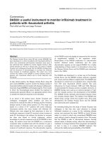
p<0.0001, respectively). The mean differences between
FD-OCT and IVUS were 0.02±0.17 mm for minimum
luminal diameter and 0.42±0.77 mm2 for minimum
luminal area, respectively, (figure 1).
Stent malapposition was detectable in all 12 patients
(100%) using FD-OCT, but in only 10 patients (83%)
using IVUS. The extent of stent malapposition detected
with FD-OCT was greater than that detected by IVUS
(20% vs 6%, p<0.001, expressed as per cent of total stent
surface area; table 3). The maximum distance and
maximum area of malapposition detected using FD-OCT
were greater than those obtained with IVUS (1.1
±0.34 mm vs 0.57±0.32 mm, p<0.001 and 2.65±1.88 mm2
vs 0.88±1.09 mm2, p<0.001, respectively; table 3).
An increase in-stent luminal areas (minimum in-stent
luminal area: 8.15±1.90 vs 7.30±1.62 mm2, p<0.05) and a
reduction in the extent of stent malapposition (19% vs
34%, p<0.005, expressed as % of total stent surface area)
were observed following postdilation when assessed with
FD-OCT, but not IVUS (table 4).
DISCUSSION
This is the first clinical study to examine systematically
the utility of FD-OCT and IVUS in the setting of extensive coronary artery calcification. Consistent with published data in more conventional atherosclerotic
populations,9 10 17 FD-OCT generally described smaller
lumen dimensions and detected acute stent malapposition more frequently when compared to IVUS. Indeed,
acute stent malapposition was detectable to some extent
in all patients using FD-OCT, with the amount of stent
Table 1 Patient demographics
N=12
Age, years
Male, n (%)
Acute presentation, n (%)
Previous MI, n (%)
Hypertension, n (%)
Hyperlipidemia, n (%)
Diabetes mellitus, n (%)
Previous CABG, n (%)
History of cigarette smoking, n (%)
70±7
10 (83)
4 (33)
3 (25)
9 (75)
12 (100)
2 (17)
3 (25)
7 (58)
CABG, coronary artery bypass graft; MI, myocardial infarction.
malapposed ranging from 1% to 53% of the total stent
surface area. Finally, high-pressure postdilation with a
non-compliant balloon was associated with a significant
reduction in acute stent malapposition as detected by
FD-OCT.
Our findings that in-stent diameters and luminal areas
were smaller with FD-OCT compared to IVUS are largely
in keeping with previous clinical studies.9 17–20
Moreover, comparing dimensions obtained using
FD-OCT with those obtained using IVUS, the mean differences in lumen area observed in this study are consistent with previous work.9 10 20 The smaller catheter size
and faster pullback speed with FD-OCT have been proposed as a potential explanation for these differences.17
However, a recent study by Kubo et al demonstrated that
vessel measurements obtained using IVUS overestimated
vessel dimensions, while enhanced delineation of the
lumen-vessel interface and visualisation of stent struts
obtained with FD-OCT led to a more accurate assessment of vessel and stent dimensions.9 This is particularly
relevant in heavily calcified vessels where acoustic
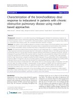
shadow and reflection may interfere with IVUS assessment (figure 2).6 Consistent with this hypothesis, previous work has demonstrated that vessel dimensions
obtained using FD-OCT are highly reproducible10 and
more accurate than IVUS.9 21 Indeed, in phantom
models and in human arteries in vitro, IVUS overestimates vessel luminal area by up to 16% and 14%,
respectively,9 22 in contrast to QCA measurement of coronary arteries, which underestimates vessel dimensions
when compared to FD-OCT.9
In keeping with more accurate delineation of the stent
and vessel interface, we found that FD-OCT detected stent
malapposition more frequently than IVUS (figure 2).
Indeed, following stent deployment but prior to postdilation, stent malapposition was detected in all patients with
FD-OCT but in only 83% patients with IVUS. With
FD-OCT, the extent of stent malapposition detected
Table 2 Procedural characteristics
N=12
Radial/femoral access, n
Guide catheter size, F
Largest rotablation burr used, mm
Total burr duration, seconds
Heparin dose, IU
Procedural success, n
Number of stents/patient, n
Number of drug eluting stents/patient, n
Mean stent diameter, mm
Total stent length, mm
Mean stent deployment pressure, atm
Postdilation balloon diameter (n=10), mm
Maximum postdilation balloon pressure
(n=10), atm
10/2
6 (6–7.5)
1.75 (1.25–2.0)
55±17
7042±2050
12 (100%)
1.1±0.3
0.9±0.5
3.3±0.5
27±15
13±3
3.8±0.8
16±5
Data are presented as median (range), mean±SD or n (%).
Gudmundsdottir I, Adamson P, Gray C, et al. Open Heart 2015;2:e000225. doi:10.1136/openhrt-2014-000225
3
Open Heart
Table 3 Comparison of IVUS and FD-OCT assessments following rotational atherectomy (n=18)
IVUS
Mean reference lumen diameter, mm
Mean reference lumen area, mm2
In-stent diameter, mm
Minimum
Maximum
Mean
In-stent area, mm2
Minimum
Maximum
Mean
Percentage of stent malapposed, %
Max stent malapposition, distance in mm
Max stent malapposition, area in mm2
FD-OCT
Difference (IVUS—FD-OCT)
p Value
3.64±0.80
10.77±1.14
3.38±0.66
9.49±3.59
0.26±0.34
1.28±1.69
0.330
0.388
2.51±0.53
4.38±0.82
3.38±0.62
2.48±0.48
3.81±0.67
3.25±0.60
0.02±0.17
0.57±0.28
0.13±0.17
0.586
<0.001
<0.005
7.19±2.62
12.81±4.61
9.54±3.48
5.5±5.0
0.57±0.32
0.88±1.09
6.77±2.18
10.48±3.65
8.78±2.91
19.6±15.1
1.10±0.34
2.65±1.88
0.42±0.77
2.33±2.25
0.82±1.15
−14.1±12.4
−0.53±0.33
−1.77±1.35
<0.05
<0.001
<0.01
<0.001
<0.001
<0.001
FD-OCT, frequency domain optical coherence tomography; IVUS, intravascular ultrasound.
(expressed as % of the total stent surface area) ranged
from 1% to 53%, compared with 1–21% for IVUS. The
extent of malapposition is in keeping the underlying
nature of calcific coronary artery disease, where even with
the use of ablative procedures such as rotational atherectomy and high-pressure postdilation, stent strut malapposition frequently persists.13 The circumferential extent but
not the depth of calcification in the vessel wall as defined
by IVUS has previously been shown to predict stent
malapposition.23
While the resolution of FD-OCT permits stent strut
level analysis,24 this is not the case for IVUS. Previous
studies have employed a number of techniques to
permit comparison between these two imaging
Figure 1 Bland Altman analyses
comparing minimum luminal area
(MLA; upper panel) and diameter
(MLD; lower panel) obtained
using FD-OCT and IVUS.
FD-OCT, frequency domain
optical coherence tomography;
IVUS, intravascular ultrasound.
4
Gudmundsdottir I, Adamson P, Gray C, et al. Open Heart 2015;2:e000225. doi:10.1136/openhrt-2014-000225
Interventional cardiology
Table 4 Effect of postdilation on stent dimensions assessed by FD-OCT and IVUS (N=6)
IVUS
Pre
In-stent diameter, mm
Minimum
Maximum
In-stent area, mm2
Minimum
Maximum
Percentage of stent malapposed (%)
Post
p Value
FD-OCT
Pre
Post
p Value
2.55±0.42
4.43±0.63
2.70±0.50
4.77±0.85
0.057
0.152
2.60±0.41
3.84±0.48
2.82±0.34
4.25±0.65
0.036
0.031
7.73±2.20
13.02±3.41
7±7
7.95±2.72
14.77±5.45
7±4
0.551
0.184
0.984
7.30±1.62
10.72±2.55
34±12
8.15±1.90
13.00±3.62
19±10
0.023
0.040
0.004
FD-OCT, frequency domain optical coherence tomography; IVUS, intravascular ultrasound.
techniques: for example, expressing malapposition as
mean malapposed area per cross-sectional slice17 or in
binary form as the presence or absence of any stent
malapposition.9 More refined methods include an
attempt to quantify the extent of malapposition, either
as the maximum number of consecutive frames with
malapposed struts25 or as the percentage malapposed
struts expressed as per cent of total number of struts.26
To address this issue and allow a clinically meaningful
comparison between the techniques, we calculated the
surface area of the stent that was malapposed and
expressed this as a percentage of the total stent surface
area. We believe this affords a robust and clinically
meaningful method by which to compare stent malapposition as detected using these two techniques.
In this study, postdilation of the implanted stent with a
non-compliant balloon inflated to high pressure was
associated with a significant increase in the minimal
in-stent luminal diameter and area, and a halving in the
extent of malapposition observed using FD-OCT. While
high-pressure postdilation of coronary stents has yielded
mixed outcomes in clinical studies,27–29 our data provide
some evidence to support this strategy as routine in
patients with extensive coronary calcification where stent
malapposition is a frequent finding. Although, mean
stent and lumen dimensions obtained using IVUS were
numerically greater following postdilation, this did not
achieve statistical significance. As discussed above, we
believe that this reflects the difficulties in delineating
the stent-lumen interface in the presence of extensive
vascular calcification.
Clinical relevance
Our findings suggest that in patients with extensive coronary artery calcification, FD-OCT is superior to IVUS at
detecting acute stent underexpansion and malapposition. This, in combination with a more rapid pullback
speed (up to 40 mm/s) of FD-OCT resulting in less
ischaemic burden,9 and previous data suggesting more
accurate assessment of lesion dimensions with FD-OCT,
support a clinical utility for FD-OCT in the evaluation of
percutaneous coronary intervention in patients with
extensive coronary artery calcification. Most of the contemporary data supporting a role for intravascular
imaging in this area relate to IVUS30–32 reflecting the
temporal evolution of these two technologies. However,
Figure 2 (A) Matched IVUS and (B) FD-OCT cross-sectional images following coronary stent implantation in a patient treated
with rotational atherectomy. An arc of calcification is visible in the vessel wall from the 12 o’clock position around to the 6 o’clock
position. Malapposed stent struts are clearly visible in this area with FD-OCT ( panel B, arrows) but the interface between stent
strut and vessel wall is poorly delineated in the corresponding IVUS image ( panel A). An area of thrombus adherent to the
luminal surface of the stent is also visible (T). FD-OCT, frequency domain optical coherence tomography; IVUS, intravascular
ultrasound.
Gudmundsdottir I, Adamson P, Gray C, et al. Open Heart 2015;2:e000225. doi:10.1136/openhrt-2014-000225
5
Open Heart
in a recent non-randomised case–control study, Prati
et al demonstrated a lower incidence of cardiac death
and myocardial infarction (6.6% vs 13%, p=0.006) in
patients undergoing percutaneous coronary intervention
guided by fluoroscopy and FD-OCT compared to fluoroscopic guidance alone.33 Large-scale randomised clinical
trials are required before we can be sure our findings
with FD-OCT will translate into improved clinical outcomes for patients.
commercially, and license their derivative works on different terms, provided
the original work is properly cited and the use is non-commercial. See: http://
creativecommons.org/licenses/by-nc/4.0/
REFERENCES
1.
2.
3.
Limitations
By definition, patients included in this study had undilatable calcific coronary artery lesions preventing delivery
of the imaging catheter to the area of interest prior to
atherectomy and treatment. This, combined with the
need for a rapid intracoronary injection of contrast
during FD-OCT imaging and the associated potential
for propagation of any atherectomy-induced dissection,
meant that intravascular imaging was performed only
following rotational atherectomy and stent implantation.
In the current study, reference vessel dimensions were
numerically smaller with FD-OCT compared to IVUS but
this did not achieve statistical significance, perhaps
reflecting the sample size. We elected to pool images
obtained following stent implantation with those
obtained following postdilation to maximise study power.
While we accept this may be a potential source of bias,
we believe that the postdilation intervention was sufficient to justify treating each run as a separate data set.
In summary, we have performed a systematic evaluation of the clinical utility of FD-OCT and IVUS in the
presence of extensive coronary artery calcification. Our
findings suggest that acute stent malapposition occurs
frequently in this setting and that FD-OCT affords
enhanced stent visualisation and detection of stent
malapposition, facilitating stent postdilation and leading
to improved stent apposition and minimal luminal areas.
4.
5.
6.
7.
8.
9.
10.
11.
12.
Contributors IG, PA, NGU and NLC contributed to the conception and design
of the study were involved in data acquisition, analysis and interpretation,
drafted the manuscript and approved the final version for publication. CG was
involved in data analysis and interpretation, contributed to manuscript drafting
and approved the final version for publication. JCS, MWB, PH, NM and DEN
were involved in data acquisition, contributed to the draft manuscript and
approved the final version for publication. All of the authors agree to be
accountable for all aspects of the work in ensuring that questions related to
the accuracy or integrity of any part of the work are appropriately investigated
and resolved.
13.
14.
15.
16.
Funding This work was funded by the Edinburgh and Lothians Health
Foundation. NLMC is supported by a National Health Service Research
Scotland Career Researcher Award.
17.
Competing interests None declared.
Ethics approval West of Scotland Ethics Committee.
Provenance and peer review Not commissioned; externally peer reviewed.
18.
Data sharing statement No additional data are available.
Open Access This is an Open Access article distributed in accordance with
the Creative Commons Attribution Non Commercial (CC BY-NC 4.0) license,
which permits others to distribute, remix, adapt, build upon this work non-
6
19.
Uren NG, Schwarzacher SP, Metz JA, et al. Predictors and
outcomes of stent thrombosis: an intravascular ultrasound registry.
Eur Heart J 2002;23:124–32.
Hong MK, Mintz GS, Lee CW, et al. Intravascular ultrasound
predictors of angiographic restenosis after sirolimus-eluting stent
implantation. Eur Heart J 2006;27:1305–10.
Doi H, Maehara A, Mintz GS, et al. Impact of post-intervention
minimal stent area on 9-month follow-up patency of paclitaxel-eluting
stents: an integrated intravascular ultrasound analysis from the
TAXUS IV, V, and VI and TAXUS ATLAS Workhorse, Long Lesion,
and Direct Stent Trials. JACC Cardiovasc Interv 2009;2:1269–75.
Attizzani GF, Capodanno D, Ohno Y, et al. Mechanisms,
pathophysiology, and clinical aspects of incomplete stent apposition.
J Am Coll Cardiol 2014;63:1355–67.
Klersy C, Ferlini M, Raisaro A, et al. Use of IVUS guided coronary
stenting with drug eluting stent: a systematic review and
meta-analysis of randomized controlled clinical trials and high quality
observational studies. Int J Cardiol 2013;170:54–63.
Mintz GS, Anderson WD, Bailey SR, et al. American College of
Cardiology Clinical Expert Consensus Document on Standards for
Acquisition, Measurement and Reporting of Intravascular Ultrasound
Studies (IVUS). A report of the American College of Cardiology Task
Force on Clinical Expert Consensus Documents developed in
collaboration with the European Society of Cardiology endorsed by
the Society of Cardiac Angiography and Interventions. Eur J
Echocardiogr 2001;2:299–313.
Prati F, Regar E, Mintz GS, et al. Expert review document on
methodology, terminology, and clinical applications of optical
coherence tomography: physical principles, methodology of image
acquisition, and clinical application for assessment of coronary
arteries and atherosclerosis. Eur Heart J 2010;31:401–15.
Bezerra HG, Costa MA, Guagliumi G, et al. Intracoronary optical
coherence tomography: a comprehensive review clinical and
research applications. JACC Cardiovasc Interv 2009;2:1035–46.
Kubo T, Akasaka T, Shite J, et al. OCT compared with IVUS in a
coronary lesion assessment: the OPUS-CLASS study. JACC
Cardiovasc Imaging 2013;6:1095–104.
Okamura T, Gonzalo N, Gutierrez-Chico JL, et al. Reproducibility of
coronary Fourier domain optical coherence tomography: quantitative
analysis of in vivo stented coronary arteries using three different
software packages. EuroIntervention 2010;6:371–9.
Mehanna E, Bezerra HG, Prabhu D, et al. Volumetric
characterization of human coronary calcification by
frequency-domain optical coherence tomography. Circ J
2013;77:2334–40.
Kume T, Okura H, Kawamoto T, et al. Assessment of the coronary
calcification by optical coherence tomography. EuroIntervention
2011;6:768–72.
Tanigawa J, Barlis P, Di Mario C. Heavily calcified coronary lesions
preclude strut apposition despite high pressure balloon dilatation and
rotational atherectomy: in-vivo demonstration with optical coherence
tomography. Circ J 2008;72:157–60.
Girassolli A, Carrizo S, Jimenez-Valero S, et al. Utility of optical
coherence tomography and intravascular ultrasound for the
evaluation of coronary lesions. Rev Port Cardiol 2013;32:925–9.
Tomey MI, Kini AS, Sharma SK. Current status of rotational
atherectomy. JACC Cardiovasc Interv 2014;7:345–53.
Mintz GS, Nissen SE, Anderson WD, et al. American College of
Cardiology Clinical Expert Consensus Document on Standards for
Acquisition, Measurement and Reporting of Intravascular Ultrasound
Studies (IVUS). A report of the American College of Cardiology Task
Force on Clinical Expert Consensus Documents. J Am Coll Cardiol
2001;37:1478–92.
Bezerra HG, Attizzani GF, Sirbu V, et al. Optical coherence
tomography versus intravascular ultrasound to evaluate coronary
artery disease and percutaneous coronary intervention. JACC
Cardiovasc Interv 2013;6:228–36.
Yamaguchi T, Terashima M, Akasaka T, et al. Safety and feasibility
of an intravascular optical coherence tomography image wire system
in the clinical setting. Am J Cardiol 2008;101:562–7.
Capodanno D, Prati F, Pawlowsky T, et al. Comparison of optical
coherence tomography and intravascular ultrasound for the
assessment of in-stent tissue coverage after stent implantation.
EuroIntervention 2009;5:538–43.
Gudmundsdottir I, Adamson P, Gray C, et al. Open Heart 2015;2:e000225. doi:10.1136/openhrt-2014-000225
Interventional cardiology
20.
21.
22.
23.
24.
25.
26.
27.
Okamura T, Onuma Y, Garcia-Garcia HM, et al. First-in-man
evaluation of intravascular optical frequency domain imaging (OFDI)
of Terumo: a comparison with intravascular ultrasound and
quantitative coronary angiography. EuroIntervention
2011;6:1037–45.
Tahara S, Bezerra HG, Baibars M, et al. In vitro validation of new
Fourier-domain optical coherence tomography. EuroIntervention
2011;6:875–82.
Chae JS, Brisken AF, Maurer G, et al. Geometric accuracy of
intravascular ultrasound imaging. J Am Soc Echocardiogr
1992;5:577–87.
Lindsay AC, Paulo M, Kadriye K, et al. Predictors of stent strut
malapposition in calcified vessels using frequency-domain optical
coherence tomography. J Invasive Cardiol 2013;25:429–34.
Barlis P, Dimopoulos K, Tanigawa J, et al. Quantitative analysis of
intracoronary optical coherence tomography measurements of stent
strut apposition and tissue coverage. Int J Cardiol 2010;141:151–6.
Kawamori H, Shite J, Shinke T, et al. Natural consequence of
post-intervention stent malapposition, thrombus, tissue prolapse, and
dissection assessed by optical coherence tomography at mid-term
follow-up. Eur Heart J Cardiovasc Imaging 2013;14:865–75.
Im E, Kim BK, Ko YG, et al. Incidences, predictors, and clinical
outcomes of acute and late stent malapposition detected by optical
coherence tomography after drug-eluting stent implantation. Circ
Cardiovasc Interv 2014;7:88–96.
Dirschinger J, Kastrati A, Neumann FJ, et al. Influence of balloon
pressure during stent placement in native coronary arteries on early
and late angiographic and clinical outcome: a randomized evaluation
of high-pressure inflation. Circulation 1999;100:918–23.
28.
29.
30.
31.
32.
33.
Frobert O, Sarno G, James SK, et al. Effect of stent inflation
pressure and post-dilatation on the outcome of coronary artery
intervention. A report of more than 90,000 stent implantations. PLoS
ONE 2013;8:e56348.
Goldberg SL, Di Mario C, Hall P, et al. Comparison of aggressive
versus nonaggressive balloon dilatation for stent deployment on late
loss and restenosis in native coronary arteries. Am J Cardiol
1998;81:708–12.
Casella G, Klauss V, Ottani F, et al. Impact of intravascular
ultrasound-guided stenting on long-term clinical outcome:
a meta-analysis of available studies comparing intravascular
ultrasound-guided and angiographically guided stenting. Catheter
Cardiovasc Interv 2003;59:314–21.
Ahn JM, Kang SJ, Yoon SH, et al. Meta-analysis of outcomes after
intravascular ultrasound-guided versus angiography-guided
drug-eluting stent implantation in 26,503 patients enrolled in three
randomized trials and 14 observational studies. Am J Cardiol
2014;113:1338–47.
Zhang Y, Farooq V, Garcia-Garcia HM, et al. Comparison of
intravascular ultrasound versus angiography-guided drug-eluting
stent implantation: a meta-analysis of one randomised trial and ten
observational studies involving 19,619 patients. EuroIntervention
2012;8:855–65.
Prati F, Di Vito L, Biondi-Zoccai G, et al. Angiography alone versus
angiography plus optical coherence tomography to guide
decision-making during percutaneous coronary intervention: the
Centro per la Lotta contro l’Infarto-Optimisation of Percutaneous
Coronary Intervention (CLI-OPCI) study. EuroIntervention
2012;8:823–9.
Gudmundsdottir I, Adamson P, Gray C, et al. Open Heart 2015;2:e000225. doi:10.1136/openhrt-2014-000225
7