báo cáo hóa học:" The value of SPECT in the detection of stress injury to the pars interarticularis in patients with low back pain" doc
Bạn đang xem bản rút gọn của tài liệu. Xem và tải ngay bản đầy đủ của tài liệu tại đây (1002.7 KB, 6 trang )
REVIEW Open Access
The value of SPECT in the detection of stress
injury to the pars interarticularis in patients with
low back pain
Katherine Zukotynski
1,4
, Christine Curtis
2
, Frederick D Grant
3,4
, Lyle Micheli
2,4
, S Ted Treves
3,4*
Abstract
The medical cost associated with back pain in the United States is considerable and growing. Although the differ-
ential diagnosis of back pain is broad, epidemiological studies suggest a correlation between adult and ad olescent
complaints. Injury of the pars interarticularis is one of the most common identifiable causes of ongoing low back
pain in adolescent athletes. It constitutes a spectrum of disease ranging from bone stress to spondylolysis and
spondylolisthesis. Bone stress may be the earliest sign of disease. Repetitive bone stress causes bone remodeling
and may result in spondylolysis, a non-displaced fracture of the pars interarticularis. A fracture of the pars interarti-
cularis may ultimately become unstable leading to spondylolisthesis. Results in the literature support the use of
bone scintigraphy to diagnose bone stress in patients with suspected spondylolysis. Single photon emission com-
puted tomography (SPECT) provides more contrast than planar bon e scintigraphy, increases the sensitivity and
improves anatomic localization of skeletal lesions without exposing the patient to additional radiation. It also pro-
vides an opportunity for better correlation with other imaging modalities, when necessary. As such, the addition of
SPECT to standard planar bone scintigraphy can result in a more accurate diagnosis and a better chance for effi-
cient patient care. It is our expectation that by improving our ability to correctly diagnose bone stress in patients
with suspected injury of the posterior elements, the long-term cost of managing this condition will be lowered.
Introduction
The economic burden of back pain is estimated to be
more than $90 billion per year in the United States
[1,2]. Costs may be due to a variety of facto rs including
primary care, diagnostic imaging, inpatient services, phy-
sical therapy and lost work productivity. Recent epide-
miological studies suggest a correlation between adult
and adolescent complaints [3,4].
The differential diagnosis for back pain is broad and
includes degenerative disease, infection, inflammation,
tumors and trauma [5-7]. Injury of the pars interarticu-
laris is one of the most common identifiable c auses of
ongoing low back pain in adolescent athletes [ 6,8,9]. It
constitutes a spectrum of disease from bone stress
through spondylolysis and spondylolisthesis. Bone stress
may be the earliest sign. It is most common at L5,
which is particularly vulnerable to micro-trauma from
repetitive flexion, extension or rotational forces. Repeti-
tive bone stress may result in spondylolysis, a non-dis-
placed fracture of the pars interarticularis. Ultimately
spondylolisthesis, or sli ppage of o ne vertebral body on
another, may occur.
The Diagnosis and Treatment of Spondylolysis
Athletes comprise the majority of patients presenting
with spondylolysis [10-12]. Sport specific maneuvers
with repetitive twisting rotation and extension increase
load on the spine, and may result in stress injury
[13,14]. The most frequently presenting complaint is
low back pain; either localized or diffuse [8,9,15]. In
more severe cases, muscle spasms from difficulty in gait
and posture may result.
The medical history should include duration of sy mp-
toms, modifying and alleviating factors, level and inten-
sity of sport participation as well as changes in muscle,
bowel and bladder function. Physical examination
involves inspection and palpation of the spine as well as
examination of range of motion [9]. Inspection of the
* Correspondence:
3
Division of Nuclear Medicine, Department of Radiology, Children’s Hospital
Boston, Boston, MA, USA
Zukotynski et al. Journal of Orthopaedic Surgery and Research 2010, 5:13
/>© 2010 Zukotynski et al; licensee BioMed Central Ltd. This is an Open Access article distributed under the terms of the Creative
Commons Attribution License ( which permits unrestricted use, distribut ion, and
reproduction in any medium, pro vided the original work is properly cite d.
spine may reveal hyperlordosis. Palpation for tenderness
is useful to identify area(s) of stress, fracture, or slippage
[9].Rangeofmotionisfrequentlymorecompromised
and painful in extension. The stork test may reveal pain
on the contralateral side when standing on one leg.
While this test is not specific for pars stress injury, it is
highly suggestive of some type of derangement of the
posterior elements of the spine [16]. Imaging studies
used to evaluate patients with low back pain include:
radiographs, bone s cintigraphy, computed tomography
(CT) and magnetic resonance imaging (MRI).
Radio graphs of the spine have limited sensitivity com-
pared with other imaging modalities in detecting bone
stress and acute spondylolysis. Furthermore, radio-
graphic defects of the pars interarticularis may not be
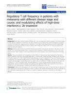
symptomatic [17,18]. Figure 1 illustrates the radio-
graphic appearance of a long standing pars interarticu-
laris defect.
Bone scintigraphy is very sensitive for the detection of
bone stress. Repetitive stress causes local bone remodel-
ing and abnormal uptake of scintigraphic tracer. Single
photon emission computed tomography (SPECT) has
10-20 times more contrast than planar bone
scintigraphy and is more sensitive than radiography and
planar bone scans. Furthermore, scintigraphic abnormal-
ities have been found to correlate with painful lesions of
the pars interarticularis [18-21]. The diagnosis of spon-
dylolisthesis is not made with scintigraphy. Once spon-
dylolisthesis develops, bone stress may be absent at the
site of spondylolysis. However, in this case, bone remo-
deling and tracer uptake may occur at the pars interarti-
cular is immediately above or below the level of fracture.
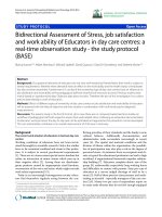
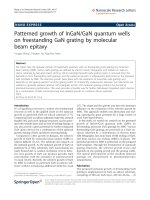
Figure 2 illustrates stress of the pars interarticularis on
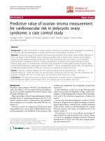
bone scintigraphy. Figure 3 presents an example where
pars stress is identified on SPECT b ut not on planar
bone scintigraphy.
CT demonstrates detailed osseous morphology, is
more specific than bone scintigraphy and may predict
the probability of ultimate bone healing [22,23]. How-
ever, CT of the spine results in higher ionizing radiation
exposure compared to bone scintigraphy [24]. Further-
more, there are reports in the literature of a normal
spine CT in patients with abnormalities on planar bone
scintigraphy and SPECT [16,25]. This may be explained
by the fact that tracer uptake in the region of the pars
interarticularis on scintigraphic studies corresponds to
A. B.
Figure 1 Radiographic findings in a patient with L5 pars interarticular is fracture and mild L5 on S1 spondylolisthesis: AP (A) and
lateral (B) images. [Red arrow points to the fracture and blue arrow points to spondylolisthesis of L5 on S1].
Zukotynski et al. Journal of Orthopaedic Surgery and Research 2010, 5:13
/>Page 2 of 6
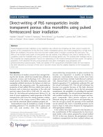
bone stress. If this stress has not yet resulted in a frac-
ture, changes may not be visible on CT. The identifica-
tion of patients with this patt ern of scintigraphic
findings is particularly important as these patients may
have the best chance of healing with early treatment [6].
Figure 4 shows a fracture of the pars interarticularis o n
CT.
MRI is not as sensitive as SPECT for identifying bone
stress of the pars interart icul aris and does not delineate
bony detail to the same extent as CT [16,25]. MRI is,
however, attractive as an imaging modality that does not
involve ionizing radiation and that is excellent in identi-
fying alternate pathology including bone edema or
abnormalities of the soft tissues, disk and spinal cord.
In general, when bone stress or spondylolysis is sus-
pected, bone scintigraphy with SPECT is recommended. If
SPECT demonstrates a pars lesion, a thin-cut CT (1 mm
axial sequence) through the area of abnormality on
SPECT, is recommended to confirm the diagnosis and
stage the lesion. If SPECT is negative, pars stress is unli-
kely to be the cause of the low back pain and MRI may be
helpful in identifying other causes of back pain [6,26].
Complete bony union offers the best long term prog-
nosis. Some patients attain a fibrous union and are conse-
quently able to return to prior activity, with favorable
short-term prognoses. Treatment often includes rest from
aggravating activities, non steroidal anti-inflammatory
medication, bracing and physical therapy emphasizing
hamstring stretching and core strengthening. The length
of activity restriction, use of bracing and type of rehabilita-
tion programs varies, reflecting a lack of consensus among
practitioners. In recalcitrant cases, electrical stimulation
may be added [9,27,28]. Prompt treatment of patients with
early pars stress has been shown to result in more predict-
able sympto m relief and less likelihood of p rogression to
spondylolisthesis [ 29-31]. Surgery is reserved for patients
who do not respond to conservative management
A.
B.
Right
Left Right Left
Anterior projection
Posterior projection
SPECT
Transverse
Sagittal Coronal
Right
tfeLthgiR
Left
Figure 2 Scintigraphic findings in a patient with right L3 pars stress on planar bone scintigraphy (A) and on SPECT (B). [Red arrows
point to the scintigraphic abnormality on SPECT].
Zukotynski et al. Journal of Orthopaedic Surgery and Research 2010, 5:13
/>Page 3 of 6
A.
B.
Right thgiRtfeL LeftRight Left
Anterior projection Posterior projection
SPECT
Figure 3 Scintigraphic findings i n a patient with right L5 pars stress on SPECT (A), not seen on planar bone scintigraphy (B).[Red
arrows point to the scintigraphic abnormality].
.B.A C.
Figure 4 CT findings in a patient with pars interarticularis fracture: Normal facet joint below fracture (A), right L3 pars interarticularis
fracture (B), normal facet joint above fracture (C). [red arrow points to the fracture and blue arrows point to normal facet joints].
Zukotynski et al. Journal of Orthopaedic Surgery and Research 2010, 5:13
/>Page 4 of 6
(approximately 5%), have progressive spondylolisthesis,
intractable pain or neurological deficits [18].
The Utility of SPECT over Planar Bone
Scintigraphy in the Evaluation of Back Pain
Studies have consistently demonstrated that SPECT is
moresensitivethanplanarbonescintigraphytoidentify
skeletal lesions [31-33]. Collier et al. compared planar bone
scintigraphy and SPECT in 19 adults with radiographic evi-
dence of spondylolysis and/or spondylolisthesis and found
that SPECT was more sensitive in identifying sites of “pain-
ful” pars interarticularis defects and that SPECT allowed
more accurate localization of the defect [19]. In a long-
term follow-up study, Bellah et al. reviewed findings on
planar and SPECT bone scintigraphy in 162 patients aged
6-32 years with symptoms of low back pain potentially
related to stress injury of the pars interarticularis. SPECT
showed an abnormal focus of radiotracer uptake in the
lumbar spine in 71 patients (44%). All abnormalities
detected on planar bone scintigraphy were detected with
SPECT. An abnormality was identified in 39 patients (24%)
on SPECT alone [20]. Even-Sapir et al. demonstrated
SPECT was more sensitive and specific than planar bone
scintigr aphy in the detection of bone metastasis in a pro-
spective study of 44 patients with prostate cancer [32].
Strobel et al. found that lesion visibility as well as the ability
to determine a specific diagnosis was significantly better for
SPECT than with planar bone scintigraphy [33].
We conducted an internal review of all patients with
low back pain or suspected spondylolysis referred to the
Division of Nuclear Medicine at Children’s Hospital, Bos-
ton for skeletal scintigraphy between October 2005 and
September 2006. Of 115 identified patients undergoing
skeletal SPECT and planar scintigraphy, SPECT identi-
fied an abnormal focus of increased tracer uptake in the
pars interarticularis in 42 patients (37%). All abnormal-
ities detected on planar bone scintigraphy were also
detected with SPECT. Planar bone scintigraphy identified
an abnormal focus of tracer uptake in the pars interarti-
cularis in 19 patients (17%). SPECT identified additional
sites of pars stress in 5 of the 19 patients with pars stress
suggested on planar bone scintigraphy (26%).
In general, SPECT increases contrast and improves
anatomic localization in comparison to planar scintigra-
phy [34]. In SPECT, images are acquired in multiple
projections with the gamma scintillation camera traver-
sing an axial orbit about the patient. Filtered back pro-
jection (FBP) or an iterative reconstruction algorithm
such as OSEM (ordered subsets expectation maximiza-
tion) is then used to create a cross-sectional image. The
cross-sectional image is a two-dimensional representa-
tion of a slice through the patient that would project
onto a single dimension on a planar bone scan. In addi-
tion, SPECT images may be displayed as a 3D
representation using a volume rendered display to pro-
vide better spatial orientation. Maeseneer et al. illu-
strated how patterns of tracer uptake in the spine on
SPECT suggested specific pathology [35]. Degenerative
disk disease might show increa sed tracer uptake cen-
tered about the disk space. Pars interarticularis stress
might show tracer uptake in the expected location of
the pars interarticula ris and metastatic disease is more
likely to involve the vertebral body with extension to the
pedicle [35,36]. Ultimately, SPECT may be fused with
CT, if needed, to help add specificity to the findings.
Conclusions
The economic burden of back pain is significant and
growing. Epidemiological studies suggest a correlation
between adult and adolescent complaints. Pars interarti-
cularis injury, a spectrum of disease ranging from bone
stress to spondylolysis and spondylolisthesis , is the most
common identifiable cause of ongoing low back pain in
adolescent athletes.
In the current era of multi-modality imaging, radio-
graphs, skeletal scintigraphy, CT and MRI all play an
important role in imaging patients with back pain. Pla-
nar bone scintigraphy has a long history in the diagnosis
of patients with suspected injury of the pars interarticu-
laris because it is more sensitive than radiographs for
localizing the site of bone stress and because CT of the
spine is associated with significant ionizing radiation.
The addition of SPECT to planar skeletal scintigraphy
increases sensitivity and improves disease localization
without exposing the patient to additional radiation.
SPECT can also identify early pars stress prior to the
development of osseous change detectable with CT. As
such, incorporation of SPECT into the standard planar
bone scintigraphy routine should lead to a more accu-
rate initial diagnosis. It is our hypothesis that by
improving our ability to promptly diagnose patients with
suspected injury of the pars interarticularis, the patients
will be better served and the long-term cost of manage-
ment can be lowered.
List of abbreviations used
SPECT: Single photon emission computed tomography;
CT: Computed tomography; MRI: Magnetic resonance
imaging.
Author details
1
Division of Nuclear Medicine, Department of Imaging, Dana-Farber Cancer
Institute, Boston, MA, USA.
2
Division of Sports Medicine, Department of
Orthopedic Surgery, Children’s Hospital Boston, Boston, MA, USA.
3
Division of
Nuclear Medicine, Department of Radiology, Children’s Hospital Boston,
Boston, MA, USA.
4
Harvard Medical School, Boston, MA, USA.
Authors’ contributions
All of the authors have read and approved the final manuscript.
Zukotynski et al. Journal of Orthopaedic Surgery and Research 2010, 5:13
/>Page 5 of 6
Authors’ Information
Katherine Zukotynski is an Instructor in Radiology at Harvard Medical School
and Christine Curtis is Team Leader in Clinical Research at the Children’s
Hospital Boston. Frederick D Grant is an Instructor in Radiology at Harvard
Medical School. Lyle Micheli is a Professor of Orthopedic Surgery at Harvard
Medical School. Ted Treves is a Professor of Radiology at Harvard Medical
School.
Competing interests
The authors declare that they have no competing interests.
Received: 11 August 2009
Accepted: 3 March 2010 Published: 3 March 2010
References
1. Dagenais S, Caro J, Haldeman S: A systematic review of low back pain
cost of illnessstudies in the United States and internationally. The Spine
Journal 2008, 8:8-20.
2. Luo X, Pietrobon R, Sun S, Liu G, Hey L: Estimates and patterns of direct
health care expenditures among individuals with back pain in the
United States. Spine 2004, 29(1):79-86.
3. Kim H, Green D: Adolescent back pain. Current Opinion in Pediatrics 2008,
20:37-45.
4. Leboeuf-Yde C, Kyvik K: At what age does low back pain become a
common problem?: A study of 29,424 individuals aged 12-41 years.
Spine 1998, 23:228-234.
5. Bernstein R, Cozen H: Evaluation of back pain in children and
adolescents. American Family Physician 2007, 76(11):1669-1676.
6. Standaert C, Herring S: Expert opinion and controversies in sports
andmusculoskeletal medicine: The diagnosis and treatment of
spondylolysis in adolescent athletes. Archives of Physical Medicine and
Rehabilitation 2007, 88:537-540.
7. Gregory P, Batt M, Kerslake R, Webb J: Single photon emission
computerized tomography and reverse gantry computerized
tomography findings in patients with back pain investigated for
spondylolysis. Clinical Journal of Sport Medicine 2005, 15(2):79-86.
8. Micheli LJ, Wood R: Back pain in young athletes. Significant differences
from adults in causes and patterns. Archives of Pediatrics and Adolescent
Medicine 1995, 149:15-18.
9. Micheli LJ, Curtis C: Stress fractures in the spine and sacrum. Clinics in
Sports Medicine 2006, 25(1):75-88.
10. Micheli LJ: Low back pain in the adolescent: differential diagnosis. The
American Journal of Sports Medicine 1979, 7:362-364.
11. Micheli LJ: Back injuries in dancers. Clinical Journal of Sports Medicine 1983,
2(3):473-484.
12. d’Hemecourt PA, Zurakowski D, Kriemler S, Micheli LJ: Spondylolysis:
returning the athlete to sports participation with brace treatment.
Orthopedics 2002, 25(6):653-657.
13. Weir MR, Smith DS: Stress reaction of the pars interarticularis leading to
spondylolysis. A cause of adolescent low back pain. Journal of Adolescent
Health Care 1989, 10(6):573-577.
14. Beutler WJ, Frederickson BE, Murtland A, Sweeney CA, Grant WD, Baker D:
The natural history of spondylolysis and spondylolisthesis: 45 year
follow-up evaluation. Spine 2003, 28(10):1027-1035.
15. Tallarico RA, Madom IA, Palumbo MA:
Spondylolysis and spondylolisthesis
in the athlete. Sports Medicine and Arthroscopy Review 2008, 16(1):32-38.
16. Masci L, Pike J, Malara F, Phillips B, Bennell K, Brukner P: Use of the one-
legged hyperextension test and magnetic resonance imaging in the
diagnosis of active spondylolysis. British Journal of Sports Medicine 2006,
40:940-946.
17. Pennell R, Maurer A, Bonakdarpour A: Stress injuries of the pars
interarticularis: radiologic classification and indications for scintigraphy.
American Journal of Roentgenology 1985, 145:763-766.
18. Standaert C, Herring S: Spondylolysis: a critical review. British Journal of
Sports Medicine 2000, 34:415-422.
19. Collier B, Johnson R, Carrera G, Meyer G, Schwab J, Flatley T: Painful
spondylolysis or spondylolisthesis studied by radiography and single-
photon-emission computed tomography. Radiology 1985, 154:207-211.
20. Bellah R, Summerville D, Treves S, Micheli L: Low-back pain in adolescent
athletes: detection of stress injury to the pars interarticularis with SPECT.
Muskuloskeletal Radiology 1991, 180:509-512.
21. Harvey C, Richenberg J, Saifuddin A, Wolman R: Pictoral review: The
radiological investigation of lumbar spondylolysis. Clinical Radiology 1998,
53:723-728.
22. Congeni J, McCulloch J, Swanson K: Lumbar Spondylolysis: A study of
natural progression in athletes. The American Journal of Sports Medicine
1997, 25(2):248-253.
23. Morita T, Ikata T, Katoh S, Miyake R: Lumbar spondylolysis in children and
adolescents. Journal of Bone and Joint Surgery 1995, 77(4):620-625.
24. Brenner D, Hall E: Computed Tomography - An Increasing Source of
Radiation Exposure. The New England Journal of Medicine 2007,
357:2277-2284.
25. Campbell R, Grainger A, Hide I, Papastefanou S, Greenough C: Juvenile
spondylolysis: a comparative analysis of CT, SPECT, and MRI. Skeletal
Radiology 2005, 34:63-73.
26. Gregory P, Batt M, Kerslake R, Scammell B, Webb J: The value of
combining single photon emission computerized tomography and
computerized tomography in the investigation of spondylolysis.
European Spine Journal 2004, 13:503-509.
27. Fellander-Tsai L, Micheli LJ: Treatment of spondylolysis with extreme
electrical stimulation and bracing in adolescent athletes: a report of 2
cases. Clinical Journal of Sport Medicine 1998, 8(3):232-234.
28. Pettine KA, Salib RM, Walker SG: External electrical stimulation and
bracing for treatment of spondylolysis. A case report. Spine 1993,
18(4):436-439.
29. Motley G, Nyland J, Jacobs J, Caborn D: The pars interarticularis stress
reaction, spondylolysis, and spondylolisthesis progression.
Journal of
Athletic Training 1998, 33(4):351-358.
30. Takemitsu M, Rassi G, Woratanarat P, Shah S: Low back pain in pediatric
athletes with unilateral tracer uptake at the pars interarticularis on
single photon emission computed tomography. Spine 2006,
31(8):909-914.
31. Anderson K, Sarwark J, Conway J, Logue S, Schafer M: Quantitative
assessment with SPECT imaging of stress injuries of the pars
interarticularis and response to bracing. Journal of Pediatric Orthopaedics
2000, 20(1):28-33.
32. Even-Sapir E, Metser U, Mishani E, Lievshitz G, Lerman H, Leibovitch I: The
detection of bone metastasis in patients with high-risk prostate cancer:
99mTc-MDP planar bone scintigraphy, single- and multi-field-of-view
SPECT, 18F-Fluoride PET, and 18F-Fluoride PET/CT. The Journal of Nuclear
Medicine 2006, 47(2):287-297.
33. Strobel K, Burger C, Seifert B, Husarik D, Soyka J, Hany T: Characterization
of focal bone lesions in the axial skeleton: performance of planar bone
scintigraphy compared with SPECT and SPECT fused with CT. American
Journal of Roentgenology 2007, 188:W467-474.
34. Sarikaya I, Sarikaya A, Holder L: The role of single photon emission
computed tomography in bone imaging. Seminars in Nuclear Medicine
2001, 31(1):3-16.
35. Maeseneer M, Lenchik L, Everaert H, Marcelis S, Bossuyt A, Osteaux M,
Beeckman P: Evaluation of lower back pain with bone scintigraphy and
SPECT. Radiographics 1999, 19:901-912.
36. Reinartz P, Schaffeldt J, Sabri O, Zimny M, Nowak B, Otswald E, Cremerius U,
Udalrich B: Benign versus malignant osseous lesions in the lumbar
vertebrae: differentiation by means of bone SPET. European Journal of
Nuclear Medicine 2000, 27(6):721-726.
doi:10.1186/1749-799X-5-13
Cite this article as: Zukotynski et al.: The value of SPECT in the detection
of stress injury to the pars interarticularis in patients with low back
pain. Journal of Orthopaedic Surgery and Research 2010 5:13.
Zukotynski et al. Journal of Orthopaedic Surgery and Research 2010, 5:13
/>Page 6 of 6