severe renal injury detected by emergency department point of care ultrasound pocus in a patient with undifferentiated hypotension
Bạn đang xem bản rút gọn của tài liệu. Xem và tải ngay bản đầy đủ của tài liệu tại đây (354.16 KB, 2 trang )
Visual Journal of Emergency Medicine 7 (2017) 1–2
Contents lists available at ScienceDirect
Visual Journal of Emergency Medicine
journal homepage: www.elsevier.com/locate/visj
Visual Case Discussion
Severe renal injury detected by Emergency Department Point of Care
Ultrasound (POCUS) in a patient with undifferentiated hypotension
MARK
⁎
Michael Halperina, , Siu Fai Lia, Andrew Shannonb
a
b
Jacobi Medical Center, Department of Emergency Medicine, Bronx, NY, USA
University of Florida, Department of Emergency Medicine, Jacksonville, FL, USA
A R T I C L E I N F O
Keywords:
Rapid Ultrasound for Shock Exam
RUSH exam
Kidney laceration
Renal laceration
Point of Care Ultrasound (POCUS)
A fifty-nine year old male was brought to the emergency department for altered mental status after being found lying in the grass. He
had alcohol on his breath and was suspected to be severely intoxicated.
Unable to provide any history, he was initially hypotensive (BP 77/44),
afebrile (97.0 F), without tachycardia or tachypnea, and normal
oxygenation on room air. The only obvious physical exam findings
were an abrasion to his left eyebrow and blood at his urethral meatus.
Given his undifferentiated hypotension, however, a RUSH (Rapid
Ultrasound for SH(ock)1 exam was performed, showing free fluid
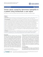
(Video) in the right upper quadrant. Additionally, a large collection
in that right upper quadrant ( Fig. 1) was thought consistent with
hematoma given the RUSH exam findings and that a similarly
prominent echogenic stripe was not noted on the left. In retrospect,
however, another possibility is that this is perinephric fat within
Gerota’s fascia.
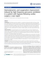
After resuscitation with one liter of normal saline, the blood
pressure stabilized and he was taken to CT scan, which revealed an
American Association for the Surgery of Trauma (AAST) grade III
laceration (no urinary extravasation)2 of the right kidney with retroperitoneal hematoma ( Fig. 2).
About 5% of renal lacerations are grade III and these generally do
not require surgical intervention because they don’t involve vascular
injuries in the renal pedicle, which can be salvaged with intra-arterial
embolization.3 His initial hemoglobin was 11.8 g/dL, which dropped to
8.0 g/dL. He was transfused 3 units of red cells, and admitted to
⁎
Fig. 1. Right upper quadrant abdominal view with clot (outlined by asterisks) in
between kidney and liver parynchema.
surgical intensive care unit. He was successfully managed non-operatively as his hemodynamics and blood counts normalized, had no
further bleeding, and was doing well at recent clinic follow up
Supplementary material related to this article can be found online
at />
Corresponding author.
E-mail address: (M. Halperin).
/>Received 14 August 2016; Received in revised form 4 October 2016; Accepted 30 October 2016
2405-4690/ © 2016 The Authors. Published by Elsevier Inc.
This is an open access article under the CC BY-NC-ND license ( />
Visual Journal of Emergency Medicine 7 (2017) 1–2
M. Halperin et al.
online version at />References
1 Perera P, Mailhot T, Riley D, Diku M. The RUSH exam: rapid ultrasound in shock in
the evaluation of the critically III. Emerg Med Clin N Am.. 2010;28:29–56.
2 Moore EE, Shackford SR, Pachter HL, et al. Organ injury scaling: spleen, liver, and
kidney. J Trauma.. 1989;29(12):1664–1666.
3 Kawashima A, Sandler CM, Corl FM, et al. Imaging of renal trauma: a comprehensive
review. Radiographics.. 2001;21(3):557–574.
Fig. 2. Axial image from CT abdomen and pelvis showing right kidney laceration with
retroperitoneal hematoma.
Appendix A. Supplementary material
Supplementary data associated with this article can be found in the
2