Báo cáo khoa học: "Abscess formation mimicking disease progression, in a patient with metastatic renal cell carcinoma during sunitinib treatment" docx
Bạn đang xem bản rút gọn của tài liệu. Xem và tải ngay bản đầy đủ của tài liệu tại đây (351.68 KB, 3 trang )
WORLD JOURNAL OF
SURGICAL ONCOLOGY
Michalaki et al. World Journal of Surgical Oncology 2010, 8:45
/>Open Access
CASE REPORT
© 2010 Michalaki et al; licensee BioMed Central Ltd. This is an Open Access article distributed under the terms of the Creative Commons
Attribution License ( which permits unrestricted use, distribution, and reproduction in
any medium, provided the original work is properly cited.
Case report
Abscess formation mimicking disease progression,
in a patient with metastatic renal cell carcinoma
during sunitinib treatment
Vasiliki Michalaki*
1
, Nikolaos Arkadopoulos
2
, Agathi Kondi-Pafiti
3
and Constantine Gennatas
1
Abstract
Background: Renal cell carcinoma (RCC) represents approximately 3% of all adult cancers and is more common in
males. Systemic treatment for RCC has improved following the introduction of tyrosine kinase inhibitors, such as
sunitinib. The molecular targets of sunitinib are receptor tyrosine kinases (RTKs). Moreover, sunitinib has an additional
anti-angiogenic effect through its inhibition of the vascular endothelial growth factor receptor activation.
Case presentation: We present a case of intra-abdominal abscess formation mimicking disease progression, in a
patient with metastatic renal cell carcinoma during sunitinib treatment.
Conclusion: In the advancing era of molecular therapy of solid tumours, sunitinib has demonstrated significant
efficacy in the post-cytokine setting treatment of metastatic renal cancer. Concurrently, however, increasing evidence
has emerged to indicate that this class of drugs exert profound immunomodulatory effects on T cells and play major
roles in immune tumor surveillance.
Background
The treatment of advanced RCC is undergoing a para-
digm shift with the recent introduction of anti-angio-
genic therapy that either directly inhibits vascular
endothelial growth factor or disrupts signal transduction
favorable to vascular development through multi-kinase
inhibitors. Angiogenic inhibitors have been found to
increase survival and are approved in advanced renal cell
carcinoma [1,2]. Consequently, most of these patients will
routinely receive tyrosine kinase inhibitors, such as suni-
tinib.
Sunitinib is an orally administered, small molecule
inhibitor of multiple receptor tyrosine kinases implicated
in tumour growth, angiogenesis, and metastatic progres-
sion. In addition, the targets of sunitinib involve vascular
endothelial growth factor receptors (VEGFR1, VEGFR2
and VEGFR3), platelet-derived growth factor receptors
(PDGFRα and PDGFRβ) and the like. We describe a case
of intra-abdominal abscess formation mimicking disease
progression during sunitinib treatment.
Case presentation
A 62-year-old patient diagnosed with a high-grade clear-
cell renal carcinoma in 1991 and was treated by left neph-
rectomy and surrenalectomy. Fourteen years later,
relapsed on the lungs and had been administered inter-
feron alfa. The patient was regularly followed up and had
regular scans that did show stabilization of the disease in
the lungs for two years. In December 2007 chest comput-
erized tomography (CT) disclosed the progression of
lung metastases. Sunitinib was initiated in January 2008
as a standard regimen (50 mg/day for 4 weeks every 6
weeks) for pulmonary metastases. Patient had a radio-
graphic response and prolonged progression free survival
of fourteen months; side effects were manageable and
included grade 2 hypertension. After five cycles, the
patient was admitted to the hospital due to complaints of
fatigue and left sided flank pain. The systolic and diastolic
blood pressures were 110 mmHg and 60 mmHg, respec-
tively, pulse rate was 90 per min and respiratory rate was
20 per min. The body temperature was 37.2°C.
Laboratory studies were conducted immediately after
the patient's arrival at the hospital. He had anemia (Hb 98
g/L) (normal range: 140-180) and thrombocytopenia (133
× 10
9
/L) (normal range: 150-450), but a WBC count was
* Correspondence:
1
Oncology Clinic, Second Department of Surgery, University of Athens,
Aretaieion Hospital, 78, V. Sofias av, 115 28, Athens, Greece
Full list of author information is available at the end of the article
Michalaki et al. World Journal of Surgical Oncology 2010, 8:45
/>Page 2 of 3
normal (6.15 × 10³/mm
3
) with 74% neutrophils. Other
laboratory findings were presented as elevated serum lev-
els of CRP (21 mg/L) (normal range:< 5), ALP (416 IU/L)
(normal range: 96-250), and slightly increased creatinine
(1.43 μmol/L) (normal range: 0.5-1.2).
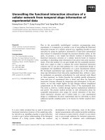
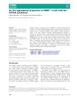
Fluorodeoxyglucose positron emission tomography
(FDG-PET-CT) scans demonstrated an area of increased
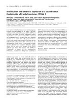
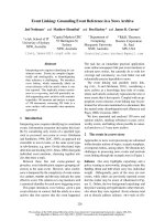
uptake in the left paravertebral area (Figure 1). MRI scan
(T2 image) demonstrated the lesion that corresponded to
the area of increased PET uptake (Figure 2).
The patient underwent a diagnostic laparoscopy in May
2009. Intra-operative biopsy of the lesion was performed;
the pathology was consistent with an abcess without evi-
dence of malignancy. After an uneventful postoperative
course, the patient was discharged on the 10th day after
surgery and chemotherapy with sunitinib was restarted.
Three months postoperatively there was no evidence of
recurrent disease.
Discussion
Renal cell cancer (RCC) is a relatively uncommon malig-
nancy. When the disease is localized is curable by sur-
gery; however, locally advanced or metastatic disease is
not curable in most cases and until recently had a limited
response to drug treatment. Historically, biologic
response modifiers or immunomodulating agents were
tested in clinical trials based on observations that some
cases of RCC can spontaneously regress. Responses have
been observed with interferon alfa, but with little effect
on overall survival.
The use of targeted therapies has substantially
improved outcomes for patients with advanced renal cell
carcinoma [3,4].
Sunitinib malate is an oral multi-kinase inhibitor tar-
geting several receptor tyrosine kinases (PDGFRalpha
and PDGFRbeta; VEGFR1, VEGFR2 and VEGFR3; KIT,
FLT3, CSF-1R and RET) that was approved by the FDA in
2006 for treatment of metastatic renal cell carcinoma. In
a randomized phase III trial, sunitinib prolonged median
progression-free survival (11 months) in comparison to
interferon-alpha (5 months); corresponding to a hazard
ratio of 0.42 (95% confidence interval: 0.32 to 0.54; P <
0.001) for patients with advanced renal cell cancer. Suni-
tinib was also associated with a higher objective response
rate than interferon-alpha (31% vs. 6%; P < 0.001) [2].
The most common toxicities with sunitinib are hand-
foot syndrome, rash, fatigue, hypertension, and diarrhea.
Concurrently, however, increasing evidence has
emerged to indicate that TKIs such as sunitinib, exert
profound immunomodulatory effects on T cells and anti-
gen-presenting cells, such as dendritic cells, which play
major roles in immune tumor surveillance. Targeted
tyrosine kinase inhibitor therapy may thus control cancer
cell growth both directly and indirectly by changing the
immunologic microenvironment. These side-effects of
therapy on normal vasculature type may lead to rare
complications such as the abscess formation in the pres-
ent case. However, physicians should have in mind that
occasionally disease extension to unexpected anatomical
sites does occur, causing unusual clinical pictures. For the
differential diagnosis between these conditions, CT scan
is considered to be the imaging study with the highest
accuracy and efficiency [5,6]. Not only can it be of great
help in diagnosis, but also in evaluating the extension of
involvement. Furthermore, an approach for drainage of
abscesses can be made on CT results. However, some-
times an exploratory laparotomy is necessary to reveal
the cause. Nonetheless, in our case, CT findings were not
sufficient for the diagnosis and the cause of imaging find-
ings was unclear until laparotomy.
In summary, a case of an intra-abdominal abscess for-
mation mimicking disease progression during sunitinib
treatment, was presented. After an uneventful postopera-
tive course, the patient was discharged on the 10th day
after surgery and chemotherapy with sunitinib was
restarted.
Figure 1 PET - CT demonstrating an area of increased uptake in
the left paravertebral area.
Figure 2 MRI scan (T2 image) demonstrating the lesion that cor-
responds to the area of increased PET uptake.
Michalaki et al. World Journal of Surgical Oncology 2010, 8:45
/>Page 3 of 3
Although stage IV disease is generally not considered
curable, the literature and clinical experience identifies
many long-term survivors, reflecting the unpredictable
nature of this malignancy. Research is directed toward
defining the optimal use of these new agents.
Consent
Written informed consent was obtained from the patient
for publication of this case report and accompanying
images. A copy of the written consent is available for
review by the Editor-in-Chief of this journal.
Competing interests
The authors declare that they have no competing interests.
Authors' contributions
Conception and design: VM, and CGG. Provision of study material: CGG, NA,
AKP. Collection and assembly of data: VM, NA. Manuscript writing: VM
Author Details
1
Oncology Clinic, Second Department of Surgery, University of Athens,
Aretaieion Hospital, 78, V. Sofias av, 115 28, Athens, Greece,
2
Second
Department of Surgery, University of Athens, Aretaieion Hospital, 78, V. Sofias
av, 115 28, Athens, Greece and
3
Histopathology Department University of
Athens, Aretaieion Hospital, 78, V. Sofias av, 115 28, Athens, Greece
References
1. Escudier B, Eisen T, Stadler WM, Szczylik C, Oudard S, Siebels M, Negrier S,
Chevreau C, Solska E, Desai AA, Rolland F, Demkow T, Hutson TE, Gore M,
Freeman S, Schwartz B, Shan M, Simantov R, Bukowski RM, TARGET Study
Group: Sorafenib in advanced clear-cell renal-cell carcinoma. N Engl J
Med 2007, 356:125-134.
2. Motzer RJ, Hutson TE, Tomczak P, Michaelson MD, Bukowski RM, Rixe O,
Oudard S, Negrier S, Szczylik C, Kim ST, Chen I, Bycott PW, Baum CM, Figlin
RA: Sunitinib versus interferon alfa in metastatic renal-cell carcinoma.
N Engl J Med 2007, 356:115-124.
3. Gollob JA, Wilhelm S, Carter C, Kelley SL: Role of Raf kinase in cancer:
therapeutic potential of targeting the Raf/MEK/ERK signal
transduction pathway. Semin Oncol 2006, 33(4):392-406.
4. Ronnen EA, Kondagunta GV, Ginsberg MS, Russo P, Motzer RJ: Long-term
Response with Sunitinib for Metastatic Renal Cell Carcinoma. Journal
of Urology 2006, 672:19-20. (s)
5. Dembry LM: Renal and perirenal abscesses. Curr Treat Options Infect Dis
2002, 4:21-30.
6. Geeting GK, Shaikh N: Renal abscess. J Emerg Med 2006, 31(1):99-100.
doi: 10.1186/1477-7819-8-45
Cite this article as: Michalaki et al., Abscess formation mimicking disease
progression, in a patient with metastatic renal cell carcinoma during suni-
tinib treatment World Journal of Surgical Oncology 2010, 8:45
Received: 30 November 2009 Accepted: 28 May 2010
Published: 28 May 2010
This article is available from: 2010 Michalaki et al; licensee BioMed Central Ltd. This is an Open Access article distributed under the terms of the Creative Commons Attribution License ( ), which permits unrestricted use, distribution, and reproduction in any medium, provided the original work is properly cited.World Journal of Surgical Oncology 2010, 8:45