Báo cáo khoa học: Reducing expression of NAD+ synthesizing enzyme NMNAT1 does not affect the rate of Wallerian degeneration ppt
Bạn đang xem bản rút gọn của tài liệu. Xem và tải ngay bản đầy đủ của tài liệu tại đây (625.03 KB, 14 trang )
Reducing expression of NAD
+
synthesizing enzyme
NMNAT1 does not affect the rate of Wallerian
degeneration
Laura Conforti
1,2,3
, Lucie Janeckova
1
, Diana Wagner
2
, Francesca Mazzola
4
, Lucia Cialabrini
4
,
Michele Di Stefano
4,
*, Giuseppe Orsomando
4
, Giulio Magni
4
, Caterina Bendotti
5
, Neil Smyth
6
and Michael Coleman
1,2
1 The Babraham Institute, Cambridge, UK
2 Center for Molecular Medicine, University of Cologne (ZMMK), Germany
3 School of Biomedical Sciences, University of Nottingham, UK
4 Dipartimento di Patologia Molecolare e Terapie Innovative, Universita’ Politecnica delle Marche, Ancona, Italy
5 Mario Negri Pharmacological Research Institute, Milan, Italy
6 School of Biological Sciences, University of Southampton, UK
Introduction
The essential role of NAD
+
in cell metabolism and
energy production has been known for over a century
and the NAD
+
synthesizing enzymes nicotinamide
mononucleotide adenylyltransferases (NMNATs) are
evolutionarily ancient and present throughout evolu-
tion, including archaebacteria. While in prokaryotes
Keywords
axon; Cre-loxP knockout; NAD(P)
+
; NMNAT;
Wallerian degeneration
Correspondence
L. Conforti, School of Biomedical Sciences,
D37c, University of Nottingham, Medical
School, Queen’s Medical Centre,
Nottingham, NG7 2UH, UK
Fax: +44 (0)115 8231476
Tel: +44 (0)115 8230142
E-mail:
*Present address
School of Biomedical Sciences, University of
Nottingham, Medical School, Queen’s
Medical Centre, Nottingham, NG7 2UH, UK
(Received 7 March 2011, revised 4 May
2011, accepted 23 May 2011)
doi:10.1111/j.1742-4658.2011.08193.x
NAD
+
synthesizing enzyme NMNAT1 constitutes most of the sequence of
neuroprotective protein Wld
S
, which delays axon degeneration by 10-fold.
NMNAT1 activity is necessary but not sufficient for Wld
S
neuroprotection
in mice and 70 amino acids at the N-terminus of Wld
S
, derived from poly-
ubiquitination factor Ube4b, enhance axon protection by NMNAT1.
NMNAT1 activity can confer neuroprotection when redistributed outside
the nucleus or when highly overexpressed in vitro and partially in Drosophila.
However, the role of endogenous NMNAT1 in normal axon maintenance
and in Wallerian degeneration has not been elucidated yet. To address this
question we disrupted the Nmnat1 locus by gene targeting. Homozygous
Nmnat1 knockout mice do not survive to birth, indicating that extranuclear
NMNAT isoforms cannot compensate for its loss. Heterozygous Nmnat1
knockout mice develop normally and do not show spontaneous neurode-
generation or axon pathology. Wallerian degeneration after sciatic nerve
lesion is neither accelerated nor delayed in these mice, consistent with the
proposal that other endogenous NMNAT isoforms play a principal role in
Wallerian degeneration.
Enzymes
NMNAT (
EC 2.7.7.1)
Abbreviations
ES cell, embryonic stem cell; KO, knockout; NAMPT, nicotinamide phosphoribosyltransferase; NMNAT, nicotinamide mononucleotide
adenylyltransferase; PARP1, poly(ADP-ribose) polymerase 1; SCG, superior cervical ganglia; VCP ⁄ p97, valosin-containing protein;
YFP, yellow fluorescent protein.
2666 FEBS Journal 278 (2011) 2666–2679 ª 2011 The Authors Journal compilation ª 2011 FEBS
and in some eukaryotes such as Drosophila only one
NMNAT isoform has been found to date, in other
simple eukaryotes such as yeast and in higher eukary-
otes including mice and humans more than one
NMNAT isoform has been identified [1]. In mammals
there are three NMNAT isoforms with different tissue
distribution and intracellular localization [2–4]. The
location of the different isoforms could be related to
specific roles played by NAD
+
and its metabolites as
second messengers in cell signalling cascades in differ-
ent environments, as recently described [5]. Higher
organisms could have evolved isoform-specific domains
mediating subcellular targeting and post-transcrip-
tional modifications responsible for NMNAT specific
functions at subcellular level [6]. Alternatively, there
could be some redundancy, for example with extranu-
clear NMNAT isoforms being able to compensate for
the nuclear isoform. Studies describing subcellular
localization of the three NMNAT isoforms are based
on the overexpression of fusion proteins which could
reach ectopic locations. The possibility also exists that
NMNATs could localize to other compartments and
act at very low levels [7]. Thus, their roles may not be
restricted to the reported locations.
Nuclear NMNAT1 synthesizes NAD
+
which is
required for the activity of histone deacetylase sirtuins
and as substrate of poly(ADP-ribose) polymerase 1
(PARP1). High levels of NAD
+
are required for life-
span extension in yeast and this response is mediated
by the activity of sirtuin family member Sir2p [8].
Another member of this family, SIRT1, also regulates
circadian rhythm in mammals [9]. Notably, nicotin-
amide phosphoribosyltransferase (NAMPT), the rate
limiting enzyme in NAD
+
synthesis, is correlated with
increased longevity in human cells [10] and is also
involved in the regulation of circadian rhythm [9].
NMNAT1 interacts with SIRT1 at target gene pro-
moters, regulating transcription of genes important for
neuronal function [11]. Nuclear NMNAT1 also regu-
lates the activity of genotoxic stress activated nuclear
protein PARP1 by providing NAD
+
[12] and by phos-
phorylation-dependent association with PARP1 [13],
thus participating in cell death pathways [14,15]. Some
debate still exists on the presence of endogenous
NMNAT1 in the axonal compartment in neurons and
on its role in axon survival [16], but targeting
NMNAT1 to axons, even at low levels, does confer
protection [17,18]. Extranuclear NAD
+
, such as that
generated by Golgi-associated NMNAT2 and by mito-
chondrial NMNAT3, is mainly used for energy pro-
duction, as a redox cofactor and as substrate of
enzymes like NAD
+
kinase, which converts NAD
+
to
NADP
+
, and NAD
+
glycohydrolases that convert
NAD
+
and NADP
+
to ADP-ribose, cyclic ADP-
ribose and nicotinic acid adenine dinucleotide phos-
phate, all of which act as second messengers in Ca
2+
release from intracellular stores.
The role of NAD
+
in Wallerian degeneration has
emerged since the discovery of the Wld
S
gene, where
the full coding sequence of Nmnat1 is fused to the
5¢ end of ubiquitination factor Ube4b giving rise to the
Wld
S
protein, a modified NMNAT1 enzyme with an
extended N-terminal sequence. Wallerian degenera-
tion, the degeneration of axons and synapses after an
injury, is delayed 10-fold by Wld
S
both in vivo and
in vitro, in organisms as diverse as mice, rats and flies
[19]. NMNAT1 enzyme activity is required for the
protective phenotype [20,21] but the N-terminal
sequences are also necessary to achieve full protection
in vivo. NMNAT1 overexpression is not sufficient to
delay axon degeneration in transgenic mice [22] and
does so only weakly in Drosophila [20]. In dorsal
root ganglia cultures, NMNAT1 confers protection
when locally transduced into axons or when highly
overexpressed [18,23]. The critical N-terminal
sequence of Wld
S
resides within the first 16 amino
acids, as their removal results in loss of neuroprotec-
tive phenotype [20,21]. Interestingly, the only known
binding partner of the N-terminal region, AAA
ATPase valosin-containing protein (VCP⁄ p97), is a
very abundant cellular protein mainly localized at the
surface of membranous intracellular organelles
[24,25]. It is possible that NMNAT1 is redistributed
to a specific location by binding to this N-terminal
region and acquires a protective function by produc-
ing or overproducing NAD
+
at that locus. As down-
regulation or rapid degradation of NMNAT2 triggers
spontaneous Wallerian degeneration, the NMNAT1
component of Wld
S
is likely to substitute for endoge-
nous NMNAT2 when this is degraded after an injury
[26].
In order to evaluate the role of endogenous
NMNAT1 in Wallerian degeneration, we inactivated
the gene by homologous recombination. Complete
inactivation of both alleles was embryonic lethal but
Nmnat1 heterozygous knockout (KO) mice were born,
developed as normal and showed reduced NMNAT1
mRNA, protein and enzyme activity levels. Wallerian
degeneration of transected sciatic nerves proceeded at
wild-type rate. These data confirm that NMNAT1 is
an essential enzyme for which NMNAT2 or NMNAT3
cannot compensate and that NAD
+
synthesis in the
nucleus is indispensable for survival. These data are
also consistent with a primary role for other endoge-
nous NMNAT isoforms such as NMNAT2 in main-
taining axon integrity.
L. Conforti et al. NMNAT1 gene inactivation and axon degeneration
FEBS Journal 278 (2011) 2666–2679 ª 2011 The Authors Journal compilation ª 2011 FEBS 2667
Results
Targeting the Nmnat1 gene
Mouse Nmnat1 is formed by four exons and spans a
148 850 kb genomic region on distal chromosome 4. In
order to allow eventual conditional deletion, we
designed a targeting construct based on the vector
pEASYFlox (a gift from W. Mu
¨
ller and K. Rajewsky)
to insert a NEO
R
selection cassette flanked by loxP
sites upstream of exon 1, within the promoter region,
approximately 600 bp 5¢ of the start ATG. A third
loxP site was placed within intron 2, between exons 2
and 3 (Fig. S1A), so that a 2.3 kb region comprising
some 5¢UTR and exons 1 and 2 was in turn flanked by
two loxP sites. After Cre-mediated recombination
between the second and the third loxP sites, part of
promoter and the first two exons of the gene would be
disrupted. Even in the unlikely event that a truncated
protein lacking these two exons was expressed, it
would not be functional because important substrate
binding sites are encoded within the first two exons.
After introduction of the targeting vector into
C57BL ⁄ 6 embryonic stem (ES) cells we verified correct
integration of the NEO
R
selection cassette and of the
third loxP site by southern blotting using 5¢ and 3¢
specific probes (Fig. S1). A 420 bp probe, placed 5¢ of
the targeting region, recognized a 9.5 kb wild-type
band on southern blots of EcoRI digested ES cell
genomic DNA. In heterozygous targeted ES cells, in
addition to the wild-type band, another band at
approximately 3 kb was found, due to the introduc-
tion of an additional EcoRI site within the NEO
R
cas-
sette. Cointegration of the third loxP site was also
verified in southern blots of ES cell genomic DNA
digested with HindIII. A 3¢, 750 bp probe recognized
an 8.7 kb band in wild-type and a 6.3 kb band in the
correctly targeted ES cells due to the introduction of a
HindIII site located immediately outside the loxP
sequence (Fig. S1).
We had a success rate of 0.26% in the generation of
correctly targeted ES cell clones, with one clone where
both NEO
R
cassette and third loxP site were correctly
integrated out of 384 total screened. We refer to the
correctly targeted allele as Nmnat1
+ ⁄ 3lox
.
Next, we transfected Nmnat1
+ ⁄ 3lox
ES cells with a
Cre recombinase expressing vector (pPGK-Cre-bpA,
kind gift of W. Mueller). Cre in vitro excised the DNA
between the loxP sites as shown in Fig. 1A. We
selected only the ES cell clones with a type II deletion
(Fig. 1A). Those clones became again sensitive to
G418 due to the excision of the NEO
R
cassette. South-
ern blot analysis of G418 sensitive ES cell genomic
DNA digested with BamHI, using a probe located out-
side the third loxP site, showed a 3.5 kb band, in addi-
tion to the wild-type 13 kb band, in cells where Cre-
mediated type II deletion and splicing of the first and
second loxP sites had occurred (Fig. 1A,B). The tar-
geted allele in these ES cells had the NEO
R
cassette
removed and only two loxP sites remaining; therefore
the cells are referred to as Nmnat1
+ ⁄ 2lox
.
Generation of heterozygous Nmnat1 knockout
mice
Nmnat1
+ ⁄ 2lox
ES cells were injected into the blast-
ocysts of 129 ⁄ J mice and chimeric mice identified by
coat colour and bred to obtain a germline transmission
of the mutant floxed allele. Germline transmission
events were confirmed by both southern blotting and
PCR of tail DNA (Fig. 1B). For PCR, primer pairs
Pr1 + Pr2 and Pr3 + Pr4 were designed to amplify
across the two loxP sites, and detected a 32 or 39 bp
wild-type band respectively that increased to 66 and
73 bp when the loxP sites were also present.
For constitutive Nmnat1 gene inactivation, we
crossed Nmnat1
+ ⁄ 2lox
male mice with C57 ⁄ BL6 K14
Cre female mice to produce heterozygous null mice on
a black background. The K14 Cre induces a full dele-
tion when bred from the female as K14 is expressed in
the oocyte [27]. The offspring of this cross had recom-
bination between the two loxP sites; therefore the
2.3 kb floxed region had been removed and only one
of the loxP sites was left behind (Fig. 1A,C). Identifi-
cation of heterozygous KO (Nmnat1
+ ⁄ )
) mice was
done by southern blot analysis and PCR using primers
Pr1 and Pr4 (see Fig. 1A). The BamHI band shifted
from 3.5 kb in the Nmnat1 floxed mice to 1.2 kb in
Nmnat1
+ ⁄ )
mice. The wild-type band of 13 kb was
still present (Fig. 1C). PCR with primers Pr1 + Pr4
gave a 2.4 kb PCR product in wild-type (not shown),
shortened to 80 bp if Cre-mediated recombination
between the two loxP sites had occurred (Fig. 1C).
When we intercrossed Nmnat1
+ ⁄ )
mice to produce
homozygous knockouts, we found no live homozygous
nulls from a total of 88 offspring that were genotyped.
Heterozygotes were born at the expected Mendelian
ratio given the absence of homozygotes (two-thirds of
total births). The remaining offspring were wild-type.
Thus, Nmnat1 is essential for embryo development.
Protein and mRNA expression analysis in
Nmnat1
+ ⁄ )
mice
We tested whether NMNAT1 expression and enzyme
activity were decreased in Nmnat1
+ ⁄ )
mice. Because
NMNAT1 gene inactivation and axon degeneration L. Conforti et al.
2668 FEBS Journal 278 (2011) 2666–2679 ª 2011 The Authors Journal compilation ª 2011 FEBS
NMNAT1 is not abundantly expressed in brain, we
first assessed protein levels in skeletal muscle, where
the protein is expressed in higher amounts [28,29].
NMNAT1 expression was significantly reduced in het-
erozygous KO mice, as shown in western blots of skel-
etal muscle homogenates probed with antibody 183
[28] (Fig. 2A). Although more difficult to visualize,
NMNAT1 band intensity was also reduced in western
blots of Nmnat1
+ ⁄ )
brain homogenates probed with
antibody 183 relative to wild-type (Fig. S2A). North-
ern blots from total brain RNA of Nmnat1
+ ⁄ )
and
wild-type mice probed with an Nmnat1 cDNA probe
also showed a reduced band intensity of Nmnat1 tran-
script (Fig. S2B). In agreement with expression data,
total NMNAT enzyme activity was significantly
reduced in brain homogenates of Nmnat1
+ ⁄ )
mice rel-
ative to wild-types (Fig. 2B). Despite the reduction in
protein levels and enzyme activity, NAD
+
levels were
not reduced in Nmnat1
+ ⁄ )
mouse brains (Fig. 2C).
In order to test whether NMNAT1 partial deletion
had any influence on the expression of the other two
NMNAT isoforms and to investigate any compensa-
tory mechanisms, we assayed isozyme-specific mRNA
expression levels in brain homogenates by real time
RT-PCR (Fig. 2D). As expected, we found that
NMNAT1 mRNA was greatly reduced in Nmnat1
+ ⁄ )
brain homogenates (Fig. 2D, left panel). However, no
significant differences were observed in NMNAT2 and
NMNAT3 mRNA relative expression levels in
Nmnat1
+ ⁄ )
brain compared with wild-type (Fig. 2D,
right panel).
We also determined the enzyme activity of each
NMNAT isoform in order to evaluate their relative
contribution to total NAD
+
formation. Isoform-spe-
HindIII
(18 670)
EcoRI
(7282)
15 00014 000 16 000 17 187
HindIII
(10 138)
BamHI
(13 390)
EcoRI
(16 760)
Sal1 Sal1 HindIII
BamHI
(1)
1
BamHI
11 00080007000
Probe 4
Wild-type (BamHI band ca 13 kb)
Type II del (BamHI band 3.5 kb)
Type I del or Cre-mediated recombination from type II del (BamHI band 1.2 kb)
1° loxP
3° loxP
–ve –ve
–ve
Wild-type allele
LoxP insertion
Wild-type allele
(13 kb)
Floxed allele
(3.5 kb)
60 61
Wild-type
allele (13 kb)
KO allele
(1.2 kb)
Pr1
Pr2
Pr3 Pr4
Pr1
Pr4
Nmnat1
+/2lox
(Floxed Nmnat1 het) Nmnat1
+/–
(KO Nmnat1 het)
NEO
1
2
3
4
loxP1
loxP2
loxP3
1
2
BamHI band
BamHI band
A
BC
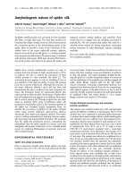
Fig. 1. Generation of heterozygous targeted mice. (A) Representation and map of Nmnat1 targeted allele and the deletion events after Cre
transfection of Nmnat1
+ ⁄ 3lox
ES cells. The expected change in the size of a BamHI band in genomic southern blots is shown in the diagram.
(B) Southern blot and PCR analysis of genomic DNA of ES cell clones after Cre-mediated recombination (type II deletion according to the dia-
gram in A). The genomic DNA was digested with BamHI and probed with probe 4. The wild-type and the recombinant band are the
expected size. PCR with primer pairs Pr1 + Pr2 and Pr3 + Pr4 shows the correct placement of the two remaining loxP sites. Exactly the
same result was shown in southern blot and PCR analysis of Nmnat1
+ ⁄ 2lox
mouse tail DNA, after blastocyst injection and coat colour screen-
ing of mice. (C) PCR and southern blot analysis of tail DNA from Nmnat1
+ ⁄ 2lox
· C57BL ⁄ 6 K14 Cre offspring. PCR was performed with prim-
ers Pr1 + Pr4 to demonstrate the correct deletion of the genomic region between the first and the third loxP site as shown by the 80-bp
product formed. The 2.4 kb wild-type PCR product cannot be distinguished on this high percentage agarose gel (left panel). The fact that the
new Cre-mediated recombination leaves only one loxP site is also demonstrated by the 1.2 kb BamHI specific band on a southern blot (right
panel).
L. Conforti et al. NMNAT1 gene inactivation and axon degeneration
FEBS Journal 278 (2011) 2666–2679 ª 2011 The Authors Journal compilation ª 2011 FEBS 2669
cific NMNAT enzyme activity was determined with a
biochemical discrimination assay based on the dis-
tinctive metal ion sensitivity of the three isoforms
(Orsomando G, Cialabrini L, Amici A, Agostinelli S,
Janeckova L, Di Stefano M, Conforti L, Coleman M,
Magni G, manuscript in preparation, adapted from
[30,31]). In agreement with mRNA and protein
expression analysis, NMNAT1 enzyme activity in
Nmnat1
+ ⁄ )
mouse brain was about half that in wild-
type (Fig. 2E). In contrast, no significant differences
were observed in NMNAT2 and NMNAT3 activity
(Fig. 2E). Despite the high brain mitochondrial content
and energy demand, NMNAT3 enzyme activity is very
low. This result was obtained in brain extracts after dis-
ruption of mitochondrial membranes, excluding the
possibility of an underestimation of NMNAT3 activity
Level of NMNAT1 protein
(arbitrary units)
0.30
0.25
0.20
0.15
*(P = 0.046)
32
A
BC
E
D
1321
NMNAT1
(31.5 kDa)
β
-actin
(42 kDa)
NAD
+
levels
(nmol·g
–1
tissue)
0
50
100
150
200
250
300
350
N.S.
C57BL/6 Nmnat1
+/–
% relative expression normalised to
β
-ACT
NMNAT1
**(P = 0.0054)
0
20
40
60
80
100
120
140
160
0
0.02
0.04
0.06
0.08
0.1
NMNAT enzyme
activity (m
U·mg
–1
)
NMNAT1 NMNAT2 NMNAT3
N.S.
N.S.
*(P = 0.0212)
Nmnat1
+/–
Wild-type
NMNAT2
N.S.
NMNAT3
0
20
40
60
80
100
120
% of each isoform expressed in Nmnat1
+/–
mice relative to wild-type
N.S.
**
NMNAT1
Nmnat1
+/–
Wild-type
C57BL/6 Nmnat1
+/–
C57BL/6 Nmnat1
+/–
0.40
0.30
0.20
0.10
0.00
NMNAT enzyme activity
(mU·mg
–1
)
C57BL/6 Nmnat1
+/–
*(P = 0.013)
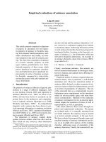
Fig. 2. NMNAT isoform expression and enzyme activity in Nmnat1
+ ⁄ )
mice. (A) Western blots of skeletal muscle homogenates from
Nmnat1
+ ⁄ )
and C57BL ⁄ 6 mice probed with antibody 183 (the antibody also reveals a non-specific upper band). The histogram represents
the integrated band intensity of the NMNAT1 band normalized to the b-actin control (n = 3, Mann–Whitney test, P = 0.046). (B) Total
NMNAT activity of brain homogenates from Nmnat1
+ ⁄ )
and C57BL ⁄ 6 mice. The enzyme activity is strongly reduced in the heterozygous
mice with respect to wild-types (n = 9, Student’s t-test, P = 0.013). (C) NAD
+
levels in wild-type and Nmnat1
+ ⁄ )
total brain homogenate
(n = 5, Student’s t -test). (D) Left panel: NMNAT1 mRNA relative expression in Nmnat1
+ ⁄ )
and wild-type brains showing strong reduction of
NMNAT1 mRNA in heterozygous KO mice. Right panel: Relative mRNA expression of each NMNAT isoform in Nmnat1
+ ⁄ )
compared with
wild-type, showing that while NMNAT1 mRNA expression is reduced, NMNAT2 and 3 mRNA relative expression is not changed. Normaliza-
tion was performed for each isoform by calculating the ratio between the expression of an individual NMNAT isoform and that of the refer-
ence gene (b-actin) in wild-type samples. The arbitrary number of 100% was assigned to this ratio for one control, and NMNAT expression
of the same isoform in the remaining controls and in Nmnat1
+ ⁄ )
brains relative to the reference gene was compared with this number.
Therefore relative mRNA expression levels can be compared between wild-type and Nmnat1
+ ⁄ )
(n = 3, Student’s t -test, **P = 0.0054). (E)
Determination of NMNAT isozyme activity in wild-type and Nmnat1
+ ⁄ )
total brain homogenates reveals highly reduced NMNAT1 activity in
heterozygous KO tissue compared with wild-type but no change in the activity of the other two isoforms. Note the very low activity of
NMNAT3 in mouse brain. (n = 3, Student’s t-test, *P = 0.0212).
NMNAT1 gene inactivation and axon degeneration L. Conforti et al.
2670 FEBS Journal 278 (2011) 2666–2679 ª 2011 The Authors Journal compilation ª 2011 FEBS
due to lack of solubilization of mitochondria during
the extraction procedure. The absence of compensatory
changes in NMNAT2 and NMNAT3 when NMNAT1
is depleted supports the model of non-redundant
functions for these isoforms.
Despite the reduction in NMNAT1 protein levels
and enzyme activity, Nmnat1
+ ⁄ )
mice are healthy,
indistinguishable from their wild-type littermates and
have a normal lifespan, suggesting that downregulation
of NMNAT1 is compatible with normal life and a
healthy nervous system, although complete inactivation
is lethal.
Wallerian degeneration rate in Nmnat1
+ ⁄ )
mice
Wld
S
neuroprotective protein contains NMNAT1 and
requires its enzyme activity to delay axon degeneration
after injury, but NMNAT1 overexpression in vivo is
not neuroprotective [21,22]. However, the role of
endogenous NMNAT1 on the rate of Wallerian degen-
eration has never been determined. To test this, we
lesioned sciatic nerves of Nmnat1
+ ⁄ )
mice and their
wild-type littermates after crossing them with YFP-H
mice [32] where some axons are labelled with the yel-
low fluorescent protein (YFP). In YFP-H positive mice
it is easy to follow axon degeneration in longitudinal
sections of lesioned sciatic nerves observed under a
fluorescent microscope [22,33]. Wallerian degeneration
of the distal stump of a sciatic nerve after an injury
follows a precise time course in wild-type mice. Axon
fragmentation begins at around 36 h, then proceeds
quickly and is complete 42 h after the lesion. In spon-
taneous mutant Wld
S
, however, Wallerian degenera-
tion is highly delayed and axon continuity is preserved
up to 3 weeks from injury [33,34]. Thus we studied
Wallerian degeneration in Nmnat1
+ ⁄ )
mice with sciatic
nerves lesioned for 30 h as a non-stringent test for
accelerated Wallerian degeneration, and for 72 h as a
non-stringent test for any delay in Wallerian degenera-
tion. Nmnat1
+ ⁄ )
X YFP-H nerves fully maintained
axon integrity 30 h after sciatic nerve lesions, similar
to wild-type nerves [Fig. 3A(a,b)]. All axons were com-
pletely fragmented 72 h after lesion, in the same way
as wild-types [Fig. 3A(d,e)]. In great contrast, axons
from Wld
S
heterozygous mice are completely preserved
at this time point [Fig. 3A(f),B]. In order to exclude an
effect on the time of onset of the degenerative process,
we also analysed axon degeneration in wild-type and
Nmnat1
+ ⁄ )
mice 36 h after sciatic nerve lesion. At this
time, axon degeneration has just begun to occur in
wild-types [34]. However, even at this time point, we
could not detect any significant difference in the num-
ber of degenerated Nmnat1
+ ⁄ )
axons compared with
wild-types (Fig. 3B). We conclude that NMNAT1
downregulation neither accelerates nor delays axon
degeneration after sciatic nerve lesion.
We tested the rate of neurite degeneration after cut
also in vitro, in superior cervical ganglia (SCG) cultures
obtained from Nmnat1
+ ⁄ )
and wild-type pups. SCG
explants were allowed to extend neurites in culture for
7 days. The neurites were then cut with a scalpel per-
pendicular to the direction of growth and observed at
different times. Axons in wild-type SCGs remain intact
3 h after cutting, but start degenerating at 6–9 h, with
degeneration complete by 24 h. Axon fragmentation in
Nmnat1
+ ⁄ )
SCG explants followed an identical time
course (Fig. 4A,B).
We determined NMNAT1 specific enzyme activity
in SCG explant extracts from wild-type and
Nmnat1
+ ⁄ )
mice (Orsomando G, Cialabrini L, Amici
A, Agostinelli S, Janeckova L, Di Stefano M, Conforti
L, Coleman M and Magni G, manuscript in prepara-
tion, adapted from [30,31]). Similarly to what was
detected in brain, NMNAT1 activity in Nmnat1
+ ⁄ )
SCG explants (0.015 mUÆmg
)1
) was half that in wild-
types (0.033 mUÆmg
)1
). NAD(P)
+
levels in SGC whole
cell extracts showed a non-significant trend towards
lower levels in heterozygous null mice relative to wild-
types (Fig. 4C). This could reflect a reduced level of
nuclear NAD
+
that is masked by the activity of extra-
nuclear NMNAT isoforms synthesizing high levels of
NAD
+
in neurites. Indeed, neurite density in these cul-
tures is very high, and NAD(P)
+
levels in wild-type
SCG neurites are around double those of their corre-
sponding cell bodies (L. Conforti, L. Janeckova and
M. Coleman, unpublished results). Thus reduction of
NAD
+
within nuclei remains possible. However, in
agreement with the result in vivo, dowregulation of
NMNAT1 expression does not affect the rate of axon
degeneration in vitro.
Discussion
These data indicate that complete NMNAT1 gene
inactivation is incompatible with the normal develop-
ment of embryos, as the extranuclear isoforms
NMNAT2 and NMNAT3 cannot compensate for
complete loss of NMNAT1. Nmnat1
+ ⁄ )
mice have
reduced NMNAT1 expression and enzyme activity;
however, they develop normally and their lifespan is
not altered. We show that the rate of Wallerian degen-
eration in vivo and in vitro in sciatic nerves and
in SCG explant cultures from Nmnat1
+ ⁄ )
mice is not
different from wild-type.
NMNAT1-generated NAD
+
in the nucleus is used
as substrate of histone deacetylase sirtuins and
L. Conforti et al. NMNAT1 gene inactivation and axon degeneration
FEBS Journal 278 (2011) 2666–2679 ª 2011 The Authors Journal compilation ª 2011 FEBS 2671
PARP1. Sirtuins have been implicated in cellular pro-
cesses such as ageing, transcription, apoptosis and
stress resistance. Yeast Sir2 and its mammalian homo-
logue SIRT1 are upregulated upon caloric restriction
and this is associated with increased lifespan [8].
SIRT1 controls the activity of genes that regulate
circadian rhythm and promotes the transcription of
NAMPT, the rate limiting enzyme in NAD
+
synthesis,
in a feedback loop that has been recently described
[35,36]. NAD
+
is substrate also for nuclear PARP1,
whose overactivation consequent to genotoxic stress
leads to NAD
+
depletion in the cytoplasm and cell
necrosis, demonstrating a communication between the
nuclear and the cytoplasmic NAD
+
pool [37].
Thus, the failure of Nmnat1 homozygous null
embryos to survive and develop may reflect perturba-
tions in gene transcription, especially sirtuin targets, or
PARP1-mediated NAD
+
depletion that cannot be
replenished locally within the nucleus. Indeed,
NMNAT1 downregulation in cell lines by small inter-
fering RNA has a profound effect on transcription of
a number of genes, some of which are important
for neuronal maintenance and normal neuronal
function [11]. Conditional homozygous inactivation of
Nmnat1 in neurons in the adult mouse will be essential
to understand whether and how transcriptional regula-
tion affects neuronal maintenance and survival.
NMNAT1 is also part of the neuroprotective protein
Wld
S
and its enzyme activity is necessary but not suffi-
cient for this protein to delay degeneration of axons
after an injury in vivo [20–22]. However, in cell cultures
and in Drosophila NMNAT1 overexpression is par-
tially neuroprotective [20,23]. Moreover, in Drosophila,
targeted disruption of NMNAT causes spontaneous
axon degeneration via a chaperone activity [38,39]. We
investigated the role of endogenous NMNAT1 in axon
protection in heterozygous null mice where we found a
strong reduction in NMNAT1 protein expression and
enzyme activity, while the other two isoforms were
expressed at wild-type levels and their enzyme activity
Nmnat1
+/–
cut t = 72h
WT cut
t = 72 h
UNCUT
WT cut
t = 30 h
Wld
S
het
cut t = 72 h
Nmnat1
+/–
cut t = 30 h
50 µm
(a)
(b) (c)
(f)(e)(d)
0
20
40
60
80
100
120
% intact axons
30 h 36 h 72 h
Wild-type
Nmnat1
+/–
Wld
S
het
N.S.
N.S.
N.S.
A
B
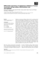
Fig. 3. Wallerian degeneration rate in
Nmnat1
+ ⁄ )
mice. (A) Tibial nerves from
Nmnat1
+ ⁄ )
mice crossed to YFP-H with
sciatic nerves lesioned for the indicated
time show a wild-type rate of Wallerian
degeneration with intact axons 30 h after
the lesion (a, b) and completely degenerated
axons 72 h after the lesion (d, e). At this
time point, Wld
S
heterozygous axons are
still completely preserved (f). Bar, 50 lm.
(B) Quantification of axon degeneration at
the indicated time points after sciatic nerve
lesions. Note that at 36 h post-lesion, when
Wallerian degeneration normally begins, the
number of degenerated axons is similar in
wild-type and Nmnat1
+ ⁄ )
.(n = 4, Student’s
t-test).
NMNAT1 gene inactivation and axon degeneration L. Conforti et al.
2672 FEBS Journal 278 (2011) 2666–2679 ª 2011 The Authors Journal compilation ª 2011 FEBS
was unchanged. Since NMNAT1 activity is predomi-
nant in brain (Fig. 2E) and NMNAT1 is also the most
catalytically efficient isoform [31], its downregulation
determines a significant reduction in total NMNAT
activity in Nmnat1
+ ⁄ )
mice that cannot be compen-
sated by NMNAT2 and ⁄ or NMNAT3. Sorci et al. [31]
reported that NMNAT2 is the predominant activity in
human brain. However, these authors used human per-
itumoural tissue for their determination of isoform-
specific NMNAT activity, whereas we used mouse half
brain homogenates. Brain has a heterogeneous cellular
composition that could influence relative abundance of
this enzyme activity; therefore our result is neither
directly comparable nor in conflict with that described
by Sorci et al. [31].
Despite NMNAT1 strong downregulation,
Nmnat1
+ ⁄ )
mice do not show any unusual phenotype
and the rate of Wallerian degeneration in these mice
or in primary neurons derived from them is unaltered.
It is possible that the maintenance of normal NAD
+
steady state levels despite the decrease in NMNAT
activity in our mutant mice underlines the lack of any
adverse phenotype. The embryonic lethality of
NMNAT1 full inactivation precludes the possibility of
testing the rate of Wallerian degeneration in the com-
plete absence of NMNAT1. However, the result
obtained in heterozygous NMNAT1 KO mice suggests
that extranuclear NMNAT activities predominantly
control the rate of Wallerian degeneration. Accord-
ingly, the two extranuclear NAD
+
-synthesizing iso-
zymes, NMNAT2 and NMNAT3, maintain wild-type
expression levels and enzyme activities in Nmnat1
+ ⁄ )
mice where Wallerian degeneration after injury pro-
ceeds at a wild-type rate.
i
0.60
Wild-type t = 0
Nmnat1
+/–
t = 0 Nmnat1
+/–
t = 3 h Nmnat1
+/–
t = 6 h Nmnat1
+/–
t = 9 h Nmnat1
+/–
t = 24 h
Wild-type t = 3 h Wild-type t = 6 h Wild-type t = 9 h
Wild-type t = 24 h
0.50
0.40
0.30
0.20
0.10
0.00
t = 0 h t = 3 h t = 6 h t = 9 h t = 24 h
N.S.
NAD(P)
+
(nmol·mg
–1
protein)
A
BC
Fig. 4. In vitro degeneration of injured axons in Nmnat1
+ ⁄ )
SCG cultures. (A) SCG explants from C57BL ⁄ 6 and Nmnat1
+ ⁄ )
mice were cul-
tured for 7 days and the extended neurites were separated from the cell body mass using a scalpel. Neurites were imaged after the cut at
the time points indicated. Bar, 10 lm. (B) Quantification of axon degeneration in SCG explant cultures after cut. The results show that there
is a time effect (P < 0.0001) but no difference between wild-type and Nmnat1
+ ⁄ )
(n = 6, two-way repeated measures ANOVA, P = 0.808).
(C) NAD(P)
+
levels in whole SCG explant cultures from C57BL ⁄ 6 and Nmnat1
+ ⁄ )
mice are similar. (n = 7, independent samples t-test,
P = 0.492).
L. Conforti et al. NMNAT1 gene inactivation and axon degeneration
FEBS Journal 278 (2011) 2666–2679 ª 2011 The Authors Journal compilation ª 2011 FEBS 2673
This is also consistent with our observation of an
increased Wld
S
protective potency when this protein is
redistributed outside the nucleus [7,17]. Moreover, we
showed lack of protection in transgenic mice overex-
pressing NMNAT1 alone and in variant-Wld
S
trans-
genic mice where an N-terminal 16 (N-16) amino acid
sequence derived from Ube4b had been removed
[21,22]. Interestingly, the only known property of the
N-16 amino acid sequence indispensable for Wld
S
action is its ability to bind the abundant cellular pro-
tein VCP ⁄ p97. This protein is involved in many cellu-
lar activities and is particularly enriched at the surface
of membranous organelles [24,25]. NMNAT activity
in mammals has become more specialized by evolving
several isoforms, each of them playing a particular
role according to its most abundant location within
the cell. Wld
S
protection may be the result of a fine
redistribution of NMNAT1, potentially via VCP bind-
ing, at a specific location inside the cell, where its
enzyme activity leads to downstream events finally
resulting in axon protection. Accordingly, cytoplasmic
Wld
S
and cytoplasmic or axonally targeted NMNAT1
are all neuroprotective [7,17,18,40]. This location
could match that of the endogenous extranuclear
NMNAT isoform NMNAT2. NMNAT2 downregula-
tion triggers spontaneous axon degeneration in pri-
mary SCG neurons [26], suggesting this may be the
endogenous NMNAT activity that normally controls
Wallerian degeneration. NMNAT3 could also be
responsible for controlling injury-induced axon degen-
eration. However, the low level of NMNAT3 activity
we detect in the nervous system and the lack of a phe-
notype when this isoform is downregulated in neuronal
cultures [26] makes it a weaker candidate. NMNAT2
is rapidly degraded after an injury and its rapid degra-
dation could trigger axon degeneration. However, the
more stable Wld
S
protein, when present, or an abnor-
mal targeting of NMNAT1 itself [17] could preserve
the injured axons by substituting for NMNAT2 [26].
The results presented here argue against functional
redundancy of the three mammalian NMNAT iso-
forms. NMNAT2 and 3 cannot compensate for loss of
NMNAT1 when this isozyme is completely inactivated,
leading to the lack of viability of null NMNAT1 KO
mice. In addition, there is no upregulation of
NMNAT2 or 3 in Nmnat1
+ ⁄ )
mice, where NMNAT1
is highly downregulated. In cultured SCGs, NMNAT1
and 3 cannot compensate for loss of NMNAT2 trig-
gered by RNA interference or by axon injury [26].
Indeed, the low level of NMNAT3 activity in brain
suggests its main functions may be predominant in
other tissues [30]. However, the various isoforms could
compensate for each other when redistributed to a dif-
ferent location. For instance NMNAT1 appears to
compensate for loss of NMNAT2 when it reaches
ectopic location by high overexpression or by re-target-
ing, therefore conferring protection to axons after cut
[17,18,22,26].
Despite the role for other NMNAT isoforms such as
NMNAT2 in controlling axonal integrity, a related role
for NMNAT1 remains possible in the absence of data
from homozygous null mice. In particular, it is possible
that the level of this enzyme activity in heterozygous
KO could remain above a threshold level needed to
significantly modify axon degeneration after an injury.
The availability of NMNAT1 floxed mice will enable
us to address this question in a future study by generat-
ing conditional KOs where the NMNAT1 gene is inac-
tivated only in neurons at postnatal stages, overcoming
the embryonic lethality of a complete null mutant.
In conclusion, NMNAT1 is indispensable for the
normal development of the embryo and NMNAT2
and 3 cannot compensate for its loss. Decreased
NMNAT1 activity in heterozygous null mice, however,
does not affect the rate of Wallerian degeneration, sug-
gesting that endogenous NMNAT1 does not have a
primary role in axon maintenance.
Materials and methods
Construction of the targeting vector
We determined the genomic sequence of the entire mouse
Nmnat1 coding region and used this to design a targeting
vector based on the plasmid pEASYFlox (a gift from W.
Mu
¨
ller and K. Rajewsky). The positive selection marker,
G418 ⁄ neomycin (NEO
R
), is flanked by two loxP sites. To
maximize the likelihood of achieving complete gene inacti-
vation, we chose to delete a region comprising the first and
second exons, including some 5¢ UTR where the promoter
is located. This region was amplified by PCR with SalI
tagged primers and cloned into the SalI site of the targeting
vector. Two additional homology regions, a 5¢ 2.3 kb
region and a 3 ¢ 4.6 kb region, were then obtained by PCR
using primers tagged with NotI ⁄ Bam HI and HindIII sites
respectively and cloned into the respective restriction sites
of pEASYFlox. We confirmed the absence of PCR and
cloning artefacts by sequencing all coding regions, the loxP
sites and most non-coding regions. The genomic locus, the
completed targeting vector and the recombination events
are shown in Fig. S1.
The primer pair sequence was as follows: 5¢ homology
arm (NotI and BamHI site underlined and italics)
5¢-AGGAAAAAA
GCGGCCGCACACTTACAGCCTGAG
GCG-3¢,5¢-CGC
GGATCCACTCCAAGGATACACTCC
GA-3¢;3¢ homology arm (HindIII site underlined and ital-
ics) 5¢-GGCCC
AAGCTTATATATTTGCCTAGGAGGGT
NMNAT1 gene inactivation and axon degeneration L. Conforti et al.
2674 FEBS Journal 278 (2011) 2666–2679 ª 2011 The Authors Journal compilation ª 2011 FEBS
C-3¢,5¢-GGCCCAAGCTTAAGACAGTGTGGAGGAGA
CCT-3¢. The targeted region (SalI site underlined and ital-
ics) was 5¢-CAACGC
GTCGACCCATGTGCTGAAAGCT
TGGT-3¢,5¢-ACTGGC
GTCGACTTGAATGTCTTAGTG
ACTGGG-3¢. All primers were purchased by Sigma-
Genosys, Haverhill, UK. All chemicals were obtained by
Sigma-Aldrich, Gillingham, UK, unless otherwise stated.
ES cell electroporation and isolation of a double
recombinant clone for blastocyst injection
The 18 kb targeting vector was linearized with NotI and elec-
troporated into Bruce 4 ES cells (from C57BL ⁄ 6 strain, kind
gift of K. Rajewsky and A. Egert). ES cell clones were posi-
tively selected 24 h post electroporation with 0.2 mgÆmL
)1
G418. Negative selection of random integration was per-
formed by addition of 2 · 10
)6
m ganciclovir to the medium.
We picked 384 clones among the ones that were resistant to
both selection agents. Southern blot analysis showed that
only one clone contained the entire targeting vector correctly
integrated at both homology arms of the genomic locus. This
clone was electroporated again in vitro with a Cre expression
vector (pPGK-Cre-bpA, kind gift of K. Rajewsky and
W. Mu
¨
ller). This allowed us to delete the NEO
R
gene and
leave a loxP flanked region amenable to conditional or con-
ventional deletion. One subclone was then isolated that had
lost the NEO
R
cassette and contained a ‘floxed’ targeted
locus (Fig. 1A). We designed primers spanning the two loxP
sites (Pr1, Pr2, Pr3, Pr4, see Fig. 1A) to confirm the presence
and the integrity of the loxP sites in the floxed clone after
Cre-mediated deletion. The PCR across the loxP sites con-
firmed the presence of both loxP sites in the targeted clone
(Fig. 1B). Furthermore, sequencing of the PCR products
confirmed that the loxP sites were correct.
Generation of targeted mice
The Bruce 4 targeted ES cell clone containing the floxed
locus was used for injection into BALB ⁄ c derived blast-
ocysts. Chimeric mice, originally identified by coat colour,
were then confirmed by southern blotting (see Fig. 1B).
Chimeric mice were backcrossed to C57BL ⁄ 6 mice and the
transmission of the mutant allele to the progeny was
revealed by coat colour analysis and southern blotting.
Nmnat1
+ ⁄ )
mice were obtained by crossing the floxed
Nmnat1 male chimerics to female C57 ⁄ BL6 K14 Cre mice
to produce heterozygous null mice on a black background
[27]. Southern blot analysis demonstrated that about 50%
of the offspring are heterozygous for the full deletion allele.
The heterozygous mice were then intercrossed in an attempt
to generate homozygous null mutants. Animal work was
performed in accordance with the relevant German and
UK government animal welfare legislation under licenses
K13, 11 ⁄ 00 (Cologne, Germany) and 80 ⁄ 1778 and 80 ⁄ 2254
(Cambridge, UK).
Preparation and analysis of DNA from ES cells,
mice and embryos
Genomic DNA was isolated using standard protocols
[21]. For southern blot analysis, genomic DNA from ES
cells was digested with EcoRI or HindIII and analysed
with a 420 bp 5¢ probe and a 750 bp 3¢ probe located
outside the targeted region (Fig. S1) and generated by
PCR from genomic DNA with the following primer
pairs: 3¢ probe, 5¢-AAT ATTTGGAA TTAGGTAA GTGT-3¢,
5¢-GTGTAAAAGACACTGTGATG-3¢;5¢ probe, 5¢-TGT
CTTAAAATGCACTTCAAAC-3¢,5¢-GTCGAGTTGCCA
TGCAGAG-3¢. Another 450 bp probe (called probe 4,
Fig. 1A) obtained by mouse genomic DNA PCR with
the primers 5¢-GGCCCAAGCTTATATATTTGCCTAG
GAGGGTC-3¢ and 5¢-TCAGACATTTATAAGTTTCG
GG-3¢ was used on southern blots of tail genomic DNA
digested with BamHI to identify both Nmnat1 floxed
mice and Nmnat1 heterozygous KO mice. PCR screening
of those mice used the following primers spanning loxP
site 1 and loxP site 2: Pr1, 5¢-TCGGAGTGTATCCTTG
GAGT-3¢; Pr2, 5¢-ACCAAGCTTTCAGCACATGG-3¢;
Pr3, 5¢-CCCAGTCACTAAGACATTCAA-3¢; Pr4, 5¢-GA
CCCTCCTAGGCAAATATA-3¢.
Western blotting, NMNAT enzyme activity assay
and NAD(P)
+
level determination
Western blotting of sagittally divided half brains was per-
formed as described previously [22]. Sagittally divided half
brains were homogenized in five volumes of RIPA buffer
[phosphate-buffered saline (PBS) containing 1% NP40,
0.5% deoxycholate, 0.1% sodium dodecylsulphate].
High-speed supernatant was diluted to approximately
0.5 mgÆmL
)1
total protein according to the Bradford assay
(BioRad, Hemel Hempstead, UK) and fractionated by
standard SDS ⁄ PAGE. After semidry blotting (BioRad,
Hemel Hempstead, UK), nitrocellulose membranes (Bio-
Rad) were blocked in PBS plus 0.02% Tween-20 and 5%
low-fat milk powder before incubation with primary anti-
body and then horseradish peroxidase conjugated second-
ary antibody (1 : 3000; Amersham Biosciences, Little
Chalfont, UK). Proteins were visualized using the ECL
detection kit (Amersham Biosciences, Little Chalfont,
UK) according to the manufacturer’s instructions. For
quantification, western blot band intensities were deter-
mined with image j software and normalized to b-actin.
NAD
+
and NAD(P)
+
levels were determined in brain or
whole cell extracts by HPLC identification or by a fluori-
metric cyclic reaction as described previously [41,42].
Total NMNAT enzyme activity was determined as
described earlier [41]. Tissue was suspended in six volumes
of 50 mm Hepes, pH 7.4, 0.5 mm EDTA, 1 mm MgCl
2
,
1mm phenylmethylsulphonyl fluoride and 0.02 mgÆmL
)1
each of leupeptin, antipain, chymostatin and pepstatin,
L. Conforti et al. NMNAT1 gene inactivation and axon degeneration
FEBS Journal 278 (2011) 2666–2679 ª 2011 The Authors Journal compilation ª 2011 FEBS 2675
and homogenized on ice (3 · 4 s with 10 s intervals at
medium speed). NMNAT activity assay was performed at
37 °C in a 0.1 mL reaction mixture containing 30 mm
Tris ⁄ HCl, pH 7.5, 2 mm nicotinamide mononucleotide
(NMN), 2 mm ATP, 20 mm MgCl
2
,10mm NaF and an
appropriate aliquot of brain homogenate. The reaction
was started by adding 4 lLof50mm NMN and stopped
by the addition of a half-volume of ice-cold 1.2 m HClO
4
.
After 10 min at 0 ° C, the mixture was centrifuged and
135 lL of supernatant was neutralized by the addition of
36 lL of 0.8 m K
2
CO
3
. NMNAT activity was calculated
after HPLC identification and quantification of the prod-
uct (NAD
+
). One unit of enzyme was defined as the
amount capable of producing 1 lmol of NAD
+
per
minute at 37 °C. The individual contribution to NAD
+
formation by each of the three NMNAT isozymes was
selectively evaluated with a method adapted from [30,31]
(Orsomando G, Cialabrini L, Amici A, Agostinelli S,
Janeckova L, Di Stefano M, Conforti L, Coleman M and
Magni G, manuscript in preparation). Statistical analysis
was performed using Student’s t-test or the Mann–
Whitney test.
Northern blotting analysis
Total RNA isolation from wild-type and Nmnat1
+ ⁄ )
mouse brains and northern blotting were performed as
described in [28] using the b-actin probe and an NMNAT1
3¢ cDNA probe [28].
Real time RT- PCR
Brains were removed from freshly killed mice, snap-frozen
in liquid nitrogen and stored at )80 °C until the time of
processing. Total RNA was extracted using TriSure (Bio-
line, London, UK) according to the manufacturer’s proto-
col. SuperScriptÔ II Reverse Transcriptase (Invitrogen,
Paisley, UK) was used to synthesize first strand cDNA fol-
lowing the manufacturer’s instructions, using 1 lg of total
RNA and oligo(d)T primer. Quantitative PCR was per-
formed using Platinum
Ò
SYBR
Ò
Green qPCR SuperMix
UDG (Invitrogen, Paisley, UK) using the following primer
pairs: NMNAT1, 5¢-TTCAAGGCCTGACAACATCGC-3¢,
5¢-GAGCACCTTC ACAGT CTCCACC- 3¢;NMNAT2,5¢-CA
GTGCGAGAGACCTCATCCC-3¢,5¢-ACACATGATGA
GACGGTGCCG-3¢; NMNAT3, 5¢-GGTGTGGAGCTGT
GTGACAGC-3¢,5¢-GCCATGGCCACTCGGTGATGG-3¢;
b-actin (reference gene), 5¢-TGTTACCAACTGGGACG
ACA-3¢,5¢-GGGGTGTTGAAGGTCTCAAA-3¢. React-
ions were performed in duplicate and standard curves using
serial dilutions of cDNA were performed for each set of
primers to establish PCR efficiencies. Relative expression
ratios in comparison with the b-actin reference gene were
determined as described in [43] and statistical analysis was
performed using the t-test.
Nerve lesion
Nerve lesions to assess the rate of Wallerian degeneration
were performed as described in [7,21] in 2–10 months old
wild-type or Nmnat1
+ ⁄ )
mice crossed to YFP-H mice.
Mice were anaesthetized with a mixture of 100 mgÆkg
)1
ketamine (Fort Dodge Animal Health, Southampton, UK)
and 5 mgÆkg
)1
xylazine (Pfizer, Sandwich, UK). Right sci-
atic nerves were transected at the upper thigh and mice
were killed by cervical dislocation at the indicated time
points. The swollen first 2 mm of distal nerve was dis-
carded, and the remaining sciatic and tibial nerve stump
was removed for confocal microscopy.
At 30, 36 or 72 h post-lesion mice were humanely killed
and sciatic and tibial nerves distal to the site of the lesion
were quickly dissected for confocal microscopy.
Acquisition and processing of confocal images
Confocal fluorescent images were acquired according to
[7,21] using a confocal microscope system (LSM 510 Meta;
Carl Zeiss, Welwyn Garden City, UK) built around an Axi-
overt 200 (Carl Zeiss, Welwyn Garden City, UK), and z
series were merged using algorithms from LSM Software
Release 3.2 (Carl Zeiss, Welwyn Garden City, UK). Tissue
preparations were mounted in Vectashield medium (Vector
Laboratories, Peterborough, UK). The fluorophore used
was YFP. Axon degeneration was quantified as described
in [21] by counting all (intact and fragmented) fluorescent
axons and calculating the percentage of intact axons in
three different fields per nerve explant examined. Statistical
analysis was performed using the t-test unless specified
otherwise in the figure legends.
Assessment of axon degeneration in SCG
cultures
SCG were dissected from 0–2 day old pups and cultured as
described [44]. Axons were allowed to extend for 7 days
before separation from the cell body mass using a scalpel,
and the degeneration of the isolated neurites was followed at
different time points for 24 h after cut. Bright field images
were acquired on a microscope (IX8I; Olympus, Southend-
on-Sea, UK) coupled to a digital camera (U-TV 0.5XC;
Olympus, Southend-on-Sea, UK) using analysis software
(Soft Imaging Systems GmbH, Muenster, Germany). Axon
degeneration was quantified as described in [45]. A two-way
repeated measures analysis of variance (ANOVA) was per-
formed on the data to look for the difference in axon degen-
eration between wild-type and Nmnat1
+ ⁄ )
.
Acknowledgements
We thank Dr Anne Segonds-Pichon for statistical
advice, Mr Stefan Milde for help with axon degenera-
NMNAT1 gene inactivation and axon degeneration L. Conforti et al.
2676 FEBS Journal 278 (2011) 2666–2679 ª 2011 The Authors Journal compilation ª 2011 FEBS
tion quantification, Dr Sebastian Lukasiak for guid-
ance on real time RT-PCR, Dr Giacomo Morreale for
critically reading the manuscript and Dr Gloria
Esposito for helpful advice. This work was funded by
the BBSRC, the Centre for Molecular Medicine of the
University of Cologne (ZMMK) grant NG3 and
the Mario Negri Pharmacological Research Institute
(Milan).
References
1 Magni G, Amici A, Emanuelli M, Orsomando G,
Raffaelli N & Ruggieri S (2004) Structure and function
of nicotinamide mononucleotide adenylyltransferase.
Curr Med Chem 11, 873–885.
2 Berger F, Lau C, Dahlmann M & Ziegler M (2005)
Subcellular compartmentation and differential catalytic
properties of the three human nicotinamide mononucle-
otide adenylyltransferase isoforms. J Biol Chem 280,
36334–36341.
3 Emanuelli M, Amici A, Carnevali F, Pierella F,
Raffaelli N & Magni G (2003) Identification and
characterization of a second NMN adenylyltransferase
gene in Saccharomyces cerevisiae. Protein Expr Purif 27,
357–364.
4 Yalowitz JA, Xiao S, Biju MP, Antony AC, Cummings
OW, Deeg MA & Jayaram HN (2004) Characterization
of human brain nicotinamide 5¢-mononucleotide adenyl-
yltransferase-2 and expression in human pancreas.
Biochem J 377, 317–326.
5 Koch-Nolte F, Haag F, Guse AH, Lund F & Ziegler M
(2009) Emerging roles of NAD+ and its metabolites in
cell signaling. Sci Signal 2, mr1.
6 Lau C, Doelle C, Gossmann TI, Agledal L, Niere M &
Ziegler M (2010) Isoform-specific targeting and inter-
action domains (ISTIDs) in human nicotinamide mono-
nucleotide adenylyltransferases (NMNATs). J Biol
Chem 285, 18868–18876.
7 Beirowski B, Babetto E, Gilley J, Mazzola F, Conforti
L, Janeckova L, Magni G, Ribchester RR & Coleman
MP (2009) Non-nuclear Wld(S) determines its neuro-
protective efficacy for axons and synapses in vivo.
J Neurosci 29, 653–668.
8 Guarente L & Picard F (2005) Calorie restriction –
the SIR2 connection. Cell 120, 473–482.
9 Ramsey KM, Yoshino J, Brace CS, Abrassart D,
Kobayashi Y, Marcheva B, Hong HK, Chong JL, Buhr
ED, Lee C et al. (2009) Circadian clock feedback cycle
through NAMPT-mediated NAD+ biosynthesis.
Science 324, 651–654.
10 van der Veer E, Ho C, O’Neil C, Barbosa N, Scott R,
Cregan SP & Pickering JG (2007) Extension of human
cell lifespan by nicotinamide phosphoribosyltransferase.
J Biol Chem 282, 10841–10845.
11 Zhang T, Berrocal JG, Frizzell KM, Gamble MJ,
DuMond ME, Krishnakumar R, Yang T, Sauve AA &
Kraus WL (2009) Enzymes in the NAD+ salvage
pathway regulate SIRT1 activity at target gene
promoters. J Biol Chem 284, 20408–20417.
12 Chiarugi A (2002) Inhibitors of poly(ADP-ribose) poly-
merase-1 suppress transcriptional activation in lympho-
cytes and ameliorate autoimmune encephalomyelitis in
rats. Br J Pharmacol 137, 761–770.
13 Berger F, Lau C & Ziegler M (2007) Regulation of
poly(ADP-ribose) polymerase 1 activity by the phos-
phorylation state of the nuclear NAD biosynthetic
enzyme NMN adenylyl transferase 1. Proc Natl Acad
Sci USA 104, 3765–3770.
14 Alano CC, Garnier P, Ying W, Higashi Y, Kauppinen
TM & Swanson RA (2010) NAD+ depletion is neces-
sary and sufficient for poly(ADP-ribose) polymerase-1-
mediated neuronal death. J Neurosci 30, 2967–2978.
15 Liu D, Gharavi R, Pitta M, Gleichmann M & Mattson
MP (2009) Nicotinamide prevents NAD+ depletion
and protects neurons against excitotoxicity and cerebral
ischemia: NAD+ consumption by SIRT1 may endan-
ger energetically compromised neurons. Neuromolecular
Med 11, 28–42.
16 Kitaoka Y, Hayashi Y, Kumai T, Takeda H, Mun-
emasa Y, Fujino H, Ueno S, Sadun AA & Lam TT
(2009) Axonal and cell body protection by nicotinamide
adenine dinucleotide in tumor necrosis factor-induced
optic neuropathy. J Neuropathol Exp Neurol 68 ,
915–927.
17 Babetto E, Beirowski B, Janeckova L, Brown R, Gilley
J, Thomson D, Ribchester RR & Coleman MP (2010)
Targeting NMNAT1 to axons and synapses transforms
its neuroprotective potency in vivo
. J Neurosci 30 ,
13291–13304.
18 Sasaki Y & Milbrandt J (2010) Axonal degeneration is
blocked by nicotinamide mononucleotide adenylyltrans-
ferase (Nmnat) protein transduction into transected
axons. J Biol Chem 285, 41211–41215.
19 Coleman MP & Freeman MR (2010) Wallerian degen-
eration, Wld(S), and Nmnat. Annu Rev Neurosci 33,
245–267.
20 Avery MA, Sheehan AE, Kerr KS, Wang J & Freeman
MR (2009) Wld S requires Nmnat1 enzymatic activity
and N16–VCP interactions to suppress Wallerian degen-
eration. J Cell Biol 184, 501–513.
21 Conforti L, Wilbrey A, Morreale G, Janeckova L,
Beirowski B, Adalbert R, Mazzola F, Di Stefano M,
Hartley R, Babetto E et al. (2009) Wld S protein
requires Nmnat activity and a short N-terminal
sequence to protect axons in mice. J Cell Biol 184,
491–500.
22 Conforti L, Fang G, Beirowski B, Wang MS, Sorci L,
Asress S, Adalbert R, Silva A, Bridge K, Huang XP
L. Conforti et al. NMNAT1 gene inactivation and axon degeneration
FEBS Journal 278 (2011) 2666–2679 ª 2011 The Authors Journal compilation ª 2011 FEBS 2677
et al. (2007) NAD(+) and axon degeneration
revisited: Nmnat1 cannot substitute for Wld(S) to
delay Wallerian degeneration. Cell Death Differ 14,
116–127.
23 Araki T, Sasaki Y & Milbrandt J (2004) Increased
nuclear NAD biosynthesis and SIRT1 activation
prevent axonal degeneration. Science 305, 1010–
1013.
24 Laser H, Conforti L, Morreale G, Mack TG, Heyer M,
Haley JE, Wishart TM, Beirowski B, Walker SA, Haase
G et al. (2006) The slow Wallerian degeneration pro-
tein, WldS, binds directly to VCP ⁄ p97 and partially
redistributes it within the nucleus. Mol Biol Cell 17,
1075–1084.
25 Morreale G, Conforti L, Coadwell J, Wilbrey AL &
Coleman MP (2009) Evolutionary divergence of valo-
sin-containing protein ⁄ cell division cycle protein 48
binding interactions among endoplasmic reticulum-
associated degradation proteins. FEBS J 276, 1208–
1220.
26 Gilley J & Coleman MP (2010) Endogenous Nmnat2 is
an essential survival factor for maintenance of healthy
axons. PLoS Biol 8, e1000300.
27 Hafner M, Wenk J, Nenci A, Pasparakis M, Scharffet-
ter-Kochanek K, Smyth N, Peters T, Kess D, Holtkot-
ter O, Shephard P et al. (2004) Keratin 14 Cre
transgenic mice authenticate keratin 14 as an oocyte-
expressed protein. Genesis 38, 176–181.
28 Conforti L, Tarlton A, Mack TG, Mi W, Buckmaster
EA, Wagner D, Perry VH & Coleman MP (2000) A
Ufd2 ⁄ D4Cole1e chimeric protein and overexpression of
Rbp7 in the slow Wallerian degeneration (WldS) mouse.
Proc Natl Acad Sci USA 97, 11377–11382.
29 Fernando FS, Conforti L, Tosi S, Smith AD & Cole-
man MP (2002) Human homologue of a gene mutated
in the slow Wallerian degeneration (C57BL ⁄ Wld(s))
mouse. Gene 284, 23–29.
30 Di Stefano M, Galassi L & Magni G (2010) Unique
expression pattern of human nicotinamide mononucleo-
tide adenylyltransferase isozymes in red blood cells.
Blood Cells Mol Dis 45, 33–39.
31 Sorci L, Cimadamore F, Scotti S, Petrelli R, Cappellac-
ci L, Franchetti P, Orsomando G & Magni G (2007)
Initial-rate kinetics of human NMN-adenylyltransferas-
es: substrate and metal ion specificity, inhibition by
products and multisubstrate analogues, and isozyme
contributions to NAD+ biosynthesis. Biochemistry 46,
4912–4922.
32 Feng G, Mellor RH, Bernstein M, Keller-Peck C,
Nguyen QT, Wallace M, Nerbonne JM, Lichtman JW
& Sanes JR (2000) Imaging neuronal subsets in trans-
genic mice expressing multiple spectral variants of GFP.
Neuron 28, 41–51.
33 Beirowski B, Berek L, Adalbert R, Wagner D, Grumme
DS, Addicks K, Ribchester RR & Coleman MP (2004)
Quantitative and qualitative analysis of Wallerian
degeneration using restricted axonal labelling in YFP-H
mice. J Neurosci Methods 134, 23–35.
34 Beirowski B, Adalbert R, Wagner D, Grumme DS,
Addicks K, Ribchester RR & Coleman MP (2005) The
progressive nature of Wallerian degeneration in wild-
type and slow Wallerian degeneration (WldS) nerves.
BMC Neurosci 6,6.
35 Eckel-Mahan K & Sassone-Corsi P (2009) Metabolism
control by the circadian clock and vice versa. Nat Struct
Mol Biol
16, 462–467.
36 Nakahata Y, Sahar S, Astarita G, Kaluzova M &
Sassone-Corsi P (2009) Circadian control of the
NAD+ salvage pathway by CLOCK-SIRT1. Science
324, 654–657.
37 Chiarugi A & Moskowitz MA (2002) Cell biology.
PARP-1 – a perpetrator of apoptotic cell death? Science
297, 200–201.
38 Zhai RG, Cao Y, Hiesinger PR, Zhou Y, Mehta SQ,
Schulze KL, Verstreken P & Bellen HJ (2006)
Drosophila NMNAT maintains neural integrity inde-
pendent of its NAD synthesis activity. PLoS Biol 4,
e416.
39 Zhai RG, Zhang F, Hiesinger PR, Cao Y, Haueter CM
& Bellen HJ (2008) NAD synthase NMNAT acts as a
chaperone to protect against neurodegeneration. Nature
452, 887–891.
40 Sasaki Y, Vohra BP, Baloh RH & Milbrandt J (2009)
Transgenic mice expressing the Nmnat1 protein mani-
fest robust delay in axonal degeneration in vivo.
J Neurosci 29, 6526–6534.
41 Balducci E, Emanuelli M, Raffaelli N, Ruggieri S,
Amici A, Magni G, Orsomando G, Polzonetti V & Nat-
alini P (1995) Assay methods for nicotinamide mononu-
cleotide adenylyltransferase of wide applicability. Anal
Biochem 228, 64–68.
42 Billington RA, Travelli C, Ercolano E, Galli U, Roman
CB, Grolla AA, Canonico PL, Condorelli F &
Genazzani AA (2008) Characterization of NAD uptake
in mammalian cells. J Biol Chem 283, 6367–6374.
43 Pfaffl MW, Horgan GW & Dempfle L (2002) Relative
expression software tool (REST) for group-wise
comparison and statistical analysis of relative
expression results in real-time PCR. Nucleic Acids Res
30, e36.
44 Buckmaster EA, Perry VH & Brown MC (1995) The
rate of Wallerian degeneration in cultured neurons from
wild type and C57BL ⁄ WldS mice depends on time in
culture and may be extended in the presence of elevated
K+ levels. Eur J Neurosci 7, 1596–1602.
45 Sasaki Y, Vohra BP, Lund FE & Milbrandt J (2009)
Nicotinamide mononucleotide adenylyl transferase-med-
iated axonal protection requires enzymatic activity but
not increased levels of neuronal nicotinamide adenine
dinucleotide. J Neurosci 29, 5525–5535.
NMNAT1 gene inactivation and axon degeneration L. Conforti et al.
2678 FEBS Journal 278 (2011) 2666–2679 ª 2011 The Authors Journal compilation ª 2011 FEBS
Supporting information
The following supplementary material is available:
Fig. S1. Targeting of the Nmnat1 gene.
Fig. S2. NMNAT1 expression analysis in wild-type
and Nmnat1
+ ⁄ )
mouse brain.
This supplementary material can be found in the
online version of this article.
Please note: As a service to our authors and readers,
this journal provides supporting information supplied
by the authors. Such materials are peer-reviewed and
may be reorganized for online delivery, but are not
copy-edited or typeset. Technical support issues arising
from supporting information (other than missing files)
should be addressed to the authors.
L. Conforti et al. NMNAT1 gene inactivation and axon degeneration
FEBS Journal 278 (2011) 2666–2679 ª 2011 The Authors Journal compilation ª 2011 FEBS 2679